Neuroinflammation: 2021 Update
←
→
Page content transcription
If your browser does not render page correctly, please read the page content below
Free Neuropathology 2:1 (2021)                                                                                                                                                    Hans Lassmann
doi: https://doi.org/10.17879/freeneuropathology-2021-3166                                                                                                                          page 1 of 14

                                                                                                                                                                                     Review

Neuroinflammation: 2021 Update
Hans Lassmann1
1
    Center for Brain Research, Medical University of Vienna, Austria

Address for correspondence:
Hans Lassmann · Center for Brain Research · Medical University of Vienna · Spitalgasse 4 · 1090 Wien · Austria
hans.lassmann@meduniwien.ac.at

Submitted: 18 December 2020                       ·      Accepted: 08 January 2021                    ·    Copyedited by: Aivi Nguyen                    ·      Published: 12 January 2021

Abstract
Key requirements for the validity of a neuropathological study are the inclusion of large numbers of biopsy or
autopsy cases and proper controls, the rigorous classification of the basic neuropathology and the selection of
the most suitable technologies for investigation. Whether the studies are performed with the fanciest, new,
and state of the art technology or with rather conventional methodology is of minor importance. Following
these criteria, a spectrum of neuropathological studies has been published in 2020, which provides new in-
sights on important questions related to neurological disease. They include the pathological substrate of brain
disease in COVID-19 infected patients, the nature of the adaptive and innate inflammatory response, or the
type and mechanisms of tissue injury and repair in multiple sclerosis, and diagnostically relevant or mechanistic
new insights into antibody-mediated diseases of the central nervous system. Other studies describe in detail
the dynamic changes of brain inflammation in patients with trisomy 21 as a disease model for Alzheimer’s dis-
ease, or the presence and consequences of vascular comorbidities in a chronic inflammatory disease, such as
multiple sclerosis. All these contributions have provided important, highly relevant clues for basic and transla-
tional neuroscience.

Keywords: Brain inflammation, COVID-19, Multiple sclerosis, Alzheimer’s disease, MOGAD

1) What was the brain pathology in                                                                         neuropathology is that the nervous system, too,
                                                                                                           can be a target, reflected for instance by anosmia
patients who died during the course of                                                                     and ageusia, encephalopathy, focal ischemic stroke,
COVID-19?                                                                                                  encephalitis, meningitis, or polyneuritis (Li et al.
                                                                                                           2020, Paterson et al. 2020, Liu et al. 2020, Hernan-
     The COVID-19 pandemic has been a major                                                                dez-Fernandez et al. 2020). To what extent these
challenge for society and particularly for health                                                          neurological deficits are due to direct SARS-Cov2
care institutions during the last year. Although                                                           infection of the nervous tissue, immune-mediated
COVID-19 primarily affects the respiratory system                                                          brain damage in the course of a systemic cytokine
and the major cause of death is pneumonia with                                                             storm, or are secondary complications of respirato-
respiratory failure, other organs, such as the renal,                                                      ry failure, the intensive care setting, or comorbidi-
cardiovascular system, or the digestive tract, are                                                         ties is not clear (Liu et al. 2020). Neuropathology
affected (Paterson et al. 2020). Most relevant for                                                         plays a critical role in answering these questions.

Copyright: © 2021 The author(s). This is an open access article distributed under the terms of the Creative Commons Attribution 4.0 International License (https://creativecommons.org/licenses/by/4.0/),
which permits unrestricted use, distribution, and reproduction in any medium, provided the original author and source are credited, a link to the Creative Commons license is provided, and any changes
are indicated. The Creative Commons Public Domain Dedication waiver (https://creativecommons.org/publicdomain/zero/1.0/) applies to the data made available in this article, unless otherwise stated.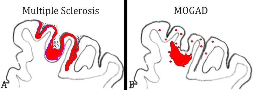
Free Neuropathology 2:1 (2021)                                                                       Hans Lassmann
doi: https://doi.org/10.17879/freeneuropathology-2021-3166                                             page 2 of 14

      The first approach to answer these questions           19 patients and is, thus, very important and
was to analyze whether molecules, which are in-              groundbreaking. However, it also has important
volved in cellular virus infection or propagation, are       limitations. It is based on autopsies of 43 patients,
expressed within the nervous system. Angiotensin             which is likely not sufficient to cover the entire
concerting enzyme 2 (ACE2) is one of the cellular            neuropathological spectrum of the disease. Anoth-
receptors recognized by SARS-Cov2, when docking              er limitation is that it does not contain data regard-
to the cell surface. Thus, the presence of ACE2 on           ing the neurological status of the patients. Thus, it
brain cells, such as neurons, glia or cerebral endo-         remains unclear, to what extent patients with spe-
thelial cells suggests that brain infections is possible     cific neurological disease manifestations have been
(Kabbani and Olds 2020, Kanwar et al. 2020). An-             included. The third limitation is that it does not
other docking molecule for SARS-Cov2 is Neuropilin           include a proper patient control group and, particu-
1 (NRP1) and a high expression of this molecule has          larly, patients with similarly severe systemic im-
been found in virus infected cells in the nasal cavity       mune activation. This particular question has been
in COVID-19 patients (Cantuti-Castelvetri et al.             addressed in another study on COVID-19 neuropa-
2020).                                                       thology, which revealed that the degree of inflam-
                                                             mation and microglia activation seen in COVID-19
      Early neuropathological studies in the COVID-          patients is similar to that of patients who died un-
19 pandemic were restricted to single case reports           der septic conditions (Deigendesch et al. 2020),
or investigations in very small patient cohorts, and         thus suggesting that it may at least in part be a
they were generally restricted to some basic neu-            secondary consequence of systemic immune acti-
ropathological investigations (Kantonen et al. 2020,         vation. Another study, also based on a large sample
Reichard et al. 2020, Jensen et al. 2020, Younger            of autopsies, mainly focused on the possible routes
2020, Liu et al. 2020). The results were diverse and         of CNS infection (Meinhardt et al. 2020). It shows
controversial, mainly providing evidence for brain           high virus load in the olfactory epithelium, includ-
damage that was not directly linked to SARS-Cov2             ing the olfactory sensory neurons, and in the olfac-
infection. The first systematic study on this issue          tory pathways within the CNS, supporting the view
appeared on October 2020 in Lancet Neurology                 of a neuronal route of virus entry into the CNS.
(Matschke et al. 2020), and this study combined              Additionally, however, virus antigen was also pre-
classical neuropathology with virology and molecu-
                                                             sent in cerebral endothelial cells, associated with
lar studies on gene expression. In a first set of data       micro-thrombosis and cerebral microinfarcts.
the authors describe that different molecules, in-
volved in virus docking and propagation, show a                    In summary, current pathological studies show
preferential expression in different cells of the cen-       that the central nervous system can be infected
tral nervous system. For example, ACE2 was mainly            with SARS-Cov2, that local infection can be associ-
found in oligodendrocytes. The highest expression            ated with nervous system damage, but the extent
of transmembrane protease serine subtype 2 and 4             of infection is low. A large part of the neuropatho-
(TMPRSS2 / 4) was seen in neurons, and NRP-1 was             logical changes seems to be secondary to systemic
mainly present in endothelial cells and astrocytes.          immune activation and cytokine storm, critical-
These data suggest different mechanisms of infec-            illness related encephalopathy, or hypoxia and
tion in different brain cells. The neuropathological         comorbidities.
studies documented the presence of focal and dif-
fuse ischemic lesions and diffuse astrogliosis. Mi-          2) What new insights have emerged
croglia activation with microglia nodules and in-
flammation were mainly seen in the lower brain
                                                             into phenotype and disease mecha-
stem. In about half of the patients, virus was found         nisms of antibody mediated autoim-
either by PCR or immunocytochemistry. However,               mune diseases of the central nervous
the virus load was very low and by immunohisto-              system?
chemistry only a very small number of virus infect-
ed cells became apparent. This study is the first to             The discovery that autoantibodies against
describe the basic neuropathology seen in COVID-             neuronal ion channels or neurotransmitter recep-
Free Neuropathology 2:1 (2021)                                                                       Hans Lassmann
doi: https://doi.org/10.17879/freeneuropathology-2021-3166                                             page 3 of 14

tors are associated with a spectrum of acute and             antibodies themselves may not be pathogenic, but
chronic neurological diseases, has revolutionized            just represent a diagnostically useful marker for an
neurological disease research (Höftberger and                autoimmune attack of cytotoxic T-cells, as it occurs
Lassmann 2017). It has been shown that these dis-            in many classical paraneoplastic diseases. The pa-
eases, which had before been regarded as func-               thology, described in a biopsy of one of the cases, is
tional diseases, neurodegenerative disorders or              in line with this assumption, showing infiltrating T-
toxic conditions, are immune-mediated and can be             cells in close contact with neurons. However, the
successfully treated with immunosuppression. The             study further describes neurophysiological experi-
number of diseases falling into this category has            ments, which show that the antibodies induce al-
profoundly increased during the last years (Höft-            tered neuronal activity patterns and increased fir-
berger and Lassmann 2017). One reason, which                 ing and bursting rates in neuronal networks in
may in part explain the increase in prevalence, is           vitro. These data suggest that auto-antibodies di-
the introduction of immunological checkpoint in-             rected against a cytoplasmic protein in synapses
hibitor therapies in oncology. Immunological                 may reach their specific target and induce func-
checkpoints are critical steps in T-cell activation,         tional changes. The authors suggest that extensive
which prohibit the development of auto-reactive T-           exocytosis and endocytosis, which takes place in
cells and auto-antibodies. When these checkpoints            active synapses, facilitates the entry of the antibod-
are inhibited, immune reactions against antigens of          ies into the intracellular compartment, but this
malignant neoplasms can be triggered, but this may           proposed mechanism does not explain how the
occur at the expense of autoimmunity, which is               antibodies leave the endosomal compartment and
frequently directed against neuronal antigens                access the cytosol. This important cell biological
(Sechi et al. 2020).                                         question with important disease relevance has to
                                                             be clarified in future studies.
      On this basis it is not surprising that auto-
antibody-associated diseases of the central nervous
system remained in the focus of research interest            3) What is the pathological difference
in 2020. The respective studies provided novel in-           between MOG antibody associated
sights into the function of NMDA and glycine recep-          inflammatory demyelinating disease
tor directed antibodies in relation to the clinical
disease spectrum (Matute et al. 2020, Carceles-
                                                             (MOGAD) and multiple sclerosis (MS)?
Cordon et al. 2020, Rauschenberger et al. 2020) or
                                                                  Focal plaques of demyelination with axonal
how autoantibodies against IgLON 5 may trigger
                                                             preservation and reactive gliosis developing in the
intracellular accumulation of tau-tangles (Landa et
                                                             context of a chronic inflammatory reaction in the
al. 2020).
                                                             central nervous system has been regarded as the
     Here, I focus on one study, which deals with            specific hallmark of MS pathology. However, a new
an interesting but also controversial aspect of these        disease entity has recently emerged, which is an
diseases. Pitsch et al. (2020) identified antibodies         inflammatory demyelinating disease associated
against the postsynaptic actin binding protein               with antibodies directed against myelin oligoden-
drebrin in patients with severe epileptic seizures,          drocyte glycoprotein (Reindl and Waters 2019). Not
which were associated with encephalopathy and                all anti-MOG antibodies are pathogenic, but only
neuropsychiatric symptoms, including depression              those that are directed against a conformational
and cognitive impairment. The antibodies, identi-            epitope, which is accessible on the surface of native
fied by selective binding to the neuropil and by             oligodendrocytes or on MOG-transfected cell lines.
western blot and sequencing, were specific for               Using these diagnostic tests, it became clear that
these patients, and the specificity for the target           MOG antibody-associated disease (MOGAD) differs
antigen was proven by the lack of binding in                 from MS by its clinical manifestation and course as
drebrin knock out mice. Drebrin is an intracellular          well as by its response to immunomodulatory
antigen, which is not exposed on the extracellular           treatment (Fujihara and Cook 2020). To explain
surface of neurons or synapses. For this reason, the         these clinical differences, a neuropathological
                                                             comparison of these diseases is mandatory, but
Free Neuropathology 2:1 (2021) Hans Lassmann
doi: https://doi.org/10.17879/freeneuropathology-2021-3166 page 4 of 14
respective knowledge on MOGAD was restricted to ed immunization with brain tissue (Höftberger et al.
few biopsies with very limited tissue samples. This 2015). New molecular biology technologies allowed
changed in 2020, with two studies describing the resurrection of a pathogenic auto-antibody from
neuropathological changes in large cohorts of the archival formaldehyde fixed and paraffin em-
MOGAD patients (Höftberger et al. 2020, Takai et bedded tissue, which was found to be directed
al. 2020). The lesions in MOGAD differ from those against a conformational epitope of MOG and in-
seen in MS in many aspects, including their topo- duced demyelination after transfer in vivo (Beltran
graphical distribution in the CNS, the type of demy- et al. 2020).
elination, and the nature of the inflammatory re-
sponse. In MS, new lesions are formed by simulta- 4) Is subpial demyelination a unique
neous demyelination in large, focal areas or by the
peripheral expansion of pre-existing lesions located
feature of multiple sclerosis patholo-
throughout the brain and spinal cord. In contrast, gy?
MOGAD demyelination occurs by confluence of
small perivenous lesions, generally resulting in a For a long time, MS has been regarded a de-
demyelination pattern similar to that seen in acute myelinating disease affecting the white matter.
disseminated encephalomyelitis (Figure 1). Demye- However, with the availability of highly sensitive
lination in MOGAD is associated with complement immunocytochemical methods, which allow stain-
deposition at the site of active myelin injury, but ing of the very thin myelinated fibers within the
the degree of complement activation is much less grey matter (Peterson et al. 2000), it became clear
compared to that seen in patients with aquaporin 4 that demyelination in the grey matter in the MS
antibody associated neuromyelitis optica (NMO). In brain is extensive (Kutzelnigg et al. 2005) and may
both conditions, MS as well as MOGAD, the basic even give rise to cortico-spinal or pure cortical vari-
lesion is characterized by inflammatory demye- ants of the disease in a subset of patients (Trapp et
lination, partial axonal preservation and reactive al. 2018). Three types of cortical lesions have been
astrocytic gliosis. However, the inflammatory reac- identified: the cortico-subcortical lesions (Type 1),
tion is fundamentally different. While in MS the the intracortical lesions (Type 2), and the subpial
dominant inflammatory reaction is seen around the lesions (Type 3) (Bo et al. 2003). The third type is
larger drainage veins in the periventricular tissue most abundant in MS and is related to focal in-
and the meninges, in MOGAD the smaller veins and flammatory aggregates in the leptomeninges (Mag-
venules are mainly affected. Finally, in MOGAD, liozzi et al. 2010; Figure 2). Several previous studies
infiltrating lymphocytes are mainly CD4+ T-cells have indicated that subpial demyelinated lesions in
with low numbers of CD8+ T-cells and B-cells; the the cortex may be specific for MS (Moll et al. 2008,
dominant lymphocytes in active MS lesions are Fischer et al. 2013), but this view was based on a
tissue resident CD8+ effector memory T-cells and B- rather limited sample of neuropathological condi-
cells / plasma cells. All these data indicate that tions other than MS. This has now been changed in
MOGAD and MS are fundamentally different dis- a very comprehensive study by Junker et al. (2020),
eases. in which nearly every thinkable neuropathological
condition has been investigated for cortical demye-
Interestingly, the pathology of MOGAD closely lination. Interestingly, subpial demyelination was
resembles the pathology seen in experimental exclusively present in MS and not seen in any of the
models of autoimmune encephalomyelitis (EAE), other conditions. The only key diagnosis missed in
induced by immunization of rats, guinea pigs, or this case series was MOGAD. However, this was
primates with CNS tissue myelin or recombinant also specifically addressed in the systematic studies
MOG (Höftberger et al. 2020). Thus, EAE appears to of MOGAD pathology outlined above (Höftberger
be a very suitable model for MOGAD, but much less et al. 2020, Takai et al. 2020), describing intracorti-
for MS. Support of this view is provided in a recent cal and cortico-subcortical lesions but absent sub-
study, on archival material from a patient, who pial cortical lesions. In rare instances, these intra-
died in the 1950s with an acute MS-like inflamma- cortical lesions can fuse and give rise to large focal
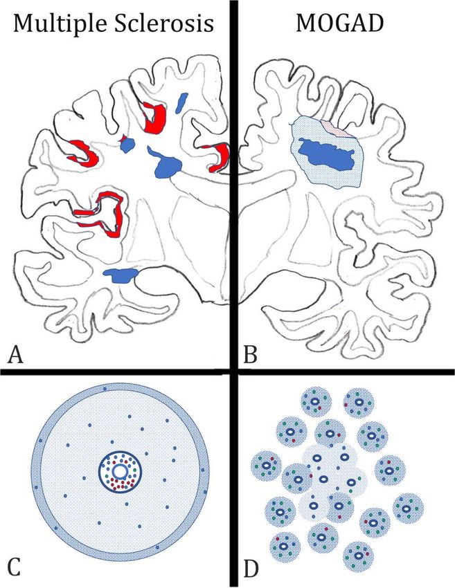
tory demyelinating disease after misguided repeat- confluent plaques of demyelination. Thus, so far itFree Neuropathology 2:1 (2021) Hans Lassmann doi: https://doi.org/10.17879/freeneuropathology-2021-3166 page 5 of 14 Figure 1: Key neuropathological features, distinguishing multiple sclerosis (MS) from the inflammatory demyelinating disease, which is associated with auto-antibodies against Myelin Oligodendrocyte Glycoprotein (MOGAD) in the forebrain. A) In MS, the lesions (blue areas) are located around large cerebral veins with a predilection site around the ventricle and the subcorti- cal white matter. They are sharply demarcated from the surrounding peri-plaque white matter and frequently display finger like perivenous extensions into the deep white matter. In the cortex, band like subpial lesions (red areas) dominate, mainly located in the deep sulci of the cortical ribbon. B) In MOGAD, lesions are mainly located in the optic nerves and spinal cord, but their structural details have so far not been clearly outlined. When lesions are present in the hemispheres of the forebrain, a type of tissue injury is seen, which resembles acute dissemi- nated encephalomyelitis. It consists of perivenous sleeves of demyelination around small veins and venules (blue dotted areas), which show confluence in the lesion center (dark blue area). When the cortex is affected small perivenous intra-cortical demyelination is seen most frequently (red dotted areas). C) In MS, the lesions form around larger veins with prominent perivascular spaces. The perivascular spaces contain mainly B-cells (red dots) and CD8+ T-lymphocytes (blue dots), the latter also disperse into the plaque parenchyma. CD4 + T-cells (green dots) are very sparse. A characteristic feature of active MS lesions is the radial expansion, reflected by a rim of activated macrophages at the lesion edge (blue dotted area). D) In contrast, in MOGAD, numerous small perivenous sheaths of demyelination are present, which arise around small veins and venules (blue dotted areas). In the inflammatory infiltrates the CD4+ T-cells dominate, while there is a much lower contribution of CD8 + T-cells (blue dots) or B-cells and plasma cells (red dots). Confluence of the perivenous lesions results in larger demyelinated plaques.
Free Neuropathology 2:1 (2021)                                                                                               Hans Lassmann
doi: https://doi.org/10.17879/freeneuropathology-2021-3166                                                                     page 6 of 14

seems that subpial cortical demyelination is a                             most effective therapeutic success is seen with
unique feature of MS pathology, but the evidence                           drugs that target T-cells and B-cells together or
for its absence in MOGAD is thus far based on a                            even B-cells alone. Thus, it is still an unresolved
rather small sample of cases. Nevertheless, the                            question, as to what cells and immune mechanisms
presence of a subpial cortical lesion strongly sup-                        trigger or drive inflammation in MS.
ports a neuropathological diagnosis of MS.
                                                                                 Pathology can help clarify this issue by provid-
                                                                           ing a concise account of the types of inflammatory
5) What immune cells drive inflamma-                                       cells seen in different stages in the evolution of MS
tion in multiple sclerosis?                                                lesions. Until a few years ago, information about
                                                                           the composition of inflammatory infiltrates in MS
      Our understanding of the immune mecha-                               was restricted to small studies performed on a lim-
nisms driving inflammation in MS is largely biased                         ited number of patients and lesions. Since 2017,
by immunological studies of experimental autoim-                           however, there are now three large studies availa-
mune encephalomyelitis, which now turns out to                             ble, which performed a phenotyping of inflamma-
have more relevance for MOGAD than for MS. Key                             tory cells in an overall sample of more than 200 MS
mechanisms of inflammation, defined from such                              patients and which included all stages of the dis-
EAE studies, such as the key role of MHC Class II                          ease (Van Nierop et al. 2017, Machado Santos et al.
restricted T-cells responses, the involvement of                           2018, Fransen et al. 2020). All studies reached a
CD4+ Th17 cells, or the central role of GM-CSF driv-                       similar conclusion that the dominant lymphocytes
en myeloid cell activation (Schreiner and Becher                           in the MS lesions are tissue resident CD8+ effector
2015) are not convincingly supported by recent                             memory T-cells (TTRM), while infiltration with CD4+
therapeutic trials targeting the respective immuno-                        T-cells is sparse. This is the case in all types of MS,
logical pathways (Baker et al. 2017). In contrast,                         including fulminant acute MS, relapsing remitting

Figure 2: Patterns of cortical demyelination in MS and MOGAD:
A) In MS, the dominant and most specific type of cortical demyelination is the subpial lesion. It is characterized by a band of demye-
lination spanning over several gyri and sulci (red lesions). The lesions are more extensive in the invaginations of the cortex. Active corti-
cal demyelination is associated with meningeal inflammation by T-cells (blue dots) and B-cells (red dots) and is characterized by a band
of activated macrophages / microglia at the border between the cortical lesion and the surrounding normal appearing cortex (purple
bands).
B) In MOGAD, the dominant cortical pathology is the presence of small perivenous intracortical demyelinated lesions (red circles). They
arise around small cortical veins and venules with inflammatory infiltrates. On rare occasions, such intracortical perivenous lesions may
give rise to a focal confluent cortical lesion, which frequently also expands into the adjacent subcortical white matter.
Free Neuropathology 2:1 (2021) Hans Lassmann
doi: https://doi.org/10.17879/freeneuropathology-2021-3166 page 7 of 14
MS as well as primary or secondary progressive MS memory T-cells (Serafini et al. 2020). Here, the hy-
and is the same in all activity stages of the lesions. pothesis is that inflammatory activity is triggered
In active lesions, a subset of CD8+ cells show focally and propagated when the virus is activated in la-
and temporally restricted activation, and B-cells are tently infected cells and recognized by the specific
much more numerous in early stages of the disease T-cells (Serafini et al. 2020). As a note of caution,
and in active lesions than in inactive lesions at late several other groups have tried to confirm the spe-
stages (Machado Santos et al. 2018). In a recent cific presence of EBV infected B-cells in the MS
study, imaging mass cytometry was applied to the brain and lesions and have failed (Lassmann et al.
biopsy tissue of a single MS patient who suffered 2010, van Nierop et al. 2017). The reason for these
from a rebound exacerbation after cessation of discrepancies is still unresolved.
natalizumab treatment (Ramaglia et al. 2019). Also,
in this study, the infiltrates contained T-cells and B- In a similar approach, an interaction between
cells, but the relative proportion of CD4+ T-cells was brain resident CD8+ cells and B-cells has been de-
higher compared to that seen in the previous stud- scribed, where the B-cells did not express EBV (van
ies. However, activation of T-cells, visualized by the Nierop et al. 2017). This suggests that the cells rec-
expression of the transcription factor NFAT or pro- ognize a B-cell autoantigen. A possible mechanism
liferation markers, was seen only in CD8+ T-cells. for how such autoimmunity against B-cells may be
Besides their absolute numbers, the activation induced is provided by Jelcic et al. (2018). MS B-
state of leukocytes is relevant for driving the in- cells, possibly in relation to their EBV infection sta-
flammatory reaction. Similarly as before (Machado tus, expand by auto-proliferation and present a B-
Santos et al. 2018), the most frequent B-cell pheno- cell specific auto-antigen to CD4+ T-cells, which also
types were IgG+ CD38+ plasmablasts. Thus, overall, expand and infiltrate the brain and spinal cord.
the pathological data are in line with the observed Within the CNS, these T-cells are activated by anti-
effects of anti-inflammatory treatments in MS pa- gen recognition on infiltrating B-cells. In addition,
tients. the respective auto-antigen (RASGRP2) is also ex-
pressed in neurons, which may further propagate
inflammation and tissue injury. Whether this
6) What do the inflammatory cells in mechanism also accounts for activation of CD8+ T-
MS lesions recognize? cells, the dominant inflammatory cells in MS le-
sions, is unknown.
An important open question is what antigen is
recognized by the CD8+ tissue resident memory T- A different approach has been followed by
cells. Since such T-cells acquire their phenotype Konjevic Sabolek et al. (2019). In this study, the
and persist after effective clearance of their cog- interaction of CD8+ T-cells with local target cells
nate antigen and become reactivated when the was identified by the polarized location of cytotoxic
antigen re-appears, it is unlikely that these cells granules at the site of contact. When this was seen,
recognize a classical auto-antigen, which is present the target cell was isolated and its nature was de-
in the CNS in excess all the time. In line with this termined in gene expression studies and by im-
view, no reactivity of CD8+ T-cells from MS brain munohistochemistry. In this study, the only cells
lesions has been seen against the commonly tested which interacted with cytotoxic CD8+ T-cells in MS
myelin antigens (van Nierop et al. 2017). lesions were myeloid cells, expressing markers of
macrophages and microglia, suggesting these may
Several potential candidate antigens have re- harbor the target antigen(s). Finally, other studies
cently been identified. Since accumulating epide- suggest that the target antigen recognized by T-
miological evidence associates Epstein Barr Virus cells in MS lesions may be the stress protein alpha
infection with MS (Bar-Or et al. 2020, Levin et al. B crystallin, which is in MS lesions most prominent-
2010), one hypothesis is that B-cells with latent EBV ly expressed in active lesions (van Noort et al.
infection are present within the CNS of MS patients 2010).
(Serafini et al. 2007, Veroni et al. 2018) together
with EBV-reactive CD8+ tissue resident effector In summary, the new data from systematic
phenotypical and functional studies on the inflam-Free Neuropathology 2:1 (2021) Hans Lassmann
doi: https://doi.org/10.17879/freeneuropathology-2021-3166 page 8 of 14
matory response within MS lesions provide lesions in different inflammatory conditions as well
groundbreaking new insights into the pathophysi- as vascular or neurodegenerative diseases in hu-
ology of the disease and question the concept of mans (Zrzavy et al. 2017, Fischer et al. 2013). To
MS being an autoimmune disease against myelin. define the patterns of microglia activation in such
However, the results are in part very divergent and, diseases may finally result in more selective and
to some extent, contradicting. Whether this indi- efficient neuroprotective treatments.
cates heterogeneous disease mechanisms between
individual patients or patient subgroups will be This is now possible with new technologies,
seen in the future. such as single-cell RNA sequencing or immunocyto-
chemical methods, which allow the simultaneous
detection of multiple protein antigens within the
7) Which microglia or macrophage same section, such as imaging mass cytometry. In
phenotypes are associated with tissue some studies of models of autoimmune encepha-
damage in the brain? lomyelitis and MS, these techniques have been
used and confirmed in an elegant way the patterns
Microglia and recruited macrophages play a of microglia activation and macrophage recruit-
major role in the induction of tissue injury, not only ment, which have been described before with more
in inflammatory brain lesions but also in neuro- conventional techniques as summarized above
degenerative diseases. For a long time, it was diffi- (Masuda et al. 2019, Ramaglia et al. 2020). Howev-
cult to distinguish between activated microglia and er, they additionally showed that microglia and
recruited macrophages within brain lesions. This macrophages with different phenotypes are pre-
has changed with the introduction of markers, sent side-by-side in the same lesion. This may indi-
which are selectively expressed on microglia, such cate selective activation signals in specific subpopu-
as the membrane protein TMEM 119 and the lations of cells. Not surprisingly, the data showed
marker for homeostatic microglia P2RY12. These that different MS lesions, which were in the same
markers have been used in a number of studies on activity stage but still displayed some distinct mor-
various different inflammatory and neurodegenera- phological features, were infiltrated by different
tive conditions and revealed a surprisingly uniform subsets of myeloid cells (Masuda et al. 2019). How-
reaction pattern of myeloid cells in human diseases ever, these studies did not reveal the high expres-
(Zrzavy et al. 2017, 2018, Hayashida et al. 2020, sion of molecules involved in oxidative injury,
Jäckle et al. 2020). In essence, at sites of initial tis- which had been prominent in earlier more conven-
sue injury, resident microglia become activated in a tional neuropathological studies.
pro-inflammatory pattern. This initial step is fol-
A possible explanation for these discrepancies
lowed by recruitment of myeloid cells from the
is provided by an elegant study, which specifically
circulation, and the recruited cells also show pro-
focused on microglia activation and oxidative injury
inflammatory activation or, at later stages of lesion
in autoimmune encephalomyelitis (Mendiola et al.
maturation, an intermediate phenotype. The dis-
2020). The authors first defined the molecular phe-
tinction of pro- versus anti-inflammatory pheno-
notype of microglia specifically selected from areas
types is now seen in a much more critical way,
of oxidative injury. Incorporating this profile in the
since some pro-inflammatory actions, such as the
analysis of single-cell RNA sequencing data and
interaction of macrophages with target cells may
immunocytochemistry, they identified specific mi-
contribute to tissue damage but may also be essen-
croglia and macrophage subpopulations, which
tial for neuroprotection and regeneration through
trigger oxidative stress, and showed that these are
the clearance of debris (Cignarella et al. 2020). An
selective subtypes of cells within the population of
important pro-inflammatory type of activation,
activated myeloid cells. These particular cell types
which seems to play a major role in the induction
also express molecules that are important in the
of tissue injury, is the expression of proteins in-
coagulation cascade and in glutathione metabo-
volved in oxidative stress, such as the expression of
lism. In addition, they produce molecules, which
the NOX2 complex (NADPH oxidase complex 2), a
counteract oxidative stress. One of these molecules
prominent marker of microglia expressed in activeFree Neuropathology 2:1 (2021) Hans Lassmann
doi: https://doi.org/10.17879/freeneuropathology-2021-3166 page 9 of 14
Acivivin, a glutathione regulating compound, sup- (Geraldes et al. 2020). The study shows, as ex-
presses the inflammatory tissue damage in a model pected, that systemic and intracranial vascular ab-
of autoimmune encephalomyelitis. It will be in- normalities increase with age in both the MS and
strumental in the future to validate directly the the control group. Young MS patients appear to
relevance of these findings in MS lesions and also in have a moderate reduction of systemic vascular co-
vascular or neurodegenerative diseases of the cen- morbidities compared to controls, but this differ-
tral nervous system. The presence of a subtype of ence disappears with aging. Within the central
myeloid cells triggering and resolving oxidative nervous system, MS patients showed a profound
stress could reconcile the above discrepancies and increase in small arteries with increased perivascu-
provide a mechanism for the excessive oxidative lar space, perivascular hemosiderin depositions and
injury in the lesions and the associations of markers periarteriolar accumulation of inflammatory cells, a
for oxidative injury with markers for disturbed type of pathology which correlated in its extent
blood coagulation in the lesions. In the long run, with the degree of MS-related pathology. These
these data may lead to tools for the therapeutic results underline the presence of age-related vas-
blockade of one of the most detrimental aspects of cular co-morbidities in the brain of MS patients,
microglia activation in human brain disease. which may be an amplification factor for neuro-
degeneration and disease progression in aging pa-
8) How do vascular comorbidities in- tients. Additionally, they indicate that, in contrast
to the current view, vascular pathology is not re-
fluence multiple sclerosis patients? stricted to veins but also affects small arteries, and
this arterial pathology develops at least in part
Clinical epidemiology of MS has shown that
independently from systemic vascular risk factors.
patients with vascular risk factors, such as diabetes,
In particular, the mechanisms how inflammatory
hypercholesterolaemia, hypertension, or heart
infiltrates accumulate around arteries in the MS
disease have a more aggressive disability progres-
brain requires further attention.
sion in comparison to patients lacking these co-
morbidities (Marrie et al. 2010). This is also reflect-
ed by lower brain volumes (Pichler et al. 2019). 9) What is wrong with remyelination
Furthermore, persistence of inflammatory demye- in multiple sclerosis?
linating lesions and higher lesion volumes are pre-
sent in brain areas with a blood perfusion that is Primary demyelination with preservation of
lower than in other brain areas (Haider et al. 2016). axons is the pathological hallmark of inflammatory
Such a synergism in neurodegeneration may in part demyelinating diseases, such as MS. Myelin allows
be explained by shared effector mechanisms of saltatory conduction in axons and also protects the
tissue injury between vascular and inflammatory axons from neurodegeneration. Thus, major efforts
diseases, including microglia activation, oxidative in MS research have been devoted to develop
injury, and mitochondrial damage (Zrzavy et al. treatments which prevent demyelination or stimu-
2017, 2018, Mahad et al. 2015). A strategically im- late myelin repair (Lubetzki et al. 2020). These re-
portant piece in the puzzle of vascular comorbidi- pair strategies, mainly designed to provide oli-
ties and MS, which was missing so far, was the lack godendrocyte progenitor cells in the lesions and to
of a comprehensive neuropathological description stimulate their differentiation into myelinating
of systemic and intracranial vascular pathology in oligodendrocytes, have been developed and tested
patients versus controls. in experimental models of demyelinating disease
and found to be quite effective. However, none of
This information has now been provided by a the strategies have yet resulted in clinically mean-
study, which is based on a unique archival collec- ingful myelin repair in controlled clinical trials in MS
tion of MS and control autopsy cases, where de- patients. In experimental models of demyelinating
tailed information regarding systemic vascular dis- disease, profound spontaneous remyelination is
eases was recorded, and which was collected prior the rule, and remyelination stimulating therapies in
to the availability of disease modifying treatments essence accelerate myelin repair. Although sponta-Free Neuropathology 2:1 (2021) Hans Lassmann
doi: https://doi.org/10.17879/freeneuropathology-2021-3166 page 10 of 14
neous remyelination may also occur in MS patients, demyelinating disease. Thus, for test screening of
in particular in fresh lesions at early disease stages, remyelination-promoting therapies different sys-
remyelination is, in general, sparse or absent. Sev- tems have to be used, which reproduce the in-
eral different mechanisms have been suggested to flammation-associated remyelination failure.
be responsible for this remyelination failure in MS
lesions. It may be due to a genuine defect in oli- 10) What can we learn from Down
godendrocyte (progenitor) biology. In an elegant
study, Starost et al. (2020) approached this ques-
Syndrome about inflammation and
tion by studying oligodendrocytes, induced from neurodegeneration in Alzheimer’s
pluripotent stem cells from MS patients and con- disease?
trols. They show that there is no difference in the
proliferation, differentiation and myelin production A large spectrum of data coming from experi-
between such cells derived from MS patients and mental studies as well as from neuropathological
from controls, thus strongly suggesting that there is and genetic investigations suggest an important
no genuine defect of oligodendrocytes in MS pa- role of innate immune mechanisms in the patho-
tients, though with the caveat that this was only genesis of Alzheimer’s disease (Akiyama et al.
performed in cells induced to form oligodendro- 2000). In particular, microglia phenotype and func-
cytes with expression of transcriptional factors tion are associated with progression of cognitive
from three MS patients and three controls. In a decline. However, information regarding the time
parallel neuropathological approach, the same course and type of microglia pathology especially in
group analyzed the patterns or remyelination in the pre-symptomatic phases of the disease is lim-
different disease stages of MS and in different ited. Analyzing Down syndrome brain pathology of
stages of lesion formation (Heß et al. 2020). They patients may provide some answers to these ques-
found that remyelination is highly efficient in a tions, since a gene dosage effect on amyloid pre-
subset of active lesions in the early stage of the cursor protein production predictably results in
disease, and their data suggest that the remyelinat- early onset Alzheimer’s disease (Wisniewski et al.
ing cells may be derived from mature oligodendro- 1985, Ballard et al. 2016).
cytes that had survived active myelin destruction.
This has also been suggested in recent studies, This approach was followed in a recent study,
using radiocarbon methods to determine the time in which the patterns of microglia activation and
of birth of new oligodendrocytes in MS lesions the expression of pro- and anti-inflammatory cyto-
(Yeung et al. 2019). In contrast, in chronic active kines was analyzed in a large sample of brain tissue
lesions at later stages of MS, very little remye- of patients with trisomy 21 who had died at differ-
lination was seen and the lack of remyelination was ent ages (Flores Aguilar et al. 2020). Already, in
associated with pro-inflammatory activity of the juvenile patients, a long time before the first accu-
local microglia population (Heß et al. 2020). These mulation of soluble Aß or the first deposition of Aß-
data suggest that products of activated inflamma- plaques, microglia activation and the production of
tory cells in chronic MS lesions may not only lead to proinflammatory cytokines was elevated in com-
their slow expansion but also inhibit the repair of parison to age matched controls. In contrast, in
myelin. This finding is also supported by the other older patients, when initial Alzheimer’s disease
study (Starost et al. 2020), which provides convinc- pathology became apparent, microglia activation
ing evidence that pro-inflammatory mediators, in decreased and was replaced by senescent microglia
particular gamma-interferon, blocks the differenti- phenotypes as well as a reduction of the produc-
ation of inducible pluripotent stem cells into mye- tion of pro-inflammatory cytokines. These results
linating oligodendrocytes. further support the view that anti-inflammatory
treatment strategies in Alzheimer’s disease may be
The key importance of these studies is that most beneficial when applied in very early (pre-
they document that the process of remyelination symptomatic) stages of disease evolution. The data
failure in MS is complex and not fully reflected in further suggest that the inclusion of patients with
the current experimental models of inflammatory trisomy 21 in future clinical trials could provide aFree Neuropathology 2:1 (2021) Hans Lassmann
doi: https://doi.org/10.17879/freeneuropathology-2021-3166 page 11 of 14
valuable contribution to improve treatment options human disease. For this, neuropathological studies
in patients with Alzheimer’s disease. are essential, but they have to be based on system-
atic analysis of a broad spectrum of cases and con-
Conclusions trols and have to be performed with the most suit-
able molecular or immunological technology. In this
Neuroimmunology is a highly dynamic field short review the prime focus was laid on such stud-
providing fascinating new insights into the patho- ies of human disease, which were sufficiently pow-
genesis of brain inflammation and neurodegenera- ered to provide definite answers to burning ques-
tion. The bulk of data, however, describe experi- tions of neuroinflammation and inflammatory brain
mental models and, thus, an additional step is nec- diseases.
essary, which defines the relevance of the data for
References
Akiyama H, Barger S, Barnum S, Bradt B, Bauer J, Cole GM, et al.. A, Schwabe T, Tassi I, Piccio L. TREM2 activation on microglia promotes
Inflammation and Alzheimer’s disease. Neurobiol Aging 2000; 21: 383– myelin debris clearance and remyelination in a model of multiple
421. doi: 10.1016/s0197-4580(00)00124-x. sclerosis. Acta Neuropathol. 2020 Oct;140(4):513-534. doi:
10.1007/s00401-020-02193-z. Epub 2020 Aug 9. PMID: 32772264;
Baker D, Marta M, Pryce G, Giovannoni G, Schmierer K. Memory B Cells PMCID: PMC7498497.
are Major Targets for Effective Immunotherapy in Relapsing Multiple
Sclerosis. EBioMedicine. 2017 Feb;16:41-50. doi: 10.1016/ Deigendesch N, Sironi L, Kutza M, Wischnewski S, Fuchs V, Hench J,
j.ebiom.2017.01.042. Epub 2017 Jan 31. PMID: 28161400; PMCID: Frank A, Nienhold R, Mertz KD, Cathomas G, Matter MS, Siegemund M,
PMC5474520. Tolnay M, Schirmer L, Pröbstel AK, Tzankov A, Frank S. Correlates of
critical illness-related encephalopathy predominate postmortem
Ballard C, Mobley W, Hardy J, Williams G, Corbett A. Dementia in COVID-19 neuropathology. Acta Neuropathol. 2020 Oct;140(4):583-
Down’s syndrome. Lancet Neurol 2016; 15: 622–36. doi: 586. doi: 10.1007/s00401-020-02213-y. Epub 2020 Aug 26. PMID:
10.1016/S1474-4422(16)00063-6. Epub 2016 Apr 11. 32851506; PMCID: PMC7449525.
Bar-Or A, Pender MP, Khanna R, Steinman L, Hartung HP, Maniar T, Fischer MT, Wimmer I, Höftberger R, Gerlach S, Haider L, Zrzavy T,
Croze E, Aftab BT, Giovannoni G, Joshi MA. Epstein-Barr Virus in Hametner S, Mahad D, Binder CJ, Krumbholz M, Bauer J, Bradl M,
Multiple Sclerosis: Theory and Emerging Immunotherapies. Trends Mol Lassmann H. Disease-specific molecular events in cortical multiple
Med. 2020 Mar;26(3):296-310. doi: 10.1016/j.molmed.2019.11.003. sclerosis lesions. Brain. 2013 Jun;136(Pt 6):1799-815. doi:
Epub 2019 Dec 17. PMID: 31862243; PMCID: PMC7106557. 10.1093/brain/awt110. Epub 2013 May 17. PMID: 23687122; PMCID:
Beltrán E, Paunovic M, Gebert D, Cesur E, Jeitler M, Höftberger R, PMC3673462.
Malotka J, Mader S, Kawakami N, Meinl E, Bradl M, Dornmair K, Flores-Aguilar L, Iulita MF, Kovecses O, Torres MD, Levi SM, Zhang Y,
Lassmann H. Archeological neuroimmunology: resurrection of a Askenazi M, Wisniewski T, Busciglio J, Cuello AC. Evolution of
pathogenic immune response from a historical case sheds light on neuroinflammation across the lifespan of individuals with Down
human autoimmune encephalomyelitis and multiple sclerosis. Acta syndrome. Brain. 2020 Nov 18:awaa326. doi: 10.1093/brain/awaa326.
Neuropathol. 2020 Oct 29. doi: 10.1007/s00401-020-02239-2. Epub Epub ahead of print. PMID: 33206953.
ahead of print. PMID: 33242149.
Fransen NL, Hsiao CC, van der Poel M, Engelenburg HJ, Verdaasdonk K,
Bø L, Vedeler CA, Nyland HI, Trapp BD, Mørk SJ. Subpial demyelination Vincenten MCJ, Remmerswaal EBM, Kuhlmann T, Mason MRJ, Hamann
in the cerebral cortex of multiple sclerosis patients. J Neuropathol Exp J, Smolders J, Huitinga I. Tissue-resident memory T cells invade the
Neurol. 2003 Jul;62(7):723-32. doi: 10.1093/jnen/62.7.723. PMID: brain parenchyma in multiple sclerosis white matter lesions. Brain.
12901699. 2020 Jun 1;143(6):1714-1730. doi: 10.1093/brain/awaa117. PMID:
Cantuti-Castelvetri L, Ojha R, Pedro LD, Djannatian M, Franz J, 32400866.
Kuivanen S, van der Meer F, Kallio K, Kaya T, Anastasina M, Smura T, Fujihara K, Cook LJ. Neuromyelitis optica spectrum disorders and
Levanov L, Szirovicza L, Tobi A, Kallio-Kokko H, Österlund P, Joensuu M, myelin oligodendrocyte glycoprotein antibody-associated disease:
Meunier FA, Butcher SJ, Winkler MS, Mollenhauer B, Helenius A, Gokce current topics. Curr Opin Neurol. 2020 Jun;33(3):300-308. doi:
O, Teesalu T, Hepojoki J, Vapalahti O, Stadelmann C, Balistreri G, 10.1097/WCO.0000000000000828. PMID: 32374571.
Simons M. Neuropilin-1 facilitates SARS-CoV-2 cell entry and
infectivity. Science. 2020 Nov 13;370(6518):856-860. doi: Geraldes R, Esiri MM, Perera R, Yee SA, Jenkins D, Palace J, DeLuca GC.
10.1126/science.abd2985. Epub 2020 Oct 20. PMID: 33082293. Vascular disease and multiple sclerosis: a post-mortem study exploring
their relationships. Brain. 2020 Oct 1;143(10):2998-3012. doi:
Carceles-Cordon M, Mannara F, Aguilar E, Castellanos A, Planagumà J, 10.1093/brain/awaa255. PMID: 32875311.
Dalmau J. NMDAR Antibodies Alter Dopamine Receptors and Cause
Psychotic Behavior in Mice. Ann Neurol. 2020 Sep;88(3):603-613. doi: Haider L, Zrzavy T, Hametner S, Höftberger R, Bagnato F, Grabner G,
10.1002/ana.25829. Epub 2020 Jul 11. PMID: 32583480. Trattnig S, Pfeifenbring S, Brück W, Lassmann H. The topograpy of
demyelination and neurodegeneration in the multiple sclerosis brain.
Cignarella F, Filipello F, Bollman B, Cantoni C, Locca A, Mikesell R, Brain. 2016 Mar;139(Pt 3):807-15. doi: 10.1093/brain/awv398. Epub
Manis M, Ibrahim A, Deng L, Benitez BA, Cruchaga C, Licastro D, 2016 Feb 8. PMID: 26912645; PMCID: PMC4766379.
Mihindukulasuriya K, Harari O, Buckland M, Holtzman DM, RosenthalFree Neuropathology 2:1 (2021) Hans Lassmann
doi: https://doi.org/10.17879/freeneuropathology-2021-3166 page 12 of 14
Hayashida S, Masaki K, Suzuki SO, Yamasaki R, Watanabe M, Koyama S, 10.1111/bpa.12889. Epub ahead of print. PMID: 32762083; PMCID:
Isobe N, Matsushita T, Takahashi K, Tabira T, Iwaki T, Kira JI. Distinct PMC7436498.
microglial and macrophage distribution patterns in the concentric and
lamellar lesions in Baló's disease and neuromyelitis optica spectrum Kanwar D, Baig AM, Wasay M. Neurological manifestations of COVID-
disorders. Brain Pathol. 2020 Sep 9. doi: 10.1111/bpa.12898. Epub 19. J Pak Med Assoc. 2020 May;70(Suppl 3)(5):S101-S103. doi:
ahead of print. PMID: 32902014. 10.5455/JPMA.20. PMID: 32515379.
Hernández-Fernández F, Sandoval Valencia H, Barbella-Aponte RA, Konjevic Sabolek M, Held K, Beltrán E, Niedl AG, Meinl E, Hohlfeld R,
Collado-Jiménez R, Ayo-Martín Ó, Barrena C, Molina-Nuevo JD, García- Lassmann H, Dornmair K. Communication of CD8+ T cells with
García J, Lozano-Setién E, Alcahut-Rodriguez C, Martínez-Martín Á, mononuclear phagocytes in multiple sclerosis. Ann Clin Transl Neurol.
Sánchez-López A, Segura T. Cerebrovascular disease in patients with 2019 Jul;6(7):1151-1164. doi: 10.1002/acn3.783. Epub 2019 Jun 14.
COVID-19: neuroimaging, histological and clinical description. Brain. PMID: 31353869; PMCID: PMC6649540.
2020 Oct 1;143(10):3089-3103. doi: 10.1093/brain/awaa239. PMID: Kutzelnigg A, Lucchinetti CF, Stadelmann C, Brück W, Rauschka H,
32645151; PMCID: PMC7454411. Bergmann M, Schmidbauer M, Parisi JE, Lassmann H. Cortical
Heß K, Starost L, Kieran NW, Thomas C, Vincenten MCJ, Antel J, demyelination and diffuse white matter injury in multiple sclerosis.
Martino G, Huitinga I, Healy L, Kuhlmann T. Lesion stage-dependent Brain. 2005 Nov;128(Pt 11):2705-12. doi: 10.1093/brain/awh641. Epub
causes for impaired remyelination in MS. Acta Neuropathol. 2020 2005 Oct 17. PMID: 16230320.
Sep;140(3):359-375. doi: 10.1007/s00401-020-02189-9. Epub 2020 Jul Landa J, Gaig C, Plagumà J, Saiz A, Antonell A, Sanchez-Valle R, Dalmau
24. PMID: 32710244; PMCID: PMC7424408. J, Graus F, Sabater L. Effects of IgLON5 Antibodies on Neuronal
Höftberger R, Guo Y, Flanagan EP, Lopez-Chiriboga AS, Endmayr V, Cytoskeleton: A Link between Autoimmunity and Neurodegeneration.
Hochmeister S, Joldic D, Pittock SJ, Tillema JM, Gorman M, Lassmann Ann Neurol. 2020 Nov;88(5):1023-1027. doi: 10.1002/ana.25857. Epub
H, Lucchinetti CF. The pathology of central nervous system 2020 Aug 27. PMID: 32740999.
inflammatory demyelinating disease accompanying myelin Lassmann H, Niedobitek G, Aloisi F, Middeldorp JM; NeuroproMiSe
oligodendrocyte glycoprotein autoantibody. Acta Neuropathol. 2020 EBV Working Group. Epstein-Barr virus in the multiple sclerosis brain: a
May;139(5):875-892. doi: 10.1007/s00401-020-02132-y. Epub 2020 controversial issue--report on a focused workshop held in the Centre
Feb 11. PMID: 32048003; PMCID: PMC7181560. for Brain Research of the Medical University of Vienna, Austria. Brain.
Höftberger R, Lassmann H. Immune-mediated disorders. Handb Clin 2011 Sep;134(Pt 9):2772-86. doi: 10.1093/brain/awr197. Epub 2011
Neurol. 2017;145:285-299. doi: 10.1016/B978-0-12-802395-2.00020-1. Aug 16. PMID: 21846731; PMCID: PMC3170536.
PMID: 28987176. Levin LI, Munger KL, O'Reilly EJ, Falk KI, Ascherio A. Primary infection
Höftberger R, Leisser M, Bauer J, Lassmann H. Autoimmune with the Epstein-Barr virus and risk of multiple sclerosis. Ann Neurol.
encephalitis in humans: how closely does it reflect multiple sclerosis ? 2010 Jun;67(6):824-30. doi: 10.1002/ana.21978. PMID: 20517945;
Acta Neuropathol Commun. 2015 Dec 4;3:80. doi: 10.1186/s40478- PMCID: PMC3089959.
015-0260-9. PMID: 26637427; PMCID: PMC4670499. Li YC, Bai WZ, Hashikawa T. The neuroinvasive potential of SARS-CoV2
Jäckle K, Zeis T, Schaeren-Wiemers N, Junker A, van der Meer F, may play a role in the respiratory failure of COVID-19 patients. J Med
Kramann N, Stadelmann C, Brück W. Molecular signature of slowly Virol. 2020 Jun;92(6):552-555. doi: 10.1002/jmv.25728. Epub 2020 Mar
expanding lesions in progressive multiple sclerosis. Brain. 2020 Jul 11. PMID: 32104915; PMCID: PMC7228394.
1;143(7):2073-2088. doi: 10.1093/brain/awaa158. PMID: 32577755. Liu JM, Tan BH, Wu S, Gui Y, Suo JL, Li YC. Evidence of central nervous
Jelcic I, Al Nimer F, Wang J, Lentsch V, Planas R, Jelcic I, Madjovski A, system infection and neuroinvasive routes, as well as neurological
Ruhrmann S, Faigle W, Frauenknecht K, Pinilla C, Santos R, Hammer C, involvement, in the lethality of SARS-CoV-2 infection. J Med Virol. 2020
Ortiz Y, Opitz L, Grönlund H, Rogler G, Boyman O, Reynolds R, Oct 1:10.1002/jmv.26570. doi: 10.1002/jmv.26570. Epub ahead of
Lutterotti A, Khademi M, Olsson T, Piehl F, Sospedra M, Martin R. print. PMID: 33002209; PMCID: PMC7537172.
Memory B Cells Activate Brain-Homing, Autoreactive CD4+ T Cells in Lubetzki C, Zalc B, Williams A, Stadelmann C, Stankoff B. Remyelination
Multiple Sclerosis. Cell. 2018 Sep 20;175(1):85-100.e23. doi: in multiple sclerosis: from basic science to clinical translation. Lancet
10.1016/j.cell.2018.08.011. Epub 2018 Aug 30. PMID: 30173916; Neurol. 2020 Aug;19(8):678-688. doi: 10.1016/S1474-4422(20)30140-
PMCID: PMC6191934. X. PMID: 32702337.
Jensen MP, Le Quesne J, Officer-Jones L, Teodòsio A, Thaventhiran J, Machado-Santos J, Saji E, Tröscher AR, Paunovic M, Liblau R, Gabriely
Ficken C, Goddard M, Smith C, Menon D, Allinson KSJ. G, Bien CG, Bauer J, Lassmann H. The compartmentalized inflammatory
Neuropathological findings in two patients with fatal COVID-19. response in the multiple sclerosis brain is composed of tissue-resident
Neuropathol Appl Neurobiol. 2020 Sep 8. doi: 10.1111/nan.12662. CD8+ T lymphocytes and B cells. Brain. 2018 Jul 1;141(7):2066-2082.
Epub ahead of print. PMID: 32895961. doi: 10.1093/brain/awy151. PMID: 29873694; PMCID: PMC6022681.
Junker A, Wozniak J, Voigt D, Scheidt U, Antel J, Wegner C, Brück W, Magliozzi R, Howell OW, Reeves C, Roncaroli F, Nicholas R, Serafini B,
Stadelmann C. Extensive subpial cortical demyelination is specific to Aloisi F, Reynolds R. A Gradient of neuronal loss and meningeal
multiple sclerosis. Brain Pathol. 2020 May;30(3):641-652. doi: inflammation in multiple sclerosis. Ann Neurol. 2010 Oct;68(4):477-93.
10.1111/bpa.12813. Epub 2020 Feb 3. PMID: 31916298. doi: 10.1002/ana.22230. PMID: 20976767.
Kabbani N, Olds JL. Does COVID19 Infect the Brain? If So, Smokers Mahad DH, Trapp BD, Lassmann H. Pathological mechanisms in
Might Be at a Higher Risk. Mol Pharmacol. 2020 May;97(5):351-353. progressive multiple sclerosis. Lancet Neurol. 2015 Feb;14(2):183-93.
doi: 10.1124/molpharm.120.000014. Epub 2020 Apr 1. PMID: doi: 10.1016/S1474-4422(14)70256-X. PMID: 25772897.
32238438; PMCID: PMC7237865.
Marrie RA, Rudick R, Horwitz R, Cutter G, Tyry T, Campagnolo D,
Kantonen J, Mahzabin S, Mäyränpää MI, Tynninen O, Paetau A, Vollmer T. Vascular comorbidity is associated with more rapid disability
Andersson N, Sajantila A, Vapalahti O, Carpén O, Kekäläinen E, Kantele progression in multiple sclerosis. Neurology. 2010 Mar 30;74(13):1041-
A, Myllykangas L. Neuropathologic features of four autopsied COVID- 7. doi: 10.1212/WNL.0b013e3181d6b125. PMID: 20350978; PMCID:
19 patients. Brain Pathol. 2020 Aug 6:10.1111/bpa.12889. doi: PMC2848107.Free Neuropathology 2:1 (2021) Hans Lassmann
doi: https://doi.org/10.17879/freeneuropathology-2021-3166 page 13 of 14
Masuda T, Sankowski R, Staszewski O, Böttcher C, Amann L, Sagar, Pichler A, Khalil M, Langkammer C, Pinter D, Ropele S, Fuchs S,
Scheiwe C, Nessler S, Kunz P, van Loo G, Coenen VA, Reinacher PC, Bachmaier G, Enzinger C, Fazekas F. The impact of vascular risk factors
Michel A, Sure U, Gold R, Grün D, Priller J, Stadelmann C, Prinz M. on brain volume and lesion load in patients with early multiple
Spatial and temporal heterogeneity of mouse and human microglia at sclerosis. Mult Scler. 2019 Jan;25(1):48-54. doi: 10.1177/
single-cell resolution. Nature. 2019 Feb;566(7744):388-392. doi: 1352458517736149. Epub 2017 Oct 13. PMID: 29027843.
10.1038/s41586-019-0924-x. Epub 2019 Feb 13. Erratum in: Nature.
2019 Apr;568(7751):E4. PMID: 30760929. Pitsch J, Kamalizade D, Braun A, Kuehn JC, Gulakova PE, Rüber T, Lubec
G, Dietrich D, von Wrede R, Helmstaedter C, Surges R, Elger CE,
Matschke J, Lütgehetmann M, Hagel C, Sperhake JP, Schröder AS, Edler Hattingen E, Vatter H, Schoch S, Becker AJ. Drebrin Autoantibodies in
C, Mushumba H, Fitzek A, Allweiss L, Dandri M, Dottermusch M, Patients with Seizures and Suspected Encephalitis. Ann Neurol. 2020
Heinemann A, Pfefferle S, Schwabenland M, Sumner Magruder D, Jun;87(6):869-884. doi: 10.1002/ana.25720. Epub 2020 Apr 10. PMID:
Bonn S, Prinz M, Gerloff C, Püschel K, Krasemann S, Aepfelbacher M, 32196746.
Glatzel M. Neuropathology of patients with COVID-19 in Germany: a
post-mortem case series. Lancet Neurol. 2020 Nov;19(11):919-929. Ramaglia V, Sheikh-Mohamed S, Legg K, Park C, Rojas OL, Zandee S, Fu
doi: 10.1016/S1474-4422(20)30308-2. Epub 2020 Oct 5. PMID: F, Ornatsky O, Swanson EC, Pitt D, Prat A, McKee TD, Gommerman JL.
33031735; PMCID: PMC7535629. Multiplexed imaging of immune cells in staged multiple sclerosis
lesions by mass cytometry. Elife. 2019 Aug 1;8:e48051. doi:
Matute C, Palma A, Serrano-Regal MP, Maudes E, Barman S, Sánchez- 10.7554/eLife.48051. PMID: 31368890; PMCID: PMC6707785.
Gómez MV, Domercq M, Goebels N, Dalmau J. N-Methyl-D-Aspartate
Receptor Antibodies in Autoimmune Encephalopathy Alter Rauschenberger V, von Wardenburg N, Schaefer N, Ogino K, Hirata H,
Oligodendrocyte Function. Ann Neurol. 2020 May;87(5):670-676. doi: Lillesaar C, Kluck CJ, Meinck HM, Borrmann M, Weishaupt A, Doppler
10.1002/ana.25699. Epub 2020 Feb 24. PMID: 32052483. K, Wickel J, Geis C, Sommer C, Villmann C. Glycine Receptor
Autoantibodies Impair Receptor Function and Induce Motor
Meinhardt J, Radke J, Dittmayer C, Franz J, Thomas C, Mothes R, Laue Dysfunction. Ann Neurol. 2020 Sep;88(3):544-561. doi:
M, Schneider J, Brünink S, Greuel S, Lehmann M, Hassan O, Aschman T, 10.1002/ana.25832. Epub 2020 Jul 20. PMID: 32588476.
Schumann E, Chua RL, Conrad C, Eils R, Stenzel W, Windgassen M,
Rößler L, Goebel HH, Gelderblom HR, Martin H, Nitsche A, Schulz- Reichard RR, Kashani KB, Boire NA, Constantopoulos E, Guo Y,
Schaeffer WJ, Hakroush S, Winkler MS, Tampe B, Scheibe F, Lucchinetti CF. Neuropathology of COVID-19: a spectrum of vascular
Körtvélyessy P, Reinhold D, Siegmund B, Kühl AA, Elezkurtaj S, Horst D, and acute disseminated encephalomyelitis (ADEM)-like pathology. Acta
Oesterhelweg L, Tsokos M, Ingold-Heppner B, Stadelmann C, Drosten Neuropathol. 2020 Jul;140(1):1-6. doi: 10.1007/s00401-020-02166-2.
C, Corman VM, Radbruch H, Heppner FL. Olfactory transmucosal SARS- Epub 2020 May 24. PMID: 32449057; PMCID: PMC7245994.
CoV-2 invasion as a port of central nervous system entry in individuals Reindl M, Waters P. Myelin oligodendrocyte glycoprotein antibodies in
with COVID-19. Nat Neurosci. 2020 Nov 30. doi: 10.1038/s41593-020- neurological disease. Nat Rev Neurol. 2019 Feb;15(2):89-102. doi:
00758-5. Epub ahead of print. PMID: 33257876. 10.1038/s41582-018-0112-x. PMID: 30559466.
Mendiola AS, Ryu JK, Bardehle S, Meyer-Franke A, Ang KK, Wilson C, Schreiner B, Becher B. Perspectives on cytokine-directed therapies in
Baeten KM, Hanspers K, Merlini M, Thomas S, Petersen MA, Williams multiple sclerosis. Swiss Med Wkly. 2015 Oct 23;145:w14199. doi:
A, Thomas R, Rafalski VA, Meza-Acevedo R, Tognatta R, Yan Z, Pfaff SJ, 10.4414/smw.2015.14199. PMID: 26495801.
Machado MR, Bedard C, Rios Coronado PE, Jiang X, Wang J, Pleiss MA,
Green AJ, Zamvil SS, Pico AR, Bruneau BG, Arkin MR, Akassoglou K. Sechi E, Markovic SN, McKeon A, Dubey D, Liewluck T, Lennon VA,
Transcriptional profiling and therapeutic targeting of oxidative stress in Lopez-Chiriboga AS, Klein CJ, Mauermann M, Pittock SJ, Flanagan EP,
neuroinflammation. Nat Immunol. 2020 May;21(5):513-524. doi: Zekeridou A. Neurologic autoimmunity and immune checkpoint
10.1038/s41590-020-0654-0. Epub 2020 Apr 13. Erratum in: Nat inhibitors: Autoantibody profiles and outcomes. Neurology. 2020 Oct
Immunol. 2020 Sep;21(9):1135. PMID: 32284594; PMCID: 27;95(17):e2442-e2452. doi: 10.1212/WNL.0000000000010632. Epub
PMC7523413. 2020 Aug 13. PMID: 32796130; PMCID: PMC7682911.
Moll NM, Rietsch AM, Ransohoff AJ, Cossoy MB, Huang D, Eichler FS, Serafini B, Rosicarelli B, Franciotta D, Magliozzi R, Reynolds R, Cinque
Trapp BD, Ransohoff RM. Cortical demyelination in PML and MS: P, Andreoni L, Trivedi P, Salvetti M, Faggioni A, Aloisi F. Dysregulated
Similarities and differences. Neurology. 2008 Jan 29;70(5):336-43. doi: Epstein-Barr virus infection in the multiple sclerosis brain. J Exp Med.
10.1212/01.WNL.0000284601.54436.e4. PMID: 17914063. 2007 Nov 26;204(12):2899-912. doi: 10.1084/jem.20071030. Epub
2007 Nov 5. PMID: 17984305; PMCID: PMC2118531.
Paterson RW, Brown RL, Benjamin L, Nortley R, Wiethoff S, Bharucha T,
Jayaseelan DL, Kumar G, Raftopoulos RE, Zambreanu L, Vivekanandam Serafini B, Rosicarelli B, Veroni C, Mazzola GA, Aloisi F. Epstein-Barr
V, Khoo A, Geraldes R, Chinthapalli K, Boyd E, Tuzlali H, Price G, Virus-Specific CD8 T Cells Selectively Infiltrate the Brain in Multiple
Christofi G, Morrow J, McNamara P, McLoughlin B, Lim ST, Mehta PR, Sclerosis and Interact Locally with Virus-Infected Cells: Clue for a Virus-
Levee V, Keddie S, Yong W, Trip SA, Foulkes AJM, Hotton G, Miller TD, Driven Immunopathological Mechanism. J Virol. 2019 Nov
Everitt AD, Carswell C, Davies NWS, Yoong M, Attwell D, Sreedharan J, 26;93(24):e00980-19. doi: 10.1128/JVI.00980-19. PMID: 31578295;
Silber E, Schott JM, Chandratheva A, Perry RJ, Simister R, Checkley A, PMCID: PMC6880158.
Longley N, Farmer SF, Carletti F, Houlihan C, Thom M, Lunn MP,
Starost L, Lindner M, Herold M, Xu YKT, Drexler HCA, Heß K, Ehrlich M,
Spillane J, Howard R, Vincent A, Werring DJ, Hoskote C, Jäger HR, Manji
Ottoboni L, Ruffini F, Stehling M, Röpke A, Thomas C, Schöler HR, Antel
H, Zandi MS. The emerging spectrum of COVID-19 neurology: clinical,
J, Winkler J, Martino G, Klotz L, Kuhlmann T. Extrinsic immune cell-
radiological and laboratory findings. Brain. 2020 Oct 1;143(10):3104-
derived, but not intrinsic oligodendroglial factors contribute to
3120. doi: 10.1093/brain/awaa240. PMID: 32637987; PMCID:
oligodendroglial differentiation block in multiple sclerosis. Acta
PMC7454352.
Neuropathol. 2020 Nov;140(5):715-736. doi: 10.1007/s00401-020-
Peterson JW, Bö L, Mörk S, Chang A, Trapp BD. Transected neurites, 02217-8. Epub 2020 Sep 7. PMID: 32894330; PMCID: PMC7547031.
apoptotic neurons, and reduced inflammation in cortical multiple
Takai Y, Misu T, Kaneko K, Chihara N, Narikawa K, Tsuchida S, Nishida
sclerosis lesions. Ann Neurol. 2001 Sep;50(3):389-400. doi:
H, Komori T, Seki M, Komatsu T, Nakamagoe K, Ikeda T, Yoshida M,
10.1002/ana.1123. PMID: 11558796.
Takahashi T, Ono H, Nishiyama S, Kuroda H, Nakashima I, Suzuki H,
Bradl M, Lassmann H, Fujihara K, Aoki M; Japan MOG-antibody Disease
Consortium. Myelin oligodendrocyte glycoprotein antibody-associatedYou can also read