CANCER CELL BIOLOGY 2020 - Rockefeller ...
←
→
Page content transcription
If your browser does not render page correctly, please read the page content below

CANCER CELL BIOLOGY 2020

What is your superpower?
Gene functional analysis is ours.

YOUR INPUT                            YOUR BRIDGE TO DISCOVERY                               YOUR RESULTS
Cell or Animal Models                 • CRISPR / RNAi Libraries & Genetic Screens            Functionally Important Genes
Biological Samples                    • DriverMap™ Targeted RNA Expression Profiling          & Biomarkers
                                      • CloneTracker™ Barcode Libraries

Real expertise delivering results. Learn more at cellecta.com.

Who we are
Cellecta is a leading provider of genomic products and services. Our
functional genomics portfolio includes gene knockout and knockdown
screens, custom and genome-wide CRISPR and RNAi libraries, construct
services, cell engineering, NGS kits and targeted expression profiling
products and services.

We can advance your discovery efforts.
www.cellecta.com info@cellecta.com +1 877-938-3910 or +1 650-938-3910                 © 2020 Cellecta, Inc. 320 Logue Ave. Mountain View, CA 94043 USA
CANCER CELL BIOLOGY 2020

                                                T
                                                           he Journal of Cell Biology (JCB) publishes new cellular and
                                                           molecular advances in any area of basic cell biology as well
                                                           as studies that describe applied cell biology to translational
                                                           fields like cancer biology. This collection celebrates works
                                                that advance our understanding of the fundamental molecular and
                                                cellular events shaping tumors and suggest novel potential therapeutic
                                                avenues, reflecting JCB’s scope and interest in cancer research, from
                                                core mechanistic findings to translational studies. Connect with JCB
                                                (jcellbiol@rockefeller.edu) with any feedback or questions, and visit
                                                www.jcb.org.

                                                5    Tumor suppressor genes set the dividing line
                                                     The highly conserved tumor suppressor genes Scribbled and Discs large work together with 14-3-3 proteins to
                                                     control mitotic spindle positioning
                                                     Yu-ichiro Nakajima … Matthew Gibson

                                                6    Novel pro-invasive endosome feedback loop is amplified in mutant p53 cancer cells
                                                     Researchers use new technique to follow fast recycling endosomes
                                                     Ashley Lakoduk … Sandra Schmid and Ping-Hung Chen

                                                7    A druggable target for metastatic prostate cancer
                                                     Identification of the phosphatase PHLPP2’s role in controlling MYC stability could also have implications for many
                                                     other cancers
                                                     Dawid G. Nowak … Lloyd C. Trotman

                                                8    Cell death pathway provides possible anti-cancer targets
                                                     Modulating cell stress pathways that regulate levels of the pro-apoptotic protein BIK could help treat deadly triple-
                                                     negative breast cancer
                                                     Fei-Yun Chen … Ruey-Hwa Chen

                                                9    SRC targets the tumor suppressor DLC1
                                                     Interaction between DLC1 and tumor promoting kinase SRC reveals a possible new treatment route for DLC1-
                                                     positive tumors
                                                     Brajendra K. Tripathi … Douglas R. Lowy

                                                10   Hair follicles restrict cancer growth
                                                     Normal skin corrals cancer-causing mutations and reveals how tissue can subvert tumorigenesis
                                                     Cristiana M. Pineda … Valentina Greco

                                                11   Protein involved in cancer metastasis hijacks another for transport
                                                     A critical protease for metastasis uses a protein involved in intracellular transport to get to the cell surface
                                                     Takuya Miyagawa, Kana Hasegawa, Yoko Aoki, Takuya Watanabe ... Hiroki Inoue

                                                12   Cancer cells turn to cannibalism to survive chemotherapy
                                                     Senescent cancer cells can engulf and digest their neighbors, allowing them to stay alive and initiate tumor relapse
                                                     Crystal A. Tonnessen-Murray … James G. Jackson

                                                13   Importin-11 mediates nuclear import of βcatenin
                                                     Targeting this transport step may block the growth of colorectal cancers caused by mutations in APC
                                                     Monika Mis … Stephane Angers

Brochure articles: Ben Short, PhD, Christina    14   Chromosomes are a barrier to normal cell division in polyploid cells
Szalinski, PhD, and Brittany Carson, PhD             Duplicated chromosome sets is common in cancer, but the extra chromosomes act as a barrier to typical “bipolar”
                                                     cell division
Design: Christine Candia                             Alix Goupil, Maddalena Nano … Renata Basto

On the cover: “The collagen trail: Journey
to the microenvironment.” Ovarian cancer
cell migrating along a collagen fiber, imaged
using scanning electron microscopy. Image ©
Elizabeth Harper, University of Notre Dame,
Notre Dame, IN. This image was featured on
the January 2020 cover of JCB.
                                                                                                                                                                             3
Editor-In-Chief                    Editorial Board
Jodi Nunnari                       John Aitchison                Marcia Haigis                                    Mark Peifer
Executive Editor                   Johan Auwerx                  Ulrich Hartl                                     Elior Peles
Tim Spencer                        Manuela Baccarini             Rebecca Heald                                    Will Prinz
email: tspencer@rockefeller.edu    Tamas Balla                   Martin Hetzer                                    Thomas Rando
                                   Maureen Barr                  Erika Holzbaur                                   Samara Reck-Peterson
Editors
Arshad Desai                       Bill Bement                   Martin Humphries                                 Daniel B. Rifkin
Pier Paolo Di Fiore                Vann Bennett                  James Hurley                                     Michael Rout
Elaine Fuchs                       Dominique Bergmann            Fumiyo Ikeda                                     Craig Roy
Anna Huttenlocher                  Monica Bettencourt-Dias       Luisa Iruela-Arispe                              Michael Rudnicki
Ian Macara                         Joerg Bewersdorf              Nancy Ip                                         Erik Sahai
Ira Mellman                        Magdalena Bezanilla           Johanna Ivaska                                   Martin Schwartz
Ana Pombo                          Cédric Blanpain               Tarun Kapoor                                     Shu-ou Shan
Louis F. Reichardt                 Julius Brennecke              Gerard Karsenty                                  Andrey Shaw
Kenneth M. Yamada                  Tony Bretscher                Scott Keeney                                     Zu-Hang Sheng
Richard Youle                      Marianne Bronner              Alexey Khodjakov                                 Agata Smogorzewska
                                   Vivian Budnik                 Hiroshi Kimura                                   Joan Steitz
Senior Scientific Editor
                                   Valérie Castellani            Jürgen Knoblich                                  Harald Stenmark
Melina Casadio
                                   Daniela Cimini                Alberto R. Kornblihtt                            Aaron Straight
email: mcasadio@rockefeller.edu
                                   Karlene Cimprich              Thomas Langer                                    Maria-Elena Torres-Padilla
Scientific Editor, Reviews         Don W. Cleveland              Ana Maria Lennon-Dumenil                         Billy Tsai
Andrea Marat                       Nika Danial                   Andres Leschziner                                Bas van Steensel
email: amarat@rockefeller.edu
                                   Ray Deshaies                  Danny Lew                                        Patrik Verstreken
Scientific Editor                  William Earnshaw              Jens Lykke-Andersen                              Mark von Zastrow
Marie Anne O'Donnell               Jan Ellenberg                 Vivek Malhotra                                   Erwin Wagner
email: modonnell@rockefeller.edu   Scott Emr                     Brendan Manning                                  John Wallingford
Managing Editor                    Anne Ephrussi                 Joan Massague                                    Tobias Walther
Lindsey Hollander                  Jeffrey Esko                  Satyajit Mayor                                   Xiaochen Wang
Phone: 212-327-8588                Sandrine Etienne-Manneville   Frauke Melchior                                  Lois Weisman
email: jcellbiol@rockefeller.edu
                                   Marc Freeman                  Tobias Meyer                                     Min Wu
                                   Judith Frydman                Liz Miller                                       Tim Yen
                                   Hironori Funabiki             Alex Mogilner                                    Tamotsu Yoshimori
                                   Melissa Gardner               Sean Munro                                       Li Yu
                                   Larry Gerace                  Maxence Nachury                                  Xiang Yu
                                   David Gilbert                 Karla Neugebauer                                 Junying Yuan
                                   Bruce Goode                   Carien Niessen                                   Marino Zerial
                                   Yukiko Gotoh                  Eva Nogales                                      Hong Zhang
                                   Roger Greenberg               Karen Oegema                                     Yixian Zheng
                                   Sergio Grinstein              Ewa Paluch

Preflight Coordinator              Senior Production Editor      Copyright to articles published in this journal is held by the authors. Articles are published
Laura Smith                        Mary Vasquez                  by Rockefeller University Press under license from the authors. Conditions for reuse of the
                                                                 articles by third parties are listed at http://www.rupress.org/terms.
Preflight Editor                   Production Manager
Rochelle Ritacco                   Camille Clowery               Print ISSN: 0021-9525.
                                                                 Online ISSN: 1540-8140
Assistant Production Editor        Production Designer
Andrew Lo Bello                    Erinn A. Grady                Rockefeller University Press
TUMOR SUPPRESSOR GENES SET THE
DIVIDING LINE
The highly-conserved tumor suppressor genes Scribbled and Discs large work
together with 14-3-3 proteins to control mitotic spindle positioning

Tumor suppressor genes keep normal                       understand how mitotic spindle move-
cells in check, but precisely how some of                ments are controlled by these proteins in
these genes function remains unknown.                    real time, Nakajima and colleagues used
Matt Gibson, Investigator and Dean of the                live imaging in developing Drosophila
Graduate School at the Stowers Institute                 wing epithelia where either Mud, Scrib
for Medical Research, together with first                or Dlg were depleted. The knockdown
author and former postdoc Yu-ichiro                      of Scrib or Dlg in the developing wing
Nakajima, now an Assistant Professor                     disc caused random spindle movements,
at Tohoku University, used the fruit fly                 “suggesting that Scrib and Dlg control
Drosophila to provide new insights into                  spindle rotation and restrict spindle posi-
the mechanism of action of the highly                    tioning,” Nakajima says.
conserved tumor suppressor proteins
Scribbled (Scrib) and Discs large (Dlg).                 During cell division in the wing disc
                                                         epithelium, Mud accumulates at spindle
The vast majority of cells in our bodies re-             poles and is also localized to the cell
side in epithelial layers—polarized sheets               junctions, where both Scrib and Dlg
of cells that tightly stick together. During             accumulate. Though Scrib and Dlg are                            In the Drosophila wing disc epithe-
epithelial cell proliferation, each mitotic              found together, it was not known whether                        lium, Scrib (green) is found along
spindle (the microtubule structure that                  they interact at the junction, so the team                      junctions, while Mud (red) localizes at
works to separate chromosomes into two                   expressed mutated versions of Scrib with                        spindle poles during mitosis. DNA is
daughter cells) typically aligns parallel to             various domains missing. The authors                            shown in blue.
the epithelial plane to ensure that the two              found that Scrib missing all of its PDZ                         Credit: Nakajima et al., 2019
daughter cells properly integrate into the               domains reduced Dlg localization to junc-
tissue. This process is known as planar                  tions. Demonstrating a role for Scrib-Dlg
cell division. If the spindle is not aligned             interaction in planar spindle orientation,
                                                                                                                      between Scrib and Mud. “14-3-3 proteins
properly when it comes time for a cell                   cells expressing that particular Scrib
                                                                                                                      could function as a molecular link that
to divide, tumors can develop or metas-                  mutant also had abnormal planar spindle
                                                                                                                      connects the junction-associated proteins
tasis can occur. Nevertheless, exactly                   orientation.
                                                                                                                      Scrib/Dlg and the mitotic apparatus,”
how the cell controls this alignment and
                                                         A proteomic analysis using Dlg as bait                       Nakajima says.
coordinates division plane with epithelial
polarity is not well understood. Nakajima                revealed 14-3-3 proteins as potential
                                                                                                                      Because altered expression of Scribbled
and colleagues turned to Drosophila to                   binding partners. Using various 14-3-3
                                                                                                                      (Scrib), Discs large (Dlg), and 14-3-3
learn more about the mechanisms of                       loss-of-function mutants Nakajima and
                                                                                                                      is associated with epithelial tumors in
planar division.                                         colleagues were able to show that 14-3-3
                                                                                                                      humans, the next step is to confirm that
                                                         proteins are required for proper control
                                                                                                                      these proteins work the same way in
Scrib and Dlg, along with another                        of planar spindle alignment. Additionally,
                                                                                                                      mammalian tissue.
protein named Mud, are known to be                       knockdown of 14-3-3s or Dlg resulted in
important for spindle orientation. To                    the reduction of physical associations

RESEARCHER DETAILS                                                                       ORIGINAL PAPER
                                                                                         Nakajima, Y., Z.T. Lee, S.A. McKinney , S.K. Swanson, L. Florens, and M.C. Gibson.
                                                                                         2019. Junctional tumor suppressors interact with 14-3-3 proteins to control planar
                                                                                         spindle alignment. J. Cell Biol. 218:1824–1838.
                                                                                         https://doi.org/10.1083/jcb.201803116

Yu-ichiro Nakajima                       Matthew Gibson
Assistant Professor, Tohoku University   Investigator and Dean of the Graduate School
yuichiro.nakajima.d2@tohoku.ac.jp        at the Stowers Institute for Medical Research

                                                                                                                                                                              5
NOVEL PRO-INVASIVE ENDOSOME
    FEEDBACK LOOP IS AMPLIFIED IN
    MUTANT P53 CANCER CELLS
    Researchers use new technique to follow fast recycling endosomes

    Most cancer-associated deaths occur                       the signaling scaffold protein APPL1.
    due to metastasis—the process through                     These endosomes serve as hubs to
    which tumor cells acquire invasive                        modulate cell signaling and promote
    capacity and disseminate to distant and                   the rapid recycling of the cell adhesion
    often vital organs. The development of                    molecule β1 integrin and the epithelial
    novel cancer treatment requires a better                  growth factor receptor (EGFR), thereby
    understanding of the acquisition of cell                  enhancing focal adhesion turnover and
    invasiveness and of metastasis, with a                    cell migration.                                             Representative thick-TIRF immunoflu-
    more in-depth assessment of pro-met-                                                                                  orescence images of APPL1-positive
    astatic cellular processes downstream                     Although these APPL1-positive endo-                         endosomes with and without EGF
    of the well characterized genetic and                     somes can be found in both normal and                       signaling.
    epigenetic alterations.                                   tumor cells, their selective redistribution                 Credit: Lakoduk et al., 2019
                                                              to the cell perimeter is enhanced by
    A team of researchers led by Sandra                       GOF p53 through upregulation of both
    Schmid and Ping-Hung Chen at the                          dynamin-1 (Dyn1) and myosin VI. This
                                                                                                                        organization and functional diversity of
    University of Texas Southwestern                          finding further elucidates the known
                                                                                                                        early endosomes,” Schmid says.
    Medical Center used a newly developed                     link between GOF p53 mutations and
    technique to monitor rapid endocytic                      endocytosis. Formation of these AP-                       The findings of this study have yet to be
    trafficking, a process known to augment                   PL1-positive endosomes depends on                         fully explored in vivo, but the relevance
    tumor progression and metastasis. This                    Akt signaling and, as increased APPL1                     to patients and potentially patient care
    technique involves thick total internal                   scaffold formation promotes Akt signal-                   is supported by online patient databas-
    reflection fluorescence (TIRF) micros-                    ing and dynamin-1 activation, a positive                  es that have confirmed the link between
    copy and analysis using a specialized                     feedback loop is created and can be                       p53 mutations and increased Dyn1
    analysis platform, cmeAnalysis. The                       further enhanced with GOF p53.                            expression.
    ability to follow fast recycling endo-
    somes, previously a limiting factor in                    Endosomal recycling–receptor signaling                    “Our data provide mechanistic insight
    studies of endocytic trafficking, sup-                    crosstalk, in combination with feedback                   into how selective activation of endo-
    ports additional investigation into the                   loops, can therefore be activated or                      cytic protein isoforms can alter endo-
    role of early endosomes in normal and                     amplified in cancer cells.                                somal recycling and receptor signaling
    pathologic cell functions.                                                                                          to promote the adaptation required for
                                                              “In addition, our studies on the reg-
                                                                                                                        aggressive phenotypes in cancers,”
    The team identified a subpopulation of                    ulation of early endocytic trafficking
                                                                                                                        Chen says.
    endosomes at the cell edge that carry                     in cancer cells have revealed added
                                                              complexity with regard to the spatial

    RESEARCHER DETAILS                                                                       ORIGINAL PAPER
                                                                                             Lakoduk, A. M., P. Roudot, M. Mettlen, H. M. Grossman, S. L. Schmid, and P.-H.
                                                                                             Chen. 2019. Mutant p53 amplifies a dynamin-1/APPL1 endosome feedback loop that
                                                                                             regulates recycling and migration. J. Cell. Biol. 218:1928–1942.
                                                                                             https://doi.org/10.1083/jcb.201810183

    Ashley Lakoduk            Ping-Hung Chen                  Sandra Schmid
    Postdoctoral Researcher   Assistant Professor             Cecil H. Green Distinguished
    Cell Biology Department   Institute of Biochemistry and   Professor in Cellular and
    UT Southwestern Medical   Molecular Biology, College      Molecular Biology; Chair,
    Center                    of Medicine, National Taiwan    Cell Biology Department, UT
6                             University
                              pinghungchen@ntu.edu.tw
                                                              Southwestern Medical Center;
                                                              sandra.schmid@
                                                              utsouthwestern.edu
A DRUGGABLE TARGET FOR METASTATIC
PROSTATE CANCER
Identification of the phosphatase PHLPP2’s role in controlling MYC stability could also
have implications for many other cancers

The oncogenic protein MYC drives                prostate cancer cells require PHLPP2 to
cell growth and proliferation while             survive and proliferate. They discovered
enhancing cell metabolism and survival.         that PHLPP2 helps stabilize MYC by
It causes many different types of cancer        removing a phosphate group that would
but cannot be targeted by conventional          otherwise trigger MYC’s destruction.
drug therapies. “It is estimated that
450,000 Americans are diagnosed                 The researchers deleted the Phlpp2
each year with a cancer that is driven          gene in mice and found that doing so
by MYC,” says Dawid G. Nowak, an                prevented prostate cancer cells from
assistant professor at Weill Cornell            metastasizing to other organs. This is
Medicine in New York.                           significant because researchers have
                                                been unable to develop treatments
One type of cancer associated with              that directly inhibit MYC, as it does not
elevated MYC levels is metastatic               contain any features that can be easily
prostate cancer. Around one in nine             targeted with a drug.
men will be diagnosed with prostate
cancer during their lifetime. The disease       Trotman and colleagues then turned                    Red arrows indicate prostate can-
is the second leading cause of cancer           to human prostate cancer cells, which                 cer cells that have metastasized to
death among American men and is                 they treated with a drug that inhibits                the lymph nodes of a genetically
projected to kill over 30,000 people in         PHLPP2. This lowered MYC levels                       engineered mouse. This process is
                                                and caused the cells to stop proliferating            blocked in mice lacking the enzyme
2019. The vast majority of these deaths
                                                and die.                                              Phlpp2.
are the result of cancers that spread, or
metastasize, from the prostate to other                                                               Credit: Nowak et al., 2019
organs in the body.                             PHLPP2 does not appear to perform any
                                                essential functions in healthy cells, so
“The five-year survival of metastatic           the researchers think that the enzyme
prostate cancer is only 28%, whereas            could be an attractive way to indirectly
the five-year survival of prostate-             target MYC in metastatic prostate
confined disease is almost 99%,”                cancer and possibly other cancers, too.
explains Lloyd C. Trotman, a professor at
Cold Spring Harbor Laboratory.                  “Our results suggest that targeted
                                                efforts to design pharmacologically
The phosphatase PHLPP2 is also                  relevant PHLPP2 inhibitors could
elevated in metastatic prostate cancer          result in very efficient new drugs
cells, but the role of this protein             that suppress MYC-driven cancer,”
was unclear. Nowak, Trotman, and                Trotman says.
colleagues found that metastatic

RESEARCHER DETAILS                                                     ORIGINAL PAPER
                                                                       Nowak, D.G., K.C. Katsenelson, K.E. Watrud, M. Chen, G. Mathew, V.D. D’Andrea,
                                                                       M.F. Lee, M.M. Swamynathan, I. Casanova-Salas, M.C. Jibilian, C.L. Buckholtz,
                                                                       A.J. Ambrico, C.-H. Pan, J.E. Wilkinson, A.C. Newton, and L.C. Trotman. 2019. The
                                                                       PHLPP2 phosphatase is a druggable driver of prostate cancer progression. J. Cell
                                                                       Biol. 218:1943–1957.
                                                                       https://doi.org/10.1083/jcb.201902048

Lloyd C. Trotman      Dawid G. Nowak
Professor             Assistant Professor
Cold Spring Harbor    Weill Cornell Medicine
Laboratory            dgn2001@med.cornell.edu
trotman@cshl.edu
                                                                                                                                                           7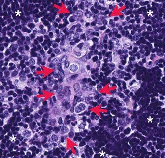
CELL DEATH PATHWAY PROVIDES
    POSSIBLE ANTI-CANCER TARGETS
    Modulating cell stress pathways that regulate levels of the pro-apoptotic protein BIK
    could help treat deadly triple-negative breast cancer

    When a cell is stressed, a complex net-          via a process known as ubiquitination.
    work of signals determine whether the            Chen and colleagues began searching
    cell will survive. Regulating these path-        for the protein involved in flagging it
    ways can offer anti-cancer strategies.           and identified Cul5-ASB11 as the ubiq-
    Fei-Yun Chen, Ruey-Hwa Chen, and                 uitin ligase that modifies BIK and tar-
    colleagues at the Academia Sinica in             gets it for destruction. The researchers
    Taiwan and National Taiwan University            found that, in response to endoplasmic
    reveal a new understanding of what               reticulum stress, ASB11 is transcrip-
    drives cell death under different stress         tionally activated by an effector of the
    conditions and how these pathways                stress-sensing protein IRE1α, promot-
    can be targeted to reduce tumor size in          ing BIK degradation and cell survival.
    triple-negative breast cancer (TNBC)             However, DNA damage–induced stress                      In a mouse model of triple-negative
    models.                                          caused the tumor suppressor p53 to                      breast cancer, overexpressing an
                                                                                                             active BIK mutant (VISA-BIKDD) in
                                                     repress ASB11 through IRE1α, stabiliz-
    Cell death regulation is crucial during                                                                  combination with an IRE1α inhibitor
                                                     ing BIK and promoting cell death.
                                                                                                             (STF) showed enhanced anti-tumor
    development and for maintaining
                                                                                                             effects compared to either treatment
    healthy tissue. When cell death is sup-          “BIK ubiquitination and degrada-
                                                                                                             by itself.
    pressed, cells can grow out of control,          tion are enhanced by ER stress and
                                                                                                             Credit: Chen et al., 2019
    causing tumors to form. Regulating cell          reduced by DNA damage, thereby
    death pathways can influence cancer              oppositely regulating cell life/death de-
    cells’ sensitivity to anti-tumor treat-          cisions in the two stressed conditions,”
    ments. A family of proteins known as             Ruey-Hwa Chen says. This presents                    inhibitor showed enhanced anti-tumor
    Bcl-2 determine whether cells commit             “an intriguing crosstalk between differ-             effects compared to either treatment
    to death, and one of the proteins in             ent cellular stress pathways.”                       by itself.
    the family is BIK. BIK is considered a
    pro-death protein; its presence signals          In TNBC, a highly aggressive disease                 “Targeting the BIK degradation path-
    a cell to die, but how BIK is regulated          with limited treatment options, the an-              way in combination with the adminis-
    and its physiological functions were             ti-tumor strategy of expressing an ac-               tration of an active BIK mutant could
    not well understood, nor was it known            tive BIK mutant is ineffective because               offer an effective anti-cancer strategy,”
    what stressors trigger BIK to promote            the mutant is prone to degradation,                  Chen says.
    cell death.                                      which helps keep TNBC cells alive. In
                                                     both cell lines and a mouse model of
    BIK protein has a short half-life and is         TNBC, overexpressing the active BIK
    thought to be flagged for destruction            mutant in combination with an IRE1α

    RESEARCHER DETAILS                                                     ORIGINAL PAPER
                                                                           Chen, F.Y., M.Y. Huang, Y.M. Lin , C.H. Ho , S.Y. Lin , H.Y. Chen , M.C. Hung, and R.H.
                                                                           Chen. 2019. BIK ubiquitination by the E3 ligase Cul5-ASB11 determines cell fate
                                                                           during cellular stress. J. Cell Biol. 218:3002–3018.
                                                                           https://doi.org/10.1083/jcb.201901156

    Fei-Yun Chen                     Ruey-Hwa Chen
    Postdoctoral Fellow              Associate Professor
    Academia Sinica, Taiwan          Academia Sinica, Taiwan
    National Taiwan University       National Taiwan University
                                     rhchen@gate.sinica.edu.tw
8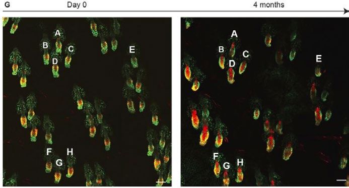
SRC TARGETS THE TUMOR SUPPRESSOR
DLC1
Interaction between DLC1 and tumor promoting kinase SRC reveals a possible new
treatment route for DLC1-positive tumors

Personalized treatments hold promise              To explore a possible mechanistic rela-
for cancers that evade generic chemo-             tionship between SRC, RhoA-GTP, and
therapies. Research by Brajendra Trip-            DLC1, they treated two DLC1-positive
athi, Douglas Lowy, and colleagues at             and two DLC1-negative non–small cell
the National Cancer Institute indicates           lung cancer lines with the SRC inhibitor
that it may be possible to target a sub-          Saracatinib, which reduced RhoA-GTP
set of cancers that express the tumor             in both DLC1-positive lines, but not in
suppressor and focal adhesion protein             the DLC1-negative lines. These results
DLC1, a component of focal adhesions              indicate SRC kinase can increase RhoA-
that can switch off the active GTPase             GTP in DLC1-positive cells, and the SRC
RhoA by acting as a GTPase-activat-               inhibitor can reverse the process.
ing protein (GAP). They establish, for                                                                    Colocalization between DLC1 (red)
the first time, a connection between              Using co-immunoprecipitation assays,
                                                                                                          and SRC (green) in a DLC1-positive
DLC1 and the tumorigenesis-promoting              they found that SRC and DLC1 interact                   non–small cell lung cancer cell line.
kinase SRC, revealing SRC inhibition              and that the localization of SRC to focal
                                                                                                          Credit: Tripathi et al., 2019
can be an effective part of the tumor             adhesions depends on the presence of
treatment in a DLC1-positive cancer               DLC1 protein. Their results show that
model and that reactivation of the tumor          SRC binds directly to DLC1 and phos-
suppressor can be a potent anti-tumor             phorylates it at residues Y451 and Y701.             activity,” Tripathi, Lowy, and colleagues
approach.                                         Phosphorylation analysis indicated that              said. A SRC inhibitor by itself reduced
                                                  DLC1 can also be phosphorylated on                   the size of DLC1-expressing tumors in
SRC is a kinase that controls many                S129 by the kinase ERK, which increas-               mice by 64%, and combining it with an
tumor-promoting processes, including              es both the binding of SRC to DLC1 and               inhibitor of AKT, which also phosphory-
regulating the cytoskeleton and a cell’s          SRC-dependent phosphorylation of                     lates and attenuates the tumor suppres-
ability to move and attach, by changing           DLC1. Phosphorylation of DLC1 by SRC                 sor activity of DLC1, reduced tumor size
the expression of oncogenic and tumor             attenuates DLC1’s Rho-GAP activity, the              by 89%.
suppressor genes. SRC also regulates              researchers discovered. Cells with DLC1
the RhoA GTPase, a protein that is fre-           mutations that are deficient for SRC                 “One possible way to increase the
quently activated in advanced cancer.             phosphorylation lacked well-formed                   proportion of tumors for which the
While surveying protein expression in             stress fibers, like those found in cells             therapeutic targeting of DLC1 could be
cancer-derived cell lines, Tripathi, Lowy,        with an active DLC1.                                 clinically beneficial might be to use a
and colleagues found a strong and un-                                                                  suitable inhibitor to reverse an epigene-
expected correlation between the levels           “Given the reversibility of the SRC-de-              tic change that has resulted in reduced
of RhoA-GTP, total SRC protein, and               pendent DLC1 phosphorylation, we                     or silenced DLC1 expression... and to
SRC activity and an inverse correlation           evaluated whether SRC inhibitors                     combine this treatment with inhibition
with DLC1 protein levels.                         might have therapeutic efficacy in a                 of SRC and/or AKT kinase activities,”
                                                  DLC1-positive tumor that had high SRC                Tripathi, Lowy, and colleagues say.

RESEARCHER DETAILS                                                         ORIGINAL PAPER
                                                                           Tripathi, B.K., M.F. Anderman, X. Qian, M. Zhou, D. Wang, A.G. Papageorge, and D.R.
                                                                           Lowy. 2019. SRC and ERK cooperatively phosphorylate DLC1 and attenuate its Rho-
                                                                           GAP and tumor suppressor functions. J. Cell Biol. 218:3060–3076.
                                                                           https://doi.org/10.1083/jcb.201810098

Brajendra K. Tripathi             Douglas R. Lowy
Senior Scientist                  Chief, Laboratory of Cellular Oncology
Center for Cancer Research        Center for Cancer Research
National Cancer Institute         National Cancer Institute
tripathib@mail.nih.gov            lowyd@mail.nih.gov
                                                                                                                                                                 9
HAIR FOLLICLES RESTRICT CANCER
     GROWTH
     Normal skin corrals cancer-causing mutations and reveals how tissue can subvert
     tumorigenesis

     Normal aged skin contains cancer-caus-
     ing mutations, recent research has
     shown, but how these mutations are
     prevented from forming tumors was a
     mystery. Christiana M. Pineda, Valentina
     Greco, and colleagues at Yale University
     used unique live-tissue imaging to track
     mouse skin cells after inducing can-
     cer-causing mutations. They found that
     protection against skin cancer comes
     from a surprising place: hair follicles.

     About 30% of all cancers contain a Ras
     mutation, but these same mutations
     have been found in non-cancerous
     skin epithelia. Pineda and colleagues
     induced Ras mutations in mouse hair
     follicle cells along with a glowing red
     reporter to track the mutant stem cells            Two-photon images of the ear skin from the same mouse at day 0 and 4 months
     and their progeny. The cells persisted in          after Ras activation reveal normal follicular architecture despite the persistent pres-
     the epithelium, revealing that the body            ence of mutant cells (red).
     doesn’t just eliminate mutant cells.               Credit: Pineda et al., 2019

     Pineda and colleagues found that when
     they induced mutations in hair follicle                                                               “Our results indicate that the hair
                                                      induce some tumors, typically at sites
     stem cells, they outcompeted wild-type                                                                follicle has a unique ability to cope with
                                                      of high grooming or scratching. Imag-
     neighboring cells. They still responded                                                               Ras-activated cells. This organ is able
                                                      ing revealed that those tumors arose
     to normal tissue constraints, such as                                                                 to integrate the mutant epithelial cells
                                                      after an injury caused them to exit the
     resting phase cues, however. Even after                                                               while remaining clinically normal,” Pine-
                                                      follicular niche. To test whether injury
     a year, the transformed cells did not de-                                                             da says. There’s still much to learn about
                                                      can promote tumorigenesis within the
     velop into tumors. In contrast, targeting                                                             what’s going on in the skin that could be
                                                      follicle, they ablated hair follicle bulbs
     the Ras mutation to the upper non-cy-                                                                 applied to other cancers. “Manipulation
                                                      and the double mutant cells showed
     cling region of the skin epithelium led to                                                            of certain cell types or signaling path-
                                                      rapid, and normal, regeneration. “Once
     benign outgrowths.                                                                                    ways may enable and/or enhance the
                                                      out of the follicular niche, Hras mutant
                                                      cells can no longer be controlled and                ability of other epithelial tissues to also
     Introducing a second mutation that
                                                      contained through hair regeneration                  suppress oncogenic growth.”
     results in the loss of TGFβ signaling into
                                                      programs,” Pineda says.
     Ras-mutant hair follicle stem cells did

     RESEARCHER DETAILS                                                      ORIGINAL PAPER
                                                                             Pineda, C.M., D.G. Gonzalez, C. Matte-Martone , J. Boucher , E. Lathrop , S. Gallini ,
                                                                             N.R. Fons, T. Xin , K. Tai, E. Marsh , D. X. Nguyen , K.C. Suozzi , S. Beronja , and V.
                                                                             Greco. 2019. Hair follicle regeneration suppresses Ras-driven oncogenic growth. J.
                                                                             Cell Biol. 218:3212-3222.
                                                                             https://doi.org/10.1083/jcb.201907178

     Cristiana M. Pineda   Valentina Greco
     Graduate Student      Professor
     Yale University       Yale University
                           valentina.greco@yale.edu

10
PROTEIN INVOLVED IN CANCER
METASTASIS HIJACKS ANOTHER FOR
TRANSPORT
A critical protease for metastasis uses a protein involved in intracellular transport to
get to the cell surface
Metastasis is often a death sentence is localized in a unique organelle and
for cancer patients. When a cancer forms specific complexes with cognate
cell begins to invade surrounding SNAREs to ensure membrane fusion
tissue, it forms a protrusion called an specificity,” Miyagawa says. The team
invadopodium. Matrix metalloprotein- identified Bet1 as the SNARE that is
ases (MMPs) are critical regulators of required for extracellular matrix degra-
this process. Takuya Miyagawa, Kana dation. Bet1 was previously shown to be
Hasegawa, Yoko Aoki, Takuya Wata- involved in transport from the ER to the
nabe, Hiroki Inoue, and colleagues at Golgi apparatus. Bet1 (green) colocalizes with MT1-
Tokyo University of Pharmacy and Life MMP (red) in invasive breast cancer
They found Bet1 colocalized with the cells.
Sciences reveal a novel mechanism for
how one of those MMPs, membrane Golgi, but surprisingly also with MT1- Credit: Miyagawa et al., 2019
type 1–MMP (MT1-MMP), is delivered to MMP in late endosomes. In invasive
invadopodia. cells, the team found that Bet1 was
able to reach the cell membrane, but in Bet1 function for its own transport,”
Many MMPs involved in cancer cell non-invasive cells, it stayed in the Golgi. Miyagawa says. Also, the team showed
invasion are secreted, soluble enzymes, Bet1 appears to be involved in MT1- that in invasive cells Bet1 is localized in
but MT1-MMP is membrane bound. MMP delivery to the cell surface, as MT1-MMP–positive endosomes as well
MT1-MMP is synthesized in the endo- Bet1 knockdown decreased the amount as the Golgi apparatus, and it forms a
plasmic reticulum (ER) and transported of MT1-MMP that reached the mem- novel SNARE complex with syntaxin 4
in vesicles to invadopodia, which are brane, whereas Bet1 overexpression and endosomal SNAREs.
structures with the ability to degrade increased MT1-MMP delivery. Addi-
the extracellular matrix, but the molecu- tionally, in invasive cells, Bet1-GFP was “MT1-MMP changes the function of Bet1
lar mechanism underlying this intracel- found in areas associated with invado- for its efficient transport to invadopodia.
lular transport was not fully understood. podia maturation. Bet1 is critical for the formation of func-
tional invadopodia that degrade ECM,
One crucial family of proteins involved Together, the team’s data indicates that and therefore could be a novel target
in vesicle trafficking specificity are MT1-MMP diverts Bet1 from its function for diagnosis, treatment, and prognosis
SNAREs, which act like twist ties when in ER to Golgi transport, to promote prediction of the disease,” Miyagawa
they meet on opposing membranes, MT1-MMP trafficking to the cell surface, says.
driving the fusion of a vesicle with its including to invadopodia in invasive
destination membrane. “Each SNARE breast cancer cells. “MT1-MMP hijacks
RESEARCHER DETAILS ORIGINAL PAPER
Takuya Miyagawa Miyagawa, T., K. Hasegawa, Y. Aoki , T. Watanabe, Y. Otagiri , K. Arasaki, Y. Wakana, K.
Kana Hasegawa Asano, M. Tanaka, H. Yamaguchi , M. Tagaya, and H. Inoue. 2019. MT1-MMP recruits
Yoko Aoki the ER-Golgi SNARE Bet1 for efficient MT1-MMP transport to the plasma membrane.
Takuya Watanabe
J. Cell Biol. 218: 3355-3371.
Graduate students https://doi.org/10.1083/jcb.201808149
Tokyo University of Pharmacy and Life Sciences
Hiroki Inoue
Assistant Professor
Tokyo University of Pharmacy
and Life Sciences
hirokii@toyaku.ac.jp
11CANCER CELLS TURN TO CANNIBALISM
TO SURVIVE CHEMOTHERAPY
Senescent cancer cells can engulf and digest their neighbors, allowing them to stay
alive and initiate tumor relapse
Chemotherapy drugs like doxorubicin bicin or other chemotherapy agents,
kill cancer cells by damaging their DNA, breast cancer cells that become senes-
but cells that survive initial treatment cent often engulf neighboring cancer
can soon give rise to new tumors. This cells. The researchers observed this
is a particular problem in breast cancers surprising behavior not only in cancer
that retain a normal copy of the TP53 cells in vitro, but also in orthotopic
gene. Instead of dying in response to tumors growing in mice. Lung and bone
chemotherapy-induced DNA damage, cancer cells are also capable of en-
these cancer cells generally just stop gulfing their neighbors after becoming
proliferating and enter a dormant but senescent, the researchers discovered.
metabolically active state known as
senescence. In addition to surviving Tonnessen-Murray and colleagues
chemotherapy, these senescent cancer found that senescent cancer cells
cells produce large amounts of inflam- upregulate a group of genes that are in-
matory molecules and other factors that volved in phagocytosis and are normal-
A breast tumor formed in mice and
can promote tumor regrowth. Chemo- ly active in macrophages. After “eating”
treated with doxorubicin contains
therapy-treated breast cancer patients their neighbors, senescent cancer cells some cancer cells (red nuclei) that
with normal TP53 genes are therefore digest them by delivering them to lyso- have been engulfed by other cancer
prone to relapse and have poor survival somes, degradative organelles that are cells (green cell membrane).
rates. also highly active in senescent cells.
Credit: Tonnessen-Murray et al., 2019
“Understanding the properties of these Importantly, the researchers deter-
senescent cancer cells that allow their mined that this process helps senes-
survival after chemotherapy treatment cent cancer cells stay alive. Senescent “Inhibiting this process may provide
is extremely important,” says Crystal cancer cells that engulfed a neighboring new therapeutic opportunities, because
A. Tonnessen-Murray, a postdoctoral cell survived in culture for longer than we know that it is the breast cancer
research fellow in James G. Jackson’s senescent cancer cells that didn’t. The patients with tumors that undergo
laboratory at the Tulane University researchers suspect that consuming TP53-mediated senescence in response
School of Medicine. their neighbors may provide senescent to chemotherapy that have poor re-
cancer cells with the energy and mate- sponse and poor survival rates,” Jackson
Tonnessen-Murray and colleagues rials they need to survive and produce says.
found that, after exposure to doxoru- the factors that drive tumor relapse.
RESEARCHER DETAILS ORIGINAL PAPER
Tonnessen-Murray, C.A., W.D. Frey, S.G. Rao, A. Shahbandi, N.A. Ungerleider, J.O.
Olayiwola, L.B. Murray, B.T. Vinson, D.B. Chrisey, C.J. Lord, and J.G. Jackson. 2019.
Chemotherapy-induced senescent cancer cells engulf other cells to enhance their
survival. J. Cell Biol. 218:3827–3844.
Jackson (back left) and Tonnes- https://doi.org/10.1083/jcb.201904051
sen-Murray (back right) pictured
with co-authors Joy Olayiwola (front
left) and Sonia Rao (front right).
James G. Jackson Crystal A. Tonnessen-
Assistant professor Murray
Tulane School of Medicine Postdoctoral fellow
jjacks8@tulane.edu Tulane School of Medicine
12IMPORTIN-11 MEDIATES NUCLEAR
IMPORT OF βCATENIN
Targeting this transport step may block the growth of colorectal cancers caused by
mutations in APC
Around 80% of colorectal cancers are manner. Due to their high βcatenin
associated with mutations in the APC levels, most cells die in response to
gene that stabilize the transcription Caspase-9 induction. But cells will sur-
factor βcatenin and lead to the protein’s vive if they lack genes required to main-
accumulation in the cell nucleus, where tain βcatenin transcriptional activity.
it can activate numerous genes that “The DEADPOOL platform constitutes
drive cell proliferation and promote the a robust system to conduct genetic
growth and maintenance of colorectal suppressor screens for the identification
tumors. But how βcatenin enters the of genes involved in signaling systems,”
cell nucleus after its levels rise is poorly Mis says.
understood.
One of the top hits in a CRISPR-based
“Because the molecular mechanisms screen of the DEADPOOL cells was
underlying βcatenin nuclear transport IPO11, which encodes a protein called
remain unclear, we set out to identify Importin-11 that is known to be involved
genes required for continuous βcatenin in nuclear import. Angers and col-
activity in colorectal cancer cells har- leagues found that Importin-11 binds to
Compared with a control (top left),
boring APC mutations,” says Stephane βcatenin and escorts it into the nucleus
removal of βcatenin (top right) or
Angers, a professor in the Department of colorectal cancer cells with muta- Importin-11 (bottom left and bottom
of Pharmaceutical Sciences at the Uni- tions in APC. Removing Importin-11 from right) reduces the growth of colorec-
versity of Toronto’s Leslie Dan Faculty of these cells prevented βcatenin from tal cancer cells carrying a mutation
Pharmacy. entering the nucleus and activating its in APC.
target genes. Credit: Mis et al., 2019
Angers and colleagues, including first
author Monika Mis, developed a new The researchers discovered that Impor-
technique that allowed them to screen tin-11 levels are often elevated in human
the human genome for genes that sup- colorectal cancers. Moreover, remov- more about how Importin-11 transports
port βcatenin’s activity in APC-mutant ing Importin-11 inhibited the growth of βcatenin into the nucleus may help
colorectal cancer cells. The DEADPOOL tumor organoids formed by APC mutant researchers develop new therapies
technique uses a colon cancer cell line cancer cells isolated from patients. that block this process and reduce the
engineered to express an inducible growth of colorectal cancers caused by
form of the apoptosis-inducing protease “We conclude that Importin-11 is mutations in APC.
Caspase-9 in a βcatenin-dependent required for the growth of colorectal
cancer cells,” Angers says. Learning
RESEARCHER DETAILS ORIGINAL PAPER
Mis, M., S. O’Brien, Z. Steinhart, S. Lin, T. Hart, J. Moffat, and S. Angers. 2020. IPO11
mediates βcatenin nuclear import in a subset of colorectal cancers. J. Cell Biol.
219:27–39.
https://doi.org/10.1083/jcb.201903017
Monika Mis Stephane Angers
Graduate student Professor
University of Toronto University of Toronto
stephane.angers@utoronto.ca
13CHROMOSOMES ARE A BARRIER TO
NORMAL CELL DIVISION IN POLYPLOID
CELLS
Duplicated chromosome sets is common in cancer but the extra chromosomes act as a
barrier to typical “bipolar” cell division
Polyploidy, when an entire duplicated clusters at each side of the cell. But
chromosome set is maintained within a things get even more complicated when
cell, is common in human tumors. How there are extra chromosomes as well as
cells maintain this unwieldy extra DNA, extra centrosomes.
especially when it comes time to divide,
was not well understood. Alix Goupil, Goupil and colleagues focused on cells
Maddalena Nano, Renata Basto, and with polyploidy and extra centrosomes
colleagues at the Institut Curie in Paris, due to errors in cytokinesis. They in-
France studied how polyploid cells duced cytokinesis failure in Drosophila
divide and found that the chromosomes neural stem cells and found that the
act as a barrier to typical “bipolar” cell extra centrosomes clustered in more Computer simulations with variable
than two groups, while the chromo- amounts of DNA (red) show that
division, but microtubule stability pro-
somes adopted a multilobed arrange- increase in DNA promote multipolar
motes bipolar cell division.
spindle arrangements.
ment within a multipolar spindle. Most
Though polyploidy is associated with polyploid anaphases were multipolar Credit: Goupil et al., 2020
poor prognosis in cancer, it is also a and generated several nuclei at mitotic
programmed and regulated process exit. The extra chromosomes of poly-
that normally occurs in early mamma- ploid cells seemed to prevent spindle tion of a bipolar spindle, the researchers
lian development. Paradoxically, cells bipolarity. looked at ovarian cancer cells lacking
that acquire an extra set of chromo- the microtubule-depolymerizing kinesin
somes through normal regulation have Similarly, in an in silico program de- MCAK. “This microtubule stabilization
a hard time proliferating, but tumors signed to simulate cell division with resulted in a considerable improve-
can form when polyploid cells arise various numbers and arrangements of ment in spindle bipolarity, and a large
from errors in cell division. In a normal centrosomes and DNA, bipolar spindle majority of polyploid cells divided in a
“bipolar” arrangement before cell divi- assembly was inhibited by the presence bipolar manner,” says Goupil. “In light
sion, two centrosomes line up and serve of extra DNA. In an ovarian cancer cell of our findings and knowing that whole
as microtubule-organizing centers at line with polyploidy induced by cytoki- genome duplications are frequent in
either side of the cell. The microtubules netic failure, the researchers confirmed cancer, it is possible that clonal expan-
then attach to the chromosome’s kine- that chromosomes acted as a barrier to sion of polyploid cancer cells is favored
tochores, pull the chromosomes apart, spindle pole coalescence. in conditions of increased microtubule
and the cell splits in two, in a process stability.”
known as cytokinesis. Cells with more Because their simulations also showed
than two centrosomes can still form a that stabilizing microtubules improved
bipolar spindle by forming centrosome centrosome clustering and the forma-
RESEARCHER DETAILS ORIGINAL PAPER
Goupil, A., M. Nano, G. Letort, S. Gemble, F. Edwards, O. Goundiam, D. Gogendeau,
C. Pennetier, and R. Basto. 2020. Chromosomes function as a barrier to mitotic
spindle bipolarity in polyploid cells. J. Cell Biol. 219:1824–1838.
https://doi.org/10.1083/jcb.201908006
Alix Goupil Maddalena Nano Renata Basto
PhD Student Postdoctoral Fellow Team Leader
Institut Curie, Paris, France University of California, Institut Curie
Santa Barbara renata.basto@curie.fr
mnano@ucsb.edu
14WHY SUBMIT
TO JCB?
AN EDITORIAL PROCESS GUIDED
BY YOUR COMMUNITY 97% OF INVITED REVISIONS
ARE ACCEPTED
At Journal of Cell Biology, all editorial decisions on research
manuscripts are made through collaborative consultation between
professional scientific editors and the academic editorial board.
INITIAL DECISION IN 5 DAYS
Submission or transfer Academic and scientific
editors review
27% PAPERS ACCEPTED AFTER
ONE ROUND OF REVIEW
If appropriate, sent to Academic and scientific Informed decisions
Peer Review editors review comments sent to authors
TIME IN PEER REVIEW: 31 DAYS
Revised manuscript Academic and scientific 97% of invited
submission and rereview editors review revisions accepted*
FINAL DECISIONS ON
TRANSFER MANUSCRIPTS**:
2 WEEKS
Published online 1-2 days after Circulated via alerts and social media
author proofs returned to readers around the world
*Median 2019
**Data on manuscripts transferred with previous reviewer
comments.
Format Neutral Transfer Policy Fair and Fast Open Access Options
We welcome submissions that include We limit rounds of revision, Our options include
You may submit your
reviewer comments from another journal. and we strive to provide Immediate Open
papers in ANY format.
You may also request manuscript transfer clear, detailed decisions that Access (CC-BY) and open
between Rockefeller University Press illustrate what is expected in access 6 months after
journals, and we can confidentially send the revisions. Articles appear publication (CC-BY-NC-SA).
reviewer reports and identities to another online in one to two days after
journal beyond RUP. author proofs are returned.Executive Director
Susan King
Assistant Director, Finance
Laura Bisberg
Director of Editorial Development
Teodoro Pulvirenti
Director of Publishing Technologies
Robert J. O'Donnell
Director of Communications and Marketing
Rory Williams
Executive Assistant to Executive Director;
Office Administrator
Demantie (Sati) Motieram
Financial Analyst
Sarah S. Kraft
Marketing Associate
Laraine Karl
Senior Science Writer
Ben Short
Journal of Cell Biology (ISSN 0021-9525) is published monthly by
Rockefeller University Press, 950 Third Avenue, New York, NY 10022.
Periodical postage paid at New York, NY and additional mailing offices.
2020 Subscription Rates
Institutional Rates
Tier 1 Tier 2 Tier 3 Tier 4
Online $2,590 $3,290 $4,040 $5,160
Print + Online $5,810 $6,480 $7,040 $8,990
For more information, please contact our subscription office.
Phone: +1 860-350-0041
Fax: +1 860-350-0039
email: subs@rockefeller.org
Advertising Requests
Phone: 201-767-4170
email: rupads@rockefeller.edu
Permission Requests
email: permissions@rockefeller.edu
Media Requests
email: news@rockefeller.edu
Postmaster
Send address changes to Journal of Cell Biology, Subscription Office, The
Bleachery, 143 West Street, New Milford, CT 06776.
16Interface with a World of Discovery
BioLegend provides a total workflow solution supporting immunology research with expertise
in a wide range of applications and platforms.
For nearly two decades, BioLegend has been a trusted partner With thousands of off-the-shelf reagents and countless
with researchers across the world, providing the applications customization options, we are equipped to help you
that have helped yield over 70,000 peer-reviewed publications. characterize immune cells, discover cellular functions,
and investigate new mechanisms of action.
Learn more at: biolegend.com/en-us/immunobiology
BioLegend is ISO 13485:2016 Certified
Toll-Free Tel (US & Canada): 1.877.BIOLEGEND (246.5343)
Tel: 858.768.5800
biolegend.com
08-0084-06
World-Class Quality | Superior Customer Support | Outstanding ValueYou can also read