Disruption of CK2 in Embryonic Neural Stem Cells Compromises Proliferation and Oligodendrogenesis in the Mouse Telencephalon䌤
←
→
Page content transcription
If your browser does not render page correctly, please read the page content below
MOLECULAR AND CELLULAR BIOLOGY, June 2010, p. 2737–2749 Vol. 30, No. 11
0270-7306/10/$12.00 doi:10.1128/MCB.01566-09
Copyright © 2010, American Society for Microbiology. All Rights Reserved.
Disruption of CK2 in Embryonic Neural Stem Cells Compromises
Proliferation and Oligodendrogenesis in the Mouse Telencephalon䌤†
Emmanuelle Huillard,1 Léa Ziercher,2,3,4 Olivier Blond,2,3,4 Michael Wong,1
Jean-Christophe Deloulme,5 Serhiy Souchelnytskyi,6 Jacques Baudier,2,3,4
Claude Cochet,2,3,4 and Thierry Buchou2,3,4*
Department of Pediatrics and Eli and Edythe Broad Institute for Stem Cell Research and Regeneration Medicine, University of
California, San Francisco, 513 Parnassus Avenue, San Francisco, California 941431; INSERM, U873, F-38054 Grenoble,
France2; CEA, iRTSV/LTS, F-38054 Grenoble, France3; Université Joseph Fourier, Grenoble, France4; INSERM, U836,
Institut des Neurosciences de Grenoble, F-38042 Grenoble, France5; and Karolinska Biomics Center,
Department of Oncology-Pathology, Karolinska Institutet, Stockholm, Sweden6
Downloaded from http://mcb.asm.org/ on May 15, 2021 by guest
Received 7 December 2009/Returned for modification 23 January 2010/Accepted 21 March 2010
Genetic programs that govern neural stem/progenitor cell (NSC) proliferation and differentiation are
dependent on extracellular cues and a network of transcription factors, which can be regulated posttransla-
tionally by phosphorylation. However, little is known about the kinase-dependent pathways regulating NSC
maintenance and oligodendrocyte development. We used a conditional knockout approach to target the murine
regulatory subunit (beta) of protein kinase casein kinase 2 (CK2) in embryonic neural progenitors. Loss of
CK2 leads to defects in proliferation and differentiation of embryonic NSCs. We establish CK2 as a key
positive regulator for the development of oligodendrocyte precursor cells (OPCs), both in vivo and in vitro. We
show that CK2 directly interacts with the basic helix-loop-helix (bHLH) transcription factor Olig2, a critical
modulator of OPC development, and activates the CK2-dependent phosphorylation of its serine-threonine-rich
(STR) domain. Finally, we reveal that the CK2-targeted STR domain is required for the oligodendroglial
function of Olig2. These findings suggest that CK2 may control oligodendrogenesis, in part, by regulating the
activity of the lineage-specific transcription factor Olig2. Thus, CK2 appears to play an essential and
uncompensated role in central nervous system development.
Casein kinase 2 (CK2) is a conserved serine/threonine ki- cell cycle progression in cultured mammalian cells (42). Fi-
nase with more than 300 substrates, mostly proteins related to nally, in a genetic screen for mutations affecting the central
transcription-directed signaling (27). CK2 is a heterotet- brain of Drosophila, a hypomorphic allele of D. melanogaster
rameric holoenzyme formed by two catalytic subunits, ␣ and CK2 has been isolated, and that study suggested a role for
␣⬘, that associate with a dimeric building block of regulatory  CK2 in cell proliferation or cell survival during brain devel-
subunits (␣22). CK2 modulates the substrate specificity of opment (15).
the CK2 enzymatic activity, and its architecture is consistent In the developing mouse brain, neural stem/progenitor cells
with its role as a docking partner for other interacting proteins (NSCs), which reside in the ventricular zone (VZ), give rise to
(4). We previously demonstrated that disruption of CK2 func- neurons and glial cells (astrocytes and oligodendrocytes) (23).
tion in mice results in postimplantation lethality. Moreover, Chronologically, neurons are generated first, and glial cells are
many attempts to generate CK2⫺/⫺ embryonic stem (ES) generated subsequent to neurogenesis. Oligodendrocytes orig-
cells failed, suggesting an essential role of mammalian CK2 inate from ventral neural progenitors in the VZ, and once
for ES cell viability (2). committed, they migrate laterally and dorsally as parenchymal
The function of CK2 for cell cycle progression has been precursors (oligodendrocyte precursor cells [OPCs]) to popu-
investigated, but its precise role is largely unknown. The cell late the entire embryonic telencephalon, where they terminally
proliferation function of CK2 was first characterized in hu- differentiate (reviewed in reference 29). Distinct genetic pro-
man fibroblasts, by use of antisense oligodeoxynucleotides and
grams using combinations of transcription factors govern mul-
microinjection of specific antibodies (19, 26). Recently, a ge-
tiple spatiotemporal origins of oligodendroglial specification
nome-wide survey of protein kinases required for cell progres-
(reviewed in reference 28). The basic helix-loop-helix (bHLH)
sion into cultured Drosophila melanogaster S2 cells by double-
transcription factor Olig2 plays an essential role in the embry-
stranded RNA demonstrated that CK2 is required for
onic specification of neural progenitors into the oligodendro-
centrosomal normality (1). In the same vein, downregulation
cyte lineage, and Olig2 gene function is absolutely required for
of CK2 by small interfering RNA (siRNA) results in delayed
OPC development (17, 21, 36, 43). Accordingly, Olig2 expres-
sion is detected first in ventral neural progenitors, precedes
* Corresponding author. Mailing address: INSERM, U873, CEA, OPC-specific expression markers, and is maintained at all
iRTSV/LTS, 17 Av. des Martyrs, F-38054 Grenoble, France. Phone: stages of oligodendrocyte development (29). It has been re-
33-438784046. Fax: 33-438785058. E-mail: thierry.buchou@cea.fr.
䌤 ported that the oligodendroglial function of Olig2 can be mod-
Published ahead of print on 5 April 2010.
† The authors have paid a fee to allow immediate free access to ulated by protein kinase AKT-mediated phosphorylation of its
this article. N-terminal domain (31). Interestingly, neural bHLH transcrip-
2737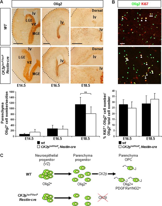
2738 HUILLARD ET AL. MOL. CELL. BIOL.
tion factors have been described as being phosphorylated and benzidine staining system secondary antibodies. Cells were stained with Hoechst
regulated by the CK2 holoenzyme (9, 39). In the mouse brain, dye 33342 (2 g/ml) to visualize nuclei. Images were acquired using a Zeiss
fluorescent microscope (Axiovert 200 M) with 16⫻ and 40⫻ objectives. Images
CK2 subunits (␣, ␣⬘, and ) are expressed in all neural cells (A. were combined for figures by using Adobe Photoshop 8.0. In situ hybridization
Kolding and B. Boldyreff, unpublished data) in which the using antisense, digoxigenin-labeled Pdgfra riboprobe was performed as de-
CK2-to-CK2␣-plus-CK2␣⬘ ratio is largely elevated compared scribed previously (20).
to that in other organs (41), suggesting a higher-threshold Neurosphere culture. E18.5 forebrains from diverse CK2 genetic back-
grounds were subjected to trypsin digestion for 15 min at 37°C. Tissues were
requirement in the amount of the holoenzyme in the central
mechanically dissociated into single-cell suspensions, filtered through a 70-m
nervous system (CNS). All together, these observations suggest cell strainer, and further treated with 10 g DNase I (Roche) for 10 min at room
that CK2 may modulate neural homeostasis. temperature. Cells from individual embryos, 2 ⫻ 106 cells/10-cm-diameter un-
In this study, we have used Cre/loxP-mediated recombina- coated dish, were cultured for primary neurosphere assays in Dulbecco’s modi-
tion to generate mice with a CK2 null allele in NSCs of the fied Eagle’s medium (DMEM)-F12 medium with B27 complement (D/F-B27)
containing 10 ng/ml epidermal growth factor (EGF; PeproTech) and 10 ng/ml
developing brain. Disruption of CK2 results in inhibition of fibroblast growth factor-2 (FGF; gift from G. Bouche, Toulouse, France). Num-
NSC proliferation and in a severe deficiency for progenitors to bers of generated primary spheres were counted after 4 days of in vitro culture
specify OPCs. In addition, we identified, in vitro, the serine- (4DIV). To assess for proliferation, 7DIV primary spheres were mechanically
Downloaded from http://mcb.asm.org/ on May 15, 2021 by guest
threonine-rich (STR) domain of Olig2 as a strict CK2-depen- dissociated and individual viable cells were counted. These cells (6 ⫻ 105 cells/
dent, CK2-targeted substrate. We also show that the STR 10-cm-diameter uncoated dish) were also used to assess for self-renewal in
secondary neurosphere assays. Olig neurospheres were generated from neural
domain of Olig2 is a modulator of its oligodendroglial activity stem/progenitor cells (NSCs) isolated from ganglionic eminences of Olig2⫹/⫺ or
when tested in neurosphere assays, suggesting that CK2 could Olig2⫺/⫺ E14.5 embryos (18). For rescue experiments, Olig2⫺/⫺ NSCs were
mediate its effect via Olig2 oligodendroglial activity. Hence, subsequently infected with HA-tagged Olig2-encoding viruses (see below) and
these findings provide insight into the genetic basis underlying selected with blasticidin (2 g/ml) 48 h after infection. Expression of HA-Olig2
proteins was confirmed by immunocytochemistry. For NSC differentiation assays,
neural progenitor proliferation and oligodendrocyte develop-
4DIV neurosphere cultures were plated on poly-L-lysine-coated glass coverslips
ment. (at a density of ⬃50 neurospheres/cm2) and shifted into D/F-B27 medium in the
presence of 3.5% fetal bovine serum (FBS; Biowest) (differentiating medium)
(11). 3DIV after plating, differentiated cultures were fixed with the PFA solution
MATERIALS AND METHODS and processed for immunocytochemistry. Additional primary antibodies were
Mouse breeding. Mice were of a C57BL/6 background. Nestin-cre transgenic mice mouse anti--tubulin III (1/500; Babco), mouse anti-O4 (1/10; gift from C. Soula,
(37) kept as heterozygotes were bred with CK2loxP/loxP mice (2). Double-heterozy- Toulouse, France), rat anti-Pdgf receptor a chain (1/100; BD PharMingen), and
gous mice (CK2loxP/wt; Nestin-cre) were bred back to CK2loxP/loxP mice to produce rat anti-HA (clone 3F10, 1/500; Roche).
the genotypes used in this study, (i) CK2loxP/wt, (ii) CK2loxP/loxP, (iii) CK2loxP/wt, Embryonic stem cell culture. CK2loxP/wt and CK2loxP/⫺ ES cell lines cul-
Nestin-cre, and (iv) mutant CK2loxP/loxP, Nestin-cre (referred to here as CK2⫺). tured in the presence of 103 units/ml leukemia inhibitory factor (LIF; Chemicon)
CK2loxP/wt, CK2loxP/loxP, and CK2loxP/wt, Nestin-cre embryonic day 18.5 (E18.5) and Cre–pMSCV-puro viral supernatant have been described previously (2). The
embryo phenotypes were identical and are referred to here as wild type (WT). PCR product corresponding to HA-tagged CK2wt coding regions was amplified
Live-born mice, postnatal day 0 (P0) pups, and E18.5 embryos were genotyped by using oligonucleotide DNA primers (5⬘ primer A [5⬘-CGGAATTCGCCGCCA
tail DNA PCR (2). Animal treatment was performed in accordance with the ethics CCATGTATCCTTATGATGTTCCTGATTATGCTATGAGCAGCTCCGAG
committee (ComEth) of Grenoble, France. GAGGTG-3⬘] and 3⬘ primer B [5⬘-GAAGATCTTCAGCGGATGGTCTTCAC
Western blot analysis. Olfactory bulb, cerebellum, and meningeal cells were G-3⬘]) and cloned into EcoRI/BglII pMSCV-neo viral vector sites (Clontech).
removed from dissected brains. Forebrain protein extracts were obtained after CK2 viral supernatants were generated by transfection of BOSC23 cells. Viral
homogenization and sonication in 25 mM Tris (pH 8.5)–1 mM dithiothreitol HA–CK2–pMSCV-neo supernatants were first used to infect 2.5 ⫻ 104/cm2
(DTT)–200 mM NaCl–5 mM EDTA. Cells were lysed in 50 mM Tris (pH 7.5)–5 CK2loxP/⫺ ES cells. To avoid chimera colonies, one day after infection, ES cells
mM EDTA–500 mM NaCl–1% Triton X-100. Extractions were done with pro- were subjected to trypsin treatment and individual cells were plated. Neomycin
tease and phosphatase inhibitor cocktails (Sigma). Samples (40 g) were ana- (Neo) selection (350 g/ml) was started 48 h after infection, and Neor clones
lyzed by 12% SDS-PAGE and processed for Western blot analysis. Antibodies were expanded and screened for expression of exogenous the HA-CK2wt pro-
used were rabbit anti-CK2 (1/500, directed against the C terminus of CK2), tein by Western blot analysis using mouse anti-HA antibody. Selected Neor
rabbit anti-CK2␣ (1/1,000; Calbiochem), mouse anti--tubulin class III isoform clones were further infected with Cre–pMSCV-puro supernatant. (CK2loxP/wt
(1/1,000; Chemicon), rabbit anti-poly(ADP-ribose) polymerase (anti-PARP) an- ES cells were used as positive controls.) One day later, individualized ES cells
tibody (1/1,000; Cell Signaling), and mouse antihemagglutinin (anti-HA) anti- were plated, and puromycin (puro) selection (1.5 g/ml) was started 48 h postin-
body (clone 12CA5, 1/1,000; Roche). fection for 6DIV or 9DIV. Individual clones were expanded and analyzed by
Histological, immunochemical, and in situ hybridization analyses. Embryos PCR analysis and Southern blot analysis for the absence of the CK2loxP allele
were perfused intracardially with 4% paraformaldehyde (PFA). Dissected brains (2) and by Western blot analysis for the absence of the endogenous wild-type
were postfixed overnight at 4°C and processed for paraffin or OCT embedding. CK2 protein. Neor puro-resistant CK2⫺/⫺ ES cells expressing only the exog-
Paraffin samples were cut at 5 m and used for hematoxylin and eosin (H&E) or enous HA-CK2wt protein were seeded in the absence of LIF (3 ⫻ 106 cells/10-
for immunochemical analysis. In order to detect proliferating cells in S phase cm-diameter uncoated plate/10 ml) to form floating embryoid bodies (EBs). In
(bromodeoxyuridine [BrdU] labeling), a 1-h short pulse was performed as de- order to stimulate neural differentiation, 5 M retinoic acid was added after
scribed previously (2). Paraffin-embedded sections were incubated with an anti- 4DIV of EB cultures and left for additional 4 days. EBs were further dissociated
BrdU rat monoclonal antibody (1/75; Harlan, Indianapolis, IN). Mitotic cell with trypsin, and individual cells (3 ⫻ 106 cells/10-cm-diameter uncoated plate/10
activities were detected with a rabbit antibody, anti-Ser10 phospho-H3 (PH3, ml) were used in neurosphere assays.
1/2,000; Upstate). Frozen sections (16 m) were used for immunostaining. Cryo- CK2 activity. Crude embryonic E18.5 forebrain extracts (adjusted to 5 mg/ml
sections were incubated in a blocking solution (phosphate-buffered saline [PBS]– of protein concentration) were assayed for CK2-dependent CK2 kinase activity
0.2% Triton X-100–5% normal goat serum) for 20 min and with a mouse IgG at 22°C for 10 min using a 3 mM concentration of the CK2-specific synthetic
blocking reagent (Vector) for 1 h. The primary antibodies used were mouse peptide RRREEETEEE (13) and [␥-32P]ATP. One unit of CK2 activity is
anti-RC2 (1/10; Hybridoma Bank, University of Iowa), rat anti-GFAP (1/500; defined as the amount of activity necessary to transfer 1 fmol of phosphate per
U.S. Biological), rabbit anti-NG2 antibody (1/100; Chemicon), rat anti-PECAM minute into the synthetic peptide. Chromatographic fractions (5 l) were assayed
antibody (1/2; gift from A. Vecchi, Milan, Italy), rabbit anti-Olig2 antibody for 3 min with a 150 M concentration of the CK2 consensus synthetic peptide
(1/20,000; gift from C. Stiles, Boston, MA), and mouse anti-Ki67 (clone MM1, RRREDEESDDEE (32) or with a purified fusion protein fragment consisting of
1/100; Novocastra). Primary antibodies were diluted in PBS–1% normal goat glutathione S-transferase (GST) and Olig2 amino acids 1 to 177 [Olig2(1-177)].
serum, followed by appropriate cyanin-3 dye-conjugated, cyanin-2 dye-conju- The PCR product corresponding to the mouse Olig2(1-177) coding region was
gated (1/500; Jackson ImmunoResearch Laboratories), Alexa488 dye-conjugated amplified from the Olig2 CMV2 template (34) by using the Expand high-fidelity
(1/500; Molecular Probe), or horseradish peroxidase (HRP) 3⬘,3⬘-diamino- PCR system (Roche) with oligonucleotide DNA primers (5⬘ primer C [5⬘-TTG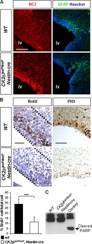
VOL. 30, 2010 CK2 AND THE CENTRAL NERVOUS SYSTEM 2739
TABLE 1. Genotypes of live-born mice and embryos from equivalent CK2 subunit (0 to 230 ng). Phosphorylated proteins were analyzed
CK2loxP/wt, Nestin-cre ⫻ CK2loxP/loxP crossingsa by SDS electrophoresis and autoradiography.
Phosphopeptide mapping and automated Edman degradation. The phosphor-
No. with genotype: ylated GST-Olig2(1-177) fusion protein was excised from the nitrocellulose
Offspring or CK2loxP/wt, CK2loxP/loxP, membrane and digested in situ with trypsin (modified sequencing grade; Pro-
embryo CK2loxP/wt CK2loxP/loxP mega). Two-dimensional phosphopeptide mapping was done using thin-layer
Nestin-cre Nestin-cre
(WT) (WT)
(WT) (CK2⫺) electrophoresis (HTLE-7000; CBS Scientific). First-dimension electrophoresis
was performed in pH 1.9 buffer (formic acid, glacial acetic acid, and water at
Live-born mice 22 18 15 0 50:156:1,794, vol/vol/vol) for 30 min at 2,000 V, and chromatography in the
P0 3 5 4 5 second dimension was performed in isobutyric acid–n-butyl alcohol–pyridine–
E14.5/E16.5/E18.5 142 146 148 154 glacial acetic acid–water (1,250:38:96:58:558, vol/vol/vol/vol/vol). After exposure,
a phosphopeptides were eluted from the plates in pH 1.9 buffer and lyophilized;
For details, see Materials and Methods.
aliquots of the samples were coupled to Sequelon-AA membrane (Millipore
Corp.) by use of carbodiimide coupling and subjected to automated Edman
degradation by using an Applied Biosystems sequencer (model 477A). Released
AATTCATATGGACTCGGACGCCAGCCT-3⬘] and 3⬘ primer D [5⬘-AACTC phenylthiohydantoin derivates from each cycle were spotted onto thin-layer
GAGTCAGTAGATCTCGCTCACCAGTC-3⬘]) and subcloned into EcoRI/ chromatography plates. The radioactivity in each spot was revealed by exposure
Downloaded from http://mcb.asm.org/ on May 15, 2021 by guest
XhoI pGEX4T2 vector sites (Amersham Biosciences) in order to generate the on a FujiX Bio-imager.
GST-Olig2(1-177) fusion protein. Purified GST-Olig2 proteins (20 g) were GST pull-down assay. Additional PCR products corresponding to coding
incubated at 22°C for 0 to 30 min with 25 M [␥-32P]ATP and 12 mM MgCl2 in regions for mouse Olig2 N-terminal amino acids 1 to 108 [N-ter(1-108)] and
the presence of 60 to 240 ng of CK2␣ subunit with increasing amounts of Olig2 bHLH amino acids 109 to 177 [bHLH(109-177)] were additionally ampli-
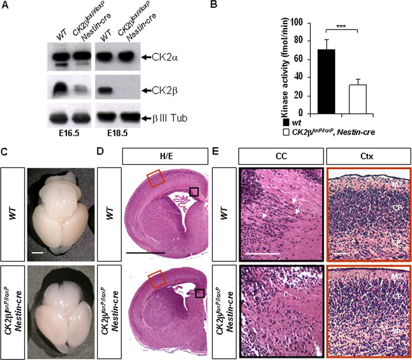
FIG. 1. CK2 loss in the central nervous system impairs telencephalon development. (A) Western blot analysis of CK2 and CK2␣ expressions
in E16.5 and E18.5 forebrain extracts. A specific neuronal (class III -tubulin [ III Tub]) antibody was used to control protein normalization.
(B) CK2-dependent CK2 activity in E18.5 CK2loxP/loxP, Nestin-cre forebrain extracts was significantly reduced (***, P ⫽ 2.7 ⫻ 10⫺9 [3 extracts
in quadruplicate per genotype]) compared to that in wild-type controls. (C) Brain morphology of E18.5 CK2loxP/loxP, Nestin-cre embryos compared
to that of the wild type. (D) Representative H&E staining of E18.5 anterior forebrains. Black and red boxed areas are shown at higher
magnification in panel E. (E) At the level of the corpus callosum (CC; black boxes), linear arrays of cells (arrows) detected in wild-type embryos
were absent in the mutants. In contrast, laminar patterns appeared similar in the cortex (Ctx; red boxes). MZ, mantle zone; CP, cortical plate zone;
SP, subplate zone. Scale bars, 1 mm (C and D) and 100 m (E).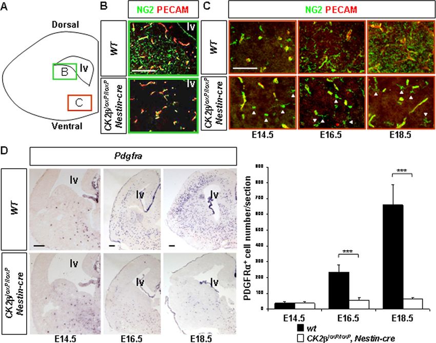
2740 HUILLARD ET AL. MOL. CELL. BIOL.
fied and subcloned into the pGEX4T2 vector as described above using 5⬘ primer
C with 3⬘ primer E (5⬘-AACTCGAGTCACTGCTGCAGCTCGGGCTCAG-3⬘)
and using 5⬘ primer F 5⬘-TTGAATTCATATGCTGCGCCTGAAGATCAACA
GCCGCGA-3⬘ with 3⬘ primer D, respectively. Interaction assays were performed
using prebound GST or GST-Olig2 glutathione Sepharose beads (Amersham
Biosciences) equilibrated in PBS–0.05% Tween 20–1 mg/ml bovine serum albu-
min (BSA) and incubated in the presence of [35S]methionine-labeled CK2 (2 ⫻
105 cpm) at 22°C for 90 min. Bound CK2 subunits were analyzed by SDS
electrophoresis and autoradiography.
Construction of HA-tagged mouse Olig2 vectors. A PCR product correspond-
ing to the HA-tagged mouse Olig2(1-177) coding region was amplified using 5⬘
primer G (5⬘-TTGAATTCTGGGCCATGTACCCATACGATGTTCCAGATT
ACGCTATGGACTCGGACGCCAGCCT-3⬘) and 3⬘ primer H (5⬘-GAAGAT
CTCGCTCACCAGTCGCT-3⬘) and subcloned into EcoRI/BglII sites of the
Olig2 CMV2 vector (34) to generate HA-tagged full-length mouse Olig2wt(1-
323). The CMV2 vector with HA-Olig2 STR amino acids 77 to 94 (STR77-94)
deleted (HA-Olig2⌬STR) was generated by site directed mutagenesis using sense
Downloaded from http://mcb.asm.org/ on May 15, 2021 by guest
primer I (5⬘-TGGGCGGCGGTGGCTTCAAGAAGAAAGACAAGAAGCAG
AT-3⬘) and antisense primer J (5⬘-ATCTGCTTCTTGTCTTTCTTCTTGAAG
CCACCGCCGCCCA-3⬘). Construction of pWZL-BLAST viral vectors encod-
ing HA-Olig2wt and HA-Olig2⌬STR proteins were generated after subcloning
EcoRI/XhoI PCR fragments obtained after amplification with respective CMV2
templates by using a GC-rich DNA polymerase (Invitrogen) with 5⬘ primer G
and 3⬘ primer K (5⬘-AACTCGAGTCACTTGGCGTCGGAGGTGAG-3⬘).
Cos7 cell transfection and coimmunoprecipitation assay. For coimmunopre-
cipitations, a Flag-tagged CK2 construct was produced after PCR amplification
using 5⬘ primer L (5⬘-TTGGATCCGCCGCCACCATGGACTACAAGGACG
ACGACGACAAGATGAGCAGCTCCGAGG-3⬘) and 3⬘ primer M (5⬘-GAAG
ATCTTCAGCGGATGGTCTTCACG-3⬘) and cloned into BamHI/BglII pSG5
vector sites. The Flag-CK2 PSG5 and the HA-tagged full-length mouse
Olig2wt(1-323) CMV2 constructs were transiently Lipofectamine-transfected
into Cos7 cells cultured in DMEM–10% fetal calf serum (FCS). Transfected cells
were lysed in radioimmunoprecipitation assay (RIPA) buffer (48 h posttransfec-
tion) in the presence of protease inhibitors, and for each extract, protein expres-
sion levels were analyzed by Western blot analysis. Five micrograms of a mono-
clonal antibody against the Flag epitope (Sigma) were added in precleared
lysates for 1 h at 4°C. Next, 50 l of RIPA-equilibrated “mouse true blot” beads
(eBioscience) was added and incubated under constant rotation for 1 h. After the
beads were washed with RIPA buffer, the precipitated proteins were analyzed by
SDS electrophoresis and Western blotting.
Statistical measures. For kinase activities, three forebrain-derived extracts per
genotype were tested in quadruplicate. For telencephalon cell counts, at least
three embryos per genotype were used for each day postcoitum (dpc) indicated.
Cells were counted on three or more nonadjacent sections per telencephalon.
For in vitro CK2 neurosphere assays, sphere and cell counts were performed
from three or six embryo-derived cultures per genotype. For in vitro neurosphere
differentiation assays, cell counts were performed from four or more embryo-
derived cultures per genotype with six glass coverslips per culture (⬃50 neuro-
spheres per coverslip). To evaluate the proliferation rate of neural stem cell-
derived embryonic stem cells, cell counts were performed from seven
differentiation cultures. Data are presented as averages ⫾ standard errors. Sta-
tistical comparisons were established using Student’s t test.
RESULTS
CK2 ablation in the central nervous system causes telen-
cephalon defects during late development. To gain insight into
functions of CK2 in the developing central nervous system
(CNS), we ablated CK2 in embryonic neural stem/progenitor
FIG. 2. CK2 positively controls forebrain NSC proliferation but cells (NSCs), since loss of CK2 is lethal to mouse embryos (2).
did not interfere with NSC specification. (A) Analysis of RC2 (red) We crossed CK2loxP/loxP mice with hemizygous Nestin-cre trans-
and GFAP (green) expressions by immunohistochemistry (IHC) in genic mice (37) to generate conditional mutant CK2loxP/loxP,
E18.5 wild-type and CK2loxP/loxP, Nestin-cre telencephalons. Cell nu-
clei were stained with Hoechst dye 33342 (blue). (B) There was a
significant reduction (***, P ⫽ 2.7 ⫻ 10⫺12 [3 embryos per genotype
and 3 nonadjacent sections per ventricular zone {VZ}]) of BrdU⫹
S-phase NSCs in CK2loxP/loxP, Nestin-cre VZs (outlined with a dotted
line) of lateral ventricles compared to the number in wild-type con- control represents the staurosporine-treated (1 M, 3 h) NIH 3T3
trols, as well as a near absence of PH3-positive mitotic cells. positive extract; cleaved PARP (arrow) serves as a marker of cells
(C) Caspase-3-dependent apoptosis analysis in E18.5 forebrains. West- undergoing caspase-3-dependent apoptosis. Scale bars, 100 m (A)
ern blot analysis of cleaved PARP in E18.5 forebrain extracts. The and 50 m (B).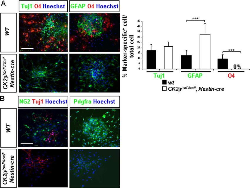
VOL. 30, 2010 CK2 AND THE CENTRAL NERVOUS SYSTEM 2741
Downloaded from http://mcb.asm.org/ on May 15, 2021 by guest
FIG. 3. Defective OPC development in CK2loxP/loxP, Nestin-cre telencephalons. (A) Schematic of a coronal telencephalon section. The green
and red boxes indicate the regions analyzed for the detection of parenchymal OPCs in panel B and of ventral OPCs in panel C, respectively.
(B) Analysis of NG2 (green) and PECAM (red) expressions by immunohistochemistry (IHC) in E18.5 wild-type and CK2loxP/loxP, Nestin-cre
telencephalons. Note the normal appearance of multiprocessed NG2⫹ parenchymal OPCs in the wild type and their absence in CK2loxP/loxP,
Nestin-cre embryos. The pattern of NG2 staining in vessel pericytes parallels PECAM endothelial cell labeling (yellow) and was unaffected.
(C) NG2 and PECAM expression analysis (IHC) revealed the presence of ventral NG2⫹ OPCs (arrowheads) in CK2loxP/loxP, Nestin-cre fetal
(E14.5) and late embryonic (E16.5 to E18.5) telencephalons. (D) In situ hybridization (ISH) analysis of Pdgfra expression in fetal (E14.5) and late
embryonic (E16.5 to E18.5) telencephalons. There was a significant reduction (***, P values of 2.5 ⫻ 10⫺10 at E16.5 and 1.0 ⫻ 10⫺6 at E18.5 [4
embryos per dpc and per genotype and 3 nonadjacent sections per telencephalon]) of Pdgfra⫹ OPCs in CK2loxP/loxP, Nestin-cre telencephalons
compared to the number in wild-type controls. lv, lateral ventricle. Scale bars, 100 m (B and C) and 200 m (D).
Nestin-cre mice (referred to here as CK2⫺). CK2⫺ mutant pups cells present in dissected forebrains, in which Cre recombinase is
were born with a Mendelian ratio (Table 1) but did not feed and not active (10). We conclude that expression of CK2 and, thus,
died shortly after birth. Therefore, the phenotype of embryos was the activity of CK2-dependent phosphorylation are effectively
analyzed. As expected, the CK2 protein was absent in CK2⫺ impaired in CK2⫺ E18.5 forebrains.
mutant E18.5 forebrain extracts (Fig. 1A). However, the CK2 To investigate the effects of loss of the CK2 regulatory
protein was still detected in CK2⫺ mutant E16.5 extracts, al- subunit on the development of the central nervous system,
though Cre-mediated recombination occurs at E9.5 to E10.5 (10). we analyzed the morphologies of E18.5 mutant brains.
This observation is in agreement with the stability of the CK2 Gross morphologies of wild-type and CK2⫺ E18.5 brains
protein integrated in the structure of tetrameric CK2 holoen- appeared similar (Fig. 1C). However, further histological
zymes (22) and could explain the late CK2⫺ mutant phenotype. analyses showed defects in CK2⫺ telencephalons: an ab-
Importantly, the expression of the CK2␣ catalytic subunit was sence at the corpus callosum (CC) level of cells that formed
unchanged upon CK2 ablation. We also measured the CK2- linear, pearls-on-a-string arrays characteristic of oligoden-
dependent CK2 activity in E18.5 forebrain extracts by using phos- droglial cells (Fig. 1E) (33) and an absence of the emerging
phorylation of the CK2-specific synthetic peptide RRREE hippocampal dentate gyrus (data not shown). In contrast,
ETEE (13). In line with ablation of CK2 in CK2⫺ mutant laminar patterns and cellular density were not affected in
forebrains, CK2-dependent CK2 activity was significantly re- the cortex (Ctx; Fig. 1D and E), suggesting that cortical
duced (⬃60%) in CK2⫺ mutant extracts compared to wild-type neurogenesis occurred normally in CK2⫺ mutants. Thus,
controls (Fig. 1B). The remaining activity in mutant extracts could CK2 loss in embryonic NSCs leads to morphology defects
be related to holoenzyme molecules derived from blood vessel during late development of the brain.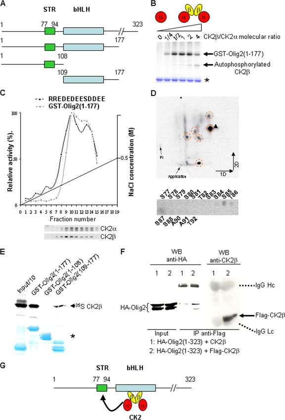
2742 HUILLARD ET AL. MOL. CELL. BIOL.
FIG. 4. Olig2⫹ progenitor analysis in fetal and late embryonic telencephalons. (A) IHC analysis of Olig2 expression in embryonic telenceph-
Downloaded from http://mcb.asm.org/ on May 15, 2021 by guest
alons. Parenchymal Olig2⫹ progenitors were detected in CK2loxP/loxP, Nestin-cre E14.5, E16.5, and E18.5 telencephalons. lv, lateral ventricle; vz,
ventricular zone; MGE, medial ganglionic eminence; LGE, lateral ganglionic eminence. There was a small decrease (**, P ⫽ 3.4 ⫻ 10⫺3 [4 embryos
per dpc and per genotype and 3 nonadjacent sections per telencephalon]) in parenchymal Olig2⫹ progenitor numbers in CK2loxP/loxP, Nestin-cre
E18.5 telencephalons compared to the number in wild-type controls. (B) Proliferation analysis of parenchymal Olig2⫹ progenitors/OPCs by double
labeling for Ki67 (red) and Olig2 (green). The ratios (percentages) of proliferating cells (Ki67⫹) coexpressing Olig2 in the parenchyma (yellow and
arrowheads) to total parenchymal Olig2⫹ cells were not different between CK2loxP/loxP, Nestin-cre and wild-type E16.5 and E18.5 telencephalons
(3 embryos per dpc and per genotype and 4 nonadjacent sections per telencephalon). (C) CK2 regulates OPC specification of parenchymal Olig2⫹
progenitors. Scale bars, 100 m (A) and 50 m (B).
CK2 disruption compromises forebrain stem/progenitor (P0) transition, radial glia gives rise to adult NSCs and pro-
cell proliferation. To understand the causes of the defects gressively loose RC2 expression in favor of GFAP (23). In
observed in mutant brains, we studied NSCs. In mice, NSCs in CK2-ablated E18.5 embryos, neither RC2⫹ radial glia nor
the VZs of developing brains possess radial glia morphology GFAP⫹ NSCs were substantially altered (Fig. 2A).
and express RC2. During the late embryonic (E18.5)/neonatal Previous results indicated that during mouse embryogenesis,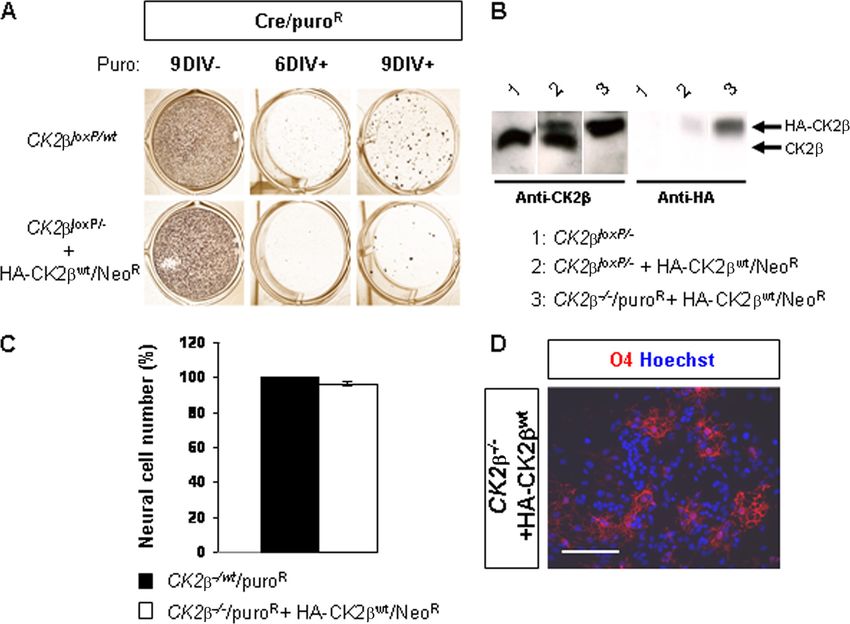
VOL. 30, 2010 CK2 AND THE CENTRAL NERVOUS SYSTEM 2743
CK2 is mainly involved in proliferation rather than in the
prevention of apoptosis (2). Therefore, we examined at E18.5
the incorporation of a short pulse of BrdU in the germinal
compartment of the telencephalon, the VZ of lateral walls (lw)
of lateral ventricles (lv). CK2⫺ VZs displayed a significant
reduction (⬃60%) of NSCs in S phase as determined by the
extent of BrdU-labeled nuclei compared to wild-type controls
(Fig. 2B). In addition, the BrdU⫹ mutant nuclei showed a
rounded morphology instead of an elongated morphology. To
further address the effects of CK2 loss on the cell cycle,
sections were stained with the mitosis marker phospho-histone
H3. Only few CK2⫺ NSCs were found phospho-histone H3
positive (Fig. 2B). It is possible that the reduced NSC number
represents a dysregulation of the cell cycle or an increase in cell
Downloaded from http://mcb.asm.org/ on May 15, 2021 by guest
death. We did not detect substantial apoptosis by terminal
deoxynucleotidyltransferase-mediated dUTP-biotin nick end
labeling (TUNEL) assay staining in CK2⫺ E18.5 forebrains
(data not shown). Accordingly, cleaved PARP, a hallmark of
caspase-3-dependent apoptosis in vivo, was undetectable in
CK2⫺ E18.5 forebrain protein extracts (Fig. 2C). Taken to-
gether, these results suggest that CK2 is important for the
proliferation, but not the genesis, of forebrain NSCs.
CK2 loss in NSCs impairs oligodendroglial differentiation
in developing telencephalons. Next, we examined whether
CK2 regulates oligodendrogenesis, as suggested by disorga-
nization of the corpus callosum of CK2⫺ brains (Fig. 1E). We
FIG. 5. CK2 regulates NSC proliferation in neurosphere cultures.
first analyzed the expression of the oligodendroglial precursor Primary neurospheres (N1; 4DIV and 7DIV) derived from E18.5 fore-
(OPC) marker NG2 (25) by immunohistochemistry (IHC). At brain NSCs are shown. Compared to wild-type controls, CK2loxP/loxP,
E18.5, parenchymal NG2⫹ cells with OPC morphology were Nestin-cre primary neurospheres in 7DIV cultures became smaller and
absent in CK2⫺ telencephalons, and only NG2⫹ pericytes did not generate secondary neurospheres (N2) after dissociation
associated with blood vessel were detected (Fig. 3B). However, (7DIV). The ratio (percentage) of generated primary neurospheres to
E18.5 forebrain-dissociated cells (6 primary neurosphere cultures per
in CK2⫺ brains, we were able to detect some NG2⫹ OPCs in genotype) in CK2loxP/loxP, Nestin-cre 4DIV cultures was not different
a ventral location (Fig. 3C, E18.5 panel). In the mouse telen- from that of wild-type controls. The number of cells obtained from
cephalon, early OPCs (E12.5 to E14.5) are of ventral origin dissociated CK2loxP/loxP, Nestin-cre primary neurospheres in 7DIV
(reviewed in references 28 and 29). Thus, it was possible that cultures was significantly reduced compared to that from wild-type
controls (***, P ⫽ 4.3 ⫻ 10⫺7 [3 primary neurosphere cultures per
the remaining E18.5 ventral OPCs in CK2⫺ brains were gen- genotype]). Scale bar, 100 m.
erated earlier during early embryogenesis, a period when the
CK2 protein is not completely ablated (Fig. 1A). Figure 3C
shows that early ventral NG2⫹ OPCs were generated in
CK2⫺ mutant E14.5 telencephalons, but in contrast to wild- both neural progenitors and OPCs (27, 28) and (ii) the de-
type controls, they did not expand during the late embryonic crease in the number of parenchymal Olig2⫹ cells is lower than
period (E16.5 to E18.5). Because NG2 cytoplasmic immuno- the decrease in the number of Pdgfra⫹ OPCs in CK2⫺ brains
staining is unsuitable for quantification, we analyzed the ex- (0% versus ⬃75% at E16.5 and ⬃30% versus ⬃90% at E18.5),
pression of another OPC marker, Pdgfra (12, 25), by in situ parenchymal Olig2⫹ cells in CK2⫺ brains may represent
hybridization. At E14.5, the numbers of Pdgfra⫹ OPCs in wild- stalled neural progenitors unable to progress along the oligo-
type and CK2⫺ brains were similar (Fig. 3D). However, there dendrocyte lineage. Alternatively, these Olig2⫹ cells may rep-
was a sharp decrease (⬃75%) in the number of Pdgfra⫹ OPCs resent abnormal glia. To investigate whether the decrease in
in E16.5 mutant telencephalons, despite incomplete inactiva- OPC numbers was due to defective proliferation of parenchy-
tion of the CK2 protein. By E18.5, this difference was more mal Olig2⫹ progenitors as described for NSCs (Fig. 2B), we
pronounced (⬃90%). determined the ratio of Ki67⫹ Olig2⫹ cells to total Olig2⫹ cells
We also investigated the expression of Olig2, a basic helix- in the parenchyma of wild-type and CK2⫺ telencephalons at
loop-helix (bHLH) transcription factor required for OPC de- E16.5 and E18.5 (Fig. 4B). Proliferation of parenchymal
velopment and maintained at all stages of oligodendrocyte Olig2⫹ cells was unchanged in CK2⫺ brains and is therefore
development (17, 21, 29, 36, 43). In contrast to the defect unlikely to account for OPC deficiency. Because parenchymal
observed for NG2⫹ and Pdgfra⫹ OPCs, analysis of Olig2 ex- Olig2⫹ progenitors originate from Olig2⫹ neuroepithelial cells
pression revealed identical numbers of parenchymal Olig2⫹ in the VZ (28, 29), the decrease in the number of Olig2⫹ cells
cells in CK2⫺ telencephalons and wild-type controls at E14.5 in the parenchyma of CK2⫺ brains may be a consequence of
and E16.5 (Fig. 4A). However, by E18.5, a small decrease a defective proliferation of Olig2⫹ cells in the VZ (Fig. 4C).
(⬃30%) in the number of parenchymal Olig2⫹ cells was ob- The discrepancy observed between the effects of CK2 disrup-
served in CK2⫺ telencephalons. Because (i) Olig2 identifies tion on the proliferation of NSCs in the VZ and that of Olig2⫹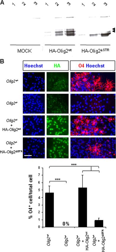
2744 HUILLARD ET AL. MOL. CELL. BIOL.
Downloaded from http://mcb.asm.org/ on May 15, 2021 by guest
FIG. 6. CK2 disruption impairs OPC specification in neurosphere cultures. (A) Primary neurospheres derived from E18.5 forebrains were
tested for their multipotency after differentiation. Cells were immunolabeled with the neuronal marker Tuj1 (green), the astrocytic marker GFAP
(green), and the oligodendroglial marker O4 (red). The ratio (percentage) of cells expressing lineage-specific markers to the total cell number
evaluated by Hoechst-stained nuclei (blue) showed an absence of O4⫹ cells (***, P ⫽ 1.3 ⫻ 10⫺8 [12 neurosphere cultures per genotype])
concomitant with a significant increase of GFAP⫹ astrocytes (***, P ⫽ 2.9 ⫻ 10⫺4) in CK2loxP/loxP, Nestin-cre cultures compared to the number
in wild-type controls. The percentages of Tuj1⫹ neurons did not differ between wild-type and mutant cultures. (B) CK2 loss altered the
specification of NG2⫹ (green) and Pdgfra⫹ (red) OPCs in differentiation cultures. Scale bars, 100 m.
progenitors in the parenchyma may reflect different mechanisms formed -tubulin III-positive neurons and GFAP⫹ astrocytes
involved in the control of the proliferation of progenitors down- but failed to generate O4⫹ differentiating OPCs (Fig. 6A). To
stream of the stem compartment. Collectively, these data sug- confirm whether this defect was the result of a failure of OPC
gest that CK2 loss leads to a defect in OPC specification of specification as observed in vivo, we examined NG2 and Pdgfra
Olig2⫹ progenitors during telencephalon development (Fig. 4C). expression in differentiated neurosphere cultures. In striking
CK2 is required for neural stem cell proliferation and contrast to control cultures, CK2⫺ neurospheres did not gen-
oligodendroglial differentiation in neurosphere culture. Em- erate any NG2⫹ or Pdgfra⫹ OPCs (Fig. 6B). To further ad-
bryonic NSCs located in the telencephalic VZ have been iden- dress the requirement of CK2 in NSC proliferation and in
tified by their ability in vitro to generate neurospheres in re- oligodendrogenesis, we expressed an HA-tagged CK2 protein
sponse to FGF2 and EGF stimulation (38). In the absence of in CK2⫺/⫺ NSC-derived embryonic stem (ES) cells (Fig. 7)
growth factors, neurosphere cells differentiate into neurons, (2). Such CK2⫺/⫺ NSCs expressing HA-CK2 were able to
astrocytes, and oligodendrocytes. To confirm whether CK2 proliferate and to generate O4⫹ differentiating OPCs, exclud-
loss affects NSC maintenance as shown in vivo, we cultured ing a genetic side effect associated with targeting of the CK2
neurospheres derived from CK2⫺ E18.5 forebrains and in- locus. Taken together, these results suggest that the cell-au-
duced their differentiation. There was no significant difference tonomous defect(s) caused by CK2 disruption results in de-
in the numbers of primary neurospheres generated from E18.5 ficient NSC proliferation and OPC specification.
wild-type and CK2⫺ forebrains in response to FGF2-EGF after Olig2 is an interacting partner of CK2 subunit and a strict
4 days of in vitro culture (4DIV) (Fig. 5). As observed in 7DIV CK2-dependent substrate in vitro. Given the essential roles
cultures, wild-type cells formed large neurospheres, whereas of Olig2 and CK2 for OPC specification in fetal and late
CK2⫺ neurospheres did not expand. This was reflected by a embryonic development (17, 21, 36, 43; this study) and the
significant reduction (⬃95%) in the number of cells obtained regulation of the oligodendroglial activity of Olig2 by phosphor-
from dissociated CK2⫺ neurospheres. In addition, we were ylation (31), we investigated whether Olig2 may be a potential
unable to generate CK2⫺ secondary neurospheres, suggesting CK2 substrate. Amino acid sequence analysis of mouse Olig2
that the self-renewing potency of CK2⫺ NSCs was severely revealed that most putative CK2 phosphorylation sites (S/T;
compromised. After differentiation, CK2⫺ neurosphere cells E/D/S n ⫺ 1, n ⫹ 1, n ⫹ 2, and/or n ⫹ 3) are present within an
VOL. 30, 2010 CK2 AND THE CENTRAL NERVOUS SYSTEM 2745
Downloaded from http://mcb.asm.org/ on May 15, 2021 by guest
FIG. 7. Exogenous CK2 expression in NSC-derived CK2⫺/⫺ embryonic stem (ES) cells promotes proliferation and oligodendroglial differ-
entiation. (A) Clonal selection of CK2⫺/⫺, Cre–pMSCV-puro-cultured (Cre/puroR) ES cells expressing exogenous HA-CK2wt/Neor protein.
CK2⫺/wt, Cre/puroR ES cell clones were generated in parallel and served as positive controls. (B) Western blot analysis with anti-CK2 (left panel)
and anti-HA (right panel) antibodies of CK2loxP/⫺ ES cells, CK2loxP/⫺ and HA-CK2wt/Neor ES cells, and CK2⫺/⫺, Cre/puroR, and HA-
CK2wt/Neor ES cells. Note the complete absence of the endogenous CK2 protein in the CK2⫺/⫺ ES cell extract. (C) Proliferation analysis of
NSC-derived ES cells. The ratio (percentage) of the number of cells present in neurosphere-derived CK2⫺/⫺ ES cells expressing the exogenous
HA-CK2wt protein to the number of cells present in control neurosphere-derived CK2⫺/wt ES cells was unchanged when normalized to the
CK2⫺/wt ES cell line (7 differentiation cultures). (D) O4⫹ differentiating OPCs were identified from neurosphere-derived CK2⫺/⫺ ES cells
expressing the exogenous HA-CK2wt protein and allowed to differentiate. Scale bar, 100 m.
N-terminal fragment encompassing M1 to Y177. We designed a labeled phosphopeptides revealed multiple spots. A major spot
GST-Olig2(1-177) fusion protein that includes mouse Olig2 was suitable for Edman degradation. The released detectable
amino acids 1 to 177 (Fig. 8A) and determined the CK2- radioactivity was observed mainly after cycles 9 and 11. Only
dependent incorporation of [␥-32P]ATP into the purified frag- one Olig2(1-177) tryptic peptide had serine/threonine residues
ment. As shown in Fig. 8B, the Olig2(1-177) fragment was at positions 9 and 11, allowing the identification of major
directly phosphorylated by CK2 only in the presence of the phosphorylated sites at Ser85 and Ser87 in a serine-threonine-
regulatory subunit CK2. We further investigated CK2-de- rich domain (STR77-94, tryptic peptide 77SSSSSTSSSTSSAAT
pendent phosphorylation of Olig2 by analyzing CK2 and Olig2 SSTK95) highly conserved in the human sequence. It should be
phosphorylation activities in fractions obtained after ion-ex- noted that minor phosphorylation sites were also detected at
change chromatography of wild-type E18.5 forebrain extracts Ser84, Thr86, and Ser88. Thus, these results identify the Olig2
(Fig. 8C). Phosphorylation experiments and Western blot anal- STR sequence as an unconventional major domain of multiple
ysis revealed coelution of CK2 and Olig2-dependent kinase. acceptors for phosphorylation by the CK2 holoenzyme.
This coelution occurred in fractions containing the highest To investigate the regulation of Olig2 phosphorylation by
CK2/CK2␣ subunits ratio. Moreover, no coelution of CK2 CK2, we tested the capacity of CK2 to interact with Olig2.
and Olig2-dependent kinase was observed when the chroma- GST pull-down assays showed that CK2 interacts with the
tography was performed with CK2⫺ forebrain extracts (data bHLH fragment of Olig2 [Olig2(109-177)] but fails to interact
not shown). In addition, Olig2 phosphorylation was abolished with the fragment containing the STR domain [Olig2(1-108)]
in the presence of 10 M TBB, a specific in vitro CK2 inhibitor (Fig. 8E). Surprisingly, this Olig2(1-108) fragment was not
(data not shown). Taken together, these results suggest that phosphorylated by the CK2 holoenzyme (data not shown).
the CK2 tetrameric holoenzyme (␣22) is an Olig2 kinase. Preincubation of CK2 with CK2␣ subunits which generate
To delineate which particular motif of the mouse Olig2(1- the tetrameric holoenzyme did not prevent its interaction with
177) fragment was involved in the phosphorylation by the CK2 the bHLH Olig2(109-177) fragment (data not shown). We
holoenzyme, we performed two-dimensional tryptic phos- confirmed that CK2 can interact with Olig2 by coimmuno-
phopeptide mapping (Fig. 8D). Analysis of the maps of 32P- precipitation of Flag-tagged CK2 with HA-tagged full-length2746 HUILLARD ET AL. MOL. CELL. BIOL.
Downloaded from http://mcb.asm.org/ on May 15, 2021 by guest
FIG. 8. CK2 positively modulates CK2-dependent phosphorylation of the Olig2(1-177) fragment in vitro. (A) Primary structure of mouse
Olig2 and schematic representation of the Olig2 fragments used in this study. Represented are the serine-threonine-rich domain sequence (STR,
amino acids 77 to 94), and the basic helix-loop-helix sequence (bHLH, amino acids 109 to 164). (B) Autoradiography of the purified recombinant
GST-Olig2(1-177) fusion fragment after in vitro phosphorylation by the CK2 catalytic subunit (␣) alone or in combination with increasing amounts
of the CK2 regulatory subunit (), which generate increased amounts of the holoenzyme (␣22). Note that CK2, an autosubstrate of the catalytic
␣ subunit, competed in the reaction for the CK2 phosphorylation of Olig2 when present in excess. Protein normalization was controlled by
Coomassie blue staining (*). (C) DEAE-Sephacel chromatography of crude wild-type E18.5 forebrain protein extracts. Each fraction was analyzed
for consensus relative CK2 activity with the RRREDEESDDEE synthetic substrate/peptide and for relative CK2-dependent Olig2(1-177) kinaseVOL. 30, 2010 CK2 AND THE CENTRAL NERVOUS SYSTEM 2747
mouse Olig2(1-323) after transient expression in Cos7 cells nuclear morphology of BrdU⫹ CK2⫺ NSCs in the ventricular
(Fig. 8F). Together, these data suggest that the bHLH domain zone (Fig. 2B) is reminiscent of a cell cycle arrest event and
of Olig2 is a docking site to promote the recruitment of CK2 suggests an interkinetic nuclear migration defect (40). During
via physical interaction with its regulatory CK2 subunit for G1, the nuclei of NSCs ascend to the basal side of the VZ,
efficient Olig2 phosphorylation on its STR sequence (Fig. 8G). where S phase is completed. Nuclei descend back toward the
The CK2-targeted STR domain is required for the oligoden- ventricular surface during G2, and this is where they stay dur-
droglial activity of Olig2 in neurosphere culture. We next ing mitosis. The present study suggests that CK2 may control
asked whether the CK2-phosphorylated STR domain is in- this process in the neural stem compartment, leading to a block
volved in mediating Olig2 biological function. Because of the in the G1/S phase in CK2⫺ NSCs. Therefore, CK2⫺ NSCs
many serine and threonine acceptor residues in the Olig2 STR would not proceed through the G2 phase. Consequently, it
that could potentially compensate for inactivation of the Ser85 would explain the near absence of PH3-positive mitotic CK2⫺
and Ser87 major phosphorylation sites, we generated an Olig2 NSCs in the VZ (Fig. 2B) and the normal proliferation of the
deletion mutant that lacks the entire STR77-94 domain (HA- more downstream population of Olig2⫹ parenchymal progen-
Olig2⌬STR). Transient transfection into Cos7 cells demon- itors in CK2⫺ embryos (Fig. 4B). Recently, it has been dem-
Downloaded from http://mcb.asm.org/ on May 15, 2021 by guest
strated identical levels of expression of HA-Olig2wt (amino onstrated that nuclear migration is governed by centrosomal
acids 1 to 323; full-length) and HA-Olig2⌬STR proteins (Fig. specific microtubule-regulating proteins (40) and reported that
9A). We infected Olig2⫺/⫺ neurosphere cultures (18) with ret- CK2 is required for centrosomal normality (1). However, a
roviruses encoding HA-Olig2wt or HA-Olig2⌬STR and tested strict CK2-dependent CK2 substrate(s) involved in NSC pro-
their potential to differentiate into the oligodendroglial lineage liferation has not been described.
(Fig. 9B). As expected, expression of HA-Olig2wt in Olig2⫺/⫺ We also show that CK2⫺ NSCs are specifically devoid of
NSCs restored the production of O4⫹ differentiating OPCs. In oligodendroglial potency. Our in vivo and in vitro studies dem-
contrast, Olig2⫺/⫺ NSCs transduced with HA-Olig2⌬STR sig- onstrate that depletion of the CK2 gene in embryonic neural
nificantly failed to differentiate into O4⫹ cells. These findings progenitors blocks the production of NG2⫹ and Pdgfra⫹
demonstrate that, in vitro, the CK2-targeted STR domain is OPCs. Several observations are in favor of a cell-intrinsic reg-
required for the oligodendroglial function of Olig2. ulation by CK2 in OPC specification per se. (i) In E16.5
CK2⫺ telencephalons, the number of parenchymal Olig2⫹
progenitors was normal, despite a strong defect in NG2⫹ and
DISCUSSION
Pdgfra⫹ OPCs, and by E18.5, whereas the number of Olig2⫹
Kinase-dependent pathways regulating NSC maintenance progenitors increased, the number of OPCs in CK2⫺ telen-
and fate specification are largely unknown. Here, we inacti- cephalons remained low compared to that in wild-type controls
vated the protein kinase CK2 regulatory subunit (CK2) gene (Fig. 3 and 4). (ii) Olig2⫹ cell proliferation was unchanged in
in CNS embryonic neural progenitors and showed that CK2 CK2⫺ brains (Fig. 4B). (iii) CK2⫺ neurosphere cultures
is an important modulator of telencephalon development. recapitulated the deficiency in OPC specification observed in
Conditional CK2 mutant mice display impaired NSC prolif- vivo (Fig. 6). (iv) CK2⫺ neurospheres cultured in the pres-
eration, as well as defective OPC specification. We show that ence of other factors (PDGF-AA and insulin-like growth fac-
CK2 interacts physically with the bHLH transcription factor tor I [IGF-I]) were unable to differentiate into oligodendro-
Olig2, an essential regulator of oligodendroglial differentia- cytes (data not shown), suggesting that a defect in a particular
tion, thereby allowing its serine-threonine-rich (STR) domain extracellular instruction for NSC specification to oligodendro-
to be phosphorylated by CK2. Finally, we show that the Olig2 genesis cannot account for OPC deficiency (3, 5, 8, 14, 35). (v)
STR domain mediates the oligodendroglial activity of Olig2. CK2 is required to promote oligodendrogenesis in NSC-de-
Our results suggest that CK2 could mediate OPC specifica- rived embryonic stem (ES) cells (Fig. 7). Thus, CK2-depen-
tion through the modulation of the oligodendroglial activity of dent specification of OPCs may correspond to a molecular
Olig2. mechanism(s) shared by the multiple independent molecular,
We present the first report of an in vivo role for CK2 in regional, and temporal origins of progenitors in the forebrain
NSC proliferation in mammals. Loss of CK2 results in re- (reviewed in reference 28) and controlled by CK2.
duced numbers of dividing cells in the embryonic germinal Genetic studies in mice demonstrated that during fetal and
zone, and our in vitro neurosphere assays show that CK2 is late embryogenesis, the bHLH transcription factor Olig2 is
required for NSC proliferation and self-renewal. The altered necessary for specification of progenitors into OPCs/oligoden-
activity. Activity profiles were normalized to the activity detected in the peak fractions. In parallel, a sample of each fraction was immunoblotted
to detect the presence of CK2␣ and CK2 proteins (lower panels). (D) Two-dimensional (2D) phosphopeptide mapping analysis of CK2-
dependent Olig2(1-177) phosphorylation. The major phosphopeptide (arrowhead; upper panel) was subjected to automated Edman degradation
and released derivates from each cycle were spotted onto plates and autoradiographed (lower panel). Deduced amino acids in the Olig2 sequence
are indicated above or below the spots. (E) Recombinant GST fusion proteins containing different Olig2 fragments (1 to 177, 1 to 108, and 109
to 177) were incubated with [35S]methionine-labeled CK2. The ability of the GST fusion proteins to pull down the CK2 subunit was detected
by autoradiography after electrophoresis. On the left, one-tenth of the input is shown. Loading controls were checked after Coomassie blue staining
of the gel (*). (F) Coimmunoprecipitation of Flag-CK2 and HA-Olig2(1-323) in Cos7 cells. Flag-CK2 was immunoprecipitated (IP) with
anti-Flag antibody, and the immunoprecipitated complexes were analyzed by Western blotting (WB) with anti-HA and anti-CK2 antibodies. Hc,
heavy chain; Lc, light chain. (G) Schematic representation of the suggested CK2-Olig2 interaction.2748 HUILLARD ET AL. MOL. CELL. BIOL.
drocytes (17, 21, 36, 43). Multiple regulations such as interac-
tions (NFIA/B, SCL, and ID4) and phosphorylation (Akt/pro-
tein kinase B [PKB]) that inhibit Olig2 function dictate a loss
into the oligodendroglial lineage (6, 24, 30, 31). The increase in
astrocytogenesis observed in CK2⫺ neurosphere cultures af-
ter differentiation (Fig. 6A) is compatible with a glial switch
following loss of the oligodendroglial function of Olig2 (6, 7,
21, 24, 30, 31, 36, 43). However, whether CK2 modulation of
astrocytic instruction is direct or a consequence of a regulation
of oligodendrocyte differentiation is unclear. We show that the
Olig2(1-177) fragment is a strict CK2-dependent CK2 sub-
strate, probably through a CK2-specific Olig2 recruitment by
direct interaction with the bHLH domain, and that CK2 is an
Olig2 kinase in forebrain extracts (Fig. 8). CK2-dependent
Downloaded from http://mcb.asm.org/ on May 15, 2021 by guest
major phosphorylation occurs in the STR domain. In neuro-
sphere assays, we demonstrated that the CK2-targeted STR
domain is required for the full oligodendroglial function of
Olig2 (Fig. 9). Although we cannot exclude phosphorylation(s)
of the STR domain by one or more other serine/threonine
kinases, our results do not definitively prove, but do suggest,
that CK2-dependent CK2 activity may play a role as a deter-
minant in the combinatorial code that trigger Olig2-dependent
oligodendrocyte lineage progression. Alternatively, we cannot
rule out other CK2-dependent transcription factor substrates
important for the oligodendroglial network which could act in
synergy inside the same genetic pathway.
During CNS development, Olig2 is sequentially required for
the generation of subsets of motor neurons (E9 to E10.5) and
OPCs (E12.5 to E14.5) (21, 36, 43). If the assumption for a
defect in Olig2 function in CK2⫺ progenitors is correct, then
it is intriguing that the process of neurogenesis is not affected.
However, in CK2 mutant brains, complete ablation of CK2
does not occur before E18.5, by which neurogenesis is largely
completed. Additional studies will be needed to address the
earlier role of CK2 in neuronal development.
The essential role of CK2 for cell viability initially detected
in ES cells (2) precluded analysis of later roles for CK2 during
development. Our conditional strategy revealed unexpected
roles for CK2 in the CNS. We demonstrated that CK2
represents a unique signaling component in NSC proliferation
and self-renewal and in the oligodendroglial lineage-restricted
pathway. If CK2-dependent CK2 activity is required for can-
FIG. 9. The Olig2 STR domain is required for NSC oligodendro- cer stem cell proliferation and self-renewal as it is for NSCs,
glial potency in neurosphere cultures. (A) Transient expression anal-
ysis of the ⌯⟨Olig2⌬STR mutant protein in Cos7 cells. Comparative
then inhibitors of interaction between the CK2 regulatory
pattern expressions of full-length (mouse; amino acids 1 to 323) HA- subunit and CK2 catalytic subunits (␣ and ␣⬘) could be useful
Olig2wt and HA-Olig2⌬STR in transiently transfected Cos7 cells. An- chemotherapeutic agents. Conversely, because differentiation
ti-HA Western blots revealed shifted-up bands within the HA-Olig2wt- block of OPCs has been demonstrated to cause neurodegen-
transfected protein extracts (arrowheads), and these were not detected erative diseases, such as chronic multiple sclerosis (16), acti-
in the HA-Olig2⌬STR version. This observation is reminiscent of the
STR sequence as phosphorylated sites. “MOCK” indicates a control vation of CK2-dependent CK2 activity may be useful in pro-
experiment performed with the empty vector. Amounts of protein are moting remyelination.
as follows: 1, 4 g; 2, 8 g; 3, 16 g. (B) Olig2⫺/⫺ NSCs were trans-
duced with HA-Olig2wt(1-323) and HA-Olig2⌬STR. NSC-derived neu-
rospheres were allowed to differentiate and stained with HA (green) to ACKNOWLEDGMENTS
detect exogenous Olig2 and with O4 (red) to assess for oligodendro-
glial potency. Olig2⫹/⫺ and Olig2⫺/⫺ assays are shown in parallel. The This work was supported by grants from the Institut National de la
ratio (percentage) of cells expressing the O4 oligodendroglial marker Santé et de la Recherche Médicale (INSERM), the Commissariat à
to the total cell number evaluated by Hoechst-stained nuclei (blue) l’Energie Atomique (CEA), the Ligue Nationale Contre le Cancer
showed a significant deficiency (***, P ⫽ 5.6 ⫻ 10⫺7 [4 neurosphere (Equipe Labelisée 2007), and the Institut National du Cancer (grant
cultures per genotype]) in the oligodendroglial activity of the HA- number 57).
Olig2⌬STR mutant compared to that of the HA-Olig2wt control. Scale We thank D. Rowitch for support, suggestions, and comments on
bars, 50 m. the manuscript regarding in vivo analysis. We also thank I. Marechal
and S. Bama for their efficiency with mouse breeding, C. Wernstedt forYou can also read