RESEARCH HANDBOOK 2020- 2021 - Boston University
←
→
Page content transcription
If your browser does not render page correctly, please read the page content below
RESEARCH
HANDBOOK 2020- 2021

1
Research is an integral component of Boston University Henry M. Goldman School of Dental Medicine (GSDM)’s
mission, goals, and objectives. The School’s mission statement begins: “The Boston University Henry M. Goldman
School of Dental Medicine will be the premier academic dental institution promoting excellence in dental education,
research, oral health care, and community service to improve the overall health of the global population”. In
addition, the mission states: ”We will shape the future of the profession through scholarship, creating and
disseminating new knowledge, developing and using innovative technologies and educational methodologies, and
by promoting critical thinking and lifelong learning.”

What is “dental research”?

Dental research involves the use of scientific analysis, observation, and experimentation to acquire new knowledge
in the field of dental medicine.

Deadline to apply

First-year research 2 x 3: Jan 4, 2021
IREC1: Feb 1, 2021
IREC2: ongoing during second-year
IREC3: ongoing during third-year

The benefits of research:

•   become trained in the design and execution of scientific studies;
•   enhance analytical thinking abilities;
•   bring breadth and depth to their dental education;
•   have a better understanding of innovative dental techniques, materials, and tools;
•   become more informed dental clinicians.
•   contribute to the dental literature by publishing the results; and
•   improve eligibility for postgraduate specialty training programs and academic appointments.

The research environment at GSDM:

•   Department of Molecular and Cell Biology, Evans 4, 72 East Concord Street
•   Department of Periodontology, 3rd Floor, 650 Albany St
•   Departments of Endodontics, General Dentistry, Oral and Maxillofacial Surgery, Orthodontics and Dentofacial
    Orthopedics, Pediatric Dentistry, 100 East Newton Street
•   Department of Health Policy and Health Services Research, 560 Harrison Avenue
•   Department of Restorative Sciences/Biomaterials, 72 East Concord St. R 520 and 650 Albany Street
•   Center for Exocrine Disorders, Center for Clinical Research, Center for Oral Diseases, Center for Research to
    Evaluate and Eliminate Dental Disparities, Pediatric Oral Healthcare Center and Center for Behavioral Science
    Research
•   Department of Translational Dental Medicine, 700 Albany St. W201
•   Other research sites: Any other research facility approved by the Pre-doctoral Research Committee. Rotation
    needs to be conducted under prior research mentor. (Early application is necessary to complete the process of
    executing an affiliation agreement between the outside institution and Boston University.)

2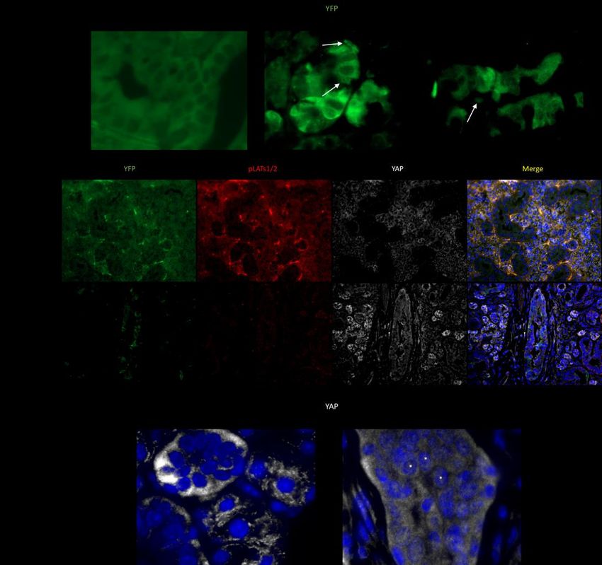
•
Research Faculty Mentors:

GENERAL DENTISTRY
   • Marianne Jurasic DMD, MPH, Clinical Associate Professor and Director of the Center for Clinical
     Research. Research Area: Public Health Utilizing Multiple Electronic Datasets to Answer Clinical and
     Epidemiological Questions
   •  Larry Dunham DMD, Clinical Assistant Professor and Director of Diversity & Multicultural Affairs.
     Research Area: Social, Economic and Cultural Influences on Global Health.
   • Garladinne Lakshmi BDS, MDS, Clinical Assistant Professor. Research area: Anatomic Variants on
     CBCT, Osteonecrosis of the Jaw, Multi-Modality Imaging Features of Maxillofacial Lesions
HEALTH POLICY & HEALTH SERVICES RESEARCH
   • Belinda Borrelli PhD, Professor and Director of Behavioral Science Research. Research Area: Motivating
     Health Behavior Change, mHealth and eHealth, Smoking Cessation and Public Health
   • Raul Garcia DMD, MMS, Professor and Chair. Research Area: Epidemiology
   • Michelle Henshaw DDS, MPH, Professor and Assistant Dean for Community Practice. Research Area:
     Public Health
   • Elizabeth Kaye MPH, PhD, Associate Professor. Research Area: Public Health
   • Astha Singhal | Profiles RNS BDS, MPH, PhD, Associate Professor, Director of Advanced Specialty
     Education Program in Dental Public Health. Research Area: Dental ER Care and Oral Health Disparities

3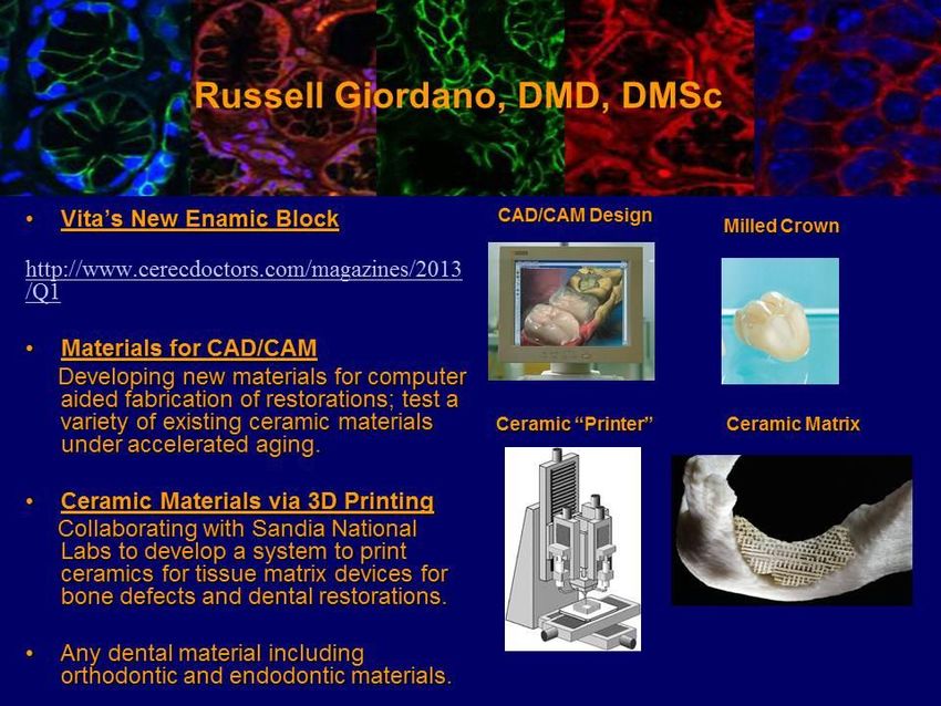
MOLECULAR & CELL BIOLOGY
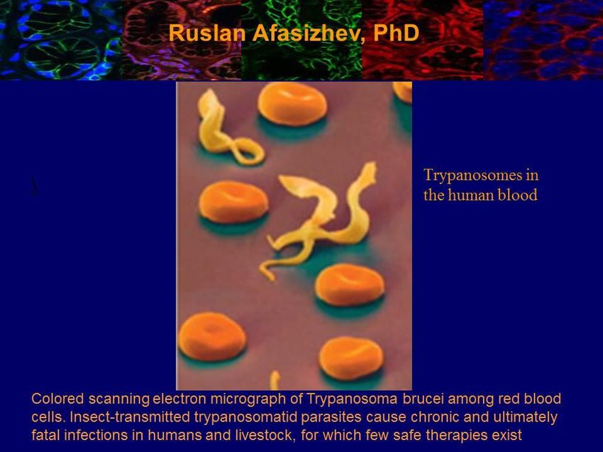
• Ruslan Afasizhev PhD, Professor. Research Area: Molecular Mechanisms of RNA Processing in
Trypanosomes
• Cataldo Leone DMD, DMSc, Professor and Associate Dean for Academic Affairs and Advanced Education
& International Programs. Research areas: Biochemistry/Periodontology
• David Levin PhD, Professor and Chair. Research Area: Biochemistry/Molecular Biology
• John Samuelson MD, PhD, Professor. Research Ares: Microbiology
• Makoto Senoo PhD, Associate Professor Research Area: Stem Cell Biology Using Molecular and Cellular
Analyses and Mouse Genetics
ORAL & MAXILLOFACIAL SURGERY
• Radhika Chigurupati DMD, MS, Associate Professor and Director of Research. Research Area: Global
Health, Early Diagnosis of Oral Cancer, Clinical Informatics
• Richard D’Innocenzo DMD, MD, Associate Clinical Professor. Research Area: Trauma, Fracture,
Maxillofacial Management and Anesthesia
• Pushkar Mehra BDS, DMD, Associate Professor and Chair. Research Area: Trauma, Fracture,
Maxillofacial Management and Orthognathic Surgery
• Vicki Noonan DMD, DMSc, Associate Professor and Director of the Clinical Oral & Maxillofacial
Pathology Practice. Research Area: Pathology/Oral Biology
• Andrew Salama DDS, MD, Assistant Professor. Research Area: EvaluatingTongue Motion and Speech
Following Reconstructive Surgery and Developing Novel Chemo-Preventive Medications for Oral Cancer
ORTHODONTICS & DENTOFACIAL ORTHOPEDICS
• Leslie Will DMD, CAGS, MSD, Professor and Chair. Research Area: Normal and Abnormal Growth,
Treatment Outcomes and Diagnostic Tools
ORTHOPEDIC SURGERY
• Louis Gerstenfeld PhD, Professor. Research Area: Cell Biology/Bone
PEDIATRIC DENTISTRY
• Athanasios Zavras DMD, DDS, MS, DrMedSc, Professor and Chair. Research Area: Epidemiology of Oral
Diseases, Health Services Research, Molecular Diagnostics and Pediatric Dentistry
PERIODONTOLOGY
• Serge Dibart DDS, DMD, Professor and Program Director. Research Area: Gingival Epithelial Cells
RESTORATIVE SCIENCES/BIOMATERIALS
• Alexander Bendayan DDS, CAGS, FICD, Clinical Associate Professor, ad Interim Chair & Associate Dean
for Digital Development and Innovation. Research Area: Biomaterials
• Laisheng Chou DMD, PhD, Professor. Research Area: Cell Biology/Oral Medicine
• Russell Giordano DMD, DMSc, Associate Professor. Research Area: Biomaterials
TRANSLATIONAL DENTAL MEDICINE
• Manish Bais, DVM, Ph.D, Research Associate Professor. Research Area: Oral Cancer/TMJ Osteoarthritis/
Bone Cartilage Regeneration
• Paola Divieti Pajevic MD, PhD, Associate Professor. Research Area: Bone Cell Biology and
Mechanotransduction.
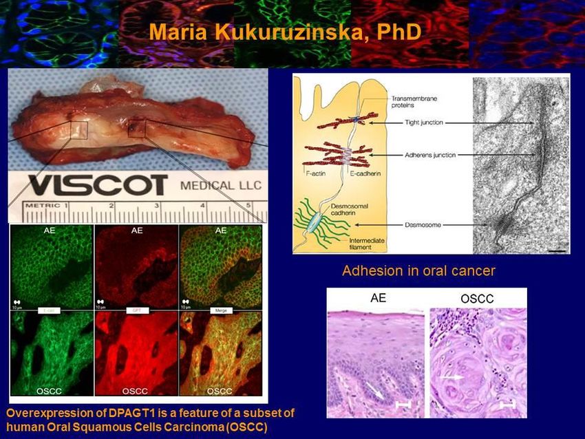
• Maria Kukuruzinska PhD, Professor and Associate Dean for Research. Research Area: Molecular and Cell
Biology/Development
• Philip Stashenko DMD, Ph.D., Associate Professor; Research Area: Oral Infection Diseases/Oral Cancer
• Philip Trackman PhD, Professor. Research Area: Cell Biology
4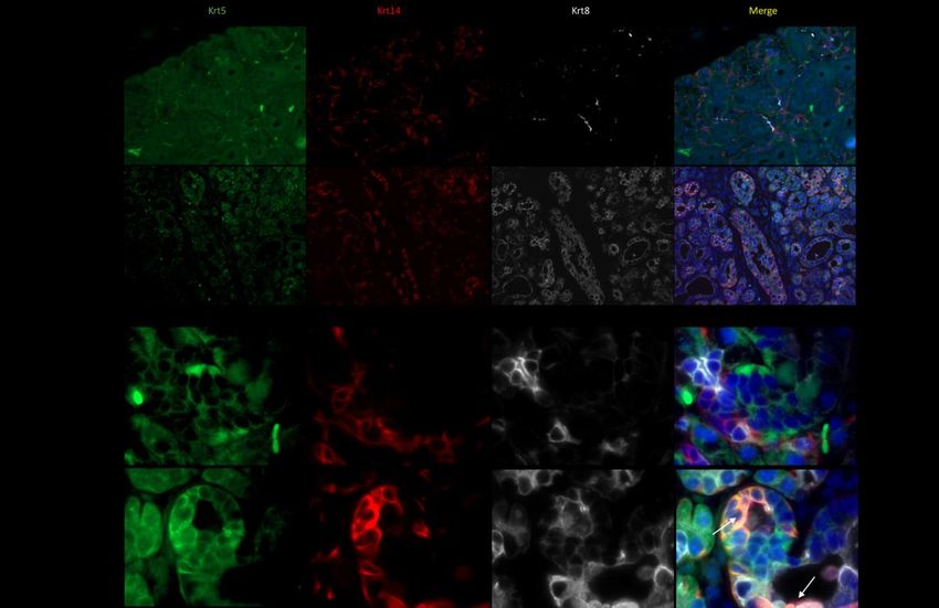
The following images represent selected research by GSDM faculty

5
6
7
Pre-doctoral Research Program (PRP)

The GSDM developed a highly successful Pre-doctoral Research Program for DMD students. The mission of the
Program is: 1) to shape the future of dental medicine and dental education through research; 2) to educate students
from diverse backgrounds about the importance of research in dental medicine; and 3) to mentor students to make
informed decisions about research career opportunities.
The PRP at the GSDM benefits individual students and the field of dental medicine. Through participation in
research students enhance their analytical thinking abilities, become trained in the design and execution of scientific
studies, gain a better understanding of innovative dental techniques, materials and tools, improve their eligibility for
postgraduate specialty training programs and academic appointments, become more informed dental clinicians, and
contribute to the dental literature by publishing their research findings.
The GSDM provides state-of-the-art research training resources. Students choose faculty mentors from 30 research
scientists involved in around 100 research projects that span broad areas of basic and applied biomedical sciences, as
well as clinical and public health research. In addition, to direct mentor-student interactions, student trainees are
expected to become important contributors to research teams and to participate in the full range of research-related
activities, including laboratory/team meetings and journal clubs. At the completion of research training, students are
expected to showcase their accomplishments at the School’s Science Day and they are encouraged to participate in
national and international scientific meetings in the areas of their research training. Information about the PRP and
the Student Research Group (SRG) can be obtained at
         http://www.bu.edu/dental/research/predoctoral
         http://www.bu.edu/dental-research/student-research/predoctoral-research/

Program Structure

Because of its unique curriculum, the GSDM offers formal research training for credit to students. Students who
maintain a 3.0 GPA or higher in their didactic and clinical courses are considered for research training. Students
selected by Committee can participate in the Program. The first-year training takes place following the completion
of the DMD didactic courses during the Apex rotation from May to July. The rotation is based on a five-day week as
follows:
     a. students dedicate two days for research training and three days for the Apex clinical assignment;
     b. students dedicate three days for research training (30 hours per week) and two days for the Apex clinical
         assignment under the Intensive Research Elective Course (IREC). Students are considered for the IREC1 if
         they have participated in research during the second semester of their dental education on a voluntary basis
         or if they have prior research experience;
     c. students can do research on a voluntary basis and are expected to spend no less than 10 hours per week in
         research training. Advanced Standing students can start research during the second semester of their dental
         education.

Prior to engaging in research training, the Pre-doctoral Research Office meets with the applicants to advise them of
their assignments and to inform them of the prerequisites to research training including CITI trainings in the
Protection of Human Subjects in Research and other regulatory requirements. The students are given a copy of the
Research Handbook that contains a detailed description of the program. During research rotations, student trainees

8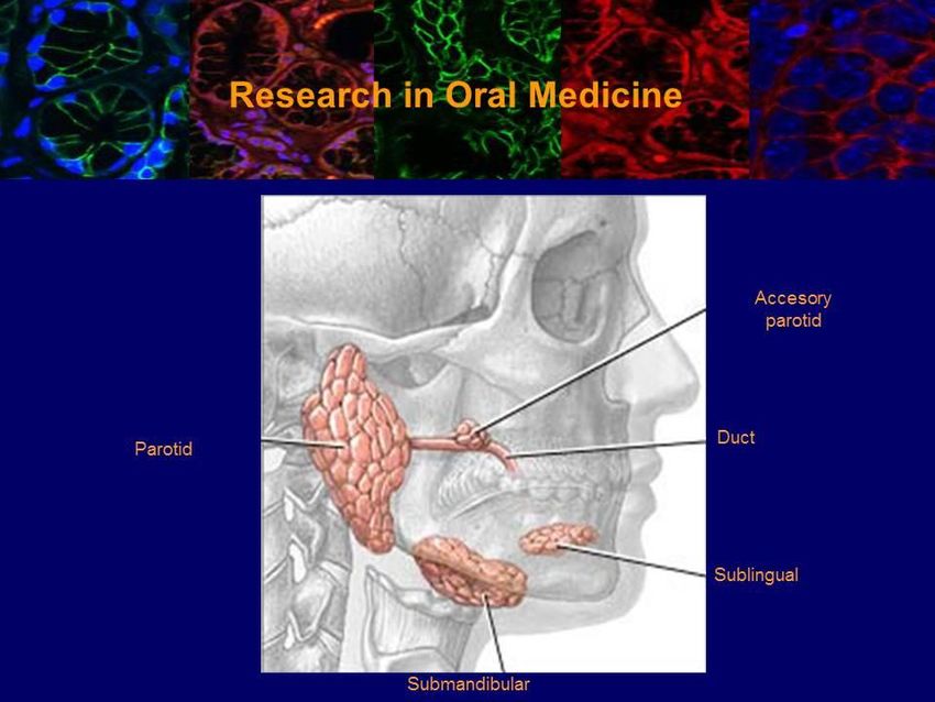
are expected to attend meetings with the Pre-doctoral Research Office and attend presentations on scientific writing
skills, presentation approaches and workshops on Communicating Science. Trainees are expected to attend seminars
relevant to their research organized by the GSDM, the School of Medicine and other research institutions in the
greater Boston area. In addition, students are required to participate in research competitions. Students are informed
of the option to do research rotations outside of Boston University that require the execution of an affiliation
agreement that governs the relationship between Boston University and the outside institution.

Student research training is overseen by the Pre-doctoral Research Committee PRC, a subset of the Research
Committee chaired by the Associate Dean for Research and Director of the PRP. The Research Committee is
composed of members of the GSDM biomedical science and clinical faculty, the Associate Dean for Academic
Affairs, and the Assistant Director of Pre-doctoral Research. The mission of the PRC is to guide and monitor
research activities among DMD students, evaluate the effectiveness of the PRP and make recommendations for
program improvements.

The Intensive Research Elective Course (IREC)

The goal of the IREC is to provide intensive and structured research experience throughout the dental school
curriculum for students who are interested in careers in oral health research.

The IREC objectives are: 1) to carry out well-defined research projects under the guidance of research mentors; 2) to
enhance critical thinking skills; 3) to participate in the full range of research-related activities, including scientific
meetings and journal clubs. Scientific meetings will provide platforms for discussions of research findings, for
troubleshooting research strategies and methodologies and for critiquing results and their interpretation; 4) to train in
the design and execution of scientific studies, gain better understanding of innovative dental techniques, materials
and tools, develop analytical thinking abilities, contribute to the dental literature by publishing results, showcase
accomplishments at local, national and international scientific meetings, become more informed dental clinicians
and improve eligibility for academic appointments; and 5) to contribute to the discovery of new knowledge.

The IREC components include mentored research and a completed project. Students need to complete the mentored
project for the section and report the results at Science Day and at other scientific events. The project could be
ongoing throughout the IREC training.

There are three options to IREC:

IREC1 - Intensive Research DMD year 1 under Apex;
IREC2 - Intensive Research DMD year 2 (2 credits);
IREC3 - Intensive Research DMD year 3 (2 credits).

9
Research Training Eligibility

Students who maintain a 3.0 GPA or higher in their didactic and clinical courses are considered for research
training. Students selected by Committee can participate in the IREC.

1) The IREC1 takes place during the first-year following the completion of the DMD didactic courses from May to
   July. The rotation is based on a five-day week as follows:
   a. Students dedicate three days for research training (30 hours per week) and two days for the Apex clinical
        assignment under the IREC. Students are considered for the IREC1 if they have participated in research
        during the second semester on a voluntary basis or if they have prior research experience.
   b. IREC1 trainees are graded by the end of the Apex rotation.

2) The IREC2 takes place during DMD year 2. Students who completed IREC1 training or those with prior
   research experience can apply.
   a. The expected number of hours is 100 contact hours minimum in the laboratory or in the clinical setting.
       The activities outlined below need to be accomplished outside the contact hours.
   b. Students need to complete the mentored project and present it at Science Day and at other scientific events.
       The project could be an ongoing product carried from IREC1.
   c. IREC 2 trainees are graded by the end of Year 2.

3) The IREC3 takes place during DMD year 3. Students who completed IREC1 and/or IRE2 training or those with
   prior research experience can apply.
   a. The expected number of hours is 100 contact hours minimum in the laboratory or in the clinical setting.
        The activities outlined below need to be accomplished outside the contact hours.
   b. Students need to complete the mentored project and report the results at Science Day and at other scientific
        events. The project could be an ongoing product throughout the IREC training.
   c. IREC3 trainees are graded by the end of DMD year 3.

Activities during the IREC
Project Development
IREC trainees work together with their mentors on the preparation of research proposals through literature reviews,
analyses of preliminary data and pilot studies. Project description includes concept definition, formulation of
specific hypotheses, aims and timelines, as well as expected outcomes. Mentors assigned to train IREC students
assume the responsibility for supporting the students through the selection, design and execution of a project. Once
the project is completed, students are expected to present at local, national or international meetings.

Seminar Series
The PRP office organizes a seminar series through which IREC trainees learn about different scientific
methodologies and approaches. These seminars enrich the trainees’ research experience by exposing them to the
latest scientific findings and facilitate development of personal relationships among peers.

Journal Club
Each trainee is required to attend a least one journal club directed at developing skills in the critical evaluation of
literature by critiquing research papers.

10
Scientific Writing and Presentation Skills The PRP Office assists the IREC trainees in the presentation of the research accomplishments at scientific meetings. An emphasis is made on improving oral presentation and writing skills. Students will attend a workshop adapted from Stonybrook Alan Alda Center for Communicating Science to improve improvisation and distilling messages. Scientific Events The PRP Office supports the IREC trainees to present their research projects at the IADR/AADR meetings, the Hinman Research Symposium, the Yankee Dental Symposium and the annual GSDM Science Day. Instructions in the Responsible Conduct of Research (RCR) Prior to Apex training, The PRP Office informs trainees of their responsibilities that include CITI training courses on the Protection of Human Subjects in Research, HIPAA, Good Clinical Practice and training in RCR. The activities include discussion of standards of good practice and policies for handling misconduct allegations. The training program on RCR consists of a series of lectures, seminars and workshops on several major issues that include Human Subjects, Research Notebooks, Authorship Responsibility, Institutional Policies on Scientific Misconduct, Proper Application of Statistical Analysis and Conflict of Interest. Training and Assessment Each research mentor is expected to provide guidance and supervision to the trainee through formal and informal meetings and interactions. The IREC trainee’s progress is determined by an evaluation questionnaire completed by the research mentor to provide an assessment of the trainee’s degree of research progress and knowledge of the specific subject area. A final grade is issued and an assessment summary upon completion of training is provided to the trainee with a comprehensive overview of his/her performance. Program Evaluation Assessment of the educational outcome is used by measuring the initial baseline through a pre-program questionnaire. A post-program questionnaire is used to quantify changes in knowledge, skills and career choices. Feedback gathered through evaluation is documented and used to improve the quality of the Program. The evaluation helps in the adjustment of goals and objectives of the research training to improve the Program outcome. Benefits while in the PRP • AADR membership • IADR/AADR annual meeting • Poster/Oral Presentations at Science Day, Hinman Symposium, Yankee Dental Congress, etc. • Publishing opportunity • AAAS/Science membership • Regulatory and ethical conduct of research training • Medical Research Scholars Program • NIDCR Summer Dental Student Award 11
Expectations during first-year research rotation

Meeting I (during the second week of rotation)
Students are advised on principles in conducting research. Expectations are emphasized regarding presentation of
their work at scientific meetings and the GSDM Science Day. Students are expected to report on their project and
research experience. Information on end of rotation presentations, American Association for Dental Research
(AADR) memberships and AADR meeting attendance are discussed. Information on mentor end of rotation
assessment is given.

Meeting II (during the second month of rotation)
• Students attend a seminar on “Scientific Writing Skills.” Information on optional seminars on “Presentation
   Approaches” is discussed.

Meeting III (end of rotation)
• present orally a summary of their research to their colleagues (first-year rotation) and present orally or as a
   poster during GSDM Science Day
• complete a program evaluation and a detailed report of the research experience.

Evaluation criteria:
• Mentor evaluation: 50% (mentor)
• Other assignments: 30% (mentor)
• Presentations: 10% (PRP office)
• Meeting attendance: 10% (PRP office)
    Mentor evaluation criteria include research science aptitudes, report writing, research skills, and
    interpersonal/communication skills.

12
Pre-doctoral Research Schedule 2020-2021
August 2020 GSDM Student Research Group Research Fair
AADR Abstract Deadline October 2020
……………… Hatton Award/Competition Deadline October 2020
October 2020 Journal Club 1
November 2020 Hinman Symposium Memphis TN
Abstract Submission Deadline YDC20
December 2020 Notification Letters sent to Presenters
APEX Research Rotation Deadline 1/4/21
AADR Student Research Fellowship Deadline 1/15/2021
Presenter Pre-registration Deadline 1/15/21
January 2021 NIDCR Student Summer Award Submission Deadline 1/15/21
NIH MRSP Medical Research Scholars Program Application Deadline
1/15/21
Yankee Dental Congress Student Table Clinics Boston, MA 1/30/21
February 2021 Application to Intensive Research Elective Course IREC1 02/01/21
GSDM Science Day 2021 Abstract Submission Deadline 02/21/21
Journal Club 2
GSDM Science Day 2021 Hiebert Lounge Thursday 3/04/21
March 2021 AADR Meeting, Boston, MA March 17-20, 2021
ADA/Dentsply Sirona Student Clinician Program March 17-20, 2021
April 2021 Student & Resident Research Recognition Luncheon
Orientation Pre-doctoral Student Research
Communication Science Mini Workshop
May 17 –
July 09, 2021 Apex Year 1 Research Rotation
Aug’20 –July’21 Intensive Research Elective Courses IREC2 + IREC3
13Rotation prerequisites
• Research Rotation Approval Form (Online form – email will be sent to student)
• Research outline
• CITI Courses for the protection of human subjects in research and the
HIPAA training at http://www.bumc.bu.edu/ocr/instructions-for-taking-
bumc-citi-courses/.
Required training - Human Subjects Protection Training – Initial Certification
1) Human Subjects Protection – Scroll down and take
2) BUMC Hipaa module
http://www.bumc.bu.edu/ohra/required-training/good-clinical-practice-gcp-certification/
3)Good Clinical Practice (GCP) training and certification
Training Programs (Affiliates with Boston University)
4)Responsible Conduct of Research RCR
Download a completion certificate for each course and send copies to ahourani@bu.edu
• Laboratory safety training for lab settings. Go to https://bu.bioraft.com/ and use your
login name and password. Click on training and course directory then on lab safety
training which you can take on blackboard and download or print a certificate.
http://www.bu.edu/researchsupport/training-how- to/environmental-health-and-
safety-training/. To set up classroom training email oehs@bu.edu
• Animal training for students working with animals. Mentor needs to add the trainee to the
protocol. http://www.bu.edu/researchsupport/forms-policies/iacuc-personnel-training-and-
qualifications-policy/ . IACUC certification
http://www.bu.edu/researchsupport/compliance/animal-care/housing/facility-access/
Research Occupational Health Program ROHP Initial Health Questionnaire (IHQ)
IACUC Trainings: https://bu.bioraft.com/ IACUC Orientation and Working with IACUC
Laboratory (Universal) Safety Training
BUASC New Researcher Orientation (NRO) Lecture
BUASC New Researcher Orientation Facility Training Tour
Be listed on an IACUC approved protocol
Submit a Security Access Application to BUASC@bu.edu . Your information will be verified and
you will be contacted to set up the appropriate facility tour. Access will be granted within 48
hours.
• Students who will be working in direct contact with the subjects and/or identifiable data must
be added to the IRB protocol.
• Students who will be handling Human-Derived samples (including cell lines) or recombinant
DNA need to do the IBC laboratory specific training at
http://www.bu.edu/researchsupport/compliance/ibc/training/. PI needs to add the
student’s name to the IBC form.
• For more information about doing clinical research:
Blackboard Site: www.learn.bu.edu – Clinical Research Resources
Consultations: Schedule appointments sasohm@bu.edu or
ksmaelli@bu.edu; BU Clinical Research Resources Office (CRRO):
http://www.bumc.bu.edu/crro/
14Information about research: www.bu.edu/dental-research;
www.bu.edu/dental/research/predoctoral;
http://profiles.bu.edu/search/
SRG: gsdmsrg@bu.edu
The Student Research Group (SRG)
Interested students are encouraged to participate in the School's SRG, a local chapter of the American Association
for Dental Research (AADR) Student Research Group. The national SRG was established in 1980 as a means by
which the AADR could foster a major source of future researchers from the ranks of dental students.
The SRG at GSDM was established in 1992 and is a component of the AADR in the Boston/Connecticut Region.
This region includes: Boston University, Tufts University, Harvard School of Dental Medicine, and the University
of Connecticut. The AADR strongly urges schools within a region to work together to promote student research
activity, and to share experiences inclusive of: competitions, conferences and interaction with research faculty.
Intercampus events have been established. All students are invited to attend.
SRG Officer’s Duties
Motivated students involved with the SRG are encouraged to run for officer’s positions. Democratic elections are
held annually.
President
• runs SRG meetings and officers’ meetings
• conducts elections
• assures that other officers’ duties are carried out
• organizes required tasks of the SRG, including official school recognition
• directs SRG-dental school relations and visibility
• writes the welcome letter to incoming freshman students before school
• starts; and acts as AADR contact with help from the faculty advisor
Vice President
• organizes big brother/sister assignment for new student researchers
• assists the president when necessary
• assists the secretary in completing applications; and
• assists the treasurer when necessary
Secretary
• records minutes from all officers’ meetings and distributes them; and
• ensures that all students have completed AADR membership applications
Treasurer
• authorizes and records all monetary transactions of the SRG; and
• ensures that SRG budget is balanced and appropriately distributed
15Sample Outline Student: Mentor/Department: Date: Goal: Determine the Role of Monocyte Chemoattractant Protein-1(MCP-1) in the Formation of Lesions of Endodontic Origin. Rationale: Inflammation resulting from tissue injury or exposure to pathogenic stimuli is known to cause release of inflammatory mediators. The release of one of these mediators, IL-1, subsequently stimulates the osteoblasts to express the monocyte chemoattractant protein-1(MCP-1) gene. In several studies, MCP-1 has been documented to attract monocytes, memory T-lymphocytes, and natural killer cells. In models of inflammation, MCP-1 deficient mice were unable to recruit monocytes, suggesting that MCP-1 plays an integral and unique role in attracting monocytes to the sites of inflammation. The expression of this gene has been documented in several disorders characterized by mononuclear infiltrates, and has been shown to contribute to the inflammatory component of these diseases. In this study, we will determine the functional significance of the expression of MCP-1 as related to the lesions of endodontic origin. Specific Aims: In this study, we will examine the effect of MCP-1 deletion in transgenic mice on endodontic lesion as measured by the following factors: 1) The size of the lesion 2) The recruitment of monocytes 16
Sample Abstract

Role of E-cadherin Junctions in Sjogren's Disease
Donna Afshar1, Sheede Khalik1, L. Ban2, Denise Faustman2, and Maria Kukuruzinska1
1Boston University, Boston, MA, USA, 2Harvard University, Charlestown, MA, USA

Objectives: Sjogren disease is an autoimmune systemic inflammatory disorder that affects a number of organs
including salivary glands. Current understanding is that altered cell-cell adhesion of autoimmune target organs
occurs prior to the establishment of lymphocytic infiltrates. The goal of this study was to gain insights into the cell
biology of the developing submandibular glands (SMG) from a NOD mouse, a model for diabetes and Sjogren-like
disease. Our hypothesis is that dysfunctional cell-cell adhesion in the developing SMG renders it a target for
lymphocytic infiltration. Here, we investigated E-cadherin, the major salivary cell-cell adhesion receptor, in the
embryonic staged SMGs from the NOD mouse to assess their functional status and effect on branching
morphogenesis and cytodifferentiation. Methods: Submandibular gland rudiments were dissected from E13.5 and
E18 NOD mice and cultured. Isolated glands were fixed, permeabilized and blocked overnight. The glands were
then stained for E-cadherin and actin cytoskeleton using the indirect immunofluorescence staining method. Primary
antibody to E-cadherin was obtained from BD Transduction and the secondary antibody, AfiiniPure Fab fragment
goat anti-mouse IgG from Jackson ImmunoReseach Laboratories. Phallodin, a stain for filamentous actin (F-actin),
was purchased from Molecular Probes. The slides were analyzed using confocal microscopy. Results: E13.5 SMGs
from NOD mice displayed altered morphology. Indirect immunofluorescence staining of E-cadherin showed
mislocalized distribution of E-cadherin junctional complexes with a pronounced lack of targeting to the apical lateral
cell-cell borders. Phalloidin staining for F-actin revealed disorganization of the actin cytoskeleton and this correlated
with the loss of salivary cell polarity. Similarly, a population of SMGs at E18 displayed discohesive morphology,
altered acinar structures and an apparent collapse of ductal structures. Conclusion: Our studies show that impaired
cell-cell adhesion in the embryonically developing SMG may explain the susceptibility of this tissue to
autoimmunity. Supported by grants PHS RO1 DE10183 and RO1 DE14437.

17
Sample Research Report
The Role of the Hippo Signaling Pathway in the Etiology of Sjögren’s Syndrome
Ariana Dela Cruz, Weihao Wang and Maria Kukuruzinska
Department of Molecular and Cell Biology, Boston University Henry M. Goldman School of Dental Medicine
ABSTRACT
Salivary glands, in particular the submandibular salivary gland (SMG), are a complex system of tissues that play a
major role in maintaining oral health. Sjögren’s syndrome (SS) is an autoimmune exocrinopathy characterized by
dysfunctional secretary acinar units, ultimately leading to xerostomia. The prognosis of SS is poorly understood, as
symptoms generally fail to appear until later disease stages. We intend to establish a reliable study model using
conditional knockout mice, which potentially would help us to better understand the early pathology of SS. We have
previously demonstrated that the Hippo signaling pathway plays a crucial component in controlling cell proliferation,
cell differentiation and stem cell renewal in human SMGs. The nuclear accumulation of Yap has been found in salivary
gland tissues derived from SS patients. Lats1/2 kinases are upstream regulators of Yap, which modulates Hippo
signaling through phosphorylation events to finely coordinate organ development. Here we observed loss of Lats1/2
drastically changed tissue morphology: there was a robust increase in connective tissue between SMG lobules and
increased appearance of disorganized luminal structures. We identifed abundant CD45+ immune cells inside the SMG
stromal region in Lats1/2 conditional KO mouse tissues, which resembles the immune infiltration pathology typical
of late SS disease stages. The Lats1/2 conditional KO mouse study model provides us a promising way to better
understand potential early disease mechanisms in SS.
INTRODUCTION
The salivary glands of mammals serve the important function of producing saliva, which plays diverse roles in
digestion, buffering, lubrication, water-balance, and protection against microorganism invasion. There are three pairs
of major salivary glands—parotid, submandibular (SMG), and sublingual—each of which differ in saliva flow rate
and resulting saliva composition. These exocrine glandular tissues are made up clusters of spherical acini that
synthesize components to ultimately produce serous, mucous, or mixed saliva. The salivary products are then
conducted towards the oral cavity mucosa through an intricate series of lumen and ducts, modifying the saliva results
in a hypotonic fluid. Salivary glands are comprised of multiple cell types including epithelial, myoepithelial,
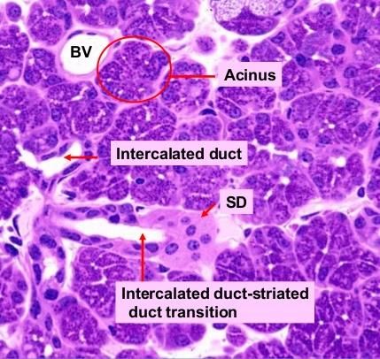
mesenchymal, neuronal, lymphatic, and endothelial cells (Figure 1). Polar epithelial cells form the parenchyma of the
salivary gland, which is comprised of epithelial ducts (intercalated, striated and excretory) and acinar cells. Between
the basal lamina and acinar and ductal cells lie myoepithelial cells (MECs), which form a basket-like network
surrounding spherical acini and ducts. MECs are star-shaped cells that structurally resemble epithelial and smooth
muscle cells, and facilitate salivary secretion through contractile processes that reduce luminal volume and prevent
acinar distension. Complex interactions among these cells types orchestrate salivary gland development, function, and
maintenance. Thus, alteration of salivary gland architecture or cellular organization can impair their important
functions in the oral cavity.
18Figure 1. Histological sections of SMG illustrating the multiple cell types present and an illustration demonstrating the architecture of the epithelial components of the SMG. BV: blood vessel, SD: serous deminlune, StD: striated duct, SA: serous acini, MA: mucous acini, MC: myoepithelial cells. Dysfunction of salivary secretion causes xerostomia (dry mouth), which can sequentially lead to dental caries and oral mucosal disorders. Xerostomia and hyposalivation are major side effects of long-term medication use and radiation therapy, and may also be caused by the presence of tumors or systemic autoimmune diseases such as Sjögren’s syndrome (SS). SS affects an estimated 0.1-4% of the United States population and approximately 1 to 2 million people internationally. It is often considered the second most common rheumatologic disorder in the United States, after systemic lupus erythematosus. There are two classifications of SS: primary SS presents without an underlying rheumatic disorder, while patients with another major rheumatic autoimmune disease are typically categorized to have secondary SS. Clinically, SS symptoms vary from mild characterized by xerostomia, dry eye, other xeroses, and neuropathies, to more severe systemic symptoms involving multiple organ systems. While majority of patients are women over the age of 40, SS has also been diagnosed in men, younger women, and children, significantly reducing the quality of life of those affected. Difficulties in speaking and eating, rampant dental caries, fatigue, and blindness are only a few of the clinical consequences observed. Currently, there is an enormous research database pertaining to the clinical aspects, diagnostic criteria, and treatment protocols for SS, however understanding the initiation and progression of the disease remains elusive. At the cellular level, SS is characterized by an increase in ductal cell population or ductal dysplasia and acinar cell loss. As well, there have been documented decreases in myoepithelial cell numbers that surround acini. A major and prominent feature of diagnosed SS is focal lymphocytic infiltration in salivary glands. Specifically, mononuclear cell numbers are increased in these glands, accompanied by high serum titres of Ro/SSA, La/SSB, and rheumatoid factor autoantibodies. T-cells, B-cells, macrophages, and dendritic cells also populate the affected salivary glands, indicating the presence of inflammation. Although the immune response is a major contributor to the clinical manifestations of SS, the existence of a pre-autoimmune phase of autoimmune diseases suggests that salivary epithelial cell dysfunction in SS is independent of presence of inflammatory cells in the gland. The pre-autoimmune phase, in which patients exhibit serologic abnormalities before clinical presentation, drives research in this field towards understanding the underlying role of epithelial cells in the development of clinical pathology. Intracellular signaling dynamics have the capacity to alter the highly polarized, functionally important structure of salivary epithelial cells and direct salivary gland morphology. In recent years, our group and others have demonstrated that the Hippo signaling pathway, and especially cellular localization of the Yes-associated protein (YAP) transcriptional regulator, plays key roles in SMG ductal cell maturation, branching morphogenesis, apoptotic pathway modulation, and that its dysregulation is associated with SS-like disease. The Hippo pathway is involved in restraining cell proliferation and promoting apoptosis through a series of kinase-mediated signaling events as outlined in Figure 2. 19
Figure 2. Simplified schematic of the Hippo signaling pathway. Numerous upstream mediators can modulate Hippo signaling
G proteins and junctional signaling. Activation of the hippo pathway involves MST1/2 phosphorylation, leading to activation of
Lats1/2 kinases, followed by phosphorylation of YAP/TAZ and its sequestration in the cytoplasm or degradation.
Dephosphorylated YAP/TAZ translocates to the nucleus where they interact with TEAD1-4 to induce transcription, promote cell
proliferation, and inhibit apoptosis.

Ultimately, activation of Lats 1 and Lats 2 Kinases (Lats1/2) phosphorylates YAP on a conserved serine residue,
which facilitates binding to 14-3-3 and YAP cytoplasmic sequestration. This restricts YAP from nuclear entry and
therefore reduces YAP transcriptional activity and also promotes cell differentiation. Thus, most of the genes involved
in Hippo signalling are recognized as tumor suppressors. Loss of Lats1/2-mediated YAP activity has been reported to
lead to defective organ morphogenesis and function. When unphosphorylated, YAP binds with transcriptional
coactivator with PDZ-binding motif (TAZ) and translocates to the nucleus. Together, YAP/TAZ are considered
oncogenes as they have the capacity to reprogram cancer cells into cancer stem cells and elevated YAP levels have
been described in some human cancers. Thus, changes in Hippo signaling can have deleterious effects on epithelial
cell homeostasis and drive changes in salivary gland function and morphology.

To examine the in vivo consequence of altered Hippo signaling in adult salivary glands, we have collaborated with
colleagues who have generated mice with conditional knockout of Lats1/2 kinases in keratin 8 (Krt8)-expressing cells.
In this model, tamoxifen drives the expression of Cre-recombinase in Krt8-expressing ductal cells. The Cre
recombinase splices sites containing Lats1/2 kinases genes and also splices out a stop codon signaling sequence
inserted between a YFP transgene. Thus, YFP-positive cells indicate Krt-8 positive cells in which Cre activity is
present and presumably cells in which Lats1/2 kinases are also downregulated (Last1/2-cnull). This animal model
allows us determine the effects of reduced Lats1/2 after the salivary glands have already developed, potentially
mimicking acquired human salivary gland disorders over inherited diseases. In this pilot experiment, we used a variety
of histological techniques to identify and characterize changes in SMG architecture, composition, and cellularity in
Lats1/2-cnull mice compared to tamoxifen-treated genetic control mice. We hypothesize that loss of Lats1/2 kinases
in adult ductal epithelial cells of the SMG drives nuclear YAP activity, leading to ductal dysplasia and a loss of acinar
structures reminiscent of SS.

20
METHODS

Animals

Lats1/2-cnull mice were generated in the laboratory of Xaralabos Varelas. Mice were generated by crossing Krt8-
CreER mice with Lats1/2 loxP/loxP; LSL-YFP mice, in which inducible knockout of Lats1/2 would be achieved upon
administration of tamoxifen. Female mice aged 6 weeks of age were treated with intraperitoneal tamoxifen (75mg/kg)
injections for 5 days, followed by an 8-day recovery period before sacrifice. A single Lats1/2-cnull was used for
subsequent analysis compared to an age- and sex-matched tamoxifen-treated control mouse (CON; Lats1/2; LSL-
YFP).

Hematoxylin and Eosin (H&E) Staining

Mouse SMGs were dissected and fixed in 4% PFA overnight prior to paraffin embedding. Paraffin-embedded tissues
were sectioned 5-um-thickness using a rotary microtome. Sectioned slides were then mounted on Superfrost plus
microscope slides and cured on a 50oC heat block overnight for adhesion. Slides were deparaffinized and rehydrated
the next day beginning with 4 washes incubated at 3 minutes each in Xylene, followed by 2 washes of 5 minutes in
100% ethanol, and then 1 wash each of 95%, 85%, 70%, 50% ethanol for 1 minute. After deparaffinization was
complete, slides were incubated in hematoxylin for 7 minutes at room temperature in a fume hood. Next, slides were
washed under running tap water for 10 minutes. Each slide was then dipped in acid alcohol to initiate hematoxylin
binding to lysine residues in nuclear histones, followed by 3 minutes of incubation in Scott’s tap water. Slides were
washed in dH2O for 2 minutes, and then dipped in 95 % ethanol. Eosin staining was performed by placing slides
directly into an eosin solution and incubated for 2 minutes, and then quickly dipped in 95% ethanol and 100% ethanol
respectively. Lastly, slides were placed in xylene for 2 minutes and then mounted with Cytoseal mounting media.

Immunofluorescence (IF) Microscopy for Paraffin Embedded Tissue

Paraffin-embedded SMG tissues were sectioned, mounted, deparaffinized as previously described in the H&E
microscopy section. Slides were then washed in dH2O for 2 minutes before performing antigen retrieval. Citrate buffer
(Vector Labs H-3300) was diluted 1:100 in dH2O to make the antigen retrieval solution, and then the solution was
heated to its boiling point in a microwave. Next, slides were added to sub-boiling antigen retrieval solution maintained
by a microwave for an additional 10 minutes. After incubation, the solution was cooled with the addition of 500 mL
water. Then the solution was then cooled for an additional 30 minutes, followed by 2 washes of 1xPBS for 5 minutes
each. An endogenous biotin blocking step was performed for the mouse primary antibody to reduce non-specific
binding. A few drops of streptavidin were added to slides and incubated for 15 minutes, followed by 2 washes in 0.1%
TBS-Tween for 5 minutes (TBS-T). The same procedure was repeated with biotin and then the slides were washed
twice with TBS-T for 5 minutes. Each tissue was then blocked in 5% donkey serum in 0.1% PBS-Tween (5% DS-
PBS-T) or 5% DS-PBS-T with MOM blocking solution for 1 hour at room temperature in a humidifier chamber.
Primary incubation was performed after blocking, and relevant primary antibodies [YFP, phospho-Lats1/2, YAP,
alpha-smooth muscle actin (a-SMA), aquaporin 5 (AQP5), keratin 5 (Krt5), keratin 14 (Krt14), keratin 8 (Krt8)] their
optimized dilutions in 5% DS-PBS-T or 5% DS-PBS-T + MOM was applied on the tissue slides and incubated at 4oC
overnight. The next day, slides were washed 4 times in TBS-T for 10 minutes each. Then secondary antibodies
solutions at optimized dilutions and 1 uM DAPI were applied on slides and incubated for 1 hour at room temperature
in a dark humidified chamber. Tissue slides were washed 4 times in TBST for 10 minutes each without exposure to
light. Slides were spun down inside a centrifuge to remove excess solution prior to mounting with antifade mounting
media.

Immunohistochemistry for paraffin embedded tissue

Paraffin-embedded SMG tissues were sectioned, mounted, deparaffinized and antigen unmasked as previously
described in the H&E microscopy section. Then tissues were blocked by adding a few drops of Dako duo enzyme

21
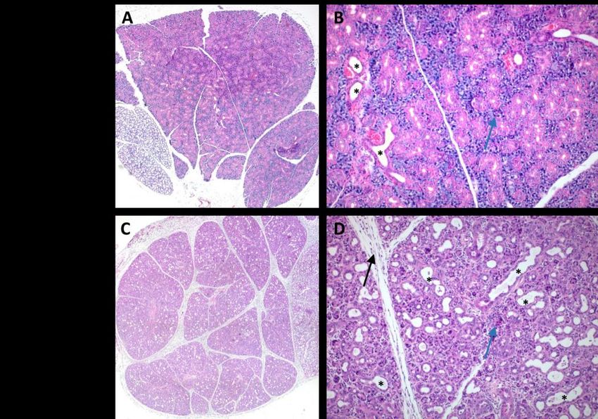
blocker (Dako #S2003) to slides for 15 minutes at room temperature in a humidified chamber and then washed with
TBS-T twice for 5 minutes. Tissues were by blocked with 5% goat serum in 0.1% PBS-Tween, and then slides were
washed twice with TBS-T for 5 minutes each. Primary antibodies (CD45, Ki67) were diluted to their optimal dilutions
in SignalStain Antibody Diluent (CST #8112) and applied on SMG tissues for 1 hour at room temperature. After
primary incubation, slides were washed 3 times with TBS-T for 5 minutes. A SignalStain Boost Detection kit was
then applied to each slide respective to each primary antibody species for 30 minutes at room temperature. Each slide
was then exposed to DAB substrate at their optimized exposure times. Next, sections were rinsed in dH2O twice for
2 minutes each. Hematoxylin counterstain was performed at room temperature for 8 minutes, followed by bluing
nuclear stain using 0.1% sodium bicarbonate solution. Dehydration was then performed with a graded ethanol series
beginning from 2 rinses in 95% ethanol for 15 seconds, 2 rinses in 100% ethanol for 1 minute and 2 rinses in xylene
for 3 minutes. After dehydration, slides were mounted with xylene-compatible mounting media.

Imaging

Immunofluorescent images were captured using an inverted epifluorescent microscope, at 20x, 40x, and 100x
magnification as indicated in the figures. The H&E and IHC images were captured on an Episcope microscope at 5x
and 10x magnification.

RESULTS

Alterations in submandibular gland morphology after conditional deletion of Hippo pathway kinases Lats1/2
in Keratin 8-expressing cells

To gain insight into the roles of Lats1/2 kinase in the adult mouse submandibular gland, we studied a mouse model
generated in which intraperitoneal administration of tamoxifen activates Cre recombinase expression under control of
the Keratin 8 (Krt8) promoter, resulting in Lats1/2 kinase conditional deletion in Krt8-expressing luminal epithelial
cells. In an effort to recapitulate clinical findings in humans, we used female mice because majority of SS patients are
females. Lats1/2 deletion was initiated at 6 weeks of age, the age in which mice reach sexual maturity, with daily
injections for 5 days, followed by an 8-day recovery period before sacrifice and tissue collection.

Histological analysis of tamoxifen control (CON) and conditional Lats1/2 deletion (Lats1/2-cnull: K8-Cre; Lats1/2
null; LSL-YFP) submandibular glands by H&E staining revealed striking consequences of attenuated Lats1/2 kinase.
In contrast to the normal salivary architecture seen in CON glands, the Lats1/2-cnull displayed three major changes:
increased connective tissue septa between the epithelial parenchyma lobules, accumulation of duct-like structures
throughout the lobules, and areas of disorganized acini (Figure 3A-D). The ductal changes were accompanied the
appearance of disorganized lumen, which lacked lining cells in some regions. These observations suggest that loss of
Lats1/2 kinases driven by keratin 8 either increase the number of ductal structures or expand the salivary gland acini,
while acinar structures were diminished.

22
Figure 3. H&E staining of CON and Lats1/2-cnull submandibular glands. A) Whole section of CON mouse submandibular
gland (5X objective); B) Whole section of Lats1/2-cnull mouse submandibular gland (5X objective); C) CON mouse submandibular
gland (10X objective); D) Lats1/2-cnull mouse submandibular gland (10X objective). Shown within C & D are ductal structures
(asterisks), acinar structures (blue arrows), and connective tissue (black arrows).

Expression of YFP, phospho-Lats1/2, and Yap localization in Lats1/2-cnull mice submandibular glands

         We next performed multiple immunofluorescence staining on serial sections of the salivary glands to analyze
changes in cellularity based on well-known salivary cell markers. As the LSL-YFP system was employed in this
animal model, YFP staining reveals active regions of Cre-recombinase activity and therefore indicates cells with loss
of Lats1/2 kinase. We found that YFP staining was strongest in ductal cells of Lats1/2-cnull SMGs (Figure 4A-B) and
that some of the YFP+ cells appeared to have cytoplasmic processes reminiscent of extrusion-like structures (Figure
4A). We also verified that knockdown of Lats1/2 was achieved in Lats1/2-cnull mice, as phospho-Lats 1/2 staining
was decreased (Figure 4B). We visualized staining of Yap, a downstream effector phosphorylated by Lats 1/2 kinases,
which localizes to the nuclei in unphosphorylated form (Figure 4B). At a higher magnification, nuclear Yap was
present in Lats1/2-cnull nuclei while Yap tended to be retained in the cytoplasm (Figure 4C). Altogether, these findings
support successful knockdown of Lats 1/2 kinases in our animal model.

23
Figure 4. Multiple immunofluorescence staining of YFP, phospho-Lats1/2 (pLats1/2), and Yap in submandibular glands.
A) YFP stain, white arrows showing YFP+ cells with extrusion-like elements, white arrows (100X objective); B) YFP, pLats1/2,
and Yap staining of submandibular glands (20X objective); C) CON mouse submandibular gland (100X objective); D) Lats1/2-
cnull mouse submandibular gland (100X objective) showing regions of nuclear Yap stain (asterisks).
Reduced and ectopic aquaporin 5 staining in Lats1/2-cnull acinar cells
Multiple immunofluorescence stain was also performed to identify aquaporin 5- and alpha-smooth muscle
actin-positive cells. Aquaporin 5 (AQP5), a water channel expressed in pro-acinar cells, is mainly localized to the
apical membrane. Lats1/2-cnull submandibular glands displayed weaker AQP5 expression, with some regions of
ectopic expression observed (Figure 5A-B). While CON mice had distinct and characteristic AQP5-positive apical
regions, AQP5 was largely decreased and slightly more diffuse, indicating abnormal or disruption of apical
morphology of Lats1/2-cnull cells acinar cells (Figure 5B). In some regions, AQP5 stain appeared to face the lining
of luminal regions, suggesting expansion of the apical aspect of acini (Figure 5C). Alpha-smooth muscle actin, a
marker of myoepithelial cells, had only slightly decreased expression in Lats1/2-cnull cells compared to CON (Figure
5A-B). We used vimentin, which stains cells of mesenchymal origin, to confirm the accumulation of mesenchymal-
derived connective tissue in Lats1/2-cnull submandibular glands (Figure 5D).
24Figure 5. Multiple immunofluorescence staining of YFP, alpha-smooth muscle actin (-SMA), aquaporin 5 (AQP5), and vimentin in submandibular glands. A-B) multiple staining of YFP, -SMA, and AQP5 in CON and Lats1/2-cnull mouse submandibular glands (40X and 100X objective, as labelled). C) Merged stain images of Lats1/2-cnull salivary cells showing diffuse AQP5 (white arrows) and AQP5 stain facing luminal regions (blue arrows). D) Vimentin staining of mesenchymal-derived cells. Analysis of differentiation in Lats1/2-cnull salivary cells by keratin staining To identify alterations in salivary acinar cell differentiation of Lats1/2-cnull mice, we stained the sections with keratin 5 (Krt5), keratin 14 (Krt14), and Krt8 (Figure 6A-B). Krt5 and Krt14 are intermediate filament proteins expressed in basal progenitor cells, and their expression is down-regulated as cells differentiate. Krt8, a marker of luminal cells in duct-like structures, also forms intermediate filaments in the cytoplasm of epithelial cells. We found no difference of Krt5 and Krt14 expression between CON and Lats1/2-cnull submandibular glands. However, Lats1/2-cnull glands exhibited increased Krt8 staining that was less punctate than CON glands and more regions with Krt5-Krt8-Krt14 overlapping expression (Figure 6A-B). Note that Krt5-Krt14 also changed from its characteristic basolateral localization in CON sections to a more diffused fashion. Collectively, these findings suggest that although acinar cell differentiation may be unchanged in response to Lats 1/2 knockdown, Krt8 in Lats1/2-cnull may associate with progenitor cells. 25
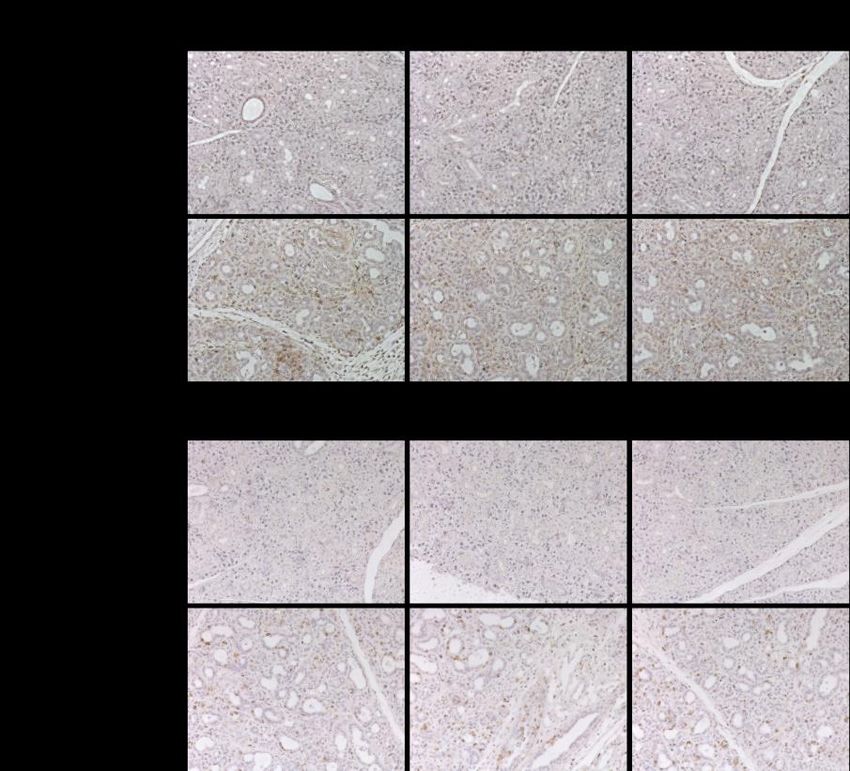
Figure 6. Multiple immunofluorescence staining of keratin 5 (Krt5), keratin 12 (Krt14), and keratin 8 (Krt8) in
submandibular glands. A) Stained glands imaged with 20X objective; B) Stained glands imaged with 100X objective, showing
regions of Krt5-Krt8 and Krt5-Krt14 overlap (white arrows).

Lats1/2-cnull salivary glands exhibit dramatic increases in lymphocyte presence and proliferation

As SS is highly characterized by infiltration of immune cells, we immunostained the glands for CD45, a key regulator
of antigen-mediated signaling and lymphocyte activation. We found that Lats1/2-cnull glands had dramatic increases
in CD45-positive cells throughout the glands (Figure 7A), and especially between ductal regions, while CON glands
showed little to no stain. We also stained the glands for Ki67, which is a marker that is strictly associated with cellular
proliferation. Similarly, we observed increased Ki67-positive cells in Lats1/2-cnull glands (Figure 7B). Ki67-positive
cells were mainly localized to the ductal regions suggesting active proliferation and luminal expansion in these regions.

26
Figure 7. Immunohistochemical staining of CD45 and Ki67 in submandibular glands. A) CD45-stained glands imaged with
20X objective; B) Ki67-stained glands imaged with 20X objective.

DISCUSSION

Some of our long-term goals are to study the influence of the Hippo signaling pathway in mammalian SMGs and to
develop a reliable mouse model for studying Sjogren Syndrome. Localization of Yap fine-tunes the balance in
regulating cell proliferation, differentiation and apoptosis. From previous lab work, we demonstrated that conditional
Yap knockout driven by sonic hedgehog targeting the SMG epithelium (Yap-cnull) resulted in defective ductal regions
and loss of Krt5/Krt14 expressed ductal progenitor cells. We know that both Yap expression and localization play an
important role in organogenesis. We intend to further study other upstream components in the Hippo pathway to better
understand its role in disease pathology. The Lats1/2-cnull mouse line was established to further investigate the
upstream effect of Hippo signaling in SMGs. Lats1/2 phosphorylates Yap that restricts nuclear Yap localization, which
leads to accumulation of cytoplasmic Yap. The cytoplasmic Yap later gets degraded through protein ubiquitination,
which indirectly promotes cell differentiation and tissue maturation. We previously showed that loss of Lats1/2
severely impairs branching morphogenesis in SMGs and results in upregulated Krt5/Krt14 positive progenitor cells in
ductal the region.

A major common clinical feature of SS is tissue infiltration by lymphocytes. In particular, activation of B-cells is
consistently found in majority of SS patients. Helper T cell populations predominates over other invading immune
cells. Our preliminary results in H&E staining showed distinct differences in tissue morphology between tamoxifen
control mice and Krt8-driven conditional Lats1/2 knockout mice (Figure 3A-D). The robust expansion of ductal
structures observed in Lats1/2-cnull H&E images is supported by the increase of Krt8-positive ductal cells observed
by IF microscopy (Figure 6A-B). Meanwhile, we did not observe a remarkable increase in Krt5/Krt14-expressing cells
in Lats1/2-cnull mice (at 40x magnification) compared to what we previously observed in Yap1/2-cnull embryonic
mice (data not shown), suggesting that Lats1/2 KO might have a reduced influence on Krt5/Krt14 expression as mice

27
age to 6 weeks old. However, under higher magnification (100x), we observed more diffuse Krt5/Krt14 localization
in Lats1/2-cnull tissue as the typical basal localization was severely disrupted. The diffuse Krt5/Krt14 localization
suggests loss of cell polarity in ductal basal cells as well as in some saliva-producing acinar units.

Increased connective tissue stroma in the Lats1/2-cnull SMGs may have occurred from repetitive or overwhelmed
tissue-reparative processes. Immune cells are key mediators of initiating tissue repair. Release of cytokines is
considered a fundamental step for immune cell and repair cell recruitment and trafficking. It is possible that cytokine
release is dysregulated in Lats1/2-cnull SMGs, especially near the ductal regions. This would subsequently activate a
large number of repair cells and ultimately result in excess fibrotic tissue deposition predominantly near the ductal
region in SMGs. Vimentin is a mesenchyme cell marker that is important in epithelium-to-mesenchyme transition and
tissue repair process. Previous lab work showed rapid activation of vimentin activation upon the onset of injury in
embryonic mouse SMGs. Preliminary data in our laboratory (not shown) also suggests that vimentin may initiate the
recruitment of other repair proteins (ADAM12, PDGFR-alpha) in response to injury. Therefore, the increased
expression of Vimentin in Lats1/2-cnull mice also suggests an active reparative process likely contributed to abnormal
connective tissue expansion (Figure 5D).

Based on our immunohistochemistry (IHC) results, we observed a remarkable increase in CD45-positive immune cells
in Lats1/2-cnull tissues (Figure 7A). This phenotype resembles lymphocyte infiltration in most of SS patients. The
initiation process of recruiting immune cells in this model requires further investigation. However, these findings
strongly suggest that Lats1/2 kinases partly play an essential role in recruiting immune cells. Our attempt to mimic SS
pathology in a mouse model was rather successful. We also found increased Ki67-expressing cells in Lats1/2-cnull
mouse, and they were predominantly clustered at periductal region (Figure 7B). The rationale of rapidly expanding
duct-like structures in Lats1/2-cnull tissue requires further study. Yet, with loss phosphorylation by Lats1/2 kinases,
it is reasonable to assume that nuclear Yap might be a driving factor that disturbs the balance between cell proliferation
and cell differentiation, resulting in expansion of disorganized Krt8-expressing luminal structures.

However, there are a few concerns about this mouse model. The global Lats1/2 knockout was found to be lethal at
early embryonic stage, making this model difficult for studying Lats1/2 function during embryonic development. In
addition, Lats1/2-cnull mice rapidly deteriorated after only 5 days of tamoxifen administration, indicating signs of
systemic pathology, and they presented with nutritional deficiencies. This further complicates our interpretation, as
we could not confirm if the result we observed was due to loss of Lats1/2 exclusively.

Interestingly, we also found acini structures in Lats1/2-cnull SMGs appeared to be much smaller and less integrated
than the CON mice acini. Although H&E staining provided us with distinctive detail on tissue morphology, we
performed immunofluorescence microscopy in order to evaluate the expression and localization of well-known
salivary epithelial cell markers. Aquaporin-5 (Aq5) is an apical membrane water channel protein that is crucial for
saliva formation by inducing passive water diffusion across the lipid bilayer membrane in SMG acinar cells. Based
on our findings, AQP5 architecture was severely disorganized and expression was reduced in Lats1/2-cnull cells
(Figure 5A-B). The disruption of apical AQP5 localization was likely a consequence of impaired cellular maintenance
signals resulting from Lats1/2 loss. Increased Yap activity in Lats1/2-cnull cells may have triggered the transcription
and expression of important paracrine signals that maintain the apical localization of AQP5. Alternatively, loss of cell-
to-cell adhesion (ZO-1, E-cadherin) could also be accountable for this phenotype. It is difficult to determine whether
cell-cell adhesion or disrupted cell growth initiated the immune-infiltration in Lats1/2-cnull mouse. However, we
could conclude that Lats1/2 kinase depletion resulted in a significant disruption in acini cytoskeletal structure and
clustering.

Alpha smooth muscle actin (SMA) is marker for myoepithelial cells, which is another key component of a functional
saliva-producing unit in the basal region. SMA serves as a mechanical contractile vector to guide proper saliva
production into ductal lumen regions along with passive water diffusion contributed by AQP5. In Lats1/2-cnull mouse
tissue, we observed a remarkable reduction in SMA expression at 40x magnification. Under higher magnification

28
You can also read