STEM CELL COLLECTION 2020 - Rockefeller University Press
←
→
Page content transcription
If your browser does not render page correctly, please read the page content below
STEM CELL COLLECTION 2020

WHY SUBMIT
    YOUR RESEARCH?

AN EDITORIAL PROCESS GUIDED                                                                                             EDITORS FROM THE
BY YOUR COMMUNITY                                                                                                      STEM CELL RESEARCH
                                                                                                                           COMMUNITY
At Journal of Cell Biology (JCB) and Journal of Experimental
Medicine (JEM), all editorial decisions on research manuscripts
are made through collaborative consultation between                                                                                  JCB
professional scientific editors and the academic editorial board.
                                                                                                                                  Elaine Fuchs
                                                                                                                                   Ana Pombo
                                                                                                                                 Carien Niessen
                                                                                                                                Juergen Knoblich
                                                                                                                                 Cedric Blanpain
                     Submission or transfer           Academic and scientific                                                   Michael Rudnicki
                                                          editors review                                                         Thomas Rando
                                                                                                                                  Yukiko Gotoh

                                                                                                                                     JEM
      If appropriate, sent to         Academic and scientific           Informed decisions
           peer review               editors review comments              sent to authors                                Emmanuelle Passegue
                                                                                                                            Elaine Dzierzak
                                                                                                                              Paul Frenette
                                                                                                                        Margaret (Peggy) Goodell
                                                                                                                            Bertie Gottgens
       Revised manuscript
     submission and rereview
                                     Academic and scientific
                                         editors review
                                                                 97% of invited revisions
                                                                  accepted at JCB and
                                                                                                                             Hanna Mikkola
                                                                     92% at JEM.*                                           Sean J. Morrison
                                                                                                                              Ulrich Steidl
                                                                                                                              Leonard Zon
                                                                                                                              Patricia Ernst
               Published online 1-2 days after   Circulated via alerts and social media                                       Toshio Suda
                  author proofs returned              to readers around the world

                                                                                                             *Median 2019

         Format Neutral                 Transfer Policy                                      Fair and Fast                            Open Access Options
         You may submit                 We welcome submissions that include                  We limit rounds of revision, and         Our options include
         your papers in                 reviewer comments from another journal. You          we strive to provide clear, detailed     Immediate Open Access
         ANY format.                    may also request manuscript transfer between         decisions that illustrate what is        (CC-BY) and open access
                                        Rockefeller University Press journals, and we        expected in the revisions. Articles      six months after publication
                                        can confidentially send reviewer reports and         appear online one to two days            (CC-BY-NC-SA).
                                        identities to another journal beyond RUP.            after author proofs are returned.

                                          LEARN MORE AT JCB.ORG AND JEM.ORG
STEM CELL COLLECTION 2020

                                                     J
                                                                 ournal of Cell Biology (JCB) and Journal of Experimental Medicine (JEM)
                                                                 editors are delighted to present this selection of recent, cutting-edge
                                                                 stem cell research. JCB publishes new cellular and molecular advances
                                                                 in any area of basic cell biology as well as papers that describe applied
                                                                 cell biology to translational fields such as stem cells. JEM is interested
                                                     in original findings on all aspects of disease pathogenesis, including both
                                                     hematopoietic stem cell and non-hematopoietic stem cell physiopathology. This
                                                     collection includes some of our most highly read papers and articles selected
                                                     to reflect the breadth of stem cell research published in our journals. We hope
                                                     you enjoy these articles that delve into the cell biology of regeneration, stem cell
                                                     division, fate decision, and their links to various diseases and innovative therapies.

                                                     Visit jcb.org and jem.org to learn about our editorial policies and members of our
                                                     editorial boards. We welcome your feedback and questions via Twitter
                                                     (@JCellBiol and @JExpMed). You can also email JCB at jcellbiol@rockefeller.
                                                     edu and JEM at sbhatt@rockefeller.edu (regarding general stem cell studies) and
                                                     xsun01@rockefeller.edu (for studies with a cancer research angle).

                                                     6    Myc regulates retinal regeneration
                                                          Study identifies multiple roles for Myc in controlling the induction and proliferation of Muller glia–derived progenitor
                                                          cells upon retinal injury in zebrafish
                                                          Soumitra Mitra … Rajesh Ramachandran

                                                     7    The RNA exosome restrains hESC differentiation
                                                          The nuclease complex degrades a variety of RNAs to maintain pluripotency and suppress the differentiation of
                                                          human embryonic stem cells
                                                          Cedric Belair … Sandra L. Wolin

                                                     8    Hair follicles restrict cancer growth
                                                          Normal skin corrals cancer-causing mutations and reveals how tissue can subvert tumorigenesis
                                                          Cristiana M. Pineda … Valentina Greco

                                                     9    Centromeres drive asymmetric stem cell division
                                                          Asymmetric incorporation of the centromeric histone CENP-A during G2/M regulates the fate of Drosophila female
                                                          germline stem cells
                                                          Anna Ada Dattoli … Elaine M. Dunleavy

                                                     10   NCAM regulates neural progenitor cell fate
                                                          Neural cell adhesion molecule controls the proliferation and differentiation of cortical progenitors by binding to the
                                                          actin polymerization regulator profilin2
                                                          Rui Huang, De-Juan Yuan, Shao Li … Quan-Hong Ma, Quan-Hong Ma

                                                     11   Glutamylation regulates HSC self-renewal
                                                          Reversible modification of the tumor suppressor BAP1 controls hematopoietic stem cell self-renewal and blood cell
                                                          formation
                                                          Zhen Xiong, Pengyan Xia, Xiaoxiao Zhu … Yong Tian, Zusen Fan

                                                     12   The chromatin landscape of glioblastoma
                                                          Profiling of glioblastoma stem cells and primary tumor samples identifies subgroup-specific transcriptional
                                                          regulatory circuits and novel therapeutic targets
                                                          Stephen C. Mack, Irtisha Singh, Xiuxing Wang … Charles Y. Lin, Jeremy N. Rich

                                                     13   Primitive HSCs continue cycling into adulthood
                                                          Study reveals that, contrary to a previous report, the most primitive hematopoietic stem cells do not become
Brochure articles: Ben Short, PhD and
                                                          permanently quiescent and, instead, show continuous mitotic activity in adult mice
Christina Szalinski, PhD
                                                          Mina N.F. Morcos, Thomas Zerjatke ... Alexander Gerbaulet
Design: Christine Candia
                                                     14   Distinguishing physiologic and oncogenic Wnt signals
On the cover: Two-photon images of
                                                          Profiling of isogenic human colon organoids reveals differences between oncogenic Wnt activation and normal Wnt
HrasG12V/+ hair follicles during the “second rest         signaling essential for stem cell maintenance
phase” (P55). Mosaic activation of Hras using
                                                          Birgitta E. Michels, Mohammed H. Mosa … Henner F. Farin
lentiviral-Cre delivery demonstrates that wild-
type follicles (green) within the HrasG12V/+ skin
reenter the growth phase precociously along          15   Leukemia hijacks myeloid regeneration pathways
with their Hras mutant (red) neighbors. Image             Correcting aberrant Notch and Wnt signaling in leukemic stem cells might normalize myeloid cell production in a
© 2019 Pineda et al. Read “Hair follicles restrict        number of different leukemias
cancer growth,” page 8.                                   Yoon-A Kang, Eric M. Pietras, Emmanuelle Passegué

                                                                                                                                                                                     3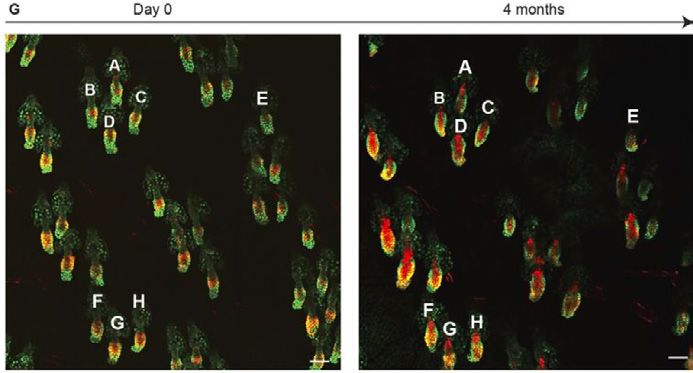
Editor-In-Chief                    Editorial Board
Jodi Nunnari                       John Aitchison                Rebecca Heald                                    Elior Peles
Executive Editor                   Johan Auwerx                  Martin Hetzer                                    Will Prinz
Tim Spencer                        Manuela Baccarini             Erika Holzbaur                                   Thomas Rando
email: tspencer@rockefeller.edu    Tamas Balla                   Martin Humphries                                 Samara Reck-Peterson
Editors                            Maureen Barr                  James Hurley                                     Daniel B. Rifkin
Arshad Desai                       Bill Bement                   Fumiyo Ikeda                                     Michael Rout
Pier Paolo Di Fiore                Vann Bennett                  Luisa Iruela-Arispe                              Craig Roy
Elaine Fuchs                       Dominique Bergmann            Nancy Ip                                         Michael Rudnicki
Anna Huttenlocher                  Monica Bettencourt-Dias       Johanna Ivaska                                   Erik Sahai
Ian Macara                         Joerg Bewersdorf              Tarun Kapoor                                     Martin Schwartz
Ira Mellman                        Magdalena Bezanilla           Gerard Karsenty                                  Shu-ou Shan
Ana Pombo                          Cédric Blanpain               Scott Keeney                                     Andrey Shaw
Louis F. Reichardt                 Julius Brennecke              Alexey Khodjakov                                 Zu-Hang Sheng
Kenneth M. Yamada                  Tony Bretscher                Hiroshi Kimura                                   Agata Smogorzewska
Richard Youle                      Marianne Bronner              Jürgen Knoblich                                  Joan Steitz
                                   Valérie Castellani            Alberto R. Kornblihtt                            Harald Stenmark
Senior Scientific Editor
                                   Daniela Cimini                Thomas Langer                                    Aaron Straight
Melina Casadio
                                   Karlene Cimprich              Ana Maria Lennon-Dumenil                         Maria-Elena Torres-Padilla
email: mcasadio@rockefeller.edu
                                   Don W. Cleveland              Andres Leschziner                                Billy Tsai
Scientific Editor, Reviews         Nika Danial                   Danny Lew                                        Bas van Steensel
Andrea Marat                       William Earnshaw              Jens Lykke-Andersen                              Patrik Verstreken
email: amarat@rockefeller.edu
                                   Jan Ellenberg                 Vivek Malhotra                                   Mark von Zastrow
Scientific Editors                 Scott Emr                     Brendan Manning                                  Erwin Wagner
Marie Anne O'Donnell               Anne Ephrussi                 Joan Massague                                    John Wallingford
email: modonnell@rockefeller.edu
                                   Jeffrey Esko                  Satyajit Mayor                                   Tobias Walther
Dan Simon
                                   Sandrine Etienne-Manneville   Frauke Melchior                                  Xiaochen Wang
email:dsimon01@rockefeller.edu
                                   Marc Freeman                  Tobias Meyer                                     Lois Weisman
Managing Editor                    Judith Frydman                Liz Miller                                       Min Wu
Lindsey Hollander                  Hironori Funabiki             Alex Mogilner                                    Tim Yen
Phone: 212-327-8588
email: jcellbiol@rockefeller.edu   Melissa Gardner               Sean Munro                                       Tamotsu Yoshimori
                                   Larry Gerace                  Maxence Nachury                                  Li Yu
                                   David Gilbert                 Karla Neugebauer                                 Xiang Yu
                                   Bruce Goode                   Carien Niessen                                   Junying Yuan
                                   Yukiko Gotoh                  Eva Nogales                                      Marino Zerial
                                   Roger Greenberg               Karen Oegema                                     Hong Zhang
                                   Marcia Haigis                 Ewa Paluch                                       Yixian Zheng
                                   Ulrich Hartl                  Mark Peifer

Senior Preflight Editor            Production Manager            Copyright to articles published in this journal is held by the authors. Articles are published
Laura Smith                        Camille Clowery               by Rockefeller University Press under license from the authors. Conditions for reuse of the
                                                                 articles by third parties are listed at http://www.rupress.org/terms.
Preflight Editor                   Production Designer
Rochelle Ritacco                   Erinn A. Grady                Print ISSN: 0021-9525.
                                                                 Online ISSN: 1540-8140
Senior Production Editor
Samantha Wolner                                                  Rockefeller University Press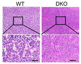
Executive Editor             Advisory Editors
Teodoro Pulvirenti           Shizuo Akira                Vijay K. Kuchroo                                 Hans Schreiber
phone (212) 327-8575         Kari Alitalo                Ralf Kuppers                                     Pamela Schwartzberg
email: jem@rockefeller.edu
                             Frederick W. Alt            Tomohiro Kurosaki                                Charles N. Serhan
Editorial Board Co-Chairs    David Artis                 Bart N. Lambrecht                                Nilabh Shastri
Carl Nathan                  K. Frank Austen             Ross Levine                                      Ethan M. Shevach
Michel Nussenzweig           Albert Bendelac             Klaus F. Ley                                     Robert Siliciano
Editors                      Christine A. Biron          Yong-Jun Liu                                     Roy L. Silverstein
Yasmine Belkaid              Hal E. Broxmeyer            Clare Lloyd                                      Hergen Spits
Jean-Laurent Casanova        Meinrad Busslinger          Tak Mak                                          Jonathan Sprent
Sara Cherry                  Arturo Casadevall           Asrar Malik                                      Janet Stavnezer
David Holtzman               Ajay Chawla                 Bernard Malissen                                 Ulrich Steidl
Susan Kaech                  Nicholas Chiorazzi          Philippa Marrack                                 Andreas Strasser
Lewis L. Lanier              Robert L. Coffman           Diane Mathis                                     Helen Su
Anne O’Garra                 Myron I. Cybulsky           Ira Mellman                                      Joseph Sun
Emmanuelle Passegué          Glenn Dranoff               Miriam Merad                                     Stuart Tangye
Alexander Rudensky           Michael Dustin              Matthias Merkenschlager                          Steven L. Teitelbaum
Arlene Sharpe                Elaine Dzierzak             Hanna Mikkola                                    Jenny Ting
David Tuveson                Mikala Egeblad              Denise Monack                                    Kevin J. Tracey
Jedd D. Wolchok              Patricia Ernst              Sean J. Morrison                                 Giorgio Trinchieri
Senior Scientific Editor     Richard A. Flavell          Muriel Moser                                     Shannon Turley
Shachi Bhatt                 Paul Frenette               Daniel Mucida                                    Emil Unanue
                             Thomas Gajewski             Charles Mullighan                                Valerie Weaver
Scientific Editors
                             Adolfo Garcia-Sastre        Cornelis Murre                                   Raymond M. Welsh
Montserrat Cols
                             Patricia Gearhart           Linda Noble-Haeusslein                           E. John Wherry
Stephanie Houston
                             Ronald N. Germain           Jeffrey Noebels                                  Thomas Wynn
Xin (Cindy) Sun
                             Daniel Geschwind            John J. O’Shea                                   Sayuri Yamazaki
Gaia Trincucci
                             Lucy Godley                 Jack Parent                                      Leonard Zon
Editors Emeriti              Margaret (Peggy) Goodell    Virginia Pascual
William A. Muller            Christopher Goodnow         Laura Pasqualucci                                Monitoring Editors
Alan Sher                    Bertie Gottgens             Erika Pearce                                     Marco Colonna
Managing Editor              Florian Greten              Fiona Powrie                                     Jason Cyster
Sylvia F. Cuadrado           Philippe Gros               Klaus Rajewsky                                   Stephen Hedrick
phone (212) 327-8575         Chyi Hsieh                  Gwendalyn J. Randolph                            Kristin A. Hogquist
email: jem@rockefeller.edu   Christopher A. Hunter       Rino Rappuoli                                    Andrew McMichael
                             Luisa Iruela-Arispe         Jeffrey Ravetch                                  Luigi Notarangelo
                             Akiko Iwasaki               Nicholas Restifo                                 Federica Sallusto
                             Jos Jonkers                 Nikolaus Romani                                  Toshio Suda
                             Johanna Joyce               David L. Sacks
                             Thirumala-Devi Kanneganti   Shimon Sakaguchi                                 Consulting Biostatistical
                             Gerard Karsenty             Matthew D. Scharff                               Editor
                             Jay Kolls                   Olaf Schneewind                                  Xi Kathy Zhou
                             Paul Kubes                  Stephen P. Schoenberger

Senior Preflight Editor      Production Manager          Copyright to articles published in this journal is held by the authors. Articles are published
Laura Smith                  Camille Clowery             by Rockefeller University Press under license from the authors. Conditions for reuse of the
                                                         articles by third parties are listed at http://www.rupress.org/terms
Preflight Editor             Production Designer
Rochelle Ritacco             Erinn A. Grady              Print ISSN 0022-1007
                                                         Online ISSN 1540-9538
Production Editor
Jennifer McGullam                                        Rockefeller University Press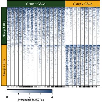
MYC REGULATES RETINAL
    REGENERATION
    Study identifies multiple roles for Myc in controlling the induction and proliferation of
    Muller glia-derived progenitor cells upon retinal injury in zebrafish

    Compared with humans and other                      ation of many cell types to form induced
    mammals, zebrafish have a much                      pluripotent stem cells.
    greater capacity to regenerate dam-
    aged tissues after injury. Damage to the            Mitra et al. found that, upon retinal injury,
    zebrafish retina, for example, causes               the two zebrafish Myc genes, myca
    Muller glial cells to dedifferentiate into          and mycb, were rapidly induced in both
    multipotent progenitor cells that prolif-           Muller glia-derived progenitor cells (MG-
    erate and eventually redifferentiate into           PCs) and neighboring cells at the injury
    many different retinal cell types, thereby          site. Knocking down or inhibiting Myc                   Four days after injury, the number
    enabling the tissue’s regeneration.                 impaired the dedifferentiation of Muller                of proliferative MGPCs (labeled
                                                        glia into MGPCs and blocked MGPC                        with GFP, green, and PCNA, red) is
    Muller glia with some stem cell–like                proliferation and retinal regeneration.                 reduced in a zebrafish retina treated
    characteristics have been identified in                                                                     with 10058-F4, a drug that inhibits
    mammalian retina, but their regener-                The researchers found that mycb pro-                    the interaction between Myc and its
    ative capacity appears to be limited.               motes the generation and proliferation                  obligatory transcriptional partner Max.
    “Unraveling the complete cascade of                 of MGPCs by inducing the expression of                  Credit: Mitra et al., 2019
    gene regulatory networks induced by                 ascl1a, leading, in turn, to the production
    retina injury in zebrafish could help in            of lin28a. (This gene regulatory cascade
    deciphering the lack of efficient regen-            is tightly controlled: while Ascl1a feeds
                                                                                                             Though the precise details remain to
    eration in mammals,” explains Rajesh                back to promote further mycb expression,
                                                                                                             be determined, Mycb therefore func-
    Ramachandran from the Indian Institute              another transcriptional target of Ascl1a,
                                                                                                             tions as both a transcriptional activator
    of Science Education and Research in                Insm1a, represses mycb).
                                                                                                             and repressor to finetune the extent of
    Mohali.
                                                        In neighboring cells, however, Mycb                  lin28a expression and MGPC formation
                                                        recruits the histone deacetylase Hdac1               in response to retinal injury.
    Ramachandran has previously identified
    important roles for several factors in              to repress the lin28a promoter and limit
                                                                                                             “Our study suggests that Mycs are part
    Muller glia dedifferentiation and retinal           lin28a expression. This may help prevent
                                                                                                             of a gene regulatory network essential
    regeneration, such as the transcriptional           MGPCs from forming throughout the ret-
                                                                                                             for retina regeneration, and provides
    activator Ascl1a and one of its targets,            ina and restrict their appearance to the
                                                                                                             insights that could ultimately help
    lin28a. In their latest study, Ramach-              immediate site of injury. The researchers
                                                                                                             improve Muller glia reprogramming in
    andran and colleagues, including first              found that Notch signaling plays a key
                                                                                                             injured human retina and enable suc-
    author Soumitra Mitra, investigated a               role in this restriction by inducing the ex-
                                                                                                             cessful repair,” Ramachandran says.
    potential role for the transcription factor         pression of an effector called her4.1 (itself
    Myc, which, among many biological                   a target for Mycb and Hdac1).
    functions, can promote the dedifferenti-

    RESEARCHER DETAILS                                                          ORIGINAL PAPER
                                                                                Mitra, S., P. Sharma, S. Kaur, M.A. Khursheed, S. Gupta, M. Chaudhary, A.J. Kurup,
                                                                                and R. Ramachandran. 2019. Dual regulation of lin28a by Myc is necessary during
                                                                                zebrafish retina regeneration. J. Cell Biol. 218:489–507.
                                                                                https://doi.org/10.1083/jcb.201802113

    Soumitra Mitra         Rajesh Ramachandran
    Graduate student       Associate Professor
    IISER Mohali           IISER Mohali
                           rajeshra@iisermohali.ac.in

6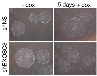
THE RNA EXOSOME RESTRAINS HESC
DIFFERENTIATION
The nuclease complex degrades a variety of RNAs to maintain pluripotency and
suppress the differentiation of human embryonic stem cells

Human embryonic stem cells (hESCs)                              EXOSC3-deficient hESCs expressed
can differentiate into all three embryon-                       increased levels of endodermal, me-
ic germ layers: the endoderm, meso-                             sodermal, and ectodermal markers,
derm, and ectoderm. This pluripotent                            suggesting that loss of the RNA exo-
state is maintained by a network of                             some expedites hESC differentiation
transcriptional regulators and epigene-                         into all three germ layers. Accordingly,
tic factors that allow the cells to self-re-                    the researchers found, wild-type hESCs
new indefinitely while remaining poised                         downregulate several subunits of the
to differentiate rapidly when required.                         nuclease complex during differentiation.
For example, the chromatin in hESCs                             Conversely, differentiated cells upreg-
is in a less condensed “open” state                             ulate these subunits when they are
amenable to gene expression.                                    reprogrammed into induced pluripotent
                                                                stem cells.
“hESCs must balance the need to keep                                                                                   Depletion of the RNA exosome has
many genes transcriptionally competent                          Wolin and colleagues identified RNAs of                little effect on the appearance (or
with the need to protect themselves                             various classes that bind to the exo-                  pluripotency) of hESC colonies. Upon
from deleterious consequences of                                some in hESCs and are upregulated                      differentiation, however, endoderm,
promiscuous transcription,” explains                            when the nuclease complex is depleted.                 mesoderm, and ectoderm markers
Sandra Wolin from the National Cancer                           These direct targets of the exosome                    are all increased in the absence of the
Institute. Various RNA surveillance and                         include LINE-1 retrotransposons and a                  exosome, indicating that the nuclease
degradation pathways might limit the                            long noncoding RNA that regulates the                  complex restrains hESC differentia-
damage of any prematurely expressed                             transcription start site of a transcription            tion into all three germ layers.
RNAs in hESCs. The RNA exosome,                                 factor important for hESC pluripotency.                Credit: Belair et al., 2019
for example, is a multiprotein nuclease                         The RNA exosome also restricts the lev-
complex involved in RNA quality control                         els of a primary microRNA, pri-miR-205,
that has also been proposed to regulate                         that is linked to endoderm develop-                 ing that the exosome restrains mesen-
the levels of specific mRNAs involved in                        ment and numerous mRNAs encoding                    doderm formation by degrading FOXH1
cell proliferation and development. But                         developmental regulators such as the                mRNA.
whether it helps maintain hESC pluripo-                         transcription factor FOXH1.
tency was unknown.                                                                                                  “Our data support a model in which the
                                                                hESCs require FOXH1 to induce forma-                exosome contributes to hESC pluripo-
Wolin and colleagues, including first                           tion of the mesendoderm, the precursor              tency by degrading RNAs that encode
author Cedric Belair, depleted the RNA                          to the mesodermal and endodermal                    key developmental regulators,” Wolin
exosome from hESCs by targeting                                 germ layers. Wolin and colleagues                   says. “Moreover, because the exosome
one of the complex’s core subunits,                             found that, in the absence of the RNA               can undergo rapid inactivation, it may
EXOSC3. hESCs lacking EXOSC3 still                              exosome, the transcriptional targets                function as a developmental switch that
expressed the pluripotency markers                              of FOXH1 are elevated during hESC                   allows hESCs to quickly increase levels
OCT4, NANOG, and SOX2. When                                     differentiation, but these increases were           of important RNAs.”
induced to differentiate, however,                              abolished by FOXH1 depletion, indicat-

RESEARCHER DETAILS                                                                     ORIGINAL PAPER
                                                                                       Belair, C., S. Sim, K.-Y. Kim, Y. Tanaka, I.-H. Park, and S.L. Wolin. 2019. The RNA
                                                                                       exosome nuclease complex regulates human embryonic stem cell differentiation.
                                                                                       J. Cell Biol. 218:2564–2582.
                                                                                       https://doi.org/10.1083/jcb.201811148

Cedric Belair                   Sandra L. Wolin
Research Fellow                 Chief, RNA Biology Laboratory
National Cancer Institute       National Cancer Institute
(Currently a Biologist at the   sandra.wolin@nih.gov
National Institute of Aging)
                                                                                                                                                                             7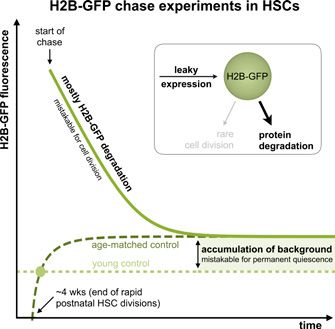
HAIR FOLLICLES RESTRICT CANCER
    GROWTH
    Normal skin corrals cancer-causing mutations and reveals how tissue can subvert
    tumorigenesis

    Normal aged skin contains cancer-caus-
    ing mutations, recent research has
    shown, but how these mutations are
    prevented from forming tumors was a
    mystery. Christiana M. Pineda, Valentina
    Greco, and colleagues at Yale University
    used unique live-tissue imaging to track
    mouse skin cells after inducing can-
    cer-causing mutations. They found that
    protection against skin cancer comes
    from a surprising place: hair follicles.

    About 30% of all cancers contain a Ras
    mutation, but these same mutations
    have been found in non-cancerous
    skin epithelia. Pineda and colleagues
    induced Ras mutations in mouse hair
    follicle cells along with a glowing red
    reporter to track the mutant stem cells            Two-photon images of the ear skin from the same mouse at day 0 and 4 months
    and their progeny. The cells persisted in          after Ras activation reveal normal follicular architecture despite the persistent pres-
    the epithelium, revealing that the body            ence of mutant cells (red).
    doesn’t just eliminate mutant cells.               Credit: Pineda et al., 2019

    Pineda and colleagues found that when
    they induced mutations in hair follicle                                                               “Our results indicate that the hair
                                                     induce some tumors, typically at sites
    stem cells, they outcompeted wild-type                                                                follicle has a unique ability to cope with
                                                     of high grooming or scratching. Imag-
    neighboring cells. They still responded                                                               Ras-activated cells. This organ is able
                                                     ing revealed that those tumors arose
    to normal tissue constraints, such as                                                                 to integrate the mutant epithelial cells
                                                     after an injury caused them to exit the
    resting phase cues, however. Even after                                                               while remaining clinically normal,” Pine-
                                                     follicular niche. To test whether injury
    a year, the transformed cells did not de-                                                             da says. There’s still much to learn about
                                                     can promote tumorigenesis within the
    velop into tumors. In contrast, targeting                                                             what’s going on in the skin that could be
                                                     follicle, they ablated hair follicle bulbs
    the Ras mutation to the upper non-cy-                                                                 applied to other cancers. “Manipulation
                                                     and the double mutant cells showed
    cling region of the skin epithelium led to                                                            of certain cell types or signaling path-
                                                     rapid, and normal, regeneration. “Once
    benign outgrowths.                                                                                    ways may enable and/or enhance the
                                                     out of the follicular niche, Hras mutant
                                                     cells can no longer be controlled and                ability of other epithelial tissues to also
    Introducing a second mutation that
                                                     contained through hair regeneration                  suppress oncogenic growth.”
    results in the loss of TGFβ signaling into
                                                     programs,” Pineda says.
    Ras-mutant hair follicle stem cells did

    RESEARCHER DETAILS                                                      ORIGINAL PAPER
                                                                            Pineda, C.M., D.G. Gonzalez, C. Matte-Martone , J. Boucher , E. Lathrop , S. Gallini ,
                                                                            N.R. Fons, T. Xin , K. Tai, E. Marsh , D. X. Nguyen , K.C. Suozzi , S. Beronja , and V.
                                                                            Greco. 2019. Hair follicle regeneration suppresses Ras-driven oncogenic growth. J.
                                                                            Cell Biol. 218:3212-3222.
                                                                            https://doi.org/10.1083/jcb.201907178

    Cristiana M. Pineda   Valentina Greco
    Graduate Student      Professor
    Yale University       Yale University
                          valentina.greco@yale.edu

8
CENTROMERES DRIVE ASYMMETRIC
STEM CELL DIVISION
Asymmetric incorporation of the centromeric histone CENP-A during G2/M regulates
the fate of Drosophila female germline stem cells
Stem cells divide asymmetrically to pro- deposited at centromeres after DNA
duce one new daughter stem cell and replication but before chromosome
one daughter cell that will subsequently segregation (i.e., during the G2/M
differentiate. One mechanism that has transition). The researchers saw similar
been proposed to contribute to this dynamics in Drosophila neural stem
asymmetry is the selective inheritance cells as well.
of sister chromatids that carry specific
epigenetic marks between the stem and Dattoli, Dunleavy, and colleagues found
daughter cell. Upon division, distinct that the cell cycle regulator CYCLIN A
epigenetic marks on sister chromatids promotes CENP-A deposition in GSCs, Superresolution image of a meta-
can result in differential gene expres- whereas CYCLIN B, and its down- phase GSC (captured using the
sion in the two resulting daughter cells. stream target HASPIN kinase, prevent H3T3P marker, green) shows asym-
According to this “silent sister hypothe- excessive CENP-A incorporation. But metric levels of CENP-A (red) at the
sis,” the centromeric regions of each sis- CENP-A isn’t deposited evenly on each centromeres of sister chromatids.
ter chromatid—which are crucial for cell set of sister chromatids: superresolution Centromeres of the chromatids
division and are epigenetically defined microscopy revealed that more CENP-A destined for the daughter GSC have
by the histone H3 variant CENP-A— is incorporated into the centromeres approximately 20% more CENP-A
of chromatids that are destined to be compared to centromeres of the
would also be distinct to facilitate their
segregated into the daughter cell that chromatids destined to be inherited
selective segregation.
by the cystoblast (CB) that will under-
maintains its stem cell identity. Accord-
go differentiation.
In human cells, CENP-A is assembled ingly, these chromatids assembled larg-
into centromeres at the end of mito- er kinetochores on their centromeres Credit: Dattoli et al., 2020
sis. “However, centromere assembly and captured more spindle microtu-
dynamics can differ among metazoans bules, providing a potential mechanism
and also among different cell types in to bias chromosome segregation. “Our results provide the first functional
the same organism,” says Elaine Dun- evidence that centromeres have a role
leavy from the National University of Overexpressing CENP-A in GSCs, in the epigenetic pathway that specifies
Ireland Galway. “To date, little is known or depleting HASPIN kinase, caused stem cell identity,” Dunleavy says. “Fur-
about centromere assembly dynamics CENP-A to be evenly deposited on thermore, our data support the silent
and functions during stem cell asym- sister chromatids. This is likely to result sister hypothesis in which centromeres
metric divisions.” in the random segregation of sister drive the selective segregation of sister
chromatids, which, according to the chromatids and the asymmetric division
Dunleavy’s team, including first author silent sister hypothesis, would promote of stem cells.”
Ada Dattoli, followed the dynamics of stem cell self-renewal. Indeed, overex-
centromere assembly in Drosophila pressing CENP-A or depleting HASPIN
female germline stem cells (GSCs) and increased the overall number of GSCs
found that, in these cells, CENP-A is in fly ovaries.
RESEARCHER DETAILS ORIGINAL PAPER
Dattoli, A.A., B.L. Carty, A.M. Kochendoerfer, C. Morgan, A.E. Walshe, and E.M.
Dunleavy. 2020. Asymmetric assembly of centromeres epigenetically regulates stem
cell fate. J. Cell Biol. 219:e201910084.
https://doi.org/10.1083/jcb.201910084
Anna Ada Dattoli (current affiliation) Elaine M. Dunleavy
Senior Postdoctoral Fellow Senior Lecturer in Biochemistry
University of Pennsylvania Perelman School Centre for Chromosome Biology, National
of Medicine University of Ireland Galway
anna.dattoli@pennmedicine.upenn.edu elaine.dunleavy@nuigalway.ie
9NCAM REGULATES NEURAL
PROGENITOR CELL FATE
Neural cell adhesion molecule controls the proliferation and differentiation of cortical
progenitors by binding to the actin polymerization regulator profilin2
Development of the cerebral cortex entiation, the researchers performed a
depends on a precisely coordinated yeast two-hybrid screen and found that
program of neural progenitor cell (NPC) an intracellular region of NCAM140, a
proliferation, migration, and differenti- splice variant of NCAM, binds to profil-
ation. The developmental program ini- in2, a key regulator of actin polymeriza-
tially generates cortical neurons before tion. Profilin2 showed a similar dynamic
switching to the production of astro- expression pattern in NPCs during
cytes and, after birth, oligodendrocytes. cortical development, and depleting
the protein altered NPC proliferation
Neural cell adhesion molecule (NCAM) and differentiation in vitro, favoring glial At embryonic day 16, there are many
has a variety of functions within the cell formation at the expense of neuron more glial fibrillary acidic protein–
nervous system and misexpression of production. positive (GFAP+) astrocytes (green)
this protein can alter the proliferation in the developing cortex of a mouse
and differentiation of NPCs. “Howev- Li and colleagues confirmed that NCAM whose NPCs lack NCAM (right).
er, it was unknown whether NCAM is regulates the proliferation and fate Credit: Huang et al., 2020
an intrinsic modulator of NPCs during of NPCs by binding to profilin2 and
cortical development,” says Shen Li promoting actin polymerization. F-actin
from Dalian Municipal Central Hospital levels were reduced in NCAM-de-
in China. ficient NPCs and an NCAM mutant tal diseases associated with abnormal
unable to bind to profilin2 was unable NCAM function,” Li says. “Understand-
Li and colleagues, including co-corre- to rescue actin dynamics or normalize ing the molecular mechanisms under-
sponding author Quan-Hong Ma from NPC proliferation and differentiation. lying these abnormalities may help in
Soochow University, found that NCAM NCAM-modulated actin dynamics may the design of future strategies aimed at
is prominently expressed in mouse influence NPCs in several ways. One correcting neural differentiation in the
NPCs during the early, neurogenic effect noticed by Li and colleagues is affected brain.”
stages of cortical development, but its that mitotic NPCs fail to round up in
levels decline during the later, gliogenic NCAM-deficient mice, which is likely to
periods when astrocytes and oligoden- impair the cells’ progression through
drocytes are produced. NPC-specific the cell cycle.
deletion of the NCAM gene caused a
transient reduction in NPC proliferation Single nucleotide polymorphisms in the
and a delay in cortical neuron genera- NCAM gene and/or defects in NCAM
tion, while astrocyte and oligodendro- proteolysis or glycosylation have been
cyte formation occurred earlier than linked to a variety of neurological
normal. disorders, including autism. “Our study
suggests that abnormalities in temporal
To understand how NCAM might NPC fate decision may contribute to the
regulate NPC proliferation and differ- pathophysiology of neurodevelopmen-
RESEARCHER DETAILS ORIGINAL PAPER
Huang, R., D.-J. Yuan, S. Li, X.-S. Liang, Y. Gao, X.-Y.
Lan, H.-M. Qin, Y.-F. Ma, G.-Y. Xu, M. Schachner, V.
Sytnyk, J. Boltze, Q.-H. Ma, and S. Li. 2020. NCAM
regulates temporal specification of neural progenitor
cells via profilin2 during corticogenesis. J. Cell Biol.
219:e201902164.
https://doi.org/10.1083/jcb.201902164
Rui Huang De-Juan Yuan Shao Li Quan-Hong Ma Shen Li
Graduate student Graduate student Professor Professor Professor
Dalian Medical Dalian Medical Dalian Medical Soochow University Dalian Municipal Cen-
University University University maquanhong@suda. tral Hospital affiliated
edu.cn with Dalian Medical
10 University
listenlishen@hotmail.
comGLUTAMYLATION REGULATES HSC SELF-
RENEWAL
Reversible modification of the tumor suppressor BAP1 controls hematopoietic stem cell
self-renewal and blood cell formation
The functions of proteins within cells The researchers identified the nuclear
can be altered by a wide range of re- deubiquitinase BAP1 as a key target of
versible, posttranslational modifications, CCP3 in HSCs. The team found that
such as methylation, phosphorylation, glutamylation of BAP1 by the enzymes
and ubiquitination. Protein glutamyla- TTLL5 and TTLL7 accelerates the pro-
tion—in which glutamate side chains tein’s degradation, whereas deglutamy-
are added to the γ-carboxyl groups of lation by CCP3 stabilizes BAP1.
glutamic acid residues—was original-
ly shown to regulate the dynamics of BAP1 is a tumor suppressor linked to
microtubules. More recently, however, a range of cancers as well as the rare
the tubulin tyrosine ligase-like (TTLL) blood disorder myelodysplastic syn-
enzymes that add glutamate side drome. Fan and colleagues found that
chains, and the cytosolic carboxypepti- BAP1 promotes HSC self-renewal and
dase (CCP) enzymes that remove them, hematopoiesis by facilitating expres- Compared with wild-type (left), the
have been shown to target a variety of sion of the transcription factor Hoxa1. bone marrow of mice lacking the
proteins and regulate a range of cellular BAP1 and Hoxa1 levels are reduced in glutamylating enzymes TTLL5 and
processes. Ccp3-deficient HSCs, limiting the stem TTLL7 (DKO, right) contains an
cells’ ability to remain quiescent and increased number of cells due to an
For example, Zusen Fan, Yong Tian, and self-renew. The HSC pool is therefore increase in BAP1 levels that promotes
colleagues at the Institute of Biophysics, soon exhausted, impairing hematopoie- HSC self-renewal and hematopoiesis.
Chinese Academy of Sciences, have sis in Ccp3-knockout mice. Credit: Xiong et al., 2020
previously shown that glutamylation
regulates the development of both In contrast, HSCs from mice lacking
megakaryocytes and group 3 innate TTLL5 and TTLL7 showed increased
lymphoid cells. “However, how glutam- levels of BAP1 and Hoxa1, promoting
ylation regulates hematopoietic stem HSC quiescence and self-renewal, as
cells (HSCs), which give rise to all blood well as boosting hematopoiesis.
cell lineages, is unclear,” says Fan.
“Therefore, glutamylation and deglu-
Fan and colleagues found that the tamylation of BAP1 play a critical role
deglutamylating enzyme CCP3 is highly in the regulation of HSC self-renewal
expressed in mouse HSCs. Inhibiting and hematopoiesis,” Fan says. “We are
this enzyme, or deleting the Ccp3 gene, currently exploring the molecular mech-
impaired HSC self-renewal and hemato- anism by which BAP1 glutamylation
poiesis, resulting in reduced HSC num- modulates Hoxa1 expression in a direct
bers in the bone marrow and decreased or indirect manner in HSCs.”
peripheral blood cell counts.
RESEARCHER DETAILS ORIGINAL PAPER
Xiong, Z., P. Xia, X. Zhu, J. Geng, S. Wang, B. Ye, X. Qin,
Y. Qu, L. He, D. Fan, Y. Du, Y. Tian, and Z. Fan. 2020.
Glutamylation of deubiquitinase BAP1 controls self-re-
newal of hematopoietic stem cells and hematopoiesis.
J. Exp. Med. 217:e20190974.
https://doi.org/10.1084/jem.20190974
Zhen Xiong Yong Tian Zusen Fan Photo not available Photo not available
Graduate student Professor Professor Pengyan Xia Xiaoxiao Zhu
Institute of Biophysics Institute of Biophysics Institute of Biophysics Postdoctoral re- Postdoctoral re-
Chinese Academy of Chinese Academy of Chinese Academy of searcher searcher
Sciences Sciences Sciences Institute of Biophysics Institute of Biophysics
ytian@ibp.ac.cn fanz@moon.ibp.ac.cn Chinese Academy of
Sciences
Chinese Academy of
Sciences
11THE CHROMATIN LANDSCAPE OF
GLIOBLASTOMA
Profiling of glioblastoma stem cells and primary tumor samples identifies subgroup-
specific transcriptional regulatory circuits and novel therapeutic targets
Glioblastoma is the most common form high expression of this GSC signature
of primary brain tumor and is largely showed increased malignancy and poor
incurable. Driven by therapy-resistant prognosis. Moreover, knocking down
glioblastoma stem cells (GSCs), glio- many of these SE-regulated genes im-
blastomas show high levels of pheno- paired GSC proliferation in vitro.
typic heterogeneity both between and
within individual tumors. In addition to this core signature
common to the vast majority of GSCs,
Cellular states are generally defined by further analysis revealed that pa-
a small number of master transcription tient-derived GSCs cluster into at
factors that bind to super-enhancers least two different groups with distinct
(SEs), driving the expression of numer- chromatin signatures defined by unique
ous genes essential for establishing SE activities and transcription factors.
cellular identity, including the master These different GSC states could also
transcription factors themselves. To be identified in primary tumor samples
define the core regulatory circuits that from glioblastoma patients.
define glioblastoma cell states, a team Mapping histone H3 acetylation
of researchers led by Jeremy Rich and Knocking down group-specific tran- reveals the distinct patterns of SE
Xiuxing Wang at the University of Cali- scription factors had group-specific activity in group 1 and group 2 GSCs.
fornia, San Diego, and Charles Lin, Ste- effects. For example, knocking down
Credit: Mack et al., 2019
phen Mack, and Irtisha Singh at Baylor RUNX2, a transcription factor uniquely
College of Medicine, profiled a large active in group 2 GSCs, inhibited the
cohort of patient-derived GSCs and growth of tumors derived from group
primary glioblastoma tumor samples. 2 GSCs, but had no effect on tumors “In sum, our data reveal the existence
derived from group 1 GSCs. of distinct GSC populations defined by
“We mapped active enhancer land- unique chromatin signatures aris-
scapes and integrated this with profiles “Transcription factors are notoriously ing from specific regulatory circuits
of gene expression, DNA methylomes, challenging to target therapeutically,” composed of SEs and SE-associated
copy number variations, and whole says Rich. “However, because these transcription factors,” Rich says. “These
exomes to identify the core transcrip- transcription factors are associated with SE programs that regulate GSC identity
tion factors and other SE-associated SEs, we were able to identify other es- (both core and group specific) can be
genes that establish and maintain GSC sential genes that are more amenable to used for the discovery of new therapeu-
identity,” Rich explains. therapeutic targeting.” For example, the tic targets.”
researchers found that group 1 GSCs
The researchers uncovered a set of show SE-mediated activation of MAPK/
SEs and genes active in >75% of GSCs, ERK signaling and can thus be spe-
allowing them to derive a core gene cifically targeted by a small molecule
signature for these cells. Tumors with inhibitor against this pathway.
RESEARCHER DETAILS ORIGINAL PAPER
Mack, S.C., I. Singh, X. Wang, R. Hirsch, Q. Wu, R.
Villagomez, J.A. Bernatchez, Z. Zhu, R.C. Gimple,
L.J.Y. Kim, A. Morton, S. Lai, Z. Qiu, B.C. Prager, K.C.
Bertrand, C. Mah, W. Zhou, C. Lee, G.H. Barnett,
M.A. Vogelbaum, A.E. Sloan, L. Chavez, S. Bao, P.C.
Scacheri, J.L. Siqueira-Neto, C.Y. Lin, and J.N. Rich.
2019. Chromatin landscapes reveal developmentally
Stephen C. Mack Charles Y. Lin Photo not available Photo not available Photo not available
encoded transcriptional states that define human
Assistant Professor Assistant Professor Irtisha Singh Xiuxing Wang Jeremy N. Rich glioblastoma. J. Exp. Med. 216:1071–1090.
Baylor College of Baylor College of Postdoc fellow Postdoctoral re- Professor of Medicine https://doi.org/10.1084/jem.20190196
Medicine Medicine Baylor College of searcher University of California,
scmack@bcm.edu charles.y.lin@bcm.edu Medicine University of California, San Diego
12 (Currently an assistant
professor at Texas
San Diego drjeremyrich@gmail.
com
A&M University)PRIMITIVE HSCS CONTINUE CYCLING
INTO ADULTHOOD
Study reveals that, contrary to a previous report, the most primitive hematopoietic
stem cells do not become permanently quiescent and, instead, show continuous
mitotic activity in adult mice
Hematopoietic stem cells (HSCs) are responding author Thomas Zerjatke,
multipotent adult stem cells that can found that in three different H2B-FP
give rise to every type of blood cell mouse strains—including the one used
throughout an individual’s lifetime. The in the 2016 study—leaky expression
mitotic activity of HSCs can be tracked even in the absence of induction causes
in vivo using an inducible “pulse” of fluorescent histone label to gradually
fluorescent histone fusion protein (H2B- accumulate in HSCs. This age-depen-
FP) which labels the cells’ chromatin. dent background fluorescence accu-
This is followed by a “chase” period mulation was particularly prominent in
during which quiescent HSCs retain quiescent HSCs with high repopulating
the fluorescent label while proliferating activity. “We argue that this accumulat-
HSCs pass it on to their daughters, ed background can be easily misinter-
gradually diluting the fluorescent signal preted as stable retention of induced
within a few cell divisions. label,” Zerjatke says.
In 2016, a high-profile study used this On the other hand, Gerbaulet and
approach in mice to identify a small colleagues found that the half-life of flu- Scheme recapitulates key features of
population of primitive HSCs containing orescent histones is within the range of pulse-chase experiments to identify
all of the HSC compartment’s long- 2–6 weeks, meaning that protein degra- quiescent HSCs retaining H2B-GFP
term repopulating activity. These cells dation represents the major contributor label. Infrequently dividing HSCs
appeared to undergo four self-renewing to loss of fluorescenct label in rarely dilute fluorescent label mostly by pro-
divisions during the first year of life dividing cells. “This precludes very long tein degradation. In parallel, continu-
before entering a state of permanent chase periods and neglecting label ous leaky background expression of
quiescence, apparently allowing them degradation will result in a massive H2B-FP progressively accumulates in
to retain fluorescent histone labeling for overestimation of the divisional activity quiescent HSCs and could be mistak-
as long as 22 months. Based on this, the of quiescent HSCs,” says Morcos. en for permanent quiescence.
authors hypothesized that HSCs count Credit: Morcos et al., 2020
and remember cell divisions. “Howev- The researchers built a new mathe-
er, their model considered neither the matical model for pulse-chase labeling
impact of continuous leaky background of HSCs with fluorescent histones, baulet and colleagues saw no evidence
expression of the fluorescent histone incorporating the impact of leaky for HSCs counting and remembering a
label, nor the division-independent loss background expression and protein set number of divisions before abrupt-
of fluorescence due to protein degrada- degradation. The model successfully re- ly entering permanent quiescence.
tion,” says Alexander Gerbaulet from the capitulated experimental observations, “Instead, our data suggest that primitive
Institute for Immunology at TU Dresden. including the preferential accumulation HSCs continue to cycle in aged mice,
of background fluorescence in more undergoing infrequent but steady mitot-
Gerbaulet and colleagues, including quiescent HSCs. Using their model to ic divisions,” Gerbaulet says.
first author Mina Morcos and co-cor- infer the mitotic activity of HSCs, Ger-
RESEARCHER DETAILS ORIGINAL PAPER
Alexander Gerbaulet Morcos, M.N.F., T. Zerjatke, I. Glauche, C.M. Munz, Y. Ge, A. Petzold, S. Reinhardt, A.
Group Leader Dahl, N.S. Anstee, R. Bogeska, M.D. Milsom, P. Säwén, H. Wan, D. Bryder, A. Roers,
Institute for Immunology, Carl Gustav Carus
and A. Gerbaulet. 2020. Continuous mitotic activity of primitive hematopoietic stem
Faculty of Medicine, TU Dresden
alexander.gerbaulet@tu-dresden.de cells in adult mice. J. Exp. Med. 217:e20191284.
Mina N.F. Morcos https://doi.org/10.1084/jem.20191284
PhD student
Institute for Immunology, Carl Gustav Carus
Faculty of Medicine, TU Dresden
Thomas Zerjatke
PhD student
Institute for Medical Informatics and Biometry,
Carl Gustav Carus Faculty of Medicine, TU
Dresden
thomas.zerjatke@tu-dresden.de
13DISTINGUISHING PHYSIOLOGIC AND
ONCOGENIC WNT SIGNALS
Profiling of isogenic human colon organoids reveals differences between oncogenic
Wnt activation and normal Wnt signaling essential for stem cell maintenance
Around 80% of colorectal cancers signaling (induced by exogenous Wnt
(CRCs) carry truncating mutations in Ad- ligands) upregulate distinct sets of genes
enoma polyposis coli (APC). Loss of the and proteins, no matter how strongly the
APC protein stabilizes the transcriptional two signaling pathways are activated.
coactivator β-catenin, a key component
of the Wnt signaling pathway, leading Many of the proteins specifically upregu-
to constitutive Wnt signaling and the lated by physiological Wnt signaling are
formation of benign adenomas that can expressed in intestinal crypts, suggest-
progress into CRC upon the acquisition ing that they could be new markers for
of further mutations. CRC cells rely on normal intestinal stem cells. On the other
Wnt signaling to continue proliferating hand, many of the proteins specifically
but, because Wnt activity also regulates upregulated in response to oncogenic
the proliferation and renewal of intestinal Wnt signaling are only expressed in tu-
stem cells critical for maintaining gut mors, and could therefore represent new
homeostasis, inhibiting this pathway has biomarkers or even therapeutic targets.
highly toxic side effects in patients.
Some studies have shown that high
Developing successful therapies might Wnt activity is associated with favorable
therefore require identifying differences outcomes in CRC, whereas other studies
between normal Wnt signaling and the have suggested that the presence of To distinguish physiological and
constitutive activation induced by onco- Wnt-active stem cells correlates with oncogenic Wnt signaling, Farin and
genic mutations in APC or other genes. increased tumor invasiveness and higher colleagues profiled isogenic pairs of
“We set out to catalog the physiological rates of relapse. “We therefore wanted colon organoids, cultured in the pres-
to test if our organoid-derived signatures ence or absence of Wnt ligands, and
and oncogenic Wnt responses in primary
are associated with distinct clinical out- carrying either wild-type or truncated
human colon epithelial cells on the
comes in CRC,” says Farin. versions of the APC gene.
transcriptome and proteome level,” says
Henner Farin from the German Cancer Credit: Michels et al., 2019
Consortium and Georg-Speyer-Haus, Farin and colleagues found that the
Goethe University Frankfurt. oncogenic Wnt signature was associated
with good prognosis in the canonical “Oncogenic and physiological Wnt
Farin and colleagues, including co-first CRC subtype CMS2, consistent with the responses therefore represent two
authors Birgitta Michels and Mohammed role of APC mutations in this subtype independent and opposing prognostic
Mosa, analyzed isogenic colon organoids and possibly reflecting an increased determinants in CRC,” Farin says. “Rather
grown from intestinal cells isolated from resemblance to benign adenomas. In than Wnt activity per se, stratification
healthy human subjects. RNA sequenc- contrast, the physiological Wnt signature of specific downstream responses may
ing and quantitative mass spectrometry predicted poor outcomes in CMS4 CRC, be more informative on the status of
revealed that oncogenic Wnt signaling possibly reflecting tumors with increased tumors.”
(induced by the CRISPR/Cas9-mediated stemness and invasiveness.
truncation of APC) and physiological
RESEARCHER DETAILS ORIGINAL PAPER
Michels, B.E., M.H. Mosa, B.M. Grebbin, D. Yepes, T. Darvishi, J. Hausmann, H.
Urlaub, S. Zeuzem, H.M. Kvasnicka, T. Oellerich, and H.F. Farin. 2019. Human colon
organoids reveal distinct physiologic and oncogenic Wnt responses. J. Exp. Med.
216:704–720.
https://doi.org/10.1084/jem.20180823
Birgitta E. Michels Mohammed H. Mosa Henner F. Farin
Graduate student Postdoctoral researcher Group Leader
German Cancer Consortium German Cancer Consortium German Cancer Consortium
Georg-Speyer-Haus Georg-Speyer-Haus Georg-Speyer-Haus
Goethe University Frankfurt Goethe University Frankfurt Goethe University Frankfurt
14 Photo © Georg-Speyer-Haus Photo © Georg-Speyer-Haus farin@gsh.uni-frankfurt.de
Photo © Georg-Speyer-HausLEUKEMIA HIJACKS MYELOID
REGENERATION PATHWAYS
Correcting aberrant Notch and Wnt signaling in leukemic stem cells might normalize
myeloid cell production in a number of different leukemias
In 2015, Emmanuelle Passegué and pathways. Passegué and colleagues
colleagues described how hematopoi- determined that, at steady state, Notch
etic stem cells (HSCs) maintain blood activity is high and Wnt activity is low
production at steady state by giving in HSCs and MPP4s. MPP3s, in con-
rise to a mix of multipotent progenitors trast, show low Notch and high Wnt
(MPPs). Some, known as MPP2 and activity. Accordingly, inhibiting Notch
MPP3, are biased toward producing or boosting Wnt signaling triggered the
myeloid lineage cells such as macro- early stages of myeloid regeneration Staining for nuclear β-catenin (red)
phages and neutrophils, while others, by promoting the production of MPP3s shows that activity of the Wnt signal-
known as MPP4, are biased toward from HSCs. ing pathway is high in myeloid-biased
lymphoid lineage cells like T and B MPP3 cells but low in normal HSCs
cells. They also showed that in times Myeloid leukemias are driven by leu- and lymphoid-biased MPP4 cells.
of stress, (following HSC transplanta- kemic stem cells (LSCs) that are often Credit: Kang et al., 2020
tion, for example), HSCs regenerate the resistant to treatment and can pro-
blood system by inducing emergency mote tumor recurrence. Passegué and
myelopoiesis pathways that gener- colleagues found that Notch activity is
new treatment options to limit aberrant
ate more MPP2 and MPP3 cells than reduced and Wnt signaling is elevated
myeloid cell production,” Passegué says.
normal, and reprogram MPP4 cells to in LSCs in mice, further supporting
“This is particularly relevant for patients
produce more myeloid lineage cells. the idea that myeloid leukemias hijack
with no identified driver mutations or
myeloid regeneration pathways and
who relapsed with resistance muta-
Together with Yoon-A Kang and Eric Pi- providing therapeutic targets. “We
tions. The next step will be to identify
etras, Passegué wondered whether this found that increasing Notch activity or
druggable targets that could appro-
myeloid regeneration pathway might decreasing Wnt activity in mouse LSCs
priately increase Notch activity while
be constitutively activated in myeloid has very similar effects in correcting
limiting Wnt activity, providing a specific
leukemias. “Using several different myeloid cell production, delaying dis-
anti-LSC therapy that could block their
mouse models, we found that MPP3 ex- ease progression, and improving overall
enhanced and overly biased myeloid
pansion and myeloid reprogramming of survival,” Passegué says.
differentiation potential.”
MPP4 are common features of myeloid
leukemia and reflect the hijacking of a Notably, similar attempts to increase
normally transiently activated pathway Notch or decrease Wnt activity in
of emergency myelopoiesis,” Passegué normal HSCs had no effect, with
says. compensatory crosstalk between the
two signaling pathways appearing to
The molecular mechanisms regulating prevent abnormal MPP function and
the production of different MPPs from blood production. “Our results identify
HSCs are unknown but are likely to a common mechanism for myeloid leu-
involve the Notch and Wnt signaling kemia development that could provide
RESEARCHER DETAILS ORIGINAL PAPER
Kang, Y.-A., E.M. Pietras, and E. Passegué. 2020. Deregulated Notch and Wnt
signaling activates early-stage myeloid regeneration pathways in leukemia. J. Exp.
Med. 217:e20190787.
https://doi.org/10.1084/jem.20190787
Yoon-A Kang Eric M. Pietras Emmanuelle Passegué
Postdoctoral Fellow Assistant Professor Professor of Genetics and
Columbia University University of Colorado An- Development
schutz Medical Campus Director, Columbia Stem Cell
Initiative
Columbia University Irving
Medical Center
15
ep2828@cumc.columbia.eduYou can also read