Adult stem cells and tissue engineering strategies for salivary gland regeneration: a review
←
→
Page content transcription
If your browser does not render page correctly, please read the page content below
Yoo et al. Biomaterials Research 2014, 18:9
http://www.biomaterialsres.com/content/18/1/9

 REVIEW                                                                                                                                       Open Access

Adult stem cells and tissue engineering strategies
for salivary gland regeneration: a review
Chankee Yoo1,2, Jeremy B Vines1, Grant Alexander1, Kyle Murdock1, Patrick Hwang1 and Ho-Wook Jun1*

  Abstract
  Saliva is an important compound produced by the salivary glands and performs numerous functions.
  Hyposalivation (dry mouth syndrome) is a deleterious condition often resulting from radiotherapy for patients with
  head and neck cancer, Sjogren’s Syndrome, or as a side effect of certain medications. Hyposalivation negatively
  affects speaking, mastication, and swallowing in afflicted patients, greatly reducing their quality of life. Current
  treatments for this pathology include modifying lifestyle, synthetic saliva supplementation, and the utilization of
  salivary gland stimulants and sialagogues. However, many of these treatments do not address the underlying issues
  and others are pervaded by numerous side effects. In order to address the shortcomings related to current
  treatment modalities, many groups have diverted their attention to utilizing tissue engineering and regenerative
  medicine approaches. Tissue engineering is defined as the application of life sciences and materials engineering
  toward the development of tissue substitutes that are capable of mimicking the structure and function of their
  natural analogues within the body. The general underlying strategy behind the development of tissue engineered
  organ substitutes is the utilization of a combination of cells, biomaterials, and biochemical cues intended to
  recreate the natural organ environment. The purpose of this review is to highlight current bioengineering
  approaches for salivary gland tissue engineering and the adult stem cell sources used for this purpose.
  Additionally, future considerations in regard to salivary gland tissue engineering strategies are discussed.
  Keywords: Hyposalivation, Mesenchymal stem cells, Tissue engineering, Salivary gland, Xerostomia

Introduction                                                                        patients with head and neck cancer [5], and results of
Saliva is a watery substance produced by the salivary                               side effects from various medications and ectodermal
glands. It consists of mucus, various electrolytes, glyco-                          dysplasias [6-8].
proteins, enzymes, and antibacterial compounds such as                                Current treatments for hyposalivation include symp-
lysozyme and IgA [1]. In relation to its excretory com-                             tom management, lifestyle changes, synthetic saliva sup-
position, saliva performs many important functions such                             plementation, and the utilization of salivary gland
as breaking down starch into maltose for digestion, aid-                            stimulants and sialagogues (e.g., the muscarinic receptor
ing in basic oral functions (e.g. speaking, mastication,                            agonists pilocarpine and cevimeline) which stimulate sal-
and swallowing), and protecting against microbial re-                               ivary secretion from residual acinar cells [9,10]. Salivary
lated pathologies such as dental caries and periodontitis.                          gland stimulation via supplementation of cytokines or
Thus, any dysfunction or cessation to saliva production                             small molecules such as pilocarpine or amifostine to in-
is a significant clinical concern. Hyposalivation, which is                         crease the secretion of saliva is the most commonly used
a characteristic of xerostomia (dry mouth syndrome),                                treatment. However, this only treats surface level symp-
can significantly reduce the quality of life for afflicted                          toms, provides temporary relief, and is plagued by mul-
patients [2,3]. Major causes of hyposalivation include                              tiple multiple side effects such as excessive sweating,
Sjögren’s Syndrome (SS) [4], γ-irradiation therapy for                              chills, dizziness, excessive tearing, flushing, voice change,
                                                                                    stuffy nose, tremor, nervousness, and diarrhea [11,12].
* Correspondence: hwjun@uab.edu                                                       Thus, development of an alternative treatment to pro-
1
 Department of Biomedical Engineering, University of Alabama at
Birmingham, Shelby Building 806, 1825 University Boulevard, Birmingham,             vide a long-term and effective solution solution is im-
AL 35294, USA                                                                       perative [13]. In order to address the limits associated
Full list of author information is available at the end of the article

                                       © 2014 Yoo et al.; licensee BioMed Central Ltd. This is an Open Access article distributed under the terms of the Creative
                                       Commons Attribution License (http://creativecommons.org/licenses/by/4.0), which permits unrestricted use, distribution, and
                                       reproduction in any medium, provided the original work is properly credited. The Creative Commons Public Domain
                                       Dedication waiver (http://creativecommons.org/publicdomain/zero/1.0/) applies to the data made available in this article,
                                       unless otherwise stated.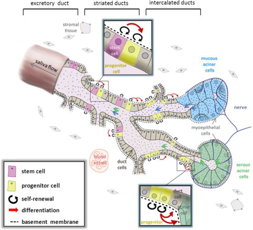
Yoo et al. Biomaterials Research 2014, 18:9                                                                          Page 2 of 12
http://www.biomaterialsres.com/content/18/1/9

with current therapies, many research groups have in-              while mucous producing acinar cells are cuboidal in
vestigated various regenerative medicine strategies utiliz-        shape and group together to form tubules. Myoepithelial
ing different stem cell sources to engineer artificial             cells are located near the ductal openings and serve to
salivary tissues that can mitigate the effects of xerosto-         contract the ducts in order to squeeze out salivary secre-
mia and hyposalivation.                                            tions (Figure 1) [18].
   Tissue engineering is defined as the application of life sci-      The effects of radiotherapy on salivary gland cells are
ences and materials engineering toward the development of          confounding. Theoretically, saliva producing acinar cells
tissue substitutes that are capable of mimicking the struc-        are not predicted to be radiation sensitive due to the fact
ture and function of their natural analogues within the body       that they are post-mitotic in nature [19]. However, des-
[14]. The general underlying strategy behind the develop-          pite this, irradiated salivary glands exhibit severe, early
ment of tissue engineered organ substitutes is the utilization     losses in saliva production [20]. In this regard, it is dis-
of a combination of cells, biomaterials, and biochemical cues      puted as to whether radiation induced hyposalivation is
intended to recreate the natural organ environment. This           a result of apoptosis or dysfunction resulting from radi-
article is intended to provide a review of the most current        ation induced damage to the membranes of acinar cells
approaches to utilizing stem cells and bioengineering princi-      [18,21-24]. During the later phases of radiotherapy in-
ples for the purpose of salivary tissue regeneration.              duced hyposalivation, functionally mature acinar cells
                                                                   cease their proliferation and are not replaced. It is sug-
Review                                                             gested that the population responsible for replacing ma-
Anatomy of the salivary glands                                     ture acinar cells, known as salivary gland progenitor cells
Within the body, there are three main sources of saliva            (SSPCs), lose their regenerative capability as a result of ra-
production: The parotid gland, the submandibular gland,            diation induced damage as well [20,25,26]. Due to this
and the sublingual gland. Additionally, there are 800–100          fact, much effort has been expended towards assessing
minor salivary glands throughout the oral cavity that pro-         various stem cell sources and materials to serve as poten-
duce a small amount of saliva, although, this amount is            tial replacements for SSPCs damaged during radiotherapy.
considerably less than the three major salivary organs [15].          The purpose of this review is to highlight current bio-
  Each of the salivary glands is located in various locations      engineering approaches for salivary gland tissue engin-
throughout the oral cavity in humans [16]. The parotid             eering and the adult stem cell sources used for this
glands are the largest of the salivary glands and are located      purpose. Some of the most currently studied adult de-
at the back of the mouth adjacent to the mandibular                rived stem cell populations for the purpose of salivary
ramus. The submandibular glands are a pair of glands lo-           gland regeneration include salivary gland-derived stem
cated superior to the diagastric muscles and below the             cells (SGSCs), mesenchymal stem cells (MSCs), and hu-
jaws, juxtaposed and perpendicular to the mandibular               man amnion epithelial stem cells (hAECs).
ramus. The saliva produced by the submandibular glands
accounts for roughly 70% of the saliva present within the
oral cavity, despite the fact that they are smaller than the       Salivary gland-derived stem cells
parotid glands. The sublingual glands are a pair of glands         Since adult stem cells are generally restricted to cell line-
located anterior to the submandibular glands and inferior          ages of the body part of origin, researchers have attempted
to the tongue and are responsible for the production of            to use stem cells derived from salivary glands (SGSCs) to
around 5% of the saliva present within the oral cavity.            reduce hyposalivation and restore natural function [17].
Each of the glands is encapsulated within connective tis-          Thus far, several approaches have been taken towards iso-
sue and arranged into lobules.                                     lating and characterizing these stem cells. Some groups
  In terms of their growth and development, each of                have sequestered cells from the parotid gland [27], the
these organs are formed from two different layers of the           submandibular gland [28], a combination of both glands
three primary embryonic germ layers [17]. The subman-              [29], and by co-culture [30].
dibular and sublingual glands are derived from the endo-             Stem cells from the human parotid gland, removed via
derm, whereas the parotid gland is of ectodermal origin.           lateral parotidectomy, have been isolated and characterized
The parotid gland produces a serous, watery secretion              in vitro [27]. Flow cytometry analysis showed that these
while the submandibular and sublingual glands produce              stem cells were strongly positive for classic MSC markers
secretions that contain mucus as well [16].                        (CD13, CD29, CD44, and CD90) and negative for key
  The salivary glands of most mammals are made up of               hematopoietic stem cell (HSC) markers (CD34, CD45).
three main cell types: serous producing acinar cells, mu-          These SG-derived stem cells further displayed MSC-like
cous producing acinar cells, and myoepithelial cells [16].         characteristics by demonstrating the ability for adipo-
Serous producing acinar cells have a pyramidal morph-              genic, osteogenic, and chondrogenic differentiation when
ology and are joined together to form spheroidal shapes            grown in their respective induction media.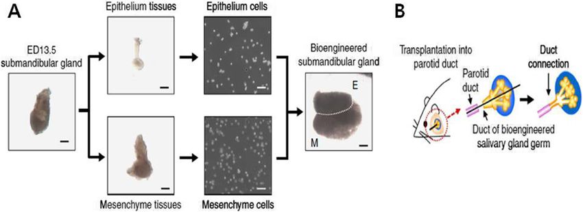
Yoo et al. Biomaterials Research 2014, 18:9                                                                              Page 3 of 12
http://www.biomaterialsres.com/content/18/1/9

 Figure 1 Schematic representation of a generic salivary gland showing component cell types and theorized stem and progenitor cell
 locations. Reproduced with permission from: [18].

   Stem cells isolated from a combination of the human              spheres. Initial H&E, Periodic Acid Schiff (PAS), CK7,
parotid and submandibular glands were revealed to have              and CK14 staining showed that cultured spheres con-
a certain capacity for in vivo recovery of salivary gland           tained acinar and ductal cells. Interestingly, acinar cells
function in radiation-damaged rat salivary glands [29].             mostly disappeared by the third day but reappeared
Prior in vitro experiments confirmed that the SGSCs                 within the existing ductal spheres by the fifth day in cul-
expressed MSC markers (CD44, CD49f, CD90, and                       ture. By day ten, acinar cells dominated sphere compos-
CD105), excluded HSC markers (CD34, CD45), differen-                ition and amylase expression, quantified using RT-PCR,
tiated into MSC lineages, and could differentiate into              increased almost 25-fold after 20 days (Figure 2).
amylase-expressing cells. Radiation-induced hyposaliva-                The in vitro results suggest that these sphere-forming
tion in rats was generated using an x-ray irradiator and            cells originate from salivary gland ducts and are able to
human SGSCs (hSGSCs) were transplanted into the                     differentiate into amylase-producing, acinar-like cells.
glands. After 60 days, the average saliva flow rate of the          To analyze the stem cell characteristics of these spheres
irradiation-damaged, hSGSC-treated group was twice                  (now termed salispheres), common stem cell markers
that of the PBS-treated, irradiation-damaged group but              (Sca-1, c-Kit, and Musashi-1) were fluorescently labeled
was still lower than the undamaged group. Treatment                 and visualized in culture. Sca-1 and c-Kit expression was
with hSGSC was also quantified by measuring the rat                 seen in excretory duct cells but not acinar cells. Results
body weight over time; the average body weight of                   from the H&E/PAS staining were confirmed by the
hSGSC-treated rats was slightly increased in comparison             fluorescent microscopy, which indicated a peak Sca-1
to the PBS-treated rats.                                            expression at 5 days.
   By using a floating sphere culture, further in vitro                Intraglandular transplantation of 3-day cultured sali-
characterization of submandibular-derived SGSCs revealed            spheres into irradiated mice resulted in the formation of
cellular expression of Sca-1, c-Kit, and Musahsi-1 [28]. Im-        ductal structures near the injection site. There was an
munohistochemical staining over a 10 day period was per-            increase in acinar cell surface area in salisphere-treated
formed to analyze the origination and development of cell           mice compared to the untreated group. Ninety days after
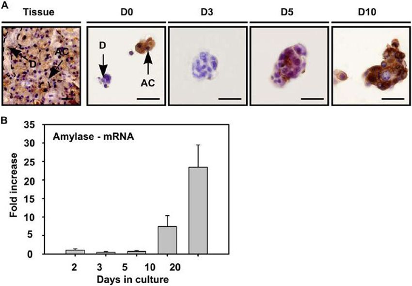
Yoo et al. Biomaterials Research 2014, 18:9                                                                                            Page 4 of 12
http://www.biomaterialsres.com/content/18/1/9

 Figure 2 Differentiation of salisphere into acinar cells. (A) Amylase expressing cells (AC) in submandibular gland tissue (Tissue) were also
 present at the onset of culture (A-D0), and were visualized in the sphere at the onset of day 5 (A-D5), whereas granulae-containing spheres
 appeared in culture at later time-points (A-D10). Antibody labeling is shown in brown, nuclei in blue. Scale bar = 50 mm. (D = duct cells, AC =
 acinar cells, D0–3– 5–10 represent days in culture). (B) Real time RT-PCR confirmed the enhanced expression of amylase during in vitro culturing
 and differentiation. Error bars represent SEM (N = 2). Amylase mRNA expression levels at 2 days of culture were normalized to one. Reproduced
 with permission from: [28].

irradiation, saliva production in salisphere-treated mice                  Bone marrow mesenchymal stem cells (MSCs)
was higher than the untreated counterparts and corre-                      Mesenchymal stem cells (MSCs) are multipotent stem
lated strongly with acinar surface area. After purifying                   cells capable of differentiating into many cell types, in-
salispheres to a c-Kit+ population, cells were capable of                  cluding chondrocytes, adipocytes, osteoblasts, acinar
differentiating into acinar cells in vitro and transplant-                 cells, and salivary epithelial cells [31-34]. Their potential
ation of a small number of cells (300–1000 per gland)                      to repair damaged tissues, anti-inflammatory effects, and
improved saliva production in 69% of irradiated mice                       low immunogenicity make MSCs strong candidates for
in vivo, after 120 days.                                                   both experimental investigations in vitro and in vivo as
  However, a major limitation for SGSC therapy in the                      well as clinical treatment of various diseases [31-35].
treatment of radiation-induced hyposalivation is the diffi-                Therefore, MSCs were investigated for regeneration and
culty with isolating autologous stem cells from a severely                 functional restoration of the salivary gland.
injured gland. To overcome this barrier, a co-culture sys-
tem of mouse embryonic stem cells (mESCs) and human                        MSC implantation and Sjögren’s syndrome
SG fibroblasts was developed to facilitate differentiation of              Two studies investigated the role of MSCs as a therapeutic
mESCs to SGSCs [30]. After 1 week in co-culture, a sig-                    option for treatment of Sjögren’s syndrome (SS), a chronic
nificant change in cell morphology was found and RT-                       autoimmune disorder that results in exocrine gland in-
PCR results showed a sudden appearance of amylase and                      flammation, impaired salivary function, and lymphocytic
bFGF. These GFP-expressing SG cells were transplanted                      infiltrates within the salivary glands [31,35]. Khalili et al.
into normal mice submandibular glands and histology was                    [35] used NOD mice with a Sjögren’s syndrome-like dis-
performed after 1 month. H&E and PAS staining of                           ease to investigate the effect of MSCs in reducing lympho-
SGSC-treated mice showed normal formation of ductal                        cytic infiltrates in the salivary gland and restoring salivary
and acinar structures. Fluorescent microscopy of the                       function (Figure 3). They found that intravenous injection
GFP-positive donor cells qualitatively confirmed the                       of MSCs reduced lymphocytic infiltrate and inflammation
cells’ ability to integrate into the existing tissue. Even                 in the salivary gland compared to untreated controls, in-
though this method is not confined by the need for au-                     cluding a 10-fold decrease in the inflammatory cytokine
tologous stem cells from a radiation-damaged gland, it                     TNF-α. MSC injection also preserved the saliva flow rate
is limited by the ethical concerns surrounding embry-                      over the 14 week post-treatment period; moreover, when
onic stem cells and their lack of availability in clinical                 MSCs were administered in conjunction with complete
settings.                                                                  Freund’s adjuvant (CFA), the salivary gland regenerative
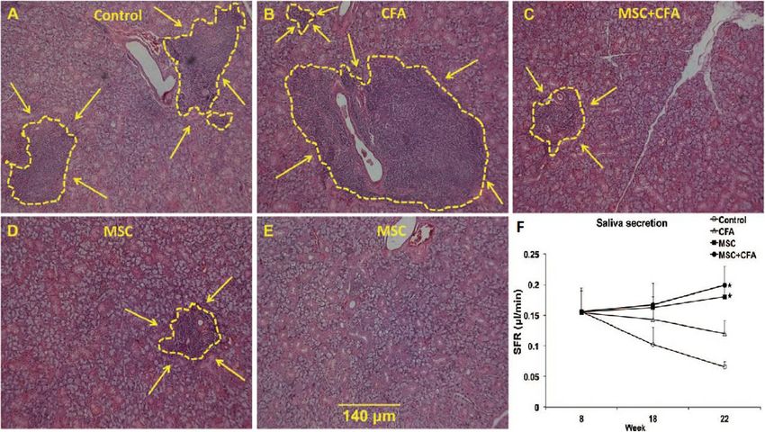
Yoo et al. Biomaterials Research 2014, 18:9                                                                                             Page 5 of 12
http://www.biomaterialsres.com/content/18/1/9

 Figure 3 Lymphocytic infiltrates in salivary glands of NOD mice. A–E: H&E staining showing lymphocytic infiltration size (shown by yellow
 line and arrows) in NOD that were: untreated (A), CFA-treated (B), MSC + CFA (C), MSC (D). In 2 of the 5 NOD mice transplanted with MSCs only,
 no lymphocytic infiltrates were noted (E). Scale bar: 140 um for all images. (F): Salivary flow rates (SFRs) of NOD mice. SFRs in MSC + CFA (black
 circle; n = 10) and MSC (black square; n = 5) groups did not decrease during the follow-up period (22 wk of age) and were significantly higher
 than SFRs of CFA-treated or control NOD groups (n = 5 per group; P < 0.05). SFRs in CFA (triangular) or control (untreated; open circle) groups
 continued to decrease during the follow-up period (*P < 0.05). Reproduced with permission from: [35].

potential increased (Figure 3). These findings indicate that                human patients with primary SS, including 11 with xeros-
MSC therapy alone reduced inflammation, but there was                       tomia [31]. Allogeneic MSC infusions were tolerated well
additional tissue repair and regeneration when adminis-                     by all 24 patients, with no adverse events reported either
tered in conjunction with CFA.                                              during or post-MSC infusion. Furthermore, all patients
   Xu et al. [31] investigated the therapeutic effects of                   displayed symptom improvements after MSC treatment,
allogeneic bone marrow-derived MSCs in both preventa-                       although the response time ranged from 2 weeks to
tive and therapeutic interventions using NOD/Ltj mice                       6 months. For the 11 patients with xerostomia, 2 weeks
with Sjögren’s syndrome-like autoimmune disorders. As                       after MSC treatment the unstimulated salivary flow rate
SS symptoms in mice typically manifest around 7-8                           significantly increased; after 1 month, it exhibited a 2-
weeks of age, preventative infusions of MSCs were given                     fold increase, and continued to rise on follow-up visits.
at an age of 6 weeks while the treatment group received                     Stimulated salivary flow rate also significantly increased
MSC infusions at 16 weeks of age. MSC infusion signifi-                     at follow-up over the course of 12 months. Overall,
cantly decreased submandibular gland inflammation in                        these findings demonstrate that the MSC treatment in
both preventative and treatment groups by supporting                        human patients was well tolerated, inhibited the inflam-
Treg and Th2 differentiation while limiting Th17 and                        matory response, significantly increased salivary flow
Tfh responses. Additionally, preventative infusions of                      rate, and improved SS disease symptoms, indicating that
MSCs resulted in sustained saliva flow rates; saliva flow                   allogeneic MSC treatment is a safe and effective therapy
rates significantly increased after 2 weeks in the MSC                      for patients with SS and xerostomia.
treatment group. These outcomes indicate that allogen-
eic MSCs were effective in both preventing and reducing                     MSC therapy for radiation damaged salivary gland
inflammatory responses as well as sustaining and restoring                  regeneration
salivary gland function in SS-like autoimmune mice [31].                    Several groups explored the effects of MSCs on radiation-
Importantly, Xu et al. also conducted a clinical investiga-                 induced damage to salivary glands [32-34]. Sumita et al.
tion in the efficacy of allogeneic MSC treatment in 24                      [32] investigated the capacity of intravenously injected
Yoo et al. Biomaterials Research 2014, 18:9                                                                       Page 6 of 12
http://www.biomaterialsres.com/content/18/1/9

MSCs to differentiate into salivary epithelial cells and re-     factors [36,37]. Kojima et al. [36] employed radiation to in-
store function to the salivary gland of mice exposed to          duce hyposalivation in mice in order to probe the regen-
head and neck irradiation. Salivary flow rate significantly      erative potential of adipose-derived stromal cells (ADSCs)
increased at 8 and 24 weeks post-radiation in MSC-               to restore salivary gland function. After percutaneous ad-
treated mice compared with untreated controls: at 8 weeks        ministration of ADSCs to the submandibular glands of ir-
it was 2-fold higher, and had increased to a level compar-       radiated mice, they found that saliva flow rate was
able to that of normal mice. Compared to untreated con-          significantly improved, recovering to about 75% of that
trols, MSC-treated mice displayed significantly reduced          found in normal mice after 5 weeks, while mice in the
cell apoptosis, a 2.5-fold increase in blood vessel percent,     sham treatment group remained hyposalivary. The ADSC
a significantly increased number of proliferative salivary       treatment group also tended to have more acinar cells,
epithelial cells, and significantly higher regeneration of ac-   blood endothelial cell recovery to levels comparable to
inar cells [32]. Moreover, transplanted MSC differentiation      those of normal mice, and alleviation of the severe inflam-
into salivary epithelial cells was observed. These results in-   matory infiltration found in the sham group. Furthermore,
dicate that MSCs have vasculogeneic and paracrine effects        ADSC treatment displayed significant increases in angio-
that increase acinar cell proliferation and inhibit cell apop-   genesis enzymes and growth factors critical to salivary
tosis, as well as the capacity to directly differentiate into    gland regeneration. These findings indicate that ADSC
salivary epithelial cells. Thus, MSCs can restore gland          treatment can restore salivary gland function after radi-
function and regenerate radiation-damaged salivary tissue.       ation damage through paracrine effects, restoration of
   Lin et al. [33] studied the therapeutic potential of MSCs     blood flow, and differentiation of ADSCs into endothelial
for salivary gland regeneration both in vitro and in vivo.       cells. However, this study is limited by the fact that no
After 3 weeks of co-culture (MSCs and acinar cells), about       ADSCs were observed to differentiate into acinar cells
half of the MSCs had differentiated into acinar-like cells,      which affect hyposalivation directly.
demonstrating MSC differentiation capacity in vitro. Both          Lim et al. [37] investigated the effects of multiple infu-
MSCs and differentiated acinar-like cells significantly in-      sions of human adipose-derived MSCs (hAdMSCs) on sal-
creased saliva production, salivary gland weight, and body       ivary gland function in radiation damaged mice. 6 hours
weight when transplanted into radiation-treated mice;            after the dose of radiation, mice were intravenously in-
these systemic and local effects indicate salivary gland re-     fused with hAdMSCs; infusions were performed again
generation. Moreover, after 43 days, transplanted MSCs           once a week for 3 consecutive weeks thereafter. At
were found to be integrated into the salivary gland and          12 weeks post-radiation, treated mice showed less peri-
transdifferentiated into acinar-like cells. Therefore, trans-    ductal and perivascular fibrosis, significantly reduced
plantation of either MSCs or differentiated acinar-like          numbers of apoptotic cells, and greater numbers of acinar
cells may aid regeneration and restore functional salivary       cells compared to the untreated group. Differentiation of
glands.                                                          hAdMSCs into salivary gland cells was observed after
   Lim et al. [34] investigated the effects of direct trans-     4 weeks in vivo as well as after co-culture in vitro with
plantation of highly homogeneous MSCs on salivary                salivary gland cells. Treatment with hAdMSCs also sig-
gland regeneration and functional restoration in mice            nificantly increased post-stimulation salivary flow rate
after neck radiation. Irradiated mice that received MSCs         compared to untreated controls, promoted regeneration
showed significant increase in saliva flow rate and im-          of salivary gland cells, and provided protection against
provement in salivary gland weight compared to irradi-           radiation damage to cells. These results show that xeno-
ated control mice that only received a PBS injection;            geneic hAdMSCs can migrate through the bloodstream
moreover, the MSC treatment group had fewer apoptotic            to radiation-damaged salivary glands and promote func-
cells, higher numbers of functional acinar cells, and an         tional recovery, indicating that hAdMSCs have potential
increase in blood microvessel density. These results indi-       for salivary gland restoration.
cate that transplanted MSCs are capable of grafting into
radiation-damaged salivary glands and preserving saliv-          Human amniotic epithelial cells
ary gland function while reducing apoptosis and increas-         Recently, much attention has been dedicated towards
ing microvessel density.                                         the study of stem cells derived from placental tissues.
                                                                 Many studies have reported the isolation and identifica-
Adipose-derived MSC therapy for radiation damaged                tion of various pluripotent and broadly multipotent cell
salivary gland regeneration                                      types from umbilical cord blood, amniotic and chorionic
Two studies explored the use of adipose-derived MSCs             membranes, wharton’s jelly, and amniotic fluid [38-43].
(AdMSCs) for salivary gland regeneration as these cells          In regard to the use of placental derived stem cells for
are readily available and are known to contribute to angio-      salivary gland regeneration, only two studies have been
genesis and to secrete multiple cytokines and growth             done. In both studies, human amniotic epithelial cells
Yoo et al. Biomaterials Research 2014, 18:9 Page 7 of 12 http://www.biomaterialsres.com/content/18/1/9 (hAECs) were isolated and utilized for salivary gland aci- and restoring saliva production in irradiated mice, nar cell regeneration [44,45]. highlighting the potential of hAECs to serve as a stem hAECs are typically derived from the top-most layer of cell source for salivary gland regeneration in clinical the amniotic membrane via trypsinization of the mem- applications. brane following its collection during cesarean section. Human amniotic epithelial cells are similar to epithelial Recent bioengineering approaches cells in the sense that they express common epithelial One of the most common treatments for hyposalivation markers such as cytokeratin 7 (CK7) and are negative is oral administration of drugs for the stimulation of sal- for CD44. However, unlike adult epithelial cells, these iva flow. Muscarinic receptor agonists, such as pilocar- cells have been demonstrated to express characteristic pine and cevimeline, have been widely used as orally markers of pluripotent stem cells such as stage specific administered drugs for hyposalivation treatment [49-51]. embryonic antigen-4 (SSEA4), octamer binding protein- However, this oral administration may cause a variety of 4 (oct-4), and Nanog [46]. Additionally, these cells have side effects including nausea, diarrhea, dyspepsia, ab- a stem-cell like character and demonstrate the capability dominal pain, dizziness, rhinitis, and hypertension [52]. to differentiate into a multitude of different lineages The side effects may lead some patients to become un- from all three embryonic germ layers such as osteocytes, comfortable with therapy and to return to palliative care. adipocytes, neurons, hepatocytes, cardiomyocytes, and Thus, a controlled release of drugs at the salivary gland pancreatic cells [47,48]. In the following studies, hAECs was considered to reduce the drug dosage which attenu- were differentiated into functional acinar cells by utiliz- ates the occurrence of side effects [53]. Controlled drug ing different methods. release systems have been developed by utilizing various In the first study, hAECs were isolated and co-cultured polymers such as hydrogels, [54] polymer based micro- with submandibular salivary gland acinar cells of sqrague chips, [55,56] nanoshells, [57,58] and microfluidics; [58] dawley rats using a double-chamber system for 1–2 weeks these systems enable drug supply to the target area with to induce their differentiation into salivary gland acinar a desired release pattern. Commercial polymer hydrogels cells [44]. At each time point, cells were analyzed with im- for a controlled release of pilocarpine have already been munohistochemistry and RT-PCR for a variety of human clinically tested in patients with Sjögren’s syndrome [53]. and salivary gland specific factors. At the initial time point, The pilocarpine-containing polymer hydrogel was placed hAECs were weakly positive for alpha amylase, however, into the buccal sulcus of the patients and it released in expression increased 3.3 fold after 1 week and 6.6 fold excess of 85% of loaded pilocarpine over 3 hours. Saliva after 2 weeks of co-culture with rat salivary gland acinar and tear production were generally increased, and oral cells. Additionally, immunofluorescent staining confirmed and ocular comfort scores assessed by visual linear cytokeratin 19 (CK19) expression in hAECs after 2 weeks. analogue scale were also generally improved [53]. Poly Both the immunofluorescent and RT-PCR analyses con- (lactic-co-glycolic acid) (PLGA) microparticles were also firmed the capability of hAECs to trans-differentiate into developed for the controlled release of drugs in the saliv- salivary gland acinar cells. ary gland and evaluated for biocompatibility with the In a separate study, isolated hAECs were injected into parotid tissue [59]. These controlled drug delivery sys- the irradiated glands of mice [45]. The glands were ana- tems potentially provide better management of salivary lyzed after 14 and 30 days using H&E and immunofluor- gland hyposalivation while having less adverse drug ef- escent staining. H&E staining revealed that irradiated fects. However, the consistency of drug release kinetics, glands treated with hAEC injections more closely re- specific targeting, and the design and shape of drug car- sembled the histological structure of the non-irradiated rier should be further verified for the effective treatment controls. Immunofluorescent staining confirmed the ex- of hyposalivation. In addition, the severity of salivary pression of MAB1281 as well as CK7, cytokeratin 14 hypofunction may be varied between patients, and pa- (CK14), and amylase. The presence of MAB1281 and tients with the most advanced stage may have little saliv- CK7/CK14 after 30 days demonstrated that the salivary ary tissue left [53]. Thus, the extent of salivary gland glands were still inhabited by human cells. These cells damage in each patient should also be carefully consid- expressed salivary gland acinar cell specific markers in- ered to determine the most effective drug therapy. dicate that they had trans-differentiated into saliva pro- Gene delivery approach has also been considered as a ducing cells. Additionally, the salivary flow rate was also potential therapeutic treatment for hyposalivation. Salivary assessed. For irradiated salivary glands treated with glands have several advantages for clinical gene delivery hAEC injection, salivary flow rate at 30 days was re- [60]. Salivary glands are easily accessible for the treat- stored to 48% of the non-irradiated controls. Overall, ment by gene-delivering vectors in a less-invasive the study determined that intra-glandularly injected manner. In addition, the gene-delivering vectors are hAECs were capable of differentiating into acinar cells well-encapsulated in the human salivary gland, which
Yoo et al. Biomaterials Research 2014, 18:9                                                                     Page 8 of 12
http://www.biomaterialsres.com/content/18/1/9

restricts the spread of vectors from the salivary gland       hAQP1 vector was limited by the gland capsule [74]. The
[60]. We can also easily assess important physiological       patients apparently had a latent Ad5 infection in the tar-
processes of salivary gland tissue, and it is not for life-   geted parotid gland which was activated after hAQP1 gene
threatening if severe unwanted complication occurs.           delivery. However, no virus or vector was detected in the
General gene delivery techniques to major salivary            patients’ serum [74]. Based on previous studies, gene de-
glands are based on cannulation of parotid or subman-         livery approaches can provide valuable translational possi-
dibular ducts, which does not require local anesthesia        bilities for hyposalivation treatment. In order to provide
and are readily injectable in the mouth [60]. Gene trans-     better clinical availability, the long-term safety of gene de-
fer into cells can be achieved using viral and non-viral      livering vectors and appropriate delivery genes should be
vectors. Viral vectors are currently the most efficient       further identified.
vectors for gene transfer, but there are some safety con-        Recently, bone marrow-derived cells (BMC) mobilization
cerns when using viral vectors; the risk of generating in-    by cytokine stimulation has also been reported for hyposa-
sertional mutagenesis, replication-competent virus, and       livation treatment [75,76]. The subcutaneous injection
immune responses may limit the clinical use of viral          of granulocyte colony stimulating factor (G-CSF) mobi-
vectors [61-63]. On the contrary, non-viral vectors have      lized BMCs to the blood stream, which resulted in BMC
less safety issues, but they show inefficiency of gene        migration to irradiated mouse salivary glands leading to
transfer in mammalian cells. For salivary glands, non-        improved morphology and function [75]. It was sug-
viral vectors are rarely used, whereas adenovirus type 5      gested that the BMC-mediated paracrine stimulation
(Ad5) and adeno-associated virus type 2 (AAV2) vectors        could enhance the glandular regeneration process. In
are most often applied [60]. Ad5 vectors efficiently          addition, the combination treatment of G-CSF, FMS-
transduce salivary gland epithelial cells in various ani-     like tyrosine kinase-3 ligand (Flt-3 L), and stem cell factor
mals, such as mice, rats, and non-human primates, and         (SCF) further increased the number of different types of
generate the expression of the delivered gene in high         mobilized BMC; this treatment not only reduced the
levels, although they are transient due to a considerable     radiation-induced hyposalivation but also ameliorated
immune response [64]. In contrast, AAV2-delivered gene        the submandibular vascular damage through BMC-
expression remains much longer because they generate          derived neovascularization [76]. This approach suggests
less host immune response, thus AAV2 vectors can be           clinical applicability for the use of BMC mobilization to
useful for studies requiring long-term expression [65].       improve radiation-induced damage. To utilize this treat-
However, AAV2 vector construction is more difficult than      ment most effectively, the molecular mechanisms be-
Ad5 vector creation, so we need to further understand its     hind the observed protection and long-term duration
biology for convenient application [60]. Various salivary     must be further explored.
gene delivery applications for hyposalivation treatment          Various current approaches including the delivery of
have been reported; their applications in animal models       stem cell and therapeutic molecules such as drugs, genes,
demonstrated the great potential for hyposalivation treat-    and cytokines, hold great promise to overcome the chal-
ment. Human aquaporin-1 (hAQP1) gene was transferred          lenge of hyposalivation; however, they provide only partial
to pigs and rats for repair of irradiated salivary glands     restoration of the damaged salivary gland and its function.
[66,67]. In addition, manganese superoxide dismutase-         Thus, to achieve the complete functional replacement
plasmid/liposome (MnSOD-PL), basic fibroblast growth          of lost or damaged tissue, a novel bioengineering ap-
factor (bFGF), and vascular endothelial growth factor         proach reconstructing a fully functional organ was pro-
(VEGF) genes were transferred to mice for the prevention      posed [15,77,78]. This approach, called the “organ germ
of radiation damage of salivary glands [68,69]. For the       method”, demonstrated the regeneration of fully func-
treatment of Sjögren’s syndrome, Interleukin-10 (IL-10),      tional salivary glands in mice, which was induced by recip-
interleukin-17 (IL-17), and vasoactive intestinal peptide     rocal epithelial and mesenchymal interactions through the
(VIP) genes were also transferred to mice [70-72]. Among      engraftment of a bioengineered salivary gland germ. The
various delivery genes, hAQP1 gene encodes a water            bioengineered gland germs were constructed with epithe-
channel membrane protein which stimulates rapid water         lial and mesenchymal single cells obtained from each
movement in response to an osmotic gradient. Thus,            gland germ at mouse embryonic day 13.5-14.5; they then
transferring hAQP1 gene to duct cells in radiation dam-       developed into a mature gland through acinar formations
aged salivary glands is expected to induce fluid secretion    (Figure 4) [78]. The bioengineered submandibular gland
by providing stimulated water permeability pathways in        showed the production of sufficient saliva in response
duct cells [60,66,73]. A human clinical trial of hAQP1        to pilocarpine administration and gustatory stimulation
gene delivery to the parotid glands of patients with radi-    by citrate and the recovery of swallowing in a salivary
ation induced hyposalivation is ongoing [73,74]. In this      gland defective mouse model [78]. The organ germ
study, it was confirmed that the spread of treated Ad5-       method provides a proof-of-concept of regeneration by
Yoo et al. Biomaterials Research 2014, 18:9                                                                                        Page 9 of 12
http://www.biomaterialsres.com/content/18/1/9

a bioengineered salivary gland as a potential treatment for               order to protect donor allogeneic cells from up-take by re-
hyposalivation. However, to realize the clinical practice                 cipient immune responses.
of this method, the identification of an appropriate cell                    The first strategy involves the use of immunoprotec-
source for a bioengineered salivary gland germ should                     tive biomaterials intended to isolate the donor stem cells
be established [15]. Thus, it will be necessary to identify               from the recipient’s innate immune cells, yet, allow the
somatic and other tissue-derived stem cell populations                    diffusion of essential nutrients and oxygen necessary to
from the patient that have the capability to reproduce                    maintain cellular viability. A few research groups have
salivary gland organogenesis via epithelial-mesenchymal                   investigated the use of various biomaterials and configu-
interactions.                                                             rations for this purpose [85-87]. A prominent example
                                                                          of this strategy in commercial development is the Encap-
                                                                          tra system, which was developed for Viacyte’s allogeneic
Conclusion                                                                cellular therapy for the treatment of diabetes, PEC-01.
Many bioengineering and adult stem cell therapies for the                 This system consists of a pouch made up of three layers
treatment of hyposalivation are currently being developed                 of polymeric meshing with decreasing porosity, with the
and investigated. Most of these treatments rely on the                    innermost layer exhibiting the smallest pore size. The
utilization of allogeneic, adult derived stem cells. While                porosity of the outermost layers are amenable to the es-
many of these therapies are promising, there are still                    tablishment of vascular networks, while the innermost
shortcomings that need to be addressed in the future. For                 layer prevents invasion by host immune cells, allowing
example, the role that allogeneic stem cells play in regen-               the diffusion of oxygen and nutrients to the encapsu-
eration has been shown to be transient in nature [79]. In                 lated therapeutic cell populations while isolating them
this regard, it is suggested that allogeneic stem cells play              from immunological attack. A second strategy, which
two main roles in regeneration. First, allogeneic stem cells              may be used in conjunction with an immunoprotective
have immunosuppressive properties that allow them to                      barrier, is to induce the overexpression of various anti-
quell inflammatory responses to encourage more robust                     inflammatory cytokines by therapeutic cell populations.
healing. Secondly, they serve to recruit endogenous stem                  In one such study, BMSCs were encouraged to overex-
cell populations to the site of injury and encourage regen-               press the anti-inflammatory cytokine IL-10 using an
eration [80-82]. However, in an environment where no en-                  mRNA transfection technology [88]. This serves to pro-
dogenous stem cell population is present (such as in the                  tect the allogeneic stem cells from the recipient’s im-
case of radiation induced xerostomia) and implanted stem                  mune response by suppressing the host’s immunological
cells are intended to serve as a functional replacement,                  cell activity, thus allowing them to carry out their thera-
this may be problematic. It has been reasonably deter-                    peutic function more efficiently.
mined that the short-lived nature of allogeneic stem cells                   An alternative solution to the problem of donor cell
is a by-product of the host immune response [83,84]. Con-                 destruction is to utilize autologous cell populations cap-
sidering this fact, several strategies have been proposed in              able of forming salivary gland acinar cells. In this sense,

 Figure 4 Generation and transplantation of a bioengineered salivary gland germ. A. Phase contrast images of the ED 13.5 submandibular
 gland germ, tissues, dissociated cells, and the bioengineered submandibular gland germ. E, epithelial cells; M mesenchymal cells. Scale bar,
 200 μm. B. Schematic presentation of the transplantation procedure using the interepithelial tissue connecting plastic method with the
 bioengineered salivary gland germ. Reproduced with permission from: [78].
Yoo et al. Biomaterials Research 2014, 18:9 Page 10 of 12
http://www.biomaterialsres.com/content/18/1/9
the ideal cellular source for this purpose is induced pluri- Author details
1
potent stem cells (iPSCs). IPSCs, which are derived from Department of Biomedical Engineering, University of Alabama at
Birmingham, Shelby Building 806, 1825 University Boulevard, Birmingham, AL
adult somatic cells of the patient via reprogramming to 35294, USA. 2Department of Otorhinolaryngology-Head and Neck Surgery,
express a subset of pluripotency genes and induce an CHA Bundang Medical Center, CHA University, 59 Yatap-ro, Bundang-gu,
embryonic-like state, would be an ideal candidate for Seongnam-si, Gyeonggi-do 463-712, South Korea.
autologous cellular therapy due to enhanced potency Received: 18 June 2014 Accepted: 9 July 2014
and immunological compatibility [89]. However, due to Published: 24 July 2014
current safety concerns and associated regulatory bar-
riers, the adoption of this technology is much more dis-
References
tant [90]. 1. Edgar W, O’Mullane D, Dawes C: Saliva and oral health. London: British
With regard to the differentiation of various stem cell Dental Association; 2004.
populations, a majority of studies induce differentiation 2. Thomson WM, Lawrence HP, Broadbent JM, Poulton R: The impact of
xerostomia on oral-health-related quality of life among younger adults.
either by co-culture with isolated salivary gland cells or Health Qual Life Outcomes 2006, 4:86.
by injection into the salivary glands of mice. In the fu- 3. Berk L: Systemic pilocarpine for treatment of xerostomia. Expert Opin Drug
ture, it would be beneficial to identify and utilize spe- Metab Toxicol 2008, 4:1333–1340.
4. Pillemer SR, Matteson EL, Jacobsson LT, Martens PB, Melton LJ 3rd,
cific growth factors to create an induction media to ease O’Fallon WM, Fox PC: Incidence of physician-diagnosed primary Sjogren
the study of various stem cell types in-vitro and allow syndrome in residents of Olmsted County, Minnesota. Mayo Clin Proc
for consistency in deriving acinar-like salivary gland 2001, 76:593–599.
5. Jensen SB, Pedersen AM, Vissink A, Andersen E, Brown CG, Davies AN,
cells. Preliminary work has been performed to elucidate Dutilh J, Fulton JS, Jankovic L, Lopes NN, Mello AL, Muniz LV, Murdoch-
some of the conditions necessary to induce the differenti- Kinch CA, Nair RG, Napenas JJ, Nogueira-Rodrigues A, Saunders D, Stirling B,
ation of progenitor cell populations into salivary gland von Bultzingslowen I, Weikel DS, Elting LS, Spijkervet FK, Brennan MT,
Salivary Gland Hypofunction/Xerostomia S, Oral Care Study G, Multinational
acinar-like cells. For example, one study found that TGF- Association of Supportive Care in Cancer /International Society of Oral O:
b1 inhibited the expression of acinar cell associated genes A systematic review of salivary gland hypofunction and xerostomia
from mesenchyme progenitors while TGF-bR1 inhibitor induced by cancer therapies: management strategies and economic
impact. Support Care Cancer 2010, 18:1061–1079.
increased their expression [91]. Further work must be 6. Pinheiro M, Freire-Maia N: Ectodermal dysplasias: a clinical classification
done in order to ascertain the specific growth factors ne- and a causal review. Am J Med Genet 1994, 53:153–162.
cessary to create a standard induction media. 7. Nordgarden H, Storhaug K, Lyngstadaas SP, Jensen JL: Salivary gland
function in persons with ectodermal dysplasias. Eur J Oral Sci 2003,
This paper is intended to provide an up to date review 111:371–376.
on the current status of salivary gland regeneration for the 8. Clauss F, Maniere MC, Obry F, Waltmann E, Hadj-Rabia S, Bodemer C,
treatment of xerostomia induced hyposalivation. Over- Alembik Y, Lesot H, Schmittbuhl M: Dento-craniofacial phenotypes and
underlying molecular mechanisms in hypohidrotic ectodermal dysplasia
all, it has been demonstrated that tissue engineering ap- (HED): a review. J Dent Res 2008, 87:1089–1099.
proaches utilizing stem cells and biomaterials have the 9. Vissink A, Mitchell JB, Baum BJ, Limesand KH, Jensen SB, Fox PC, Elting
capability to become a viable means to achieve mean- LS, Langendijk JA, Coppes RP, Reyland ME: Clinical management of
salivary gland hypofunction and xerostomia in head-and-neck cancer
ingful salivary gland regeneration. However, while pre- patients: successes and barriers. Int J Radiat Oncol Biol Phys 2010,
liminary in-vitro, animal, and human clinical studies 78:983–991.
have yielded promising results, it is clear that additional 10. Silvestre FJ, Minguez MP, Sune-Negre JM: Clinical evaluation of a new
artificial saliva in spray form for patients with dry mouth. Med Oral Patol
research is needed before salivary gland regeneration Oral Cir Bucal 2009, 14:E8–E11.
becomes a widespread therapeutic and clinical option. 11. Jansma J, Vissink A, Spijkervet FK, Roodenburg JL, Panders AK, Vermey A,
Szabo BG, Gravenmade EJ: Protocol for the prevention and treatment of
Competing interests oral sequelae resulting from head and neck radiation therapy. Cancer
The authors declare that they have no competing interests. 1992, 70:2171–2180.
12. Jellema AP, Slotman BJ, Doornaert P, Leemans CR, Langendijk JA:
Authors’ contributions Impact of radiation-induced xerostomia on quality of life after primary
CY was responsible for initial literature review and writing. JBV was radiotherapy among patients with head and neck cancer. Int J Radiat
responsible for the Abstract, Introduction, and Conclusion sections. He was Oncol Biol Phys 2007, 69:751–760.
also responsible for writing material on the Human amniotic epithelial cells 13. Nelson J, Manzella K, Baker OJ: Current cell models for bioengineering a
portion. GA was responsible for writing material covering MSCs. He also salivary gland: a mini-review of emerging technologies. Oral Dis 2013,
assisted in writing the conclusion. KM wrote the section pertaining to 19:236–244.
salivary gland-derived stem cells and their use in in vitro and in vivo 14. Langer R, Vacanti JP: Tissue engineering. Science 1993, 260:920–926.
experiments for the regeneration of salivary function. PH was responsible for 15. Hirayama M, Oshima M, Tsuji T: Development and prospects of organ
writing the recent bioengineering approaches section. HWJ was responsible replacement regenerative therapy. Cornea 2013, 32(Suppl 1):S13–S21.
for directing the overall paper organization as well as the topics. He also 16. Holsinger FC, Bui DT: Anatomy, function, and evaluation of the salivary
played a major part in the editing of the paper. All authors read and glands. In Salivary Gland Disorders. Springer; 2007:1–16. [http://link.springer.
approved the final manuscript. com/chapter/10.1007%2F978-3-540-47072-4_1]
17. Coppes RP, Stokman MA: Stem cells and the repair of radiation-induced
Acknowledgement salivary gland damage. Oral Dis 2011, 17:143–153.
This study was supported by NSF career award (CBET-0952974) and NIH 18. Pringle S, Van Os R, Coppes RP: Concise review: Adult salivary gland
1R03EB017344-01 to HWJ, Alabama EPSCoR GRSP Fellowship to PH, and stem cells and a potential therapy for xerostomia. Stem Cells 2013,
NuTech Medical Inc., to HWJ and JBV. 31:613–619.Yoo et al. Biomaterials Research 2014, 18:9 Page 11 of 12
http://www.biomaterialsres.com/content/18/1/9
19. Sreebny LM, Vissink A: Dry mouth, the malevolent symptom: a clinical guide. 40. In ’t Anker PS, Scherjon SA, Kleijburg-van der Keur C, de Groot-Swings GM,
Ames, Iowa: John Wiley & Sons; 2010. Claas FH, Fibbe WE, Kanhai HH: Isolation of mesenchymal stem cells
20. Coppes RP, Zeilstra LJ, Kampinga HH, Konings AW: Early to late sparing of of fetal or maternal origin from human placenta. Stem Cells 2004,
radiation damage to the parotid gland by adrenergic and muscarinic 22:1338–1345.
receptor agonists. Br J Cancer 2001, 85:1055–1063. 41. Fukuchi Y, Nakajima H, Sugiyama D, Hirose I, Kitamura T, Tsuji K: Human
21. Burlage FR, Coppes RP, Meertens H, Stokman MA, Vissink A: Parotid and placenta-derived cells have mesenchymal stem/progenitor cell potential.
submandibular/sublingual salivary flow during high dose radiotherapy. Stem Cells 2004, 22:649–658.
Radiother Oncol 2001, 61:271–274. 42. Takahashi K, Igura K, Zhang X, Mitsuru A, Takahashi TA: Effects of
22. Zeilstra LJ, Vissink A, Konings AW, Coppes RP: Radiation induced cell loss osteogenic induction on mesenchymal cells from fetal and maternal
in rat submandibular gland and its relation to gland function. Int J Radiat parts of human placenta. Cell Transplant 2004, 13:337–341.
Biol 2000, 76:419–429. 43. Tsai MS, Lee JL, Chang YJ, Hwang SM: Isolation of human multipotent
23. Stephens LC, Schultheiss TE, Price RE, Ang KK, Peters LJ: Radiation mesenchymal stem cells from second-trimester amniotic fluid using a
apoptosis of serous acinar cells of salivary and lacrimal glands. Cancer novel two-stage culture protocol. Hum Reprod 2004, 19:1450–1456.
1991, 67:1539–1543. 44. Huang GL, Zhang NN, Wang JS, Yao L, Zhao YJ, Wang YY:
24. Stephens LC, Schultheiss TE, Small SM, Ang KK, Peters LJ: Response of Transdifferentiation of human amniotic epithelial cells into acinar cells
parotid gland organ culture to radiation. Radiat Res 1989, 120:140–153. using a double-chamber system. Cell Reprogram 2012, 14:377–383.
25. Konings AW, Coppes RP, Vissink A: On the mechanism of salivary gland 45. Zhang NN, Huang GL, Han QB, Hu X, Yi J, Yao L, He Y: Functional
radiosensitivity. Int J Radiat Oncol Biol Phys 2005, 62:1187–1194. regeneration of irradiated salivary glands with human amniotic
26. Konings AW, Faber H, Cotteleer F, Vissink A, Coppes RP: Secondary epithelial cells transplantation. Int J Clin Exp Pathol 2013, 6:2039–2047.
radiation damage as the main cause for unexpected volume effects: 46. Murphy SV, Atala A: Amniotic fluid and placental membranes: unexpected
a histopathologic study of the parotid gland. Int J Radiat Oncol Biol Phys sources of highly multipotent cells. Semin Reprod Med 2013, 31:62–68.
2006, 64:98–105. 47. Murphy S, Rosli S, Acharya R, Mathias L, Lim R, Wallace E, Jenkin G: Amnion
27. Rotter N, Oder J, Schlenke P, Lindner U, Bohrnsen F, Kramer J, Rohwedel J, epithelial cell isolation and characterization for clinical use. Curr Protoc
Huss R, Brandau S, Wollenberg B, Lang S: Isolation and characterization Stem Cell Biol 2010, Chapter 1:Unit 1E 6.
of adult stem cells from human salivary glands. Stem Cells Dev 2008, 48. Ilancheran S, Michalska A, Peh G, Wallace EM, Pera M, Manuelpillai U:
17:509–518. Stem cells derived from human fetal membranes display multilineage
28. Lombaert IM, Brunsting JF, Wierenga PK, Faber H, Stokman MA, Kok T, differentiation potential. Biol Reprod 2007, 77:577–588.
Visser WH, Kampinga HH, de Haan G, Coppes RP: Rescue of salivary gland 49. Dawson LJ, Smith PM, Moots RJ, Field EA: Sjogren’s syndrome-time for a
function after stem cell transplantation in irradiated glands. PLoS One new approach. Rheumatology (Oxford) 2000, 39:234–237.
2008, 3:e2063. 50. Fox RI, Konttinen Y, Fisher A: Use of muscarinic agonists in the treatment
29. Jeong J, Baek H, Kim YJ, Choi Y, Lee H, Lee E, Kim ES, Hah JH, Kwon TK, of Sjogren’s syndrome. Clin Immunol 2001, 101:249–263.
Choi IJ, Kwon H: Human salivary gland stem cells ameliorate 51. Braga MA, Tarzia O, Bergamaschi CC, Santos FA, Andrade ED, Groppo FC:
hyposalivation of radiation-damaged rat salivary glands. Exp Mol Med Comparison of the effects of pilocarpine and cevimeline on salivary
2013, 45:e58. flow. Int J Dent Hyg 2009, 7:126–130.
30. Kawakami M, Ishikawa H, Tachibana T, Tanaka A, Mataga I: Functional 52. Ship JA: Diagnosing, managing, and preventing salivary gland disorders.
transplantation of salivary gland cells differentiated from mouse early ES Oral Dis 2002, 8:77–89.
cells in vitro. Hum Cell 2013, 26:80–90. 53. Gibson J, Halliday JA, Ewert K, Robertson S: A controlled release
31. Xu J, Wang D, Liu D, Fan Z, Zhang H, Liu O, Ding G, Gao R, Zhang C, Ding pilocarpine buccal insert in the treatment of Sjogren’s syndrome.
Y, Bromberg JS, Chen W, Sun L, Wang S: Allogeneic mesenchymal stem Br Dent J 2007, 202:E17. discussion 404–405.
cell treatment alleviates experimental and clinical Sjogren syndrome. 54. Tabata Y: Tissue regeneration based on growth factor release. Tissue Eng
Blood 2012, 120:3142–3151. 2003, 9(Suppl 1):S5–S15.
32. Sumita Y, Liu Y, Khalili S, Maria OM, Xia D, Key S, Cotrim AP, Mezey E, 55. West JL: Drug delivery: pulsed polymers. Nat Mater 2003, 2:709–710.
Tran SD: Bone marrow-derived cells rescue salivary gland function in 56. Richards Grayson AC, Choi IS, Tyler BM, Wang PP, Brem H, Cima MJ,
mice with head and neck irradiation. Int J Biochem Cell Biol 2011, Langer R: Multi-pulse drug delivery from a resorbable polymeric
43:80–87. microchip device. Nat Mater 2003, 2:767–772.
33. Lin CY, Chang FH, Chen CY, Huang CY, Hu FC, Huang WK, Ju SS, Chen MH: 57. Hirsch LR, Gobin AM, Lowery AR, Tam F, Drezek RA, Halas NJ, West JL:
Cell therapy for salivary gland regeneration. J Dent Res 2011, 90:341–346. Metal nanoshells. Ann Biomed Eng 2006, 34:15–22.
34. Lim JY, Yi T, Choi JS, Jang YH, Lee S, Kim HJ, Song SU, Kim YM: 58. Sershen SR, Mensing GA, Ng M, Halas NJ, Beebe DJ, West JL:
Intraglandular transplantation of bone marrow-derived clonal Independent Optical Control of Microfluidic Valves Formed from
mesenchymal stem cells for amelioration of post-irradiation salivary Optomechanically Responsive Nanocomposite Hydrogels. Adv Mater
gland damage. Oral Oncol 2013, 49:136–143. 2005, 17:1366–1368.
35. Khalili S, Liu Y, Kornete M, Roescher N, Kodama S, Peterson A, Piccirillo CA, 59. Cantin M, Miranda P, Suazo Galdames I, Zavando D, Arenas P, Velasquez L,
Tran SD: Mesenchymal stromal cells improve salivary function and Vilos C: In vivo biocompatibility of the PLGA microparticles in parotid
reduce lymphocytic infiltrates in mice with Sjogren’s-like disease. gland. Int J Clin Exp Pathol 2013, 6:2412–2418.
PLoS One 2012, 7:e38615. 60. Samuni Y, Baum BJ: Gene delivery in salivary glands: from the bench to
36. Kojima T, Kanemaru S, Hirano S, Tateya I, Ohno S, Nakamura T, Ito J: the clinic. Biochim Biophys Acta 1812, 2011:1515–1521.
Regeneration of radiation damaged salivary glands with adipose-derived 61. Woods NB, Bottero V, Schmidt M, von Kalle C, Verma IM: Gene therapy:
stromal cells. Laryngoscope 2011, 121:1864–1869. therapeutic gene causing lymphoma. Nature 2006, 440:1123.
37. Lim JY, Ra JC, Shin IS, Jang YH, An HY, Choi JS, Kim WC, Kim YM: Systemic 62. Thomas CE, Ehrhardt A, Kay MA: Progress and problems with the use of
transplantation of human adipose tissue-derived mesenchymal stem viral vectors for gene therapy. Nat Rev Genet 2003, 4:346–358.
cells for the regeneration of irradiation-induced salivary gland damage. 63. Somia N, Verma IM: Gene therapy: trials and tribulations. Nat Rev Genet
PLoS One 2013, 8:e71167. 2000, 1:91–99.
38. Romanov YA, Svintsitskaya VA, Smirnov VN: Searching for alternative 64. Kagami H, Atkinson JC, Michalek SM, Handelman B, Yu S, Baum BJ,
sources of postnatal human mesenchymal stem cells: candidate O’Connell B: Repetitive adenovirus administration to the parotid gland:
MSC-like cells from umbilical cord. Stem Cells 2003, 21:105–110. role of immunological barriers and induction of oral tolerance. Hum Gene
39. Kogler G, Sensken S, Airey JA, Trapp T, Muschen M, Feldhahn N, Liedtke S, Ther 1998, 9:305–313.
Sorg RV, Fischer J, Rosenbaum C, Greschat S, Knipper A, Bender J, Degistirici 65. Voutetakis A, Zheng C, Mineshiba F, Cotrim AP, Goldsmith CM, Schmidt M,
O, Gao J, Caplan AI, Colletti EJ, Almeida-Porada G, Muller HW, Zanjani E, Afione S, Roescher N, Metzger M, Eckhaus MA, Chiorini JA, Dunbar CE,
Wernet P: A new human somatic stem cell from placental cord blood Donahue RE, Baum BJ: Adeno-associated virus serotype 2-mediated
with intrinsic pluripotent differentiation potential. J Exp Med 2004, gene transfer to the parotid glands of nonhuman primates. Hum Gene
200:123–135. Ther 2007, 18:142–150.You can also read