Spider phylosymbiosis: divergence of widow spider species and their tissues' microbiomes
←
→
Page content transcription
If your browser does not render page correctly, please read the page content below
Dunaj et al. BMC Evolutionary Biology    (2020) 20:104
https://doi.org/10.1186/s12862-020-01664-x

 RESEARCH ARTICLE                                                                                                                                      Open Access

Spider phylosymbiosis: divergence of
widow spider species and their tissues’
microbiomes
Sara J. Dunaj1, Brian R. Bettencourt2, Jessica E. Garb1 and Robert M. Brucker3*

  Abstract
  Background: Microbiomes can have profound impacts on host biology and evolution, but to date, remain vastly
  understudied in spiders despite their unique and diverse predatory adaptations. This study evaluates closely related
  species of spiders and their host-microbe relationships in the context of phylosymbiosis, an eco-evolutionary
  pattern where the microbial community profile parallels the phylogeny of closely related host species. Using 16S
  rRNA gene amplicon sequencing, we characterized the microbiomes of five species with known phylogenetic
  relationships from the family Theridiidae, including multiple closely related widow spiders (L. hesperus, L. mactans, L.
  geometricus, S. grossa, and P. tepidariorum).
  Results: We compared whole animal and tissue-specific microbiomes (cephalothorax, fat bodies, venom glands, silk
  glands, and ovary) in the five species to better understand the relationship between spiders and their microbial
  symbionts. This showed a strong congruence of the microbiome beta-diversity of the whole spiders, cephalothorax,
  venom glands, and silk glands when compared to their host phylogeny. Our results support phylosymbiosis in
  these species and across their specialized tissues. The ovary tissue microbial dendrograms also parallel the widow
  phylogeny, suggesting vertical transfer of species-specific bacterial symbionts. By cross-validating with RNA
  sequencing data obtained from the venom glands, silk glands and ovaries of L. hesperus, L. geometricus, S. grossa,
  and P. tepidariorum we confirmed that several microbial symbionts of interest are viably active in the host.
  Conclusion: Together these results provide evidence that supports the importance of host-microbe interactions
  and the significant role microbial communities may play in the evolution and adaptation of their hosts.
  Keywords: Phylosymbiosis, Host-microbe interactions, Black widow spiders, Common house spider, Hologenome,
  Metatranscriptomics, Arthropod evolution, Microbiome

Background                                                                                microbiome samples at much lower costs and higher
Microbial communities play diverse and important roles                                    depth than in the past [5]. Consequently, the study of the
in host organism biology, including influencing host nutri-                               “Hologenome” or a host’s genome plus their microbiome
tion and metabolism [1], immune function [2], animal be-                                  as a codependent unit is expanding to include important
havior [1–3], and speciation [2–4]. Recent advances in                                    work across multiple disciplines (Human Microbiome
molecular biology techniques and Next Generation Se-                                      Project, host-microbe interactions and toxicity, arthropod
quencing (NGS) technologies have allowed for sequencing                                   evolution and adaptation) [2, 6].
                                                                                            Arthropod microbiome research also includes evidence
                                                                                          supporting arthropod speciation by symbiosis and the
* Correspondence: brucker@rowland.harvard.edu
3
 The Rowland Institute of Harvard University, Cambridge, MA, USA
                                                                                          importance of studying the host genome and its micro-
Full list of author information is available at the end of the article                    biota in its entirety [3, 6]. For instance, host-microbe
                                           © The Author(s). 2020 Open Access This article is licensed under a Creative Commons Attribution 4.0 International License,
                                           which permits use, sharing, adaptation, distribution and reproduction in any medium or format, as long as you give
                                           appropriate credit to the original author(s) and the source, provide a link to the Creative Commons licence, and indicate if
                                           changes were made. The images or other third party material in this article are included in the article's Creative Commons
                                           licence, unless indicated otherwise in a credit line to the material. If material is not included in the article's Creative Commons
                                           licence and your intended use is not permitted by statutory regulation or exceeds the permitted use, you will need to obtain
                                           permission directly from the copyright holder. To view a copy of this licence, visit http://creativecommons.org/licenses/by/4.0/.
                                           The Creative Commons Public Domain Dedication waiver (http://creativecommons.org/publicdomain/zero/1.0/) applies to the
                                           data made available in this article, unless otherwise stated in a credit line to the data.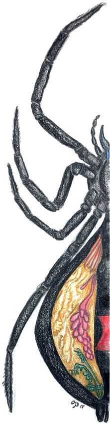
Dunaj et al. BMC Evolutionary Biology   (2020) 20:104                                                          Page 2 of 17

interactions influence host development and cause hy-         limited but intriguing evidence to suggest the presence
brid lethality in Jewel wasps (Nasonia) [4, 7]. Further,      of a diverse microbial community within Latrodectus
microbial communities within and across three closely         species and the potential for genetic exchange with their
related and environmentally controlled Nasonia species        microbial symbionts.
exhibit a phylogenetically distinct pattern that mirrors         The purpose of this study is to characterize the micro-
their hosts’ phylogeny [4]. This evolutionary host-           biomes of widow spiders using high-throughput metage-
microbe relationship provides strong evidence, along          nomic sequencing and to evaluate if there is evidence of
with other similar studies, to support a recently pro-        phylosymbiosis across closely related species in the
posed hypothesis known as phylosymbiosis [7, 8]. Phylo-       genus Latrodectus, its sister genus Steatoda and the
symbiosis describes the ecological and evolutionary           more distantly related common house spider (Parastea-
relationship of the microbiomes across related host spe-      toda tepidariorum). We hypothesize that the microbial
cies, where the microbial community profile parallels the     communities of these spider species will diverge in a pat-
phylogeny of closely related host species and maintains       tern mirroring their phylogeny and that each specialized
an “ancestral signal” [7, 8]. Recent research suggests that   tissue type will contain its own unique complement of
phylosymbiosis is common and can also play a role in          microbial community members. Specifically, the micro-
host fitness and health. For example, deer mice inocu-        bial communities will not be randomly assembled but
lated with microbial communities from more distantly          instead will be host-specific and will match the host
related species had lower food digestibility and jewel        phylogenetic pattern. Additionally, we expect that meta-
wasps that received transplants of interspecific micro-       bolically active and functional microbial symbionts
biota had reduced survival compared to those exposed          would also be detectable via RNA sequencing of these
to their own intraspecific microbiota [8].                    spider species’ specialized tissues. Therefore, analysis of
  Microbiome research on non-insect arthropods, such          publicly available RNA sequencing data for the venom,
as spiders, is limited and has mainly focused on PCR-         silk, and ovary glands from L. hesperus, L. geometricus, S.
based sequencing assays targeting specific, well charac-      grossa, and P. tepidariorum can provide support and val-
terized arthropod symbionts known to have an impact           idation for the functionally viable microbial community
on arthropod fitness, reproductive behavior and isolation     members of interest observed within the 16S sequencing
such as Wobachia, Rickettsia, Spiroplasma, and Cardi-         data. Together these comparative analyses provide im-
nium, [9–19]. Comprehensive investigation of spider           portant insights into the evolution of spider-microbe re-
microbiomes has only been conducted in a few studies          lationships and the hologenome of these medically
[12, 20], but the distinct evolutionary pattern of phylo-     significant spiders.
symbiosis and existence of evolutionarily significant
spider-microbe relationships have not been evaluated.         Results
Moreover, the degree to which spider species and differ-      Diversity, host, and tissue specificity of spider microbiota
ent tissues harbor unique microbial communities is            The microbiome of each spider species included in this
poorly known.                                                 study (5 species in family Theridiidae) was evaluated
  Black widow spiders (genus Latrodectus) provide a           from whole spider samples (3 per each species, 15 whole
particularly suitable system to understand how microbes       samples) and at a tissue-specific level (3 animal/ tissue
influence spider evolution. The availability of a well-       sets per species, 5 tissues per set – cephalothorax,
resolved Latrodectus phylogeny facilitates tests of phylo-    venom glands, ovary, silk glands, and fat tissue, 75 tissue
symbiosis [21]. Additionally, this clade of spiders is med-   samples). Crickets (12 whole), the prey items/ food
ically significant because their venom contains               source used for these spiders, were also included in this
neurotoxic latrotoxin proteins, making black widow            study to determine if their microbial communities affect
spider venom hazardous to humans [21–23]. While               the microbial composition and diversity of the host
latrotoxins are not known from spiders outside of the         spider species. In total, including controls, 109 samples
black widow family Theridiidae [23], recently Borden-         were processed through the 16S amplicon sequencing
stein and Bordenstein sequenced phage WO, that com-           and analysis workflow (Fig. S1). The results from the
monly infects Wolbachia, and concluded this viral             alpha diversity tests (Fig. S2A & C, Faith’s Phylogenetic
genome encoded a latrotoxin C-terminal domain hori-           Diversity and Shannon Index respectively) showed that
zontally transferred from the black widow spider [24].        we achieved sufficient depth of sequencing coverage
This study also confirmed the presence of Wolbachia in        (≥5000 reads per sample) to measure OTU (Operational
Latrodectus geometricus using targeted PCR. Similarly,        Taxonomic Unit) diversity, as the diversity per spider
Goodacre et al. [17] used targeted PCR to show an un-         sample plateaued by 7000 sequences. Additionally, even
identified Latrodectus species was infected with Wolba-       after filtering sequences for quality and contamination,
chia and Rickettsia, but not Spiroplasma. This provides       all but one of our spider samples had greater than 5000
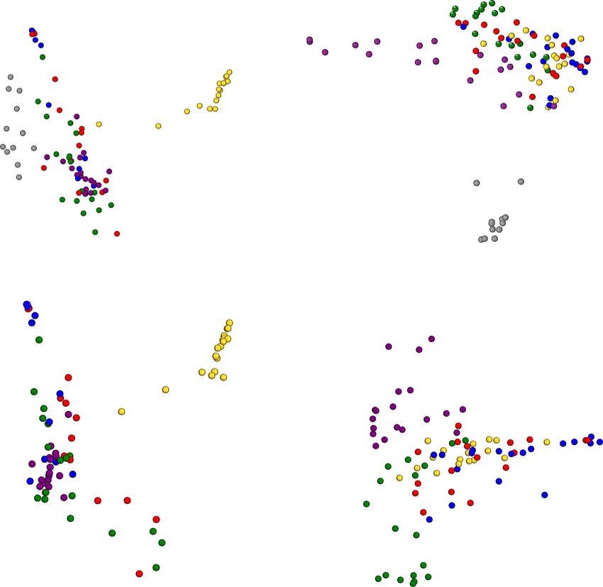
Dunaj et al. BMC Evolutionary Biology       (2020) 20:104                                                                                   Page 3 of 17

reads per sample. We utilized these results to select the                     P. tepidariorum and many of S. grossa samples tend to
depth of coverage passed into the core phylogenetic di-                       cluster separately in the un-weighted UniFrac plots (Fig.
versity plug-in command in QIIME to generate EMPeror                          1b & d). In other words, the relative abundance
PCoA clustering plots for each UniFrac (weighted and                          (weighted UniFrac) of the dominant microbial commu-
unweighted) and Bray-Curtis dissimilarity distance                            nity members in L. geometricus affects how the whole
matrices [25]. In each of the beta-diversity clustering                       spider and tissues samples cluster together for this spe-
PCoA plots, the spiders’ prey items (crickets) clearly                        cies. In contrast, the microbial community members in
cluster together and separately from all spider samples                       S. grossa and P. tepidariorum samples segregate when
(Fig. 1a & b). The L. geometricus samples (whole and                          the abundances are not taken into account (presence –
each tissue type replicate) segregate from other samples                      absence only) when generating UniFrac distance ma-
in the weighted UniFrac PCoA plots (Fig. 1a & c), while                       trixes for PCoA plotting. Also, the P. tepidariorum

 Fig. 1 Beta-diversity principal coordinate analysis plots for whole animal samples. Weighted UniFrac plot with cricket samples (a) - L. geometricus
 samples cluster together within blue oval and crickets in red oval. Un-weighted UniFrac plot with cricket samples (b) - P. tepidariorum samples
 cluster together within purple oval, over half the S. grossa samples cluster together within yellow oval, and crickets cluster separately in red oval.
 Weighted UniFrac plot without cricket samples (c) - L. geometricus samples cluster together within red oval. Un-weighted UniFrac plot without
 cricket samples (d) -P. tepidariorum samples cluster together within green oval, over half the S. grossa samples cluster together within purple oval
Dunaj et al. BMC Evolutionary Biology     (2020) 20:104                                                                                Page 4 of 17

whole spider samples clustered closely with other P.                        diversity between spider species, where P. tepidariorum
tepidariorum whole spider samples from different MiSeq                      and S. grossa samples tended to have greater overall mi-
runs. A small number (n = 7) of our negative controls                       crobial community diversity compared to the Latrodec-
had a low number of reads; of those that had some                           tus species samples (Fig. 2 & Fig. S3). This variance of
OTUs assigned, the number of reads (35–1035 reads,                          diversity is also observed in each of the alpha-diversity
and one PCR control at 5689 reads), all were well below                     boxplots (Fig. S4A - D), where P. tepidariorum and S.
the number of reads retained after rarefaction (7032 cut-                   grossa samples tend to have higher diversity indexes for
off) for diversity analyses run across all sample types and                 their microbial communities as compared to Latrodectus
thus removed from downstream analyses.                                      species samples.
   The resulting assigned taxonomies for each group of                        All of the L. geometricus samples were dominated by
spider samples were visualized with R to evaluate the                       Candidatus Rhabdochlamydia (genus), by relative abun-
composition and relative abundances of the more dom-                        dance. According to the SILVA database, this 16S se-
inant (> 2% relative abundance) microbial community                         quence is specific to brown widows. This microbial
members within each tissue per species. The resulting                       symbiont had a broad range in abundance within the L.
tissue microbial community profiles vary in the level of                    geometricus samples; at the lowest, average, relative

 Fig. 2 Taxa Plots Per Tissue Type and Species. Major microbial constituents (> 2%) within each whole spider and tissue sample for each species
 (replicates grouped via mean-ceiling). Sample types as follows: Whole Spiders, Cephalothorax, Venom Glands, Ovary, Silk Glands, and Fat Tissue.
 Widow anatomy illustration by S. Dunaj, 2018
Dunaj et al. BMC Evolutionary Biology    (2020) 20:104                                                          Page 5 of 17

abundance, 61% of the microbial community in fat tis-          Delftia, Rhizobiales, Sphingomonas, Propionibacterium,
sues and up to 95% of whole spider samples (91% of             and Sphingomonadaceae.
ovary, 83% of silk glands, 76% of venom glands, 73% of            To determine the bacterial taxa unique to specialized
cephalothorax) at the highest relative abundance. Gillia-      organs, we analyzed the replicate grouped (mean-ceiling)
mella is a major bacterial symbiont that is found across       master sample taxa tables from QIIME2–2018.4 (Level 6
many of the spider samples. This symbiont primarily            – Genera) with a custom python script that computes
dominates L. mactans and L. hesperus samples. The rela-        the unique OTUs for each tissue type. Results show that
tive abundances of Gilliamella in L. mactans samples           there are two detectable microbial community members
are as follows: 91% in ovary, 79% in whole spiders, 68%        unique to venom glands and shared across all spider
in silk glands, 62% in venom glands, 54% in fat tissue,        species: Psychrobacter and Variovorax (Table S4A lists
and 35% in cephalothorax. Gilliamella also is a major          all venom specific microbiota). There are also bacterial
microbial community member constituent within L. hes-          taxa unique to silk glands (Table S4B). One of these,
perus samples and is observed at the following relative        Candidatus Peribacteria, is detected in multiple silk
abundances: 68% in silk glands, 40% in venom glands,           glands from two different spider species (S. grossa and P.
40% in cephalothorax, 30% in ovary, 27% in whole spi-          tepidariorum). Several bacterial symbionts were ob-
ders, and 25% in fat tissue. Gilliamella is also observed      served to be unique to ovaries, including Candidatus
to be a dominant (27%) bacterial community member in           Falkowbacteria, which was detected in both L. geometri-
S. grossa fat tissue. Another prominent bacterial sym-         cus and L. mactans ovaries (see Table S5A for full listing
biont, Spiroplasma, is found to be a dominant commu-           of ovary microbiota). Lastly, Table S5B lists the micro-
nity member within L. hesperus whole spiders, with an          biota specific to fat tissue, where Gemmatimonas was
average relative abundance of 40%. Wolbachia is also ob-       detected in both P. tepidariorum and L. geometricus fat
served in all the spider species and tissue types, except      tissue samples.
for in P. tepidariorum. The average relative abundance
of Wolbachia ranges from low (< 1%) in some species to         RNA sequencing versus 16S amplicon sequencing
a high of 8% in L. hesperus ovary tissue. Wolbachia is         Our analysis of public RNA sequencing data from silk,
also observed as a rare (< 1%) microbial community             venom, and ovary glands across multiple widow-related
member within ovary samples of L. geometricus, L. mac-         species found 30 bacterial symbionts/ community mem-
tans and S. grossa. Wolbachia is also detected as a sig-       bers were concordant with our generated 16S amplicon
nificant symbiont in other tissue samples at the               sequencing dataset (Tables 1 and 2). Overall, there are
following relative abundances: 2.5% in whole L. mactans,       94 shared OTUs observed between the 16S and RNA se-
3% in L. hesperus silk glands, 3% in S. grossa silk glands     quencing datasets and a large number of bacterial reads
and cephalothorax, and 2% in L. geometricus venom              (taxonomic hits in CosmosID) within the RNA sequen-
glands (0.8% in L. hesperus and 0.3% in S. grossa venom        cing data for each sample type, ranging from 10,512 to
glands as well). Other major microbial community mem-          1,123,096 (Table 1). All of the bacterial hits/ OTUs from
bers that are found across several spider species and          the RNA sequencing data that did not match our 16S
their tissues include Bartonella, Ralstonia, Acinetobacter,    dataset are listed in Table S6. Several of the top
Table 1 Summary of OTUs observed in RNA sequencing verses 16S amplicon sequencing of Silk, Venom, and Ovary glands in
Widow related spiders
Sample ID                 Tissue Type             16 s OTUs   RNAseq OTUs           Bacterial Hits (RNAseq)     Shared OTUs
L. hesperus               Silk Glands             55          1                     10,512                      0
L. hesperus               Venom Glands            79          24                    152,364                     12
L. hesperus               Ovary                   68          20                    39,829                      6
L. geometricus            Silk Glands             53          8                     47,646                      4
L. geometricus            Venom Glands            73          18                    1,181,879                   10
L. geometricus            Ovary                   71          10                    519,983                     6
S. grosa                  Silk Glands             159         8                     1,123,096                   6
S. grosa                  Venom Glands            180         26                    1,122,123                   16
S. grosa                  Ovary                   142         32                    354,499                     16
P. tepidariorum           Silk Glands             98          12                    191,609                     5
P. tepidariorum           Venom Glands            130         34                    112,093                     13
P. tepidariorum           Ovary                   96          1                     44,772                      0
Dunaj et al. BMC Evolutionary Biology (2020) 20:104 Page 6 of 17
Table 2 RNA sequencing verses 16S amplicon sequencing of silk, venom, and ovary glands from widow-related spiders
microbial constituents that did match between these models based on the Bayesian Information Criterion -
datasets include the following: Achromobacter, Acineto- BIC). This model is also within the AIC and BIC 100%
bacter, Bradyrhizobium, Burkholderiales, Methylobacter- confidence intervals determined by JModel Test 2 [27].
ium, Propionibacteriaceae, Pseudomonas, Ralstonia, The resulting spider species phylogenetic tree was
Sphingobium, Sphingomonas, Stenotrophomonas, and rooted with the P. tepidariorum branch, based on earlier
Wolbachia (Table 2). Pseudomonas, Ralstonia and phylogenies [23] (Fig. 3).
Staphylococcus are detected in the RNA-seq data across We then compared the distances between this refer-
all four spider species. The following microbial commu- ence widow phylogenetic tree to each resulting beta-
nity members are found in both the silk and venom diversity dendrogram generated from every set of
glands of P. tepidariorum: Achromobacter, Acinetobacter, grouped tissue and whole spider samples with TreeCmp
Propionibacteriaceae, and Stenotrophomonas. Also, S. [28]. Congruency between the host phylogeny and each
grossa has twenty-one microbial community members beta-diversity dendrogram was evaluated by the resulting
found across both datasets and two of which are ob- Robinson-Foulds Cluster and Matching Cluster mea-
served across the silk, venom, and ovary glands - Methy- surements (Fig. 4 and Fig. S4), where the resulting scale
lobacterium and Staphylcoccus. The venom glands of L. for these metrics ranged from a value of 0 to 4 (with 0
geometricus has three bacterial constituents found across equating to complete congruence) [8, 28]. Congruency
both sets of sequencing data: Sphingomonas, Streptococ- arises from the corresponding/mirrored relationship be-
cus, and Veillonella. L. hesperus has both Escherichia tween the branches of the host phylogenetic tree and the
and Sphingobium within its venom glands. Within the microbial beta-diversity tree. These beta-diversity den-
16S and RNA sequencing data for L. hesperus and L. drograms are generated from the clustering of the dis-
geometricus, the venom glands for both species contain tance matrix values derived from each respective beta-
Wolbachia, Streptococcus, Staphylococcus and diversity test. This congruency is indicative of a phylo-
Pseudomonas. symbiotic microbial community assembly across related
species / host clade [3, 8]. We observe evidence for phy-
Phylosymbiotic microbial community assembly losymbiosis (host-microbiota congruency) across the fol-
In order to test for phylosymbiosis across our spider spe- lowing spider sample sets with the Bray-Curtis microbial
cies and their tissues, a host phylogeny with at least four beta-diversity dendrograms (Fig. 4): whole spiders, ceph-
species of interest is required. A maximum likelihood alothorax, venom glands, silk glands and ovaries. Host-
phylogenetic tree (Fig. 3) was generated with RAxML microbiota congruency is also observed for several of
based on the mitochondrial gene COI [26] utilizing the these tissue sets and whole spiders in the weighted Uni-
highest ranking Akaike Information Criterion (AIC) sub- Frac and Jaccard beta-diversity dendrograms to host
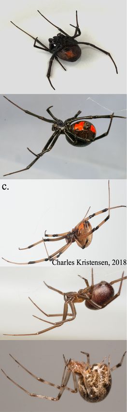
stitution model – GTR + γ (also the highest ranking phylogeny comparisons, which can be viewed in Fig. S4.Dunaj et al. BMC Evolutionary Biology (2020) 20:104 Page 7 of 17 Fig. 3 Widow Related Spider Phylogeny. Rooted (P. tepidariorum), bootstrapped (10,000 iterations) phylogenetic tree from selected spider mtCOI genes (accession numbers in each branch label). Substitution model – GTR + γ. a Photograph of L. hesperus (S. Dunaj, 2017). Photographs of L. mactans (b), L. geometricus (c), S. grossa (d) and P. tepidariorum (e) (C. Kristensen, 2018)
Dunaj et al. BMC Evolutionary Biology    (2020) 20:104                                                                             Page 8 of 17

 Fig. 4 Phylosymbiosis between Widow Phylogeny and Bray-Curtis Microbiota Dendrogram Relationships. Congruency between widow related
 spider phylogeny and beta-diversity dendrograms was measured via Robinson-Foulds Cluster (R-F Cluster) metric, where a value of 0 = complete
 congruence and 2 = incomplete incongruence. Matching Cluster (MC) Metric also accounts for incongruency between closely related branches of
 rooted trees and a value of 0 also indicates complete congruence between compared trees. Pink asterisks are utilized to denote phylosymbiotic
 relationships between the widow phylogeny and microbial dendrogram

Interestingly, phylosymbiosis is not observed across the                 samples, where the majority of these samples were seg-
fat/ mid-gut tissues, the ovary tissues in the weighted                  regated from other spider species samples (Fig. 1a-c).
UniFrac dendrogram or in any of the unweighted Uni-                      We also found that P. tepidariorum and S. grossa have a
Frac dendrograms. An Adonis PERMANOVA test indi-                         greater diversity of microbial community members
cates significant impacts of species and tissue on beta                  across whole spider and tissue specific samples, as com-
diversity (F = 8.761037291 and 1.25112113, P = 0.000999                  pared to the Latrodectus species included in this study
and 0.04495505, respectively), but no significant inter-                 (Fig. 2). Additionally, OTUs corresponding to known
action (model: diversity = species * tissue).                            arthropod symbionts were found across both 16S and
                                                                         RNA-seq datasets, validating the presence of active,
Discussion                                                               functional microbial symbionts within the venom, silk
In this study, we uncovered several key insights into                    and ovary glands from four of the spider species
widow spider microbial communities and provided sup-                     (Table 2). Furthermore, we provide support for phylo-
port for phylosymbiotic patterns of these communities                    symbiosis across this clade of spiders based on our
across several members of the Theridiidae family/widow                   observations of the host phylogeny mirroring the
spider host clade. We observed species-specific cluster-                 majority of the microbial beta-diversity trees (Fig. 4
ing of spider microbiome samples. Specifically, we de-                   and Fig. S4). Phylosymbiosis was also observed
tected a significant clustering pattern with the                         across several tissue types from these spiders, includ-
Weighted-UniFrac beta-diversity test for L. geometricus                  ing the ovaries.
Dunaj et al. BMC Evolutionary Biology (2020) 20:104 Page 9 of 17 We evaluated the microbial community diversity for Psychrobacter and Variovorax are two microbial mem- this clade of widow spiders and their specialized tissues. bers unique to venom glands that were found in two sets The PCoA beta-diversity plots tend to show a species- of spider species (P. tepidariorum and L. geometricus - specific clustering pattern for these spider microbiome Psychrobacter, S. grossa and L. mactans - Variovorax). samples. We found a significant clustering of L. geome- Psychrobacter is a gram-negative, non-motile coccoba- tricus samples within the Weighted-UniFrac beta- cilli that is exceptionally cold-tolerant [30], and has been diversity PCoA plots. We suspect that this clustering in- isolated from the guts of several fish species [31], and dicates limited microbial diversity and is due to the detected in the rectums of decomposing swine [32]. Var- microbiome of L. geometricus being dominated primarily iovorax is a genera of gram-negative bacteria found cap- by Candidatus Rhabdochlamydia a putative intracellular able of catabolizing toxic and complex compounds and bacteria. Candidatus Rhabdochlamydia was observed to found in water and soil [33]. Variovorax has also been be significantly abundant in each set of tissues for L. geo- determined to be a gut symbiont of beetle larvae (Holo- metricus with ANCOM testing, indicating that this may trichia parallela) [34], mosquitoes (Anopheles culicifa- be a species-specific microbial community member. This cies) [35] and sand flies (Phlebotomus papatasi) [36]. limited microbial diversity may be due to L. geometricus, Furthermore, our findings of a limited set of microbial a presumed African endemic, being recently introduced community members unique to Latrodectus venom to North America (Florida and California) and expand- glands is supported by recent research that demonstrates ing its range from a small populations [23]. We the significant and potent anti-microbial properties of hypothesize that the low microbial diversity of L. geome- venoms isolated from a wide range of spider species and tricus may mirror limited genetic diversity in the host the possibility that venom may elicit a “preservative” ef- population due to a bottleneck; putatively, bottlenecking fect on prey items [37, 38]. the population could have restricted the variation of bac- Additional microbial symbionts of other arthropods, terial diversity that is vertically transmitted [29]. L. geo- some with potential adaptive function, were identified metricus sampled for this study were collected from the within our spider samples. Gilliamella was found across vicinity of Sarasota, Florida and were inbred. The col- four of the five spider species and was dominant in L. onies of the S. grossa and P. tepidariorum may have been mactans and L. hesperus samples. Interestingly, Gillia- inbred as well, but the original females of these colonies mella has been determined to be a major symbiont of were possibly from more genetically diverse populations. bees and was isolated from the guts of Apis mellifera However, based on detecting phylogenetically distinct [39–41] and multiple Bombus species [40–42]. Spiro- microbial communities within each spider species’ ovar- plasma is also a well-characterized bacterial symbiont of ies, some of these microbial symbionts are possibly verti- many arthropods and was found to dominate the whole cally transmitted as observed in other systems [8]. This L. hesperus samples [43–45]. This particular symbiont is would explain the phylogenetic congruity between mi- thought to provide certain arthropod hosts protection crobial diversity and spider phylogeny. The results from against parasitoid wasps, nematodes and fungal patho- PCoA beta-diversity plots also demonstrate that the spi- gens, but is considered pathogenic in bees (A. mellifera) ders’ prey items (crickets) have a microbial community and mosquitoes (Culex tritaeniorhynchus and Aedes sol- distinct from all spider samples (Fig. 1a & c). This sug- licitans) [43]. Spiroplasma has also been documented as gests that the cricket microbiota does not contribute to a symbiont of other spider species [14, 15, 17, 20] and the dominant microbial community members within the includes spiders within the following families: Gnaphosi- spider samples and that isolation of the host subject dae, Lycosidae, Araneidae, Linyohiidae, Tetragnathidae, prior to dissections could have helped isolate the signal Scytodidae, and Linyphiidae. Goodacre et al. [17] of non-prey/host specific microbiome from the prey- screened widow spiders for Spiroplasma but this sym- item “background” microbial community profile. biont was not observed; however, only three individuals We were able to identify microbial community mem- (species not indicated) were evaluated and the authors bers in multiple types of spider tissues and determine utilized S. ixodetis specific rather than universal 16S which microbial taxa are unique to venom and silk PCR primers. Additionally, we detected several other glands. For example, we found that spider species more known arthropod symbionts across several spider species evolutionarily distant (P. tepidariorum and S. grossa) to (Bartonella [36, 46], Ralstonia [47], Acinetobacter [12, the black widow species (L. hesperus and L. mactans) 20, 47], Delftia [47], Rhizobiales [47, 48], Sphingomonas had more unique bacterial taxa in their venom and silk [20, 47], Propionibacterium [7], and Sphingomonadaceae glands (Table S4A & S4B). The fewer unique microbial [47, 49]). We also found evidence that several of the mi- community members in Latrodectus venom glands sug- crobial community members observed in P. tepidar- gest that this toxin-rich environment selected for only a iorum, S. grossa, L. geometricus and L. hesperus are small set (8 genera) of venom tolerant microbiota. possibly metabolically active, as transcripts for these
Dunaj et al. BMC Evolutionary Biology   (2020) 20:104                                                         Page 10 of 17

symbionts were found in corresponding RNA sequen-              environment, as our results suggest an “ancestral micro-
cing datasets (RNA-Seq data was unavailable for L. mac-        bial signal” being retained across the host phylogeny [7,
tans). Although we cannot discount the potential role          8]. This “signal” implies that species’ microbial commu-
that husbandry, housing, and acquisition might play in         nities are more similar to each other but diverge from
the assembly of these communities, these results support       when they share their last common ancestor; akin to
the possibility that the above-mentioned microbial sym-        homology of genetic pathways in a genome that diverge
bionts are viable and functional members of these widow        but indicate a common ancestor. Although, we do not
species’ microbiomes. The majority of these concordant         know what the genetic sequence of the common ances-
RNA transcripts and 16S detected microbiota have also          tor was, much like we cannot know what the shared an-
been reported as symbionts within other arthropod              cestral microbiome was. The same eco-evolutionary
microbiomes [47] and include the following taxa: Bra-          pattern of phylosymbiosis has also been observed across
dyrhizobium in spiders [20], Burkholderiales in ants [50],     such distant taxa as jewel wasps (Nasonia), fruit flies
Methylobacterium in jewel wasps, mosquitoes, and spi-          (Drosophila), rodents (Peromyscus, Mus, and Neotoma),
ders [4, 20, 51], Pseudomonas in mosquitoes, honey bees        mosquitoes, and wild hominids [4, 8, 51, 52]. However,
and other spiders [20, 46, 51], Sphingomonas in spiders        phylosymbiosis of host species’ tissue specific
[20], Stenotrophomonas in beetle larvae [34], Staphylo-        specialization has never been described before to our
coccus in jewel wasps [7] and Wolbachia, a well-known          knowledge. Together with these recent studies of mul-
symbiont of many arthropods. It is also possible that          tiple hosts and their microbe interactions, we add add-
some of the Wolbachia transcripts identified could have        itional support for the hypothesis of phylosymbiosis as a
originated from horizontally transferred genes expressed       biological process.
within these spider hosts, as Wolbachia and its prophage          The hologenomic theory of evolutionary theory is sup-
WO possibly have undergone HGT events with Latro-              ported through observations of phylosymbiosis of these
dectus species [24]. In such cases it is challenging to sep-   hosts’ microbiota with the resulting analysis of the ovary
arate out host verse microbial transcripts from RNA            tissues, implicating heritability of complex communities
sequencing data. Additionally, both assays detect Wolba-       of microbes from one generation to the next. However,
chia within the ovary tissues from L. geometricus. More        the genesis of the associated microbiome to the ovaries
interestingly, the sources and locality of the spiders sam-    could also be environmentally acquired or even transient
pled in the RNA sequencing studies were sampled from           representations of the microbiome. It is important to
geographical sites different from those of the spiders col-    note that vertical transmission of the spiders’ microbiota
lected to generate this study’s 16S dataset (except for S.     is not yet demonstrated, outside of the observations of
grossa). Even with these different sampling conditions,        endosymbionts Wolbachia and Cardinium, but is also
we are still able to detect multiple species of the same       not an obligatory condition for the hologenomic theory
bacterial symbionts between these datasets, indicating         of evolution (see Brooks et al. 2016). Specifically, the
these may be species-specific microbiota.                      paralleled host phylogenetic tree and the ovary specific
  We predicted that we would observe phylosymbiosis            microbial beta-diversity trees (Bray-Curtis and Jaccard)
across the host phylogeny and we found evidence of this        indicate that there are phylogenetically distinct microbial
eco-evolutionary pattern of microbial community assem-         community members present in widow spider ovaries
bly within these widow spider host species and their re-       and these symbionts could be vertically transmitted to
spective tissues (except fat tissue). This is supported by     progeny as the observations were consistent between in-
the observed congruency of the host phylogeny and mul-         dividuals within a species. We also detected unique mi-
tiple microbial beta-diversity dendrograms (Fig. S4). If       crobial community members for each species’ ovary
the microbial communities across these tissues and             tissues (Table S5A), a counter point to random associa-
whole spider species samples were stochastic/ randomly         tions of microbes in a host. Further evidence to this is
assembled, then we would have expected to observe the          the presence of the well-documented, heritable, arthro-
dendrograms to not parallel each other, have high              pod symbionts. Wolbachia, for example, was in each set
Robinson-Foulds Cluster and Matching Cluster mea-              of ovaries, except for the more distantly related P. tepi-
surements and resemble the random pattern / incom-             dariorum. Wolbachia has been evaluated in past studies
plete congruency found between the fat tissue microbial        for possibly causing sex-ratio distortion in spiders [10,
diversity trees (Fig. S4). The non-congruent fat tissue        13]. Sex-biased prevalence of Wolbachia (more females
trees could have been due to a comparative lack of host-       infected verses males) was observed in two of the 27
species-specific tissue specialization. Therefore, each        spider species surveyed by Duron et al. [13] (Meta men-
spider species and its specialized tissues appear to have      gei and Tetragnatha montana), suggesting Wolbachia’s
phylogenetically distinct microbial communities that           involvement in the sex-ratio manipulation of spiders. Re-
were not specifically acquired by their immediate              search on the dwarf spider (Oedothorax retusus) has also
Dunaj et al. BMC Evolutionary Biology   (2020) 20:104                                                          Page 11 of 17

indicated that bacterial symbionts play a role in sex-ratio    Last feeding and environmental isolation
distortion, most significantly by maternally inherited         At least six adult female spiders of each species were fed
Wolbachia [10]. Due to the dwarf spider’s smaller clutch       one cricket on the same day. After 24 h − 32 h each
sizes, it is speculated that the mechanism for this phe-       spider was transferred from the feeding vial into a sterile
nomena is the male-killing of embryos [10]. Other              housing container. Each spider was isolated in this sterile
known bacterial symbionts observed in our spider sam-          environment for 7–8 days, without any additional feed-
ples include Cardinium, which was noted by Duron               ings. The goal of this isolation procedure was to “starve”
et al. [13] to be transmitted from mother to progeny           spiders to reduce the signal/ background microbial com-
within marbled cellar spiders (Holocnemus pluchei);            munity of the spider’s prey.
however, this symbiont does not seem to skew sex-ratios          Twelve crickets were also included in this study to de-
or manipulate reproduction as observed in other arthro-        termine background / food sourced microbial commu-
pods [2, 13, 16]. Cardinium was also observed to be a          nity members. Prior to the first round of spider
major, dominant symbiont in three other spider species         dissections, crickets from the same batch underwent the
H. graminicola, U. insecticeps, and A. difficilis [20]. How-   same isolation conditions as the spiders. An additional
ever, we did not detect Cardinium in these selected            three crickets were used during the second round of
widow related spider species.                                  spider dissections (for second sequencing run) to deter-
                                                               mine the cricket microbiome at the time of the respect-
                                                               ive spiders’ last feeding. These cricket controls were
Conclusions                                                    euthanized on the same day as the last feeding and
In conclusion, we provide evidence for phylosymbiosis          stored immediately at − 80 °C.
across black widow related spiders in the Theridiidae
Family. Our results support the phylosymbiotic pattern         Aseptic spider tissue dissections
of microbial communities across whole spider samples           Prior to tissue dissections, forceps, wash containers/ bea-
and several tissue sample sets (cephalothorax, venom           kers, and dissection buffer (SSC buffer) were autoclaved
glands, silk glands and ovaries) based on congruency be-       and sterilized. The microscope and surrounding area
tween the widow phylogeny and multiple microbial               were cleaned with 10% bleach and 70% ethanol. After
beta-diversity dendrograms. This specialization of tissues     each dissection, the forceps were sterilized with a bleach
and microbiomes within a species represents a unique           wash, ethanol wash, followed by a sterile PCR water
facet of phylosymbiotic evolution not previously de-           (VWR) rinse prior to the next spider dissection. Each
scribed - tissue tropic phylosymbiosis. We also provide        spider underwent surface sterilization to remove possible
evidence for possible maternal-offspring transfer of           environmental contaminants with a 10% bleach soak for
phylogenetically distinct microbial communities based          1 min followed by two separate washes in sterile PCR
on our phylosymbiosis analyses and characterization of         water for 1 min each (adapted from Brooks, A.W., et al.
the microbiota from spider hosts’ ovary tissues. These         [8]). Three to four individual spiders of each species had
observations suggest that the diversity of symbiotic mi-       the following tissues dissected in an aseptic manner [53]:
crobial communities within and across spider species is        venom glands [54], cephalothorax (without chelicerae),
hypothetically, in part, vertically inherited. Thereby, the    silk glands, ovary, and fat / mid-gut region.
host-microbiome association is putatively evolving in re-         Each tissue was rinsed with sterile PCR-grade water
sponse to host speciation and has had the potential to         prior to collection in a sterile 1.5 mL microfuge tube.
shape host evolution.                                          Three to four individual spiders of each species (whole,
                                                               no dissection) were also surface sterilized prior to trans-
                                                               fer to a sterile 1.5 mL microfuge tube. All cricket sam-
Methods                                                        ples were transferred to 1.5 mL tubes without surface
Sample acquisition                                             sterilization. All samples and an aliquot of SSC buffer
Adult female spiders were acquired from Spider Pharm           were frozen in liquid nitrogen and then stored at − 80 °C
(Yarnell, AZ). These spiders include the following spe-        until DNA extraction.
cies: Parasteatoda tepidariorum, Steatoda grossa, Latro-
dectus geometricus, Latrodectus hesperus, Latrodectus          DNA extractions
mactans. Female spiders were confirmed to have                 DNA was extracted from each spider (3 per each spe-
reached adulthood by examining the epigynum prior to           cies), spider tissue (3 sets per species), cricket and nega-
dissections. Each spider was treated / housed in identical     tive controls (SSC buffer and DNA extraction controls
conditions and fed crickets (Vita-Bug, common brown            (reagents only) using Qiagen’s QiaAMP DNA Mini kits.
cricket – Timberline Fisheries, Marion, IL) from the           We utilized Qiagen’s protocol with the following specifi-
same batch/lot whenever possible.                              cations: liquid nitrogen to freeze samples prior to
Dunaj et al. BMC Evolutionary Biology (2020) 20:104 Page 12 of 17
homogenizing with a motorized pestle, 1.5 h lysis (vor- barcode for each sample’s 16S amplicons [49, 57, 58].
texing every 20–30 min), a centrifugation step for 30 s at We designed custom primers containing V1 and V2 re-
6000 g prior to transferring sample lysates to their re- gions following the 16S primer design protocol by
spective columns, and two elution steps (except for Kozich and Schloss [59], where the 27F and 338R
venom glands) - each with 5 min room temperature in- primers contain a unique 8 bp barcode on each primer, a
cubations. Prior to the DNA extractions of large whole short Linker/ Pad sequence and the appropriate Illumina
spiders (Latrodectus species), each individual spider was adaptor sequence (i5 or i7) (see Table S1 for list of pri-
divided in half with sterilized razor blades and forceps mer sequences). Preliminary data showed that two-step
and the mass of each half was measured. DNA was ex- PCR yielded significantly better results (consistent visible
tracted from both halves separately to avoid overloading bands from gel electrophoresis) than nested-PCR. These
the columns. Eluted DNA was combined in equal ratio multi-step PCR processes were also compared with sin-
based off of the pre-processed weight. Each sample type gle step PCR, where single-step PCR resulted in incon-
had an optimal elution volume, based on the size of the sistent and/ or low amplification of the spider
tissue or if a whole sample (spider or cricket). These elu- microbiome DNA samples.
tion volumes were as follows: whole spider = 400ul, Extracted DNA from each sample and all negative
cricket = 200ul, cephalothorax = 200ul, venom glands = controls (SCC buffer, Negative Extraction Controls, and
50ul, ovaries = 100ul, silk glands = 100ul, fat = 100ul. PCR-water (non-template control) were run through
DNA extractions were performed aseptically, with re- one round of PCR-1 using a 12.5ul reaction with Q5
directed airflow, and while wearing a facial mask to re- high fidelity master-mix (New England BioLabs, Inc.)
duce the risk contaminating the samples with exhaled with the following cycling conditions: 98 °C for 30 s, 25
bacteria. The extracted DNA was then quantified with cycles of 98 °C for 30 s (denature), 50 °C for 30 s (anneal),
ThermoFisher’s Quant-iT dsDNA High Sensitivity kit. and 72 °C for 30 s (extension), with a final extension step
at 72 °C for 10 min and end hold at 4 °C. PCR-2 included
16S rRNA gene amplicon library preparation and 2-3ul of PCR-1 product as template DNA. Four PCR-2
sequencing replicate 25ul reactions, using Q5 high fidelity master-
The standard methods for taxonomic classification of mix, were generated per sample (3 with sample PCR-1
bacteria within a microbial community utilize the small product and 1 as a non-template control). The condi-
ribosomal subunit (16S rRNA) gene. The 16S rRNA tions for PCR-2 were as follows: 98 °C for 30 s, 15 cycles
gene contains nine hyper-variable regions of various of 98 °C for 30 s (denature), 50 °C for 30 s (anneal), and
lengths. The variable regions with highest confidence of 72 °C for 30 s (extension), with a final extension step at
identifying bacteria down to the genus and species level 72 °C for 10 min and end hold at 4 °C. Each set of PCR-2
to date are the V1-V2 and V1-V3 regions [55, 56]. The product replicates were combined per sample and puri-
V1-V2 region was selected for this study because it is ~ fied with AMPure XP beads in a 1.8X bead-to-product
310 bp long and the appropriate length for higher quality ratio [60]. Each purified sample was then normalized to
paired-end sequencing with the Illumina MiSeq. Fur- the same molar mass using Qubit Fluorometric Quanti-
thermore, utilizing the V1-V2 target is 90% accurate for fication (ThermoFisher Scientific). Two final normalized,
identifying bacteria at the species level and 92% accurate pooled sample libraries and custom sequencing primers
at the genus level [55]. Prior to commencing this study, (Table S2) were sent to Cornell University’s Genomics
over twenty spider samples (whole and tissue) were used Facility (according to their protocol) for two runs of
to test different sets of universal polymerase chain reac- paired-end sequencing (2 × 250 bp), with a 10% PhiX
tion (PCR) primers that target the V1-V2 region (27F- spike in, on an Illumina MiSeq following the Kozich and
338R) and V3-V4 region (338F-786R). The 27F (5′- Schloss MiSeq protocol [59]. The concentration of se-
AGAGTTTGATCMTGGCTCA-3′ – slightly modified quencing primers was doubled for the second round of
from Brooks et al.) and 338R (5′-GCTGCCTCCC sequencing in order to increase the number of high-
GTAGGAGT-3′) universal 16S primers amplified the quality reads.
expected ~ 310 bp V1-V2 region from < 90% of test sam-
ples [8]. Microbial community data analysis
The V1 and V2 variable regions of the 16S rRNA gene Pre-processing of sequences and initial quality control
were amplified from the extracted DNA and mock com- The Quantitative Insights into Microbial Ecology (QIIM
munity DNA control (ZymoBIOMICS™ Microbial Com- E) program was utilized for pre-processing sequencing
munity Standard from Zymo Research) utilizing reads and microbial community analyses [61]. QIIME 1
universal PCR primers, 27F and 338R [8]. PCR was com- was used to add barcodes to the read files (merge_bcs_
pleted in a two-step process (PCR-1 and PCR-2) in order reads.py), extract barcode sequences from the reads (ex-
to yield significant PCR product with a unique molecular tract_barcodes.py), join overlapping paired-end readsDunaj et al. BMC Evolutionary Biology (2020) 20:104 Page 13 of 17
(join_paired_ends.py), and lastly demultiplex the joined tissue sample groups feature tables were summarized
reads according to their respective barcodes and sample (feature-table summarize command), taxa barplots gen-
IDs (split_libraries_fastq.py). After joining the paired- erated and reviewed via QIIME 2 View. The resulting
end reads (un-joined reads were removed from down- level 3 (class) and level-6 (genera) csv files were analyzed
stream analyses), the demultiplexing script also passes via R with the following packages: dplyr, tidyr, stringr,
reads through quality filtering (reads < Q20 were re- and digest [69–73]. The OTUs that made up at least 2%
moved from the dataset). A total of 896,429 reads out of or greater relative abundance across each tissue set was
3,578,685 passed initial quality filtering for the first run used to generate barplots with ggplot 2 [74].
and 5,172,436 reads out of 10,211,041 passed from the Alignments were completed on the set of representa-
second sequencing run (large percentage of reads lost to tive sequences with MAFFT (Alignment MAFFT plug-
PhiX spike-in and joining-step). in) [75] and the unconserved, highly gapped columns in
these aligned sequences were masked with the alignment
Sequence Dereplication, chimera checking, OTU picking, plug-in, mask command [76]. A phylogenetic tree was
and taxonomy assignments with QIIME then generated with the FastTree 2 tool (Phylogeny
The resulting joined, demultiplexed and high quality plug-in) using a Maximum-Likelihood method [77]. The
reads from each MiSeq run are contained in their run- resulting tree was then midpoint rooted (Phylogeny
specific seqs.fna output file and were imported into plug-in, midpoint-root option) and utilized for down-
QIIME 2 [62]. Each set of sequences were dereplicated stream beta-diversity analyses.
and de novo chimera-checked via the VSEARCH plug-in
tool [63]. Chimeric reads (i.e. PCR artifacts/biases from Mock community standard and quality assurance measures
parental strands acting as primers during PCR – hybrid The data from the first sequencing run underwent qual-
16S sequences from two different species of bacteria that ity control measures (Table S3) to ensure the sample li-
artificially affect diversity estimates) were removed from brary preparation steps and sequencing performed as
the dataset to reduce the impact of PCR errors prior to expected prior to moving forward with sample library
Operational Taxonomic Unit (OTU) clustering and di- preparation and second sequencing run for the majority
versity analyses [8, 64]. The feature-table plug-in was of the spider samples. This step was also completed to
then utilized to merge the two sets of resulting high determine if the OTU clustering threshold of 99% was
quality sequences (merge-seqs option) and feature/ OTU appropriate for the data analysis pipeline, in order to re-
tables (merge option) from each of the runs together for duce the potential of erroneously generating OTUs.
downstream analyses. The resulting feature table and se- Quality control assessments were completed by compar-
quences files were run through open-reference OTU ing the percentage of each mock community member
picking with VSEARCH utilizing SILVA’s 16S QIIME present within the first sequencing run dataset to the
formatted database (release 128–99% identity sequences) theoretical/ expected mock community composition as
at a 99% identity threshold for clustering [63, 65–67]. provided by the manufacturer, Zymo Research. The pre-
Low abundance OTUs were filtered out from the result- liminary results of the mock community analysis indi-
ing feature table, where OTUs with a frequency of less cated that the sample library preparation method and
than 10 sequences across all samples were removed. sequencing specifications were appropriate and accur-
Taxonomy assignments were also generated with SILVA ately measured the mock community composition. Fur-
rRNA Database (release 128–99% consensus taxonomy, thermore, one of the whole P. tepidariorum replicate
7 levels) by extracting out only the 16S V1-V2 regions samples (note: total of 3 replicates per sample) from the
that correspond to the 27F – 338R primers used for first sequencing run was repeated through the library
sample library preparation – truncation length of 500 bp preparation process and second MiSeq run as a positive
(feature-classifier plug-in, extract-reads option) [68]. control to test the differences between runs.
QIIME 2’s Naïve Bayes classifier was trained to these ex-
tracted V1-V2 reference sequences with the feature- Diversity analyses
classifier plug-in (fit-classifier-naive-bayes). We then uti- After OTU clustering, taxonomy assignments,
lized this V1-V2 trained classifier set to complete taxo- taxonomy-based filtering, and 16S rRNA gene align-
nomic assignments with the classify-sklearn feature- ments, the feature/ OTU table was rarefied based on the
classifier plug-in option. After assigning taxonomy to the depths of coverage per sample type. A depth of 7032
OTUs, taxa plots were generated with the taxa plug-in randomized sequences per sample was selected for the
(barplot command) and all OTUs that were classified as core-metrics beta-diversity analyses (prior to any group-
unassigned, chloroplasts, and / or mitochondria were fil- ing of replicate samples) based on this depth of coverage
tered out from the feature/ OTU table (taxa filter-table encompassed all but one spider tissue sample (this sam-
command) and representative sequences. Each of the ple contained a very high percentage of chloroplastDunaj et al. BMC Evolutionary Biology (2020) 20:104 Page 14 of 17
related OTUs, which were filtered out in upstream data spiders are as follows: P. tepidariorum (silk, venom, and
processing steps) and the alpha-diversity results from ovary) - Cologne, Germany, out-bred with P. tepidar-
running the diversity plug-in, alpha-rarefaction com- iorum spiders from Spider Pharm/ Arizona, United
mand run on spider samples (Fig. S1A & B, depth range States, L. hesperus (silk, venom and ovary) - Riverside
of 18–18,832 sequences). QIIME 2’s diversity plug-in County, California, United States, L. geometricus (silk,
core-metrics-phylogenetic command was run on all the venom, and ovary) - San Diego County, California,
filtered samples (crickets included), each set of tissue United States. S. grossa was obtained directly from
types, and then on only the spider samples (crickets re- Spider Pharm (similar to samples used for 16S sequen-
moved); in order to best resolve the resulting UniFrac cing). The following SRA files were downloaded from
distance matrix derived EMPeror PCoA plots [78]. Box- NCBI’s SRA database: SRR1219665, SRR1824489,
plots were generated, along with group significance stat- SRR5131057, SRR5131058, SRR5285094, SRR5285095,
istical testing (using the QIIME diversity alpha-group- SRR5285096, SRR5285099, SRR5285100, SRR5285114,
significance which is a Kruskal-Wallis one-way analysis SRR5285115, SRR5285118, SRR5285121, SRR5285122,
of variance), from each resulting alpha-diversity test vec- SRR5285123, SRR5285135, SRR5285136, SRR5285138,
tor file utilizing the diversity plug-in alpha-group- SRR5285141 and SRR5285142 [21, 53, 83, 84]. Fastq files
significance command [79]. Statistical analyses were were extracted from each SRA file utilizing the SRA-
completed to determine significant changes in the abun- Toolkit, fastq-dump command [85]. The extracted fastq
dances of the microbial community members between files were run through FastQC and then the adaptors
and across samples from each tissue type with ANCOM and poor quality bases were trimmed with Trimmomatic
testing [80]. Sample (biological) replicates were then [86, 87]. The Trimmomatic parameters utilized for trim-
grouped with the feature-table group command (mean- ming the raw reads are as follows: crop_length = total
ceiling, i.e. average frequency of each OTU across sam- read length – 1 (between 75 and 100 depending on li-
ple replicates) by base sample type - whole spiders, brary), seed_mismatches = 2, paired_end_seed_score =
cephalothorax, venom glands, ovaries, silk glands and fat 30, min_adapter_length = 2, keepBothReads = true, slid-
tissue (one grouped table per tissue type – resulting in 6 ing_window_size = 10, sliding_window_minimum_aver-
grouped tables - only these grouped tables, not individ- age_phred_score = 15, min_length_to_keep_reads = 36,
ual replicate samples, were used for the beta-diversity trimmomatic_threads = 8 [86, 87]. Corresponding Read 1
dendrograms for phylosymbiosis analyses). A master and Read 2 sequences for each species silk, venom or
grouped feature table was also generated for all the ovary glands were concatenated, gzipped and uploaded
spider samples – grouping each replicate sample by the to CosmosID for identification of any possible microbial
mean-ceiling option. Each of these grouped feature ta- transcripts / sequences, as Cosmos ID is capable of
bles were summarized (feature-table summarize com- uploading and analyzing raw or processed read data
mand), had taxa barplots generated and each were [88]. CosmosID takes raw, unassembled reads and
reviewed via QIIME 2 View. Each grouped tissue type matches them to the GenBook® database, utilizing statis-
feature table was then passed though the diversity beta- ticial and and computational methods [89].
rarefaction command with a selected depth of coverage
determined by the lowest sample sequence/ feature Phylosymbiosis analyses
count per each set of grouped tissue samples (Whole The following analysis methods were used to determine
Spiders = 14,509, Cephalothorax = 20,799, Venom = 20, if there is evidence of phylosymbiosis across the selected
826, Ovaries = 28,629, Silk = 28,628, Fat = 17,074) for the widow related spider phylogeny as described by Brooks
following beta-diversity tests: Bray-Curtis, Jaccard, Un- et al. [8]. Mitochondrial COI gene sequences were ob-
weighed UniFrac [81] and Weighted UniFrac [78]. Fur- tained from a previous study, where a 428–659 bp frag-
thermore, a PERMANOVA test was conducted to look ment was sequenced for each spider species represented
at pairwise differences in beta diversity (Bray-Curtis Dis- in this study [22]. The five resulting mtCOI sequences
tances) between species or tissues was conducted as de- were aligned with ClustalX v2.1 in multiple alignment
fined by the conditions of the ADONIS function within mode [90]. The aligned sequences were exported in
QIIME (Supplemental Table 8) [82]. Newick format and run through jModeltest (2.1.10
v20160303) to determine the best substitution model for
RNA sequencing Metatranscriptome analysis generating a widow spider phylogenetic tree [27, 91].
Publicly available RNA sequencing data from previous The aligned sequences were then utilized to generate a
research completed by Garb et. al, specific to silk, Maximum-Likelihood tree with RAxML v8.2.11 (GTR +
venom, and ovary glands, were acquired from NCBI for γ substitution model and 10,000 iterations) [26]. The
the following species: P. tepidariorum, S. grossa, L. geo- resulting tree was viewed and rooted with the P. tepidar-
metricus, and L. hesperus. The collection sites for these iorum branch utilizing FigTree v1.4.3 [92]. The resultingYou can also read