Animal Models in Microbeam Radiation Therapy: A Scoping Review - MDPI
←
→
Page content transcription
If your browser does not render page correctly, please read the page content below
Review

Animal Models in Microbeam Radiation Therapy: A
Scoping Review
Cristian Fernandez-Palomo 1, Jennifer Fazzari 1, Verdiana Trappetti 1, Lloyd Smyth 2,
Heidrun Janka 3, Jean Laissue 1 and Valentin Djonov 1,*
 1 Institute of Anatomy, University of Bern, 3012 Bern, Switzerland; cristian.fernandez@ana.unibe.ch (C.F.P.);
 jennifer.fazzari@ana.unibe.ch (J.F.); verdiana.trappetti@ana.unibe.ch (V.T.);
 jean-albert.laissue@pathology.unibe.ch (J.L.)
 2 Department of Obstetrics & Gynaecology, University of Melbourne, Parkville 3057, Australia;

 lloyd.smyth@unimelb.edu.au
 3 Medical Library, University Library Bern, University of Bern, 3012 Bern, Switzerland;

 heidrun.janka@ub.unibe.ch
 * Correspondence: valentin.djonov@ana.unibe.ch; Tel.: +41-31-631-8432

 Received: 24 January 2020; Accepted: 21 February 2020; Published: 25 February 2020

 Abstract: Background: Microbeam Radiation Therapy (MRT) is an innovative approach in radiation
 oncology where a collimator subdivides the homogeneous radiation field into an array of co-planar,
 high-dose beams which are tens of micrometres wide and separated by a few hundred micrometres.
 Objective: This scoping review was conducted to map the available evidence and provide a
 comprehensive overview of the similarities, differences, and outcomes of all experiments that have
 employed animal models in MRT. Methods: We considered articles that employed animal models
 for the purpose of studying the effects of MRT. We searched in seven databases for published and
 unpublished literature. Two independent reviewers screened citations for inclusion. Data extraction
 was done by three reviewers. Results: After screening 5688 citations and 159 full-text papers, 95
 articles were included, of which 72 were experimental articles. Here we present the animal models
 and pre-clinical radiation parameters employed in the existing MRT literature according to their use
 in cancer treatment, non-neoplastic diseases, or normal tissue studies. Conclusions: The study of
 MRT is concentrated in brain-related diseases performed mostly in rat models. An appropriate
 comparison between MRT and conventional radiotherapy (instead of synchrotron broad beam) is
 needed. Recommendations are provided for future studies involving MRT.

 Keywords: microbeam radiation therapy; scoping review; animal models; rat; mouse; synchrotron;
 spatial fractionation; radiotherapy; oncology

1. Introduction

1.1. Background
 In our society, cancer is an increasingly prevalent and therapeutically challenging disease. Of all
cancer patients treated curatively, half will receive radiation therapy [1]. However, despite these
efforts, more than a quarter of a million people die from cancer in the EU annually [2]. The main
factor limiting conventional radiotherapy (RT) is the toxicity induced in vital surrounding healthy
tissues, which constrains dose-escalation and effective tumour control [3]. Synchrotron X-ray
microbeams have been recognized as a unique tool to overcome this limitation [4], eliciting scientific
interest across the world in a radio-therapeutic application known as Microbeam Radiation Therapy
(MRT). MRT consists of a spatially-modulated, co-planar array of low energy X-rays delivered to

Cancers 2020, 12, 527; doi:10.3390/cancers12030527 www.mdpi.com/journal/cancers
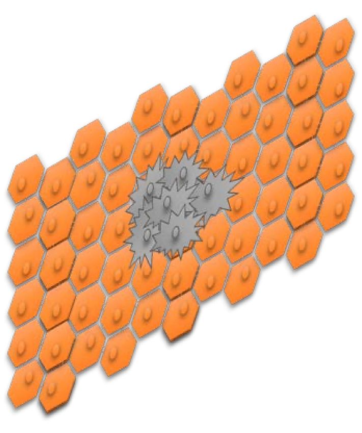
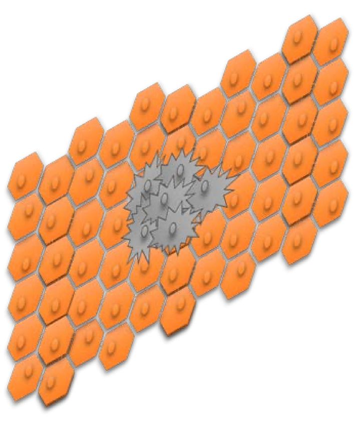
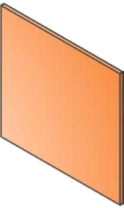
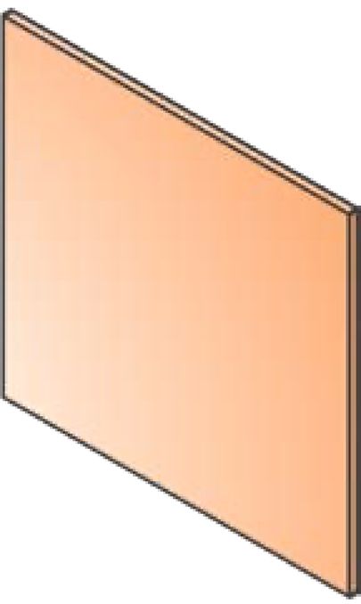
Cancers 2020, 12, 527 2 of 25

tumours [5]. These spatially fractionated beams exploit the dose-volume effect for extraordinary
normal tissue tolerance [6] and trigger a cascade of biological effects that greatly improve tumour
control [7].

1.2. Rationale
 In the last few decades, MRT research has expanded to include several synchrotron and non-
synchrotron sources around the world. The progress, however, has been relatively slow. A significant
challenge for MRT is the wide range of modifiable radiation parameters which can profoundly impact
its radiobiological effects, and ultimately, treatment outcomes. Research groups have explored these
radiation parameters using various rodent and non-rodent animal models. However, there remains
no clear consensus within the community on the optimal MRT parameters and strategy for cancer
therapy, delaying translation to human clinical trials. Comprehensively evaluating the existing
literature is essential to unifying these pre-clinical data and strategically promoting future research
to support clinical translation.

1.3. Objective
 The objective of this scoping review is to map the available evidence and provide an overview
of the similarities, differences, and outcomes of all experiments that have employed animal models
in MRT. The aim is to inform the community about progress made in the field of MRT and to identify
existing gaps in current research.

2. Methods

2.1. Protocol and Registration
 Our protocol was developed according to the scoping review methodological framework
proposed by the Joanna Briggs Institute [8], and refined by two methodological papers on how to
conduct scoping reviews published by Peters at al. [9] and Tricco et al. [10]. The draft protocol was
revised by the research team of the Institute of Anatomy of the University of Bern and by the
information specialist of the University of Bern's Medical Library. The protocol could not be
registered in the International Prospective Register of Systematic Reviews (PROSPERO), as this is not
yet an option for scoping reviews. This scoping review was written according to the Preferred
Reporting Items for Systematic Reviews and Meta-Analyses (PRISMA)-update for Scoping Reviews
[11].

2.2. Eligibility Criteria
 The review considered articles that employed animal models with the purpose of studying the
normal tissue or therapeutic effects of MRT. We included all papers that employed MRT as (i) an
array of co-planar microbeams, (ii) from any radiation source (synchrotron and non-synchrotron) and
quality (photons, protons, and others), (iii) that were applied to animals. We defined MRT as an array
of co-planar microbeams of widths equal to or lower than 100 µm, which are spaced by a few hundred
micrometres within the array. This definition of MRT has been used by other research groups, and it
is becoming the standard in the field [5,12–15]. MRT is also described as spatial fractionation of the
radiation beam, which is the main difference with conventional radiotherapy (Figure 1). We excluded
papers that employed minibeams because their width exceeds 100 µm. We also excluded publications
that employed a single microbeam and a microbeam scanning technique because these approaches
do not qualify as spatial fractionation.

Cancers 2020, 12, 527 3 of 25

2.3. Information Sources
 Literature searches were performed by an information specialist (HJ) in the following
information sources:
 • Medline (Ovid) (incl. Epub Ahead of Print, In-Process and Other Non-Indexed Citations,
 Medline Daily and Ovid Medline Versions (1946–October 02, 2019)
 • Embase (Ovid) (1947–October 02, 2019)
 • Cochrane Library (1996–October 02, 2019)
 • Scopus (1788–October 02, 2019)
 • ICTRP Trial Register (WHO)
 • ClinicalTrials.gov
 • Livivo Search portal

2.4. Search Strategy
 An initial search strategy in Medline was drafted and tested against a list of core references to
see if they were included in the search results. After refinement and consultation with the research
team, search strategies were set up for each information source, based on database-specific, controlled
vocabulary and text words. No limits have been applied in any database considering study types,
languages, publication years, etc. Beside standard bibliographic databases for medical journals like
Medline, Embase and the Cochrane Library, two interdisciplinary databases, Scopus and Livivo, have
been selected to search for other publications types than journal articles (e.g., conference proceedings,
book series, publications from university repositories). All searches were run on October 3rd, 2019.
In addition to electronic database searching, bibliographies and reference lists from relevant
publications were checked.
 For our search strategy, we decided not to focus on any particular animal species or animal
models. This allowed for maximum sensitivity in the search and ensured that publications on all
organisms on which MRT had been applied were included in our search results. Therefore, our search
strategy focused only on the concepts of “microbeam radiation therapy”, “synchrotron x-ray
microbeams” in combination with “radiotherapy”, “therapy” or similar terms. The detailed search
strategies are presented in Supplementary Materials.

2.5. Selection of Sources of Evidence
 All identified citations were collated, loaded into EndNote and duplicates were removed. The
collection was then uploaded into the software Rayyan [16]. Two independent reviewers performed
the first level screening of titles and abstracts against the inclusion criteria. Two reviewers also
performed the second level of full-text screening.

2.6. Data Charting and Synthesis of Results
 Three reviewers determined the variables to extract. The studies were charted jointly by using
Microsoft Excel Online. We grouped the studies into experimental articles, reviews, clinical
perspectives, reports, commentaries, and Letters to the Editor. Reviews, commentaries, and clinical
perspectives were used to screen for possibly missing citations. The experimental articles and reports
were charted.
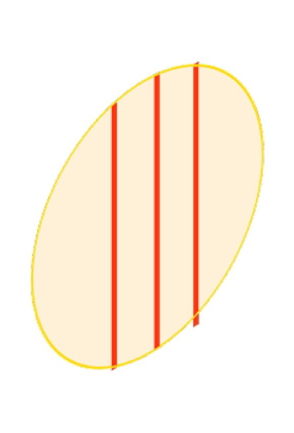
Cancers 2020, 12, 527 4 of 25

 A
 Healthy cell

 Homogenous radiation
 Conventional Radiotherapy Tumour cell

 Radiation source

 Multi-slit
 collimator

 B

 Spatial fractionation
 Microbeam Radiotherapy

 Centre-to-centre
 space 50–500 μm

 Microbeam width
 20–100 μm

 C Microbeam

 Peak
 dose
 Dose

 Valley
 dose

 Lateral extension

 Figure 1. Homogenous radiation vs spatial fractionation. Schematic representation of a homogenous
 beam of radiation (panel A) and a MRT array (panel B) targeting a malignancy. Schematic
 representation of the microbeams dose distribution (panel C).

3. Results

3.1. Literature Selection
 After duplicates were removed, a total of 5688 records were identified (Figure 2). Based on title
and abstract screening, 5534 citations were excluded, and 159 potentially relevant papers were
Cancers 2020, 12, 527 5 of 25

assessed for full-text eligibility, which includes 5 additional records identified through reference
scanning. After the full-text screening, 64 articles were further excluded, and 95 studies were finally
included in the review. The list of included references can be found in the Supplementary Materials.

 Records identified through Additional records identified
 database searching (n=7404) through other sources (n=0)

 Records after duplicates removed
 (n=5688)

 Records excluded (n=5534)

 Additional articles
 Full-text articles assessed for
 identified through
 eligibility (n=159) Full-text articles excluded (n=64)
 reference scanning (n=5)
 • Conference abstracts n=45
 • Not MRT n=15
 • Not animal model n=3
 • Duplicate n=2
 Full-text articles selected for
 data extraction (n=95)

 n=95 studies included

 n=72 Experimental articles
 n=16 Reviews
 n=2 Clinical Perspectives
 n=1 Report
 n=2 Commentary
 n=1 Book chapter
 n=1 Letter to the Editor

 Figure 2. Study Flow. Details of the flow of information during the study.

3.2. Study Characteristics
 The 95 papers studying animal models in MRT were disseminated between 1994 and 2019
(Figure 3A). The majority of the articles have been published in the last ten years, with the highest
publication record (11 papers) in 2015. From the 72 experimental articles, the most commonly used
animal models have been Rats (59.7%) and Mice (33.3%) with only a few studies using other animal
models (Figure 3B).

Cancers 2020, 12, 527 6 of 25

 Figure 3. Publication rate and frequency of animal species used as animal models in Microbeam
 Radiation Therapy (MRT). (A) Shows the yearly publication of full-text articles that described Animal
 Models in MRT (including reviews and commentaries). (B) Shows the word count and frequency of
 all animal species used (it only includes the 72 experimental articles).

3.3. Technical Parameters
 In the reviewed literature, the MRT array was created via two mechanisms. By performing
several consecutive irradiations (with horizontal translation) using a single-slit collimator, to
generate an array of co-planar microbeams at the target. Alternatively, by using a multi-slit collimator
that generates an array of co-planar microbeams simultaneously.
 Across the included literature, the width of the microbeams ranged from 20 to 100 µm, with
spacing varying from 50 up to 500µm (See Table 1; Table 2).
 MRT can be delivered as a single or multiple array(s). When multiple arrays are delivered, the
irradiation geometry can be subclassified according to the trajectory of delivery and the anatomical
plane.
 • Trajectory of delivery: if two or more arrays were delivered in the same trajectory or
 direction, they were called unidirectional. However, if the arrays were delivered in two or
 more trajectories, they were referred to as bidirectional or multidirectional.
 • Anatomical plane: if two or more MRT arrays were delivered in the same anatomical plane
 they were also called co-planar. When two co-planar arrays were delivered without overlap
 of the peak-dose regions, they were referred to as interlaced. Delivery in different planes
 was referred to as cross-planar where irradiation with a single unidirectional MRT array
 was followed by a rotation of the animal and a second (or third) MRT array was delivered
 (creating a grid-like pattern in the target volume). When arrays in this manner intersect at
 90°, they are orthogonal (irrespective of the anatomical plane or trajectory).
 The use of the word cross-fired however, was more general. It described two or more arrays, but
it did not inform consistently about the trajectory or the anatomical plane of the arrays.
 Peak-doses ranged from 3.9 Gy [17] up to 10000 Gy [18], however, the depth at which they were
measured varied across the publications from 0.3 mm (skin-entry or entrance) to several millimetres
in depth depending on the location of the target. Valley-doses were also reported at different depths,
the lowest was 0.5 Gy at skin-entry [19], and the highest was 38.5 Gy at 5 mm depth [20]. Additionally,
the peak-to-valley dose ratio (PVDR) was described as an important metric related to the therapeutic
index of MRT [21].
 Dose-rate was reported in only 27 out of 72 experimental articles and varied greatly from 0.3 to
16000 Gy/s. This large variation is relevant because dose-rate, together with the peak-dose, will
determine whether MRT will also have a FLASH effect. The FLASH effect is defined as the delivery
Cancers 2020, 12, 527 7 of 25

of radiation at ultra-high dose rates, which allows normal tissue tolerance levels to be exceeded [22–
24]. This is achieved when the whole dose of radiation (the peak-dose in the case of MRT) is delivered
in less than 200 milliseconds [24]. Therefore, not all MRT sources will be able to have a FLASH effect
since, for example, the delivery of a peak-dose of 400 Gy requires a dose rate of 2000 Gy/s as a
minimum to be delivered in 200 milliseconds.

3.4. Animal Models in MRT
 Animal models are divided into their use for investigating tumour control, non-neoplastic
diseases and normal tissue effects following MRT.

3.4.1. MRT for Cancer
 Table 1 reports all the studies that involve the use of MRT to treat tumour-bearing animals and
summarises key radiation parameters which are associated with tumour control.

3.4.2. Glioma
 The most commonly employed cancer models in MRT research were brain tumours. The first
animal model used was syngeneic 9L gliosarcoma (9LGS) implanted in rats. In the early nineties, at
the National Synchrotron Light Source of the Brookhaven National Laboratory, Laissue, Slatkin and
colleagues [25–28] showed that MRT (unidirectional and bidirectional) delayed tumour growth of
intracerebral 9LGS. Since then, 9LGS cells in the Fisher 344 strain rat model has been widely used to
test several MRT configurations and doses.
 In 2002, Dilmanian and colleagues [15] studied different unidirectional arrays on the same 9LGS
model. The array had a constant microbeam width (27 µm), three spacings (50 µm, 75 µm and 100
µm), and two peak-doses (150 Gy and 500 Gy). All MRT conditions improved survival as compared
to the control group.
 In addition, several studies conducted by Bouchet and colleagues [20,29,30], using the 9LGS
model, investigated the molecular mechanisms behind the MRT efficacy in comparison to
unirradiated controls.
 A comparison between a homogenous broad beam (BB) of radiation delivering 17.9 Gy to the
tumour with one MRT array - formed by microbeams of 50 µm width and centre-to-centre spacing
of 200 µm (hereafter defined as “50 µm; 200 ctc”) - delivering 200 Gy peak-doses to the tumour
revealed that the survival for the animals of both groups was similar [31]. However, when the
deposited peak-dose in the MRT array was increased to 400 Gy the survival of the gliosarcoma-
bearing rats was significantly increased compared to the BB irradiated animals [31].
 Several studies have shown that delivering multiple MRT arrays increased the survival of 9LGS-
bearing rats significantly compared with unirradiated controls [32], with characteristic induction of
hypoxia at the tumour site [33]. Serduc et al. [34] employed bidirectional MRT arrays to compare the
efficacy of three different microbeams widths (25, 50 and 75 µm), each delivered as two orthogonally
cross-planar intersecting arrays. In all the MRT configurations the valley-dose was kept constant (at
18 Gy each) by fixing the spacing (211 ctc), and only the peak-dose and the microbeam widths were
modified. Thus, microbeams of 25, 50, and 75 µm of width, had peak-dose of 860 Gy, 480 Gy, and 320
Gy per array, respectively. All MRT groups delayed the 9LGS tumour growth. The bidirectional MRT
of 50 µm 211 ctc had the best therapeutic index, while the bidirectional MRT of 25 µm, 211 ctc caused
early deaths in 50% of cases four days after irradiation.
 Three cross-planar MRT arrays (orthogonal to each other) were used to target 9LGS-bearing rats
in three consecutive days at the European Synchrotron [35]. Briefly, day 1 (25 µm; 211 ctc; 400 Gy),
day 2 (25 µm; 211 ctc; 360 Gy), and day 3 (25 µm; 211 ctc; 400 Gy). This strategy increased the lifespan
of the 9LGS-bearing rats by 216% in comparison to the control group.
 MRT has also been tested on 9LGS in combination with different therapeutic agents. In a study
by Régnard and colleagues [36], MRT (25 µm; 200 ctc; 625 Gy peak-dose) was combined with direct
intra-tumoral injection of the chemotherapeutic agents' Temozolomide, Cisplatin, or the contrast
Cancers 2020, 12, 527 8 of 25

agent Gadolinium. Combining Temozolomide and Cisplatin with MRT did not improve survival.
However, the concomitant administration of Gadolinium, as a result of radiation dose-enhancement,
did. The antimitotic drug Chalcone JAI-51 (an inhibitor of tubulin polymerization) improved survival
of 9LGS-bearing rats when combined with two orthogonally cross-fired MRT arrays (50 µm; 200ctc;
480 Gy peak-dose) [37]. Furthermore, Sorafenib (an anti-angiogenic drug) was tested in combination
with MRT on the same cancer model [38]. Although the tumour growth rate was similar after MRT
and the MRT+Sorafenib groups, there were appreciable changes in the tumour physiology which
included reduced tumour blood volume after MRT+Sorafenib.
 A study from 2006 shed light on the possible synergistic action between MRT and the immune
system [39] where combining MRT (27 µm; 211 ctc; 625 Gy peak-dose) with injections of irradiated
9LGS cells (transfected with the gene GM-CSF), increased the median survival of 9LGS-bearing rats.
 Other types of glioma cell lines have also been exposed to MRT. F98 glioma cells and C6 cells,
implanted respectively in Wistar and Fisher rats, were used to demonstrate that two orthogonal,
cross-planar MRT arrays (24.75 µm; 211 ctc; 350 Gy peak-dose) significantly increased survival as
compared to untreated controls [40]. This effect was amplified when MRT was preceded by the
administration of the glutathione synthesis inhibitor buthionine-SR-sulfoximine [41]. When two
orthogonal MRTs (50 µm; 200 ctc; 241 Gy peak-dose and 10.5 Gy valley-dose) were compared to two
orthogonal BB fields (10.5 Gy), MRT was more effective than BB in slowing F98 glioma growth and
increasing tumour vessel permeability [42]. The same model of F98 cells in rats was employed by
Fernandez-Palomo and colleagues, to prove that the presence of glioma causes different abscopal and
bystander effects in MRT-irradiated rats and also in unirradiated (tumour-free) cage-mate rats [43].
 C6 glioma-bearing rats were also employed by Fernandez-Palomo et al. for the study of
bystander and abscopal effects when comparing MRT with BB [19]; both MRT and BB promoted
radiation-induced bystander effects in the non-irradiated portion of the brain and in the bladder of
the tumour-bearing animals. This same model has been used to study the dose deposition of MRT
(25 µm; 200 ctc; at 35, 70 or 350 Gy peak-dose) using the marker of DNA damage -H2AX [44].
Moreover, the damage of the optic nerve was studied after treating the same C6 glioma in Wistar rat
with cross-fired MRTs (25 µm; 200 ctc, 350 Gy peak-dose) [45]. The optic nerves did not show any
significant histological difference compared to unirradiated controls.
 Rat xenografts of glioma cells implanted in the brain of nude mice were also used in MRT
studies. In a model of 9LGS cells implanted in nude mice (strain not reported), a significant increase
of tumoral apparent diffusion coefficient (ADC), consistent with an increase of vascular permeability,
was shown 24 h after the delivery of two orthogonally cross-planar MRT arrays (25 µm; 211 ctc; 500
Gy peak-dose) in irradiated tumours versus unirradiated controls [46]. In addition, athymic nude
mice (Crl:NU-Foxn1nu) were implanted with F98 glioma cells to establish that bystander and
abscopal effects can also be detected in athymic mice after MRT irradiation (50 µm; 200 ctc; 22 or 110
Gy peak-dose) [47].
 Finally, it must be mentioned that human xenografts in nude mice have also been explored. In
the study performed by Uyama and colleagues [48], U251 human glioma cells, implanted
subcutaneously into the flank of nude mice (BALB/cAJc1-nu/nu), were employed to successfully
demonstrate the efficacy of one MRT array (100 µm; 500 ctc; 124 Gy peak-dose) and two cross-planar,
but unidirectional MRT arrays of (100 µm; 500 ctc; 124 Gy peak-dose) or (20 µm; 100 ctc; 111 Gy peak-
dose) at the SPring-8 Synchrotron radiation facility in Japan.

3.4.3. Mammary Tumours
 Investigations of the effects of MRT on mammary malignancies have been made at the Japanese
and Australian Synchrotrons. The most used pre-clinical model for the study of MRT on mammary
malignancies is the subcutaneous injection of EMT-6.5 mouse mammary carcinoma cells in female
BALB/c mice. Dilmanian and colleagues [49] injected EMT6.5 cells subcutaneously in the flanks of
mice from which tumours were subsequently harvested and small portions transplanted
subcutaneously into the calves of BALB/c female mice. These mammary carcinoma-bearing mice
were targeted with either one MRT array (90 µm; 300 ctc; doses in Table 1), two cross-planar but

Cancers 2020, 12, 527 9 of 25

unidirectional arrays (90 µm; 300 ctc; doses in Table 1), or with BB of either 23, 30, 38, or 45 Gy. They
reported that even one array can control EMT-6.5 tumour growth but that the cross-planar
configuration optimally balances tumour control and tissue toxicity.
 By subcutaneously inoculating EMT-6.5 and 67NR mammary tumour cells on the right hind leg
of BALB/c mice, Crosbie and colleagues [50] showed a significant increase in median survival
compared to controls following one MRT array (25 µm; 200 ctc, 560 Gy). They also showed no signs
of tumour re-growth after two different cross-fired MRT arrays (25 µm; 200 ctc; 280 Gy peak-dose)
and (25 µm; 200 ctc; 560 Gy peak-dose).
 Using the same syngeneic model, a comprehensive transcriptomic analysis of EMT-6.5 tumours,
exposed either to MRT (25 µm; 200 ctc; 560 Gy peak-dose) or BB (11 or 22 Gy) compared to non-
irradiated tumour controls, underlined how MRT-treated tumours overexpress immune-related
genes at early time points after irradiation [51]. Similarly, MRT (25 µm; 200 ctc; 112 Gy or 560 Gy
peak-dose) delivered to EMT-6.5 tumours up-regulated genes that are involved in eosinophil
recruitment and/or functionality relative to BB (5 or 9 Gy) irradiated tumours [52]. Using the same
EMT-6.5 mouse model and same MRT and BB configurations and doses, Yang et al. [53] showed that
a different immune response is induced by MRT compared to BB, including a significant increase of
tumour-infiltrating T-lymphocytes at 48h post-MRT.
 The tissue composition of the EMT6.5 mammary model was investigated after MRT (25 µm; 200
ctc; 560 Gy peak-dose) and BB (11, 22 or 44 Gy) using Fourier-transform infrared microspectroscopy
[54]. Absorbance patterns in the nucleic acid region showed chemical shifts between the peak and
valley-dose regions [54].
 Griffin et al. [55] employed a different type of syngeneic mouse model of mammary carcinoma,
where 4T1 cells were injected subcutaneously in the limb of female BALB/c mice. They combined
MRT with the anti-angiogenic peptide anginex and showed enhanced tumour growth-delay in
combination with MRT.

3.4.4. Melanoma
 A recent publication from Potez et al. [7] described for the first time that MRT (50 µm; 200 ctc;
407.6 Gy peak-dose) is an efficient approach for the treatment of radioresistant melanomas compared
to a BB (6.2 Gy) irradiation and unirradiated control animals. In this model, B16-F10 melanoma cells
were implanted in the ears of C57BL/6J mice.

3.4.5. Squamous Cell Carcinoma
 One study from Miura and colleagues [56] employed a murine model of aggressive murine
SCCVII squamous cell carcinomas implanted subcutaneously into the left thighs of female C3H mice.
They showed that a better palliative effect was elicited by MRT irradiations (35 µm; 200 ctc; 625 Gy
or 884 Gy peak-dose and 70 µm; 200 ctc; 442 Gy peak-dose) compared to BB (25 or 35 Gy) irradiations.
Cancers 2020, 12, 527 10 of 25

 Table 1. MRT Parameters Used in Cancer Models.
 Microbeam
 Number
 Animal Cancer Type Width Spacing Peak-dose (Gy) Valley-dose (Gy) Evaluated Criteria
 of arrays
 (µm) (µm)
 Animal survival, tumour
 Gliosarcoma 400 entrance dose;
 Rat [1] 1&2 50 200 12.5 @1cm depth growth, tumour vasculature,
 (9L) 350 @1cm depth
 and cell proliferation
 Animal survival, cell cycle,
 Gliosarcoma 480 entrance dose;
 Rat [2] 2 50 200 18.6 @1cm depth and DNA distribution
 (9L) 418 @1cm depth
 patterns
 Gliosarcoma 400 entrance dose; Tumour vasculature and
 Rat [3] 2 50 200 12.5 @1cm depth
 (9L) 350 @1cm depth tumour oxygenation
 Gliosarcoma 400 dose @tumour 18 dose @tumour (i.e., Animal survival and
 Rat [4] 1 50 200
 (9L) (i.e., @7mm depth) @7mm depth) Transcriptomics
 Tumour growth,
 Gliosarcoma 400 dose @tumour 8 dose @tumour (i.e.,
 Rat [5] 1 50 200 transcriptomics, and
 (9L) (i.e., @7mm depth) @7mm depth)
 histopathology
 400 dose @tumour 17.4 dose @tumour
 Animal survival, tumour
 Gliosarcoma (i.e., @7mm depth) (i.e., @7mm depth)
 Rat [6] 1 50 200 growth, cell proliferation,
 (9L) 200 dose @tumour 8.7 dose @tumour (i.e.,
 and gene expression
 (i.e., @7mm depth) @7mm depth)
 Tumour vasculature and
 Rat [7] Glioma (F98) 2 50 200 241.4 entrance dose 10.5 @9mm depth
 tumour oxygenation
 560 entrance dose 8.5 @centre of brain
 Mammary 1 Animal survival, DNA
 Mouse 800 entrance dose 12 @centre of brain
 (EMT6.5/67N 25 200 damage, cell proliferation,
 [8] 280 entrance dose 8.5 @centre of brain
 R) 2 and apoptosis
 560 entrance dose 17 @centre of brain
 150 entrance dose;
 20 @centre of brain
 108 @centre of brain
 250 entrance dose;
 50 34 @centre of brain
 179 @centre of brain
 300 entrance dose;
 40 @centre of brain
 215 @centre of brain
 Gliosarcoma 250 entrance dose; Animal survival and
 Rat [9] 1 27 17 @centre of brain
 (9L) 179 @centre of brain histopathology
 300 entrance dose;
 75 20 @centre of brain
 215 @centre of brain
 500 entrance dose;
 33 @centre of brain
 359 @centre of brain
 500 entrance dose;
 100 19 @centre of brain
 359 @centre of brain
 800 dose @tumour 16 dose @tumour
 890 dose @tumour 18 dose @tumour
 19 dose @tumour
 970 dose @tumour
 1 90 300
 Mouse Mammary 1740 dose @tumour 35 dose @tumour
 Tumour ablation
 [10] (EMT6.5) 1820 dose @tumour 36 dose @tumour
 1900 dose @tumour 38 dose @tumour
 410 dose @tumour 16 dose @tumour
 2 90 300 520 dose @tumour 21 dose @tumour
 650 dose @tumour 26 dose @tumour
 17.5, 35, 70, 350 Bystander effects in-vivo by
 Rat [11] Glioma (C6) 1 25 200 0.51, 1.03, 2, 10.3
 entrance dose clonogenic cell survival
 35, 70, 350 entrance
 Rat [12] Glioma (C6) 1 25 200 NR DNA damage
 dose
 Bystander effects in-vivo by
 Rat [13] Glioma (F98) 1 25 200 20, 200 entrance dose NR clonogenic cell survival and
 cellular calcium fluxes
 Mouse Bystander effects in-vivo by
 nude Glioma (F98) 50 400 22, 110 entrance dose 0.5, 2.5 clonogenic cell survival and
 [14] cellular calcium fluxes
 7.5 in a 10 mm solid Tumour growth, tumour
 Mouse Mammary
 1 50 200 150 @5 mm depth water vasculature, and tumour
 [15] (4T1)
 phantom hypoxia
 Immune response by gene
 Mouse Mammary
 1 25 200 112, 560 NR expression and
 [16] (EMT6.5)
 histopathology
 Gliosarcoma Tumour vasculature, and
 Rat [17] 1 50 200 400 entrance dose NR
 (9L) tumour hypoxia
 Gliosarcoma Animal survival, tumour
 Rat [18] 1 25 100 625 entrance dose NR
 (9L) growth, and histopathology
 Mouse 442, 625, 884 entrance Animal survival, tumour
 1 35 200 NR
 [19] dose growth, and tumour ablation

Cancers 2020, 12, 527; doi:10.3390/cancers12030527 www.mdpi.com/journal/cancers
Cancers 2020, 11 of 25

 Squamous cell
 carcinoma 70 200 442 entrance dose
 (SCCVII)
 Optic nerve damage by
 Rat [20] Glioma (C6) 2 25 200 350 entrance dose NR
 histopathology
 Tumour growth, tumour
 Mouse Melanoma vasculature, cell
 1 50 200 407.6 dose @tumour 6.2 dose @tumour
 [21] (B16F10) proliferation, cell senescence,
 and immune response
 Gliosarcoma 200 625 entrance dose 12.1 dose @tumour Animal survival, tumour
 Rat [22] 1 25
 (9L) 100 625 entrance dose 36 dose @tumour growth, and histopathology
 Gliosarcoma Animal survival, tumour
 Rat [23] 1 25 200 625 entrance dose NR
 (9L) growth, and histopathology
 Biochemical changes by
 Mouse Mammary synchrotron Fourier-
 1 25 200 560 entrance dose 11
 [24] (EMT6.5) transform infrared
 microspectroscopy
 Glioma (C6, Animal survival and object
 Rat [25] 2 25 211 350 entrance dose NR
 F98) recognition
 Animal survival and
 Rat [26] Glioma (F98) 2 28 400 350 18 dose @ tumour
 cognitive dysfunction
 Mouse Animal survival, tumour
 Gliosarcoma
 nude 1 25 211 500 entrance dose 24 (cross-fired) growth, and tumour
 (9L)
 [27] vasculature
 25 860 entrance dose
 Gliosarcoma Animal survival and
 Rat [28] 2 50 211 480 entrance dose 18 @1cm depth
 (9L) histopathology
 75 320 entrance dose
 Gliosarcoma 400, 360 (+24h), 400 Animal survival and
 Rat [29] 3 50 211 15
 (9L) (+48h) entrance dose histopathology
 Animal survival,
 Gliosarcoma
 Rat [30] 1 27 211 625 entrance dose NR histopathology, and immune
 (9L)
 response
 Mouse Mammary
 1 25 200 560 11 Transcriptomics
 [31] (EMT6.5)
 Mouse 1 100 500 124 4.8 Tumour growth,
 nude Glioma (U251) 20 100 111 8.2 histopathology, and
 2
 [32] 100 500 124 9.6 apoptosis
 Mouse Mammary
 1 25 200 112, 560 NR Immune response
 [33] (EMT6.5)
 NR: Not-Reported.

3.4.6. MRT for non-neoplastic diseases
 Microbeam radiation therapy has also been applied to the treatment of several non-malignant
diseases and employed as a tool for studying Central Nervous System (CNS) injury.

Epilepsy:
 Due to the high tolerance of the central nervous system to MRT, it has been applied, pre-
clinically, as a radio-surgical tool to modulate brain networks and treat a variety of neurological
disorders including epilepsy. The size and precision of microbeams can mimic a surgical cut to the
epileptic cortex reproducing the current surgical ablation techniques but with a reduced risk of
inducing neurological dysfunction [57,58]. Microbeam transections of the sensorimotor cortex
significantly reduced seizure without inducing neurological dysfunction (100 µm; 400 ctc; 240 or 320
Gy peak-dose) [59]. This is reflected by decreased neuronal excitability of neurons in the
somatosensory cortex and reduction of spontaneous synaptic activities and synchronization [60].
Anti-epileptic activity was maintained more than 4 months post-irradiation [61] with an absence of
myelin and neurons in the microbeam-irradiated slices [62].

Restenosis:
 MRT has been implemented as a potential treatment for restenosis following angioplasty,
exploiting the vascular-sparing and wound-healing effects of MRT to interfere with the process of
cellular hyperplasia, which underlies this condition [63]. The MRT array used in this study (27 µm;
200 ctc; 150 Gy peak-dose) did not yield significant improvement, and future dose-escalation studies
are required.
Cancers 2020, 12 of 25

Spinal cord Injury:
 MRT has also been used as a tool to induce spinal cord injuries to study the mechanisms
following CNS injury [64]. Further studies have been performed, but their microbeams width fall
outside our inclusion criteria.

3.4.7. Normal Tissue Effects of MRT
 It has been well described that in addition to effective tumour control, MRT exhibits exceptional
preservation of normal tissues. This tissue-sparing effect is exemplified by the high tolerance of skin,
CNS, and vasculature following MRT doses. Table 2 provides a summary of the conservative MRT
parameters that each publication cites before the onset of pathology in the majority of test animals.
Valley-doses are indicated where provided.

Central Nervous System, Adults:
 Slatkin et al. 1995 presented the tissue-sparing effects on normal adult rat brain exposed to MRT
(20 µm; 200 ctc) [18]. Brain tissue appeared normal following peak-dose of 625 Gy, necrosis was
absent even after peak-doses of up to 5000 Gy, and was visible only after a peak-dose of 10000 Gy. In
another study, when multiple arrays were used, brain tissue damage was restricted to the cross-
irradiated regions of the two arrays (25 µm; 200 ctc; 625 Gy peak-dose) and normal tissue damage
was absent in the brain exposed to only one array [25]. Irradiation of the rat spinal cord did not impair
locomotion after 253 Gy (35 µm; 210 ctc), while paresis and foreleg paralysis developed when the
dose reached 357 Gy or greater [65]. When valley-doses at the target depth approached 18 Gy (507
Gy peak-dose), white and grey matter necrosis, necrosis of microvasculature and leukocyte
infiltration was observed.

Developing CNS:
 Employing MRT in infant patients was also pursued in pre-clinical studies. Early MRT studies
were aimed at assessing its effects on immature brain tissues with suckling rats [26], weanling piglets
[66] and unhatched duck embryos [67] as models. In fact, there was a differential dose tolerance of
the suckling rat relative to the weanling piglet, with the rat displaying neurological dysfunction when
exposed to 150 Gy (28 µm; 105 ctc)[26]. Similarly, irradiation of the CNS of the unhatched duck
embryo could withstand a peak-entrance dose of 160 Gy (27 µm; 100 ctc) before displaying signs of
ataxia within 75 days of hatching [67]. In contrast, the piglet showed no signs of neurological damage
at an dose of 600 Gy (28-30 µm; 200 ctc) [66]. These three studies had comparable beam widths,
however, they differed in the beam spacing with 100 µm and 200 µm used in the rat and piglet,
respectively. When the spacing was increased to 200 µm, the dose threshold tolerated by the suckling
rat increased to 300 Gy without signs of neurological dysfunction [26].

Eye:
 Certain target areas in the brain may result in ocular radiation exposure. The eye of the rat
developed retinal degeneration after bidirectional MRT arrays (25 µm; 211 ctc; 350 Gy peak-dose)
[68]. This may occur after exposure of the retina to valley doses of up to 14 Gy [69]. The optic nerve
was also preserved at these doses [45], however, ocular damage following MRT irradiations to the
brain have not been assessed at doses below 350 Gy. Therefore, a tolerance threshold cannot be
reported.

Vasculature:
 The normal tissue response and tissue sparing effect of MRT are dependent on the preservation
or regeneration of microvasculature. Vasculature organization of the irradiated tissue depends on the
stage of tissue maturation and as a result, determines tolerance to MRT. Models of immature
vasculature have been the regenerating fin of the zebrafish [70] and the early chick chorioallantoic
Cancers 2020, 13 of 25

membrane [71]. The susceptibility of immature vessels to MRT-induced damage is significantly
higher than the mature vessels which show very little post-MRT alterations (vascular lesions, reduced
perfusion) when the MRT spacing is kept below 200 µm [71]. Although conducted in non-malignant
tissue, the immature vasculature is representative of the disorganized, leaky and fast-growing
tumour vessel networks.
 In the mouse brain, it has been shown that the microvasculature can withstand up to a 1000 Gy
peak entrance dose from a unidirectional MRT array (25 µm; 211 ctc) [72] with only transient blood-
brain-barrier leakage. No vascular damage [73], or changes in cerebral water content were observed
at this dose [72,74]. In the beam path, there is a progressive disappearance of glial and neuronal cells,
but endothelial cells remain detectable in the irradiated tissue and tissue remains perfused. No
changes in blood volume or vascular density are observed (up to 3 months post-irradiation)
indicating rapid vascular repair [72]. Additionally, peripheral arteries in the mouse can tolerate peak
microbeam doses up to 2000 Gy (50 µm; 400 ctc) with minimal damage [75].

Skin:
Studies investigating the tolerance of the skin of rats and mice have shown that doses up to 1170 Gy
(90 µm; 300 ctc; dose-valley up to 35 Gy) are marked by the absence of moist desquamation, focal
denudation or exudation [76]. Moreover, 1740-1900 Gy peak-entry-doses showed significant damage
to the skin in both the mouse and the rat with the development of moist desquamation, epilation, and
necrosis. The valley-dose was responsible for these adverse reactions, limiting valley-doses to less
than 45 Gy for both cross-planar MRT of 2 arrays [49] and a single MRT array [76]. The peak threshold
dose is reduced to 650 Gy with a bidirectional microbeam as the volume of irradiated tissue is
increased with this irradiation geometry. The skin has been shown to efficiently repair double-strand
breaks in the microbeam path (24-84 hrs post-irradiation [pi]) with the absence of apoptosis after
MRT (25 µm; 175 ctc) [77]. Doses up to 800 Gy (25 µm; 200 ctc) exhibited similar damage to BB doses
of up to 44 Gy [78]. Histological changes following 800 Gy (25 µm or 50 µm; 200 ctc) include
thickening of the dermis and epidermis, hyperkeratosis, dermal oedema, and a loss of sebaceous
glands [6,78].

Other tissues:
 Lastly, MRT has been applied as a fertility-sparing modality for testicular irradiation and has
been able to successfully preserve spermatogenesis in the rat [79]. MRT could, therefore, also be
implemented as a treatment modality for testicular cancers or tumours in the inguinal region where
infertility is an adverse outcome of current conventional radiotherapies.

Importance of Valley-Doses:
 Normal brain tissue damage (including radiation-induced necrosis) is highly dependent on the
valley-dose, and many authors suggest that cells in these regions repopulate the tissue damage in
those regions receiving the peak-dose [25,49,66,73,80,81]. Therefore, it is important to ensure that
valley-dose does not exceed the tolerance threshold of the normal tissue [65]. Valley-doses can be
reduced either by decreasing the peak-dose, decreasing the width of the microbeam itself or
decreasing the beam field-size [34]. Delivered MRT doses attenuate with increasing tissue depth [34],
with the peak regions exhibiting a sharper fall-off than the valley regions [82].Ensuring effective dose
delivery to the target while maintaining appropriate valley-doses to preserve normal tissue, is
therefore an essential step in treatment planning. Many papers in the MRT field do not report
predicted doses received by the target tissue, but when provided, these values are reported in Table
2.
Cancers 2020, 14 of 25

 Table 2. MRT Parameters Eliciting Normal Tissue Tolerance.
 Microbeam
 Threshold Peak- Valley-Dose
 Animal Tissue Width Spacing Pathology
 Dose (Gy) (Gy)
 (µm) (µm)
 Immature
 Duck [67] 27 100 Ataxia 160 NR
 CNS
 27 75 250 17
 Rat [15] Brain Necrosis, oedema
 27 100 500 33
 Rat [73] Brain 27 200 Cell loss, demyelination 1000 NR
 Tissue damage (loss of 625 uni- &
 Rat [25] Brain 25 100 tissue structure; vascular bidirectional NR
 damage) 312 bidirectional
 Paresis/Paralysis (over 383 357 (19mm
 Rat [65] Spinal cord 35 210 12.7
 dpi) transverse depth)
 Damage to
 Mouse
 Brain 25 211 microvasculature/vasogenic 312–1000 5.8
 [72]
 oedema (up to 1 mpi)
 Rat (11–13 28 105 150
 day old) Brain 28 105 Neurological dysfunction 50 >5 Gy [69]
 [26] 25 210 300 bidirectional
 Neurological function (465 600 peak-entry 12.6 @
 Piglet [66] Brain 20–30 210
 dpi) 263 @ cerebellum cerebellum
 20 200 Necrosis (14 dpi) 5000
 37 75 Tissue damage (14–31dpi) 625
 Brain
 20 200
 Rat [18] Loss of nuclei (30-31dpi) 2000 NR
 37 75
 37 200 1250–2500
 Skin Epilation (2–4 weeks dpi)
 37 75 1250–2500
 Mouse Cerebral oedema (up to 28
 Brain 25 211
Cancers 2020, 15 of 25

paradigm (spatial fractionation). Thus, even tumour cells not directly irradiated can be eradicated
completely while damage to healthy tissue can be repaired efficiently. As a consequence, it is
anticipated that MRT will warrant higher tumour control at reduced toxicity in patients.

4.1. Potential Underlying Mechanisms of Microbeam Radiation Therapy

4.1.1. Targeted and Non-Targeted Effects of MRT
 Peak-doses cause severe radiation damage that is restricted to the cell populations in the path of
the microbeam. This can be observed in the brain of rats as early as 8h post MRT (25 µm; 200 ctc; 350
Gy peak-dose) [44], and as late as 128 days post MRT (25 µm; 100 ctc; 625 Gy peak-dose) [25]. In the
cerebellum of piglets, microbeam tracks were still visible 15 months post MRT (20–25 µm; 211 ctc;
cerebellar peak-dose 66 to 263 Gy; valley-dose 3.2 to 12.6 Gy) [66]. Sharp delimitation of damaged
tissue in the microbeam path has also been shown in Drosophila tissue where high peak doses
exceeding lethal, seamless irradiation doses affect specific morphological processes while
maintaining the survival of post-mitotic tissues and the organism as a whole [84].
 Radiation-induced bystander effects (RIBE) are relevant for MRT because tissue exposed to the
valley-dose will receive signals from neighbouring cells exposed to the peak-dose. Although falling
outside of our inclusion criteria due to its employment of a single microbeam and not an array,
Dilmanian et al [85] were the first to suggest the importance of RIBE in MRT. Their results from
irradiated rat spinal cord indicated that the repair process and the elimination of apoptotic cells in
the peak area occurred faster than expected, suggesting that restoration and proliferation was a
consequence of “beneficial” bystander factors coming from the valley area. This was further
suggested by the results of experiments by Fernandez-Palomo et al [44] performed in the brain of
rats.
 When it comes to the effects on non-irradiated tissues (outside of the microbeam array),
responses such as genotoxic effects [77] and clonogenic cell death on cells exposed to signals from the
irradiated animal [19,43] have been observed after MRT. Some of those responses have involved the
immune system [47], with some author suggesting that a functional immune system is key to observe
such genotoxic effects post MRT [86].

4.1.2. MRT Selectively Disrupts Immature Blood Vessel
 The biological effects induced by MRT go beyond direct tumour cell destruction. In fact, MRT
does not impact the morphological and functional characteristics of normal murine brain vessels even
after delivery of doses up to 1000 Gy [72]. Brain perfusion, capillary density and blood volume remain
unaffected 12h to 3 months after an anteroposterior MRT array (25 µm; 211 ctc; 312 or 1000 Gy peak-
entrance dose) [72]. No changes in animal behaviour have been observed [72]. Data from chick
chorioallantoic membrane [71] and zebrafish fin regeneration [70] models demonstrate the disruptive
vascular effect of MRT on immature blood vessels. Work in adult organisms confirmed that the
disruptive vascular effects of MRT depend on the vascular maturation status. In adult zebrafish, a
correlation between microbeam width and biological effects of MRT was identified [70]. The study
indicated that microbeam spacing between 50 to 100 µm could selectively affect mature and
immature vessels. Murine brain vessels do not tolerate beamlets wider than 100µm when peak doses
of 400 Gy are delivered [87]. The use of MRT in rodent models revealed a preferential adverse effect
on tumour vessels rather than those of healthy tissue. In a murine melanoma model, MRT
significantly reduced (24%) the perfusion of the tumour blood vessels indicating vascular disruption
[7].
 MRT preferentially lowers tumour O2 saturation levels in gliosarcoma as a result of reduced
endothelial cell density and increased inter-vessel distance following two cross-fired arrays
(anteroposterior and lateral; each 50 µm; 200 ctc; 400 Gy peak-entry-dose) leading to tumour hypoxia
observed by GLUT-1 overexpression [32,33,38]. However, the persistence of hypoxia may be dose-,
Cancers 2020, 16 of 25

time- and tumour-dependant with contrasting evidence of tumour hypoxia in a mammary carcinoma
decreasing within 14 days post-irradiation at a dose of 150 Gy [55].

4.1.3. MRT Transiently Increases Tumour Blood Vessel Permeability
 Few reports indicate that MRT causes a partial disintegration of the endothelium leading to an
early (1 to 4h) but significant increase in tumour blood vessel permeability (manuscript in
preparation). This “MRT-induced vascular permeability window” represents a potent drug delivery
strategy. Combination of MRT with chemotherapy or nanoparticles applied in the “permeability
window” results in significantly better tumour control (manuscript in preparation). A recently
published study indicated that MRT induced selective vascular permeability in tumour blood vessels
but not in normal vasculature. The tumour vascular permeability was observed from days 2 to 7 after
MRT. In contrast, it was observed only at day 7 after BB radiotherapy [42]. This study also
documented that delivering a second MRT array induced earlier, more pronounced, and more
persistent tumour vascular permeability than BB.

4.1.4. MRT Boosts the Recruitment of Circulating Immune Cells into Tumours
 Data obtained in zebrafish indicate that MRT triggers an acute inflammatory response restricted
to the regenerating tissue [70]. Six hours post-MRT, the regenerating tissue was infiltrated by
neutrophils, and thrombocytes which adhered to the cell wall locally in the beam path. The mature
tissue was not affected by microbeam irradiation [70]. In a mouse tumour model, it has been
demonstrated that BB markedly increases tumour-associated macrophages and neutrophils while
there were no increases in these populations following MRT [53]. The role of MRT in the recruitment
of immune cells represents a promising immunoprophylactic treatment strategy and should be
further explored in experimental and clinical studies. A recently performed study in melanoma-
bearing animals showed that MRT (particularly after 2 irradiations) selectively increased production
of natural killer (NK) cell- and cytotoxic T lymphocytes-attracting chemokines while, also showing,
decreased secretion of T regulatory cell-, tumour associated macrophage- and neutrophil-recruiting
chemokines (manuscript in review). The role of single and repeated MRT administration in the
recruitment of immune cells represents a promising immunoprophylactic treatment strategy and
should be further explored in experimental and clinical studies.
 In addition, increased pericyte density following MRT suggests a vascular normalization effect
[55] which may increase migration of immune cell populations to the tumour and modulate cytokine
expression that may support an anti-tumour immune response. Synergism between
immunoprophylaxis and MRT has been documented [39]. Differential modulation of genes involved
in inflammation and immunity are also marked at early timepoints between tumour and normal
tissue [29,52] and increased leukocyte infiltration into the tumour has also been observed.

4.2. Clinical Translation
 Barriers to the translation of MRT relate to a limited understanding of how to compare the
radiobiological effects of MRT with conventional BB radiotherapy, as well as, understanding the
biology behind the physical properties of the microbeams. In addition, the optimal therapeutic
strategy for MRT is still to be defined.

4.2.1. Comparative Therapeutic Effects of MRT and BB Radiotherapy
 A limited number of studies systematically compared the biological effects of MRT with BB
radiotherapy. The superiority of MRT compared to synchrotron BB treatment for tumour control has
been demonstrated in mice bearing B16-F10 melanoma [7] and rats bearing 9LGS [31]. In these
studies, the MRT valley-dose was kept equal to the BB dose; however, future studies could use the
Equivalent Uniform Dose concept (EUD) to determine more biologically comparable doses of MRT
and BB [88].
Cancers 2020, 17 of 25

 Dose-escalation studies in healthy rodents have identified doses of MRT and BB predicted to
cause equal rates of late spinal cord myelopathy [65], lethal neurotoxicity [80] and acute toxicity
following total and partial body irradiation [83]. Of these studies, only Smyth et al. [83] included a
comparison between MRT and BB radiotherapy at a conventional dose-rate (in the order of 2 Gy
min−1). This indicates the need for further studies controlling for the potential FLASH tissue-sparing
effects of synchrotron broad-beam irradiation.

4.2.2. Identifying Optimal Targets and Strategies for MRT
 A key limitation of MRT is the need for a kilovoltage energy spectrum to maintain spatial
fractionation on a microscopic scale, leading to 30–40% of the dose being attenuated within the first
few centimetres of tissue [89,90]. In addition, the PVDR, which is a metric intrinsically linked to the
therapeutic ratio of MRT, decreases with increasing tissue depth and field size [89]. The majority of
pre-clinical studies to date have been performed in rodent models (Figure 3), where this issue of beam
penetration and PVDR is less important due to the small dimensions of the target. However, human
diseases will require larger field sizes and target depths [91].
 Given that PVDRs are likely to be lower for human treatments [91], a strategy which relies on
relatively low peak-dose (100–250 Gy) by leveraging the immunomodulatory [29,53] or angio-
disruptive effects of MRT [42,70] could strike an optimal balance between toxicity and tumour
control. Previous studies have shown MRT to be effective for treating rat models of glioma when
combined with chemotherapeutic agents [36,37,41] or immunotherapy [39]. Future studies should
investigate the optimal dose, sequencing and timing of MRT in relation to chemotherapy and
immunotherapy and whether there is a role for temporally fractionating the delivery of MRT. This is
particularly relevant for combined radio-immunotherapy, where fractionated radiotherapy has been
shown to be more effective than a large single fraction for stimulating an immunogenic response
[92,93]. The combination of MRT with immunotherapy is an under-explored frontier which warrants
the attention of future pre-clinical studies. A previously established model of metastatic melanoma
[7] could be ideal in this context, given the clinical use of radio-immunotherapy for melanoma.

4.2.3. Candidate Animal Models
 Animal models for future research should reflect technically feasible and medically justifiable
clinical scenarios for a human trial of MRT. Although various brain malignancies have been the major
focus of pre-clinical MRT research and proposed as future targets for clinical MRT [94–96], the depth
of intracranial diseases, the need for beams to transverse the skull, and the potential proximity to
sensitive structures could make it a difficult target for the first clinical trials. However, lessons can be
learned from the relatively newer field of FLASH radiotherapy which has already been translated to
the clinic with the first human FLASH treatment being for cutaneous T-cell lymphoma on the forearm
[22].
 Similarly, superficial or cutaneous lesions, as well as those of the appendicular musculoskeletal
system [96], would be ideal candidates for the first human trials of MRT. Therefore, future animal
models for MRT research could include murine models of cutaneous T-cell lymphoma [97–99], locally
recurrent breast cancer [100], soft-tissue sarcoma [101,102] and osteosarcoma [103–105]. These are
diseases which can be highly radio-resistant and are often refractory to conventional radiotherapy.
Ideally, murine models should be immunocompetent in order to explore and harness the
immunomodulatory effects of MRT. Furthermore, soft-tissue sarcoma and osteosarcoma occur
spontaneously in canines and are well-established models of human disease [106,107]. Veterinary
patients would be on a size and scale more comparable to humans and could provide an intermediate
step towards a clinical trial.

4.3. Limitations of this Study
 A limitation of this study was that some articles, whose sole purpose was studying the effects of
MRT, fell outside our inclusion criteria because the researchers used a single beam instead of an array.
Cancers 2020, 18 of 25

We would like to report that in one case (properly informed in the text of this scoping review), we
cited one of these articles to complement other animal studies in MRT.

5. Conclusion and Recommendations

5.1. Appropriate Controls
 Historically the dose delivered by a synchrotron BB has been considered as a control for MRT.
However, BB originating from synchrotron sources has the potential of achieving FLASH normal
tissue sparing effect. Therefore, the therapeutic advantages of MRT over BB may have been
substantially underestimated in these studies, and appropriate BB irradiation controls, using
conventional dose-rates from clinical linear accelerators or conventional kilovoltage X-ray sources
should be used. Additionally, comparative doses should be based on biological equivalence, such as
Equivalent Uniform Dose, rather than valley-dose alone.

5.2. Standardization of the MRT Array and Doses
 Amongst the literature , there is a wide variation in the parameters used for the MRT array and
peak and valley-doses. We recommend working on establishing a method for tracking and
standardizing the effects of these variations. We hope that the two tables presented in this scoping
review can serve as a starting point when selecting radiation parameters for future studies. So that
effective tumour control with effective normal tissue sparing can be achieved. Moreover, progress is
needed to identify the optimal MRT array and doses required for adequate vascular disruption,
vascular permeability and recruitment of tumour-infiltrating immune cells in each animal model.

5.3. Preclinical Animal Models
 The most employed tumour models, especially for brain tumours, have been established in rats.
We recommend diversifying the use of animal species and including new tumour cancer models to
accelerate the clinical translation of MRT.

5.4. Veterinary Trials
 Veterinary trials, in canines or felines, will provide an important intermediary step between
small, pre-clinical animal models and human treatments. These trials will be crucial to ensure the
radiobiology and physical characteristics of MRT can be translated to humans despite the challenges
of size and beam attenuation. Furthermore, large animal models will validate a clinical workflow for
MRT, including custom dose-calculation algorithms, conformal treatment planning capabilities,
image-guidance, patient-positioning systems, and reporting and verification of dose coverage of the
target. At this stage, superficial lesions of extremities are the likely first candidates for veterinary
trials, considering their current use in translational research, their prevalence in the canine population
and a favourable anatomic location and depth for MRT.
 We believe that the consolidation of these five recommendations may result in a significant
advance and rapid adoption of MRT in the clinic.
Supplementary Materials: The detailed Search Strategy and the list of the 95 included articles can be found here

Author Contributions: Conceptualization, C.F.-P. and V.D.; database search, H.J., record screening, J.F. and C.F.-
P., data extraction, J.F., V.T. and C.F.-P. writing, C.F.-P., J.F., V.T., L.S., H.J., and V.D.; edition, J.F., L.S., C.F.-P.,
V.D., and J.L. All authors have read and agreed to the published version of the manuscript.

Funding: The present work was supported by the Swiss National Foundation (31003A_176038), Swiss Cancer
Research (KFS-4281-08-2017), and Bernische Krebsliga (grant number 190).

Acknowledgments: We dedicate this review to our dear colleague Elke Bräuer-Krisch who was an integral part
of MRT research and a proponent for its clinical application. We would also like to thank the MRT community
for all the helpful and stimulating discussions.
You can also read