Astrocyte glutathione maintains endothelial barrier stability - UZH
←
→
Page content transcription
If your browser does not render page correctly, please read the page content below
Zurich Open Repository and
Archive
University of Zurich
Main Library
Strickhofstrasse 39
CH-8057 Zurich
www.zora.uzh.ch
Year: 2020
Astrocyte glutathione maintains endothelial barrier stability
Huang, Sheng-Fu ; Othman, Alaa ; Koshkin, Alexey ; Fischer, Sabrina ; Fischer, David ; Zamboni,
Nicola ; Ono, Katsuhiko ; Sawa, Tomohiro ; Ogunshola, Omolara O
Abstract: Blood-brain barrier (BBB) impairment clearly accelerates brain disease progression. As ways
to prevent injury-induced barrier dysfunction remain elusive, better understanding of how BBB cells
interact and modulate barrier integrity is needed. Our metabolomic profiling study showed that cell-
specific adaptation to injury correlates well with metabolic reprogramming at the BBB. In particular
we noted that primary astrocytes (AC) contain comparatively high levels of glutathione (GSH)-related
metabolites compared to primary endothelial cells (EC). Injury significantly disturbed redox balance in
EC but not AC motivating us to assess 1) whether an AC-EC GSH shuttle supports barrier stability and
2) the impact of GSH on EC function. Using an isotopic labeling/tracking approach combined with Time-
of-Flight Mass Spectrometry (TOF-MS) we prove that AC constantly shuttle GSH to EC even under
resting conditions - a flux accelerated by injury conditions in vitro. In correlation, co-culture studies
revealed that blocking AC GSH generation and secretion via siRNA-mediated -glutamyl cysteine ligase
(GCL) knockdown significantly compromises EC barrier integrity. Using different GSH donors, we further
show that exogenous GSH supplementation improves barrier function by maintaining organization of
tight junction proteins and preventing injury-induced tight junction phosphorylation. Thus the AC GSH
shuttle is key for maintaining EC redox homeostasis and BBB stability suggesting GSH supplementation
could improve recovery after brain injury.
DOI: https://doi.org/10.1016/j.redox.2020.101576
Posted at the Zurich Open Repository and Archive, University of Zurich
ZORA URL: https://doi.org/10.5167/uzh-188146
Journal Article
Published Version
The following work is licensed under a Creative Commons: Attribution 4.0 International (CC BY 4.0)
License.
Originally published at:
Huang, Sheng-Fu; Othman, Alaa; Koshkin, Alexey; Fischer, Sabrina; Fischer, David; Zamboni, Nicola;
Ono, Katsuhiko; Sawa, Tomohiro; Ogunshola, Omolara O (2020). Astrocyte glutathione maintains
endothelial barrier stability. Redox Biology, 34:101576.
DOI: https://doi.org/10.1016/j.redox.2020.101576
Redox Biology 34 (2020) 101576

                                                                     Contents lists available at ScienceDirect

                                                                              Redox Biology
                                                        journal homepage: www.elsevier.com/locate/redox

                                                                                                                                                               T
Astrocyte glutathione maintains endothelial barrier stability
                            a,b                         c                             a,b                              a,d                           e
Sheng-Fu Huang , Alaa Othman , Alexey Koshkin , Sabrina Fischer , David Fischer ,
Nicola Zambonic, Katsuhiko Onof, Tomohiro Sawaf, Omolara O. Ogunsholaa,b,∗
a
  Institute for Veterinary Physiology, University of Zurich, Winterthurerstrasse 260, CH-8057, Zurich, Switzerland
b
  Zurich Center for Integrative Human Physiology, University of Zurich, Winterthurerstrasse 190, CH-8057, Zurich, Switzerland
c
  Department of Biology, Institute of Molecular Systems Biology, Eidgenössische Technische Hochschule Zürich, Otto-Stern-Weg 3, CH-8093, Zurich, Switzerland
d
  Institute of Zoology, University of Basel, Vesalgasse 1, CH-4051, Basel, Switzerland
e
  Functional Genomics Center Zurich, University of Zurich, Winterthurerstrasse 260, CH-8057, Zurich, Switzerland
f
  Department of Microbiology, Graduate School of Medical Science, Kumamoto University, 1-1-1 Honjo, Kumamoto 860-8556, Japan

A R TICL E INFO                                             A BSTR A CT

Keywords:                                                   Blood-brain barrier (BBB) impairment clearly accelerates brain disease progression. As ways to prevent injury-
Neurovascular unit                                          induced barrier dysfunction remain elusive, better understanding of how BBB cells interact and modulate barrier
Metabolic communication                                     integrity is needed. Our metabolomic profiling study showed that cell-specific adaptation to injury correlates
Barrier stability                                           well with metabolic reprogramming at the BBB. In particular we noted that primary astrocytes (AC) contain
Tight junction
                                                            comparatively high levels of glutathione (GSH)-related metabolites compared to primary endothelial cells (EC).
Blood-brain barrier
Redox balance
                                                            Injury significantly disturbed redox balance in EC but not AC motivating us to assess 1) whether an AC-EC GSH
                                                            shuttle supports barrier stability and 2) the impact of GSH on EC function. Using an isotopic labeling/tracking
                                                            approach combined with Time-of-Flight Mass Spectrometry (TOF-MS) we prove that AC constantly shuttle GSH
                                                            to EC even under resting conditions - a flux accelerated by injury conditions in vitro. In correlation, co-culture
                                                            studies revealed that blocking AC GSH generation and secretion via siRNA-mediated γ-glutamyl cysteine ligase
                                                            (GCL) knockdown significantly compromises EC barrier integrity. Using different GSH donors, we further show
                                                            that exogenous GSH supplementation improves barrier function by maintaining organization of tight junction
                                                            proteins and preventing injury-induced tight junction phosphorylation. Thus the AC GSH shuttle is key for
                                                            maintaining EC redox homeostasis and BBB stability suggesting GSH supplementation could improve recovery
                                                            after brain injury.

1. Significance                                                                                  blood-brain barrier (BBB) breakdown and/or dysfunction occurs in
                                                                                                 many neurological diseases, and significantly contributes to disease
    Improving brain vascular function to accelerate disease recovery                             progression [1–3]. Multiple studies suggest that stabilizing brain vas-
remains an unmet medical need. Here we show that astrocyte (AC)-                                 cular function in patients with neurological disease could arrest or even
derived glutathione (GSH) plays a strategic role in endothelial (EC)                             reverse the course of brain disorders [2], but ways to attain this goal
stability by suppressing EC tight junction phosphorylation and deloca-                           remain elusive. Highly specialized endothelial cells (EC) that are in
lization. Importantly, blocking AC GSH shuttling disrupts the en-                                close contact with perivascular astrocytes and pericytes, form the inner
dothelial barrier resulting in increased permeability. Thus maintaining                          wall of brain microvessels. The stabilization of EC tight junction com-
this transfer during injury scenarios is critical. Our data provides sig-                        plexes mediates barrier tightness and restricts the passage of substances
nificant insight into (patho)physiological metabolic BBB regulation and                          to and from the bloodstream thus maintaining cerebral ion and meta-
suggests GSH treatment may be an effective way to combat EC dys-                                 bolic balance [3,4]. We are convinced that better understanding of how
function and improve vascular health.                                                            perivascular cells regulate both physiological and pathological EC re-
                                                                                                 sponses will provide valuable insight into how to modulate BBB func-
2. Introduction                                                                                  tionality.
                                                                                                     Astrocyte (AC) endfeet ensheath brain microvessels as well as con-
        Vascular health underlies proper physiological function. Early                           nect to neurons thus functioning as regulators of different cellular

    ∗
        Corresponding author. Institute of Veterinary Physiology, Vetsuisse Faculty, University of Zurich, Winterthurerstrasse 260, CH-8057, Zurich, Switzerland.
        E-mail address: larao@access.uzh.ch (O.O. Ogunshola).

https://doi.org/10.1016/j.redox.2020.101576

Available online 19 May 2020
Received 1 March 2020; Received in revised form 28 April 2020; Accepted 10 May 2020

2213-2317/ © 2020 The Authors. Published by Elsevier B.V. This is an open access article under the CC BY license
(http://creativecommons.org/licenses/BY/4.0/).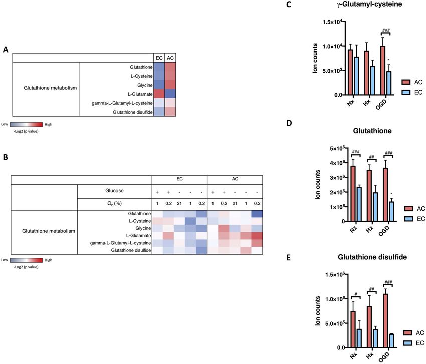
S.-F. Huang, et al.                                                                                                             Redox Biology 34 (2020) 101576

compartments. Direct contact between AC and neurons allows ex-                  glucose) and oxygen and glucose deprivation (OGD) i.e. 1% O2 without
change of multiple substances that sustain neuronal survival and                glucose (Fig. 1C–E). At baseline both cells displayed similar amounts of
function. An important example is the release of supportive metabolites         the intermediate product γ-glutamyl-cysteine with an injury-induced
such as glutathione (GSH) and glutamine. GSH is a critical antioxidant          reduction observed only in EC (Fig. 1C). Much higher levels of the end
in the brain that maintains cellular oxidative homeostasis [5]. GSH             products GSH and GSH disulfide were consistently measured in AC
metabolic crosstalk rapidly contributes to maintaining neuronal redox           (Fig. 1D and E) with EC GSH levels strongly reduced during OGD
homeostasis [6], supporting neurotransmitter recycling [7,8] and reg-           (Fig. 1D). Thus AC have comparatively better stores and greater capa-
ulating neuronal gene expression programs [9]. In contrast, little is           city to generate/use GSH in injury situations.
known of how metabolites impact BBB integrity [4], but it seems logical
that a similar AC-EC crosstalk could also support barrier function.             3.2. Oxidative homeostasis is stable in AC but not EC
     We previously performed metabolomic profiling of primary AC and
EC to obtain detailed insight into cell-specific regulation of critical             GSH efficiently maintains cellular redox balance [5]. To know
pathways during resting and different injury conditions [10]. A number          whether the redox equilibrium of AC and EC correlated with their GSH
of metabolites and pathways that were reduced in EC but elevated or             metabolic activity, we measured reactive oxygen species (ROS) levels
stabilized in AC during injury were identified. We considered that AC           for 48 h in hypoxic and ischemic cells. Within 4h hypoxia induced a 3-
secretion of such metabolites, especially those known to facilitate cell        fold increase in AC ROS activity that returned to normoxic baseline by
survival or adaptation to adverse situations, could be key in supporting        48h (Fig. 2A). Intriguingly, the severest condition of oxygen and glu-
EC function. GSH was of particular interest due to its critical anti-oxi-       cose withdrawal in AC had less dramatic effects on ROS levels and
dant role and the fact that GSH-deficiency has already been observed in         never reached those of hypoxic values (Fig. 2A). In contrast, ROS ac-
many BBB impairment-associated brain diseases in patients [11,12].              cumulation in EC (Fig. 2B) was insult severity-dependent with OGD
GSH deficit was also shown to induce barrier leakage in a rat model             inducing higher ROS levels than hypoxia. Notably OGD-induced ROS
[13] and its administration prevented endothelial oxidative imbalance           accumulation in EC began only after 6 h and remained consistently
in vitro [14,15].                                                               elevated by 2–4 fold at 48h compared to Nx baseline (Fig. 2B). These
     We hypothesized that GSH is critical for brain vascular function and       results highlight a cell-specific and time-dependent modulation of redox
its secretion by AC promotes BBB stability during injury conditions i.e.        balance during injury conditions.
when EC GSH generation is impeded. Using an isotopic labeling/
tracking approach combined with Time-of-Flight Mass Spectrometry                3.3. Injury increases GSH secretion by AC
(TOF-MS) we prove that AC secreted GSH is constantly shuttled to EC,
and this flux is accelerated by injury conditions. Next, we demonstrate             Since secretion of GSH and its related metabolites by AC could
that GSH synthesis-deficient AC (via siRNA-mediated γ-glutamyl cy-              benefit EC in addition to neurons in vivo we assessed AC intracellular
steine ligase knockdown) lose their ability to protect the barrier under        and extracellular GSH levels during normoxia, hypoxia and OGD for up
injury conditions. Finally, in the absence of AC, sole GSH supple-              to 24h. Interestingly, endogenous GSH levels were consistently main-
mentation abrogates injury-induced EC permeability. Taken together,             tained at normoxic levels with OGD tending to increase the quantities
better insight of metabolic shuttling and reprogramming may provide             (Fig. 3A). Extracellularly a time- and injury-dependent increase of more
innovative ways to modulate BBB function.                                       than two-fold was noted compared to normoxic conditions (Fig. 3B).
                                                                                Thus injury conditions stimulate GSH secretion but do not alter en-
3. Results                                                                      dogenous homeostasis consistent with GSH associated pathways being
                                                                                constantly active and modulated in AC during injury situations
3.1. Changes in glutathione-related metabolites during injury conditions        (Fig. 1B). To date, a GSH-specific exporter has not been identified but
                                                                                multidrug resistance proteins (MRP) are considered to be the major
    In a previous study we used untargeted LC-MS to obtain compre-              transporter family involved in GSH secretion [17]. In this regard injury-
hensive metabolomic profiles of primary rat brain microvascular en-             induced AC MRP2 mRNA (Supplementary information Fig. S1A) and
dothelial cells (EC) and primary rat astrocytes (AC) exposed to different       protein expression was observed (Supplementary information Fig. S1B)
conditions (Huang et al. in press). Samples were collected and extracted        whereas MRP1 and MRP4 expression was unchanged (data not shown).
after 24h normoxia (Nx), hypoxia (Hx) or near anoxia (Ax) in media              Gamma glutamyl transpeptidase (GGT) plays a key role in extracellular
with or without glucose ( ± Glc) to simulate ischemia in vitro. We noted        GSH catabolism and supporting intracellular oxidative stress home-
that regulation of key metabolites of the GSH pathway was quite dif-            ostasis. We observed that messenger RNA levels of GGT were strongly
ferent in the two cell types. A comparison relative to the baseline (Nx)        induced during hypoxia and particularly OGD (Supplementary in-
composition of each cell is shown in a heatmap with increased and               formation Fig. S1C) but no change in protein levels was detected
decreased metabolites indicated in red and blue respectively (Fig. 1A).         (Supplementary information Fig. S1D).
Major metabolites supporting GSH synthesis such as cysteine and gly-
cine were clearly more abundant in AC (Fig. 1A). Furthermore, cellular          3.4. GSH is constantly shuttled from AC to EC
GSH and GSH disulfide levels were overrepresented in AC compared to
EC (Fig. 1A).                                                                       To investigate if EC take up the GSH secreted by AC, a stable isotope
    Next we investigated how injury modulates cellular GSH metabo-              labeling approach combined with TOF-MS was used to monitor its
lism in AC and EC by comparing the injury profiles with Nx baseline             movement. As cysteine is an essential GSH building block, 34S15N-cy-
conditions. Heat maps show oxygen and glucose deprivation stimulate             steine (Cys) isotope was incorporated into AC GSH molecules as sche-
AC GSH metabolic activity whereas a clear reduction occurs in EC                matically depicted (Fig. 4A). First, endogenous GSH levels were de-
(Fig. 1B). Interestingly, under the most severe injury condition (0.2%          pleted by culturing AC in methionine/cysteine-free culture media for
O2 without glucose) GSH and glycine were strongly reduced in AC,                24h. Subsequently the AC were incubated in 34S15N-Cys-containing
whereas γ-glutamyl-cysteine, a precursor that determines the GSH                media for 24h to boost biosynthesis of endogenously labeled 34S15N-
synthesis rate [16], remained high. This implies that AC maintain GSH           GSH. The ratios of ion intensities of GSH (considering both mass ac-
metabolism even during severe conditions. In complete contrast, EC              curacy and TOF resolution) were compared in AC treated with and
GSH metabolism was diminished under virtually all conditions and                without 34S15N-Cys. Using an annotation depicting the abundance of
particularly in the absence of glucose. We directly compared levels of          isotopes, M is natural GSH monoisotopic mass and M+3 is enriched
three GSH activity indicators in AC and EC during hypoxia (1% O2 with           metabolite which is 3 Da heavier than the monoisotopic GSH. The M

                                                                            2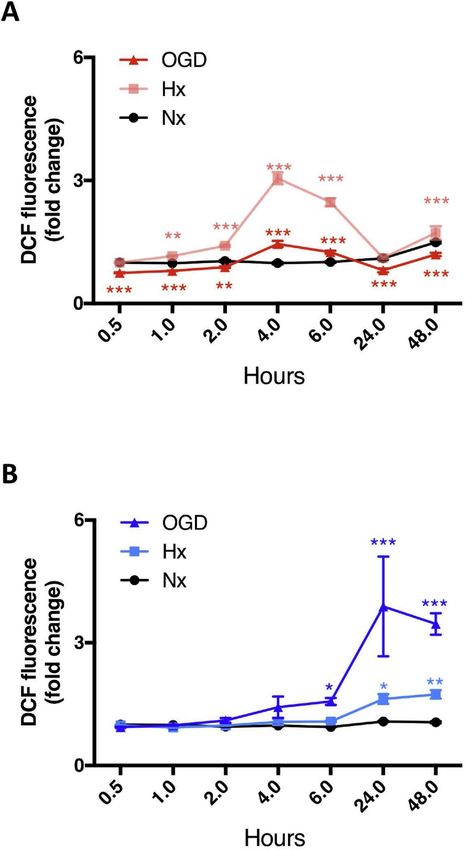
S.-F. Huang, et al.                                                                                                                   Redox Biology 34 (2020) 101576

Fig. 1. Changes in glutathione-related metabolites during injury conditions.
Levels of metabolites that participate in glutathione (GSH) metabolism in rat brain primary astrocyte (AC) and endothelial cell (EC) were measured by LC-MS after
exposure to oxygen deprivation (1% O2 and 0.2% O2) combined with or without glucose (Glc) withdrawal for 24h. (A) Heatmap shows the relative activity of the
major GSH-related metabolites comparing the two cell types under normoxia (Nx). (B) Heatmap of GSH metabolic alterations during injury was generated by
comparison with their Nx control with increased metabolite level in red and decreased metabolite level in blue. (C-E) Ion counts of three GSH metabolism indicators,
g-glutamyl-cysteine (C), GSH (D) and GSH disulfide (E), during injury is presented separately. Hypoxia (Hx, 1% O2 with glucose) and OGD (1% O2 without glucose).
The metabolite intensities were normalized to total ion counts. The -Log2(P value) for each metabolite was calculated using unpaired T-test. *P < 0.05; One-way
ANOVA compared to Nx. #P < 0.05, ##P < 0.01, ###P < 0.001; Two-way ANOVA, compared to astrocytes (AC) in the same condition. Mean ± SD. n = 4.

+3/M ratio, corresponding to the natural isotopic abundance in the                   Unexpectedly, measurement of EC intracellular metabolites after nor-
baseline samples, is expected to increase in samples treated with the                moxic (Nx) co-culture showed labeled GSH levels were increased by up
isotope (due to the inclusion of labeled GSH in the cellular GSH pool).              to 2-fold compared to baseline within 24h. Thus metabolic shuttling
Accordingly, the natural abundance of labeled GSH (M+3/M) was                        exists in the absence of injury (Fig. 4C). Injury exposure further ex-
6–7% in cells not incubated in 34S15N-Cys and exceeded 135% after 24h                pedited enrichment of labeled GSH (Fig. 4C) demonstrating increased
stimulation confirming successful incorporation into the intracellular               paracrine crosstalk. Notably, GSH shuttling occurs even during harshest
GSH pool (Fig. 4B). Next, to test our hypothesis that AC-derived GSH is              conditions as levels similar to normoxia were maintained during severe
shuttled to EC, we co-cultured labeled AC with EC in Transwells™ [18]                OGD (Ax-Glc) (Fig. 4C). Thus GSH shuttling between AC and EC is
prior to 24h injury exposure (Fig. 4A). Samples from the different                   constantly active.
compartments were then collected and analyzed to assess if transfer
occurred. Interestingly, intracellular levels of labeled GSH in both
                                                                                     3.5. Paracrine GSH shuttling is crucial for barrier maintenance during
mono- and co-cultured AC lysates always corresponded to baseline
                                                                                     injury
values (6%–8%), suggesting extended exposure results in metabolic
dilution of the M+3 enrichment (Supplementary information Fig. S2).
                                                                                        To prove GSH shuttling supports barrier function, we employed

                                                                                 3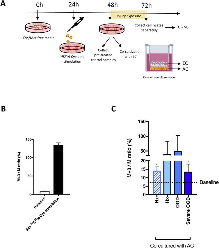
S.-F. Huang, et al.                                                                                                                 Redox Biology 34 (2020) 101576

                                                                                    Fig. 3. Injury increases GSH secretion by AC.
                                                                                    (A and B) Changes in intracellular and extracellular GSH levels in primary AC
Fig. 2. Oxidative homeostasis is stable in AC but not EC. (A and B) Oxidative       after 6h, 16h and 24h Hx and OGD were measured in cell lysates (A) and
stress measurements were performed in primary AC (A) and EC (B) exposed to          culture media (B) respectively. Concentrations were normalized to either 1 mg
hypoxia (Hx, 1% O2 with glucose) and OGD (1% O2 without glucose). Nx, 21%           total protein or 1 mL media to facilitate comparison between the different
O2 with glucose group was used as control. *P < 0.05, **P < 0.01,                   conditions and cell type. *P < 0.05, **P < 0.01, ***P < 0.001; two-way
***P < 0.001; Two-way ANOVA compared to Nx at the same time point.                  ANOVA compared to Nx baseline. #P < 0.05; Two-way ANOVA, compared to
Mean ± SD. n = 3.                                                                   injury 6h baseline. Mean ± SD. n=6.

siRNA to silence γ-glutamyl cysteine ligase (GCL) gene and disrupt the              3.6. Boosting EC GSH levels prevents injury-induced BBB breakdown
rate limiting step of GSH synthesis [5]. Post siGCL transfection, protein
levels were significantly reduced (Fig. 5A) and GSH secretion rate was                  As EC GSH metabolism is increasingly shutdown during injury, we
successfully reduced by 10–20% compared to untreated controls                       asked if barrier impairment can be prevented by providing the meta-
(Fig. 5B). GSH-deficient AC were then co-cultured with primary EC on                bolite exogenously. Permeability of confluent primary EC monolayers
Transwells™ and exposed to hypoxic/OGD for 48h. Barrier function was                treated with GSH compounds prior to hypoxic/ischemic exposure for
measured by lucifer yellow flux. In agreement with their barrier sup-               48h was measured. In untreated samples the degree of barrier perme-
portive role [4,18], co-culture of EC with untreated (UNT) AC pre-                  ability correlated with injury severity (Fig. 6A and B) as expected. Ex-
vented injury-induced barrier leakage (Fig. S3). Remarkably, when co-               citingly, enhancing GSH levels using GSHee and NAC prevented barrier
cultured with GSH-deficient AC (siGCL) significantly increased para-                leakage not only in hypoxia but also during OGD (Fig. 6A and B). No-
cellular flux was measured under both injury conditions compared to                 tably, GSHee maintained the BBB as tight as the normoxic controls in
siCTRL (Fig. 5C). Thus impeding GSH shuttling compromises AC pro-                   both injury conditions. The GSH synthesis inhibitor (BSO) showed no
tective effects and directly impairs the ability of EC to maintain their            additional negative effect compared to controls (UNT, Fig. 6A and B).
barrier function. Clearly the shuttle reinforces barrier stability.                     Using immunostaining we next tested if exogenous GSH improved
                                                                                    BBB stability by suppressing injury-induced tight and adherens junction

                                                                                4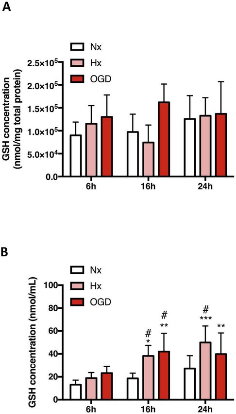
S.-F. Huang, et al.                                                                                                            Redox Biology 34 (2020) 101576

                                                                                                       Fig. 4. GSH is constantly shuttled from AC to EC.
                                                                                                       To investigate if EC take up AC-derived GSH, a stable
                                                                                                       isotope labelling approach was combined with Time-
                                                                                                       of-Flight Mass Spectrometry (TOF-MS) to specifically
                                                                                                       track AC synthesized GSH. (A) The schematic shows
                                                                                                       the experimental setup. Endogenous GSH was de-
                                                                                                       pleted by incubating with L-Cys/Met free media for
                                                                                                       24h. 34S15N-cysteine was employed to stimulate AC
                                                                                                       endogenous 34S15N-GSH generation. Isotope-labeled
                                                                                                       AC were co-cultured with EC under different condi-
                                                                                                       tions for 24h then cell lysates were analyzed by TOF-
                                                                                                       MS. (B) Levels of intracellular 34S15N-GSH was
                                                                                                       measured in untreated AC lysate (baseline) and after
                                                                                                       24h isotope stimulation. (C) After co-culture, levels
                                                                                                       of labeled GSH in EC lysates were measured.
                                                                                                       *P < 0.05, **P < 0.01, ***P < 0.001; Student’s T-
                                                                                                       test compared to baseline. Mean ± SD. n=4.

                                                                                                       Fig. 5. Paracrine GSH shuttling is crucial for barrier
                                                                                                       maintenance during injury. (A) AC were transfected
                                                                                                       with GCL small Interfering RNA (siGCL). A re-
                                                                                                       presentative immunoblot after 48h transfection
                                                                                                       shows normoxic GCL expression levels. (B) The rate
                                                                                                       of GSH release by normoxic AC after GCL knockdown
                                                                                                       was measured post transfection at 24h and 48h. (C)
                                                                                                       Permeability of the EC barrier after co-culture with
                                                                                                       transfected AC under different conditions for 48h
                                                                                                       was measured by lucifer yellow flux. **P < 0.01,
                                                                                                       compared to Nx siCTRL. #P < 0.05, ##P < 0.01;
                                                                                                       Student's T-test compared to injury control (siCTRL).
                                                                                                       n = 8. (For interpretation of the references to colour
                                                                                                       in this figure legend, the reader is referred to the
                                                                                                       Web version of this article.)

delocalization. As expected, injury severity progressively disrupted            3.7. Exogenous GSH suppresses occludin tyrosine phosphorylation
junctional localization at cell-cell borders as observed by discontinuous
and frayed organization of claudin-5 (arrows, Fig. 6C) and occludin                 The post-transcriptional modification of tight junction proteins dy-
(data not shown) in hypoxic cells compared to normoxic controls. In-            namically regulates complex assembly and localization [19,20]. Parti-
jury also disrupted β-catenin retention at cell-cell borders (arrows,           cularly tyrosine phosphorylation of occludin and claudin-5 C-terminus
Fig. 6D). Overall during injury the cells lost both morphology and close        impacts their interaction with ZO-1 and cytoskeleton, and initiates
organization with gaps increasingly visualized (asterisks). Consistent          protein internalization [21,22]. Immunoprecipitation was used to as-
with the functional data, GSH enhancers prevented tight and adherens            sess claudin-5 and occludin tyrosine phosphorylation (pTyr) status after
junction disruption in both injury conditions although NAC was con-             24h exposure to hypoxia/OGD in a human brain microvascular en-
sistently less effective than GSHee (Fig. 6C and D). Thus boosting cel-         dothelial cell line (hCMEC/D3) and primary rat brain EC. As expected
lular GSH levels prevents barrier impairment (without impacting cell            increased phosphorylation of both proteins was seen in hypoxic
survival, Supplementary information Fig. S4).                                   hCMEC/D3. GSHee strongly suppressed hypoxia-induced (Fig. 7A and

                                                                            5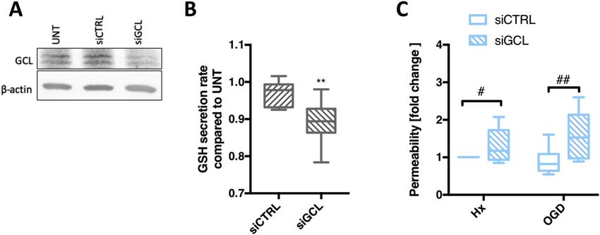
S.-F. Huang, et al.                    Redox Biology 34 (2020) 101576

                          Fig. 6. Boosting EC GSH levels prevents
                          injury-induced BBB breakdown.
                          Primary EC monolayers were either un-
                          treated (UNT) or exposed to GSH com-
                          pounds and exposed to hypoxia/OGD for
                          48h. GSH enhancers (5mM NAC and 5mM
                          GSHee) and GSH synthetase inhibitor
                          (200mM BSO) were applied. (A and B) EC
                          barrier leakage was measured under Hx
                          and OGD using lucifer yellow. (C and D)
                          Representative immunofluorescent images
                          of claudin-5 and b-catenin localization in
                          EC. Note the disconnection and delocali-
                          zation of junctions in areas marked by ar-
                          rows. Asterisks indicate inter-EC gap for-
                          mation and holes. *P < 0.05; One-way
                          ANOVA compared to Nx baseline.
                          #
                            P < 0.05, ##P < 0.01, ###P < 0.001;
                          Two-way ANOVA, compared to injury un-
                          treated (UNT). Mean ± SD. n = 5.

                      6
S.-F. Huang, et al. Redox Biology 34 (2020) 101576
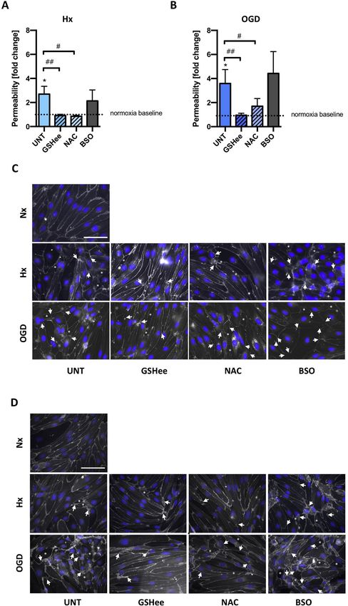
Fig. 7. Exogenous GSH suppresses occludin
tyrosine phosphorylation.
Two different cell models (human brain mi-
crovascular EC cell line (hCMEC/D3) and
primary EC) were treated with GSHee prior to
hypoxic or ischemic exposure for
24h. (A) Immunoprecipitation (I.P) using
phospho-tyrosine conjugated (p-Tyr) beads
was performed followed by immunoblot ana-
lysis of tight junction proteins in hCMEC/
D3. (B and C) Densitometric quantification
of pTyr-occludin (p-Occludin)(B) and pTyr-
claudin-5 (p-Claudin-5)(C) was subsequently
carried out. (D and E) Similar modulations
were further confirmed in primary EC, shown
in the representative immunoblots (D) and
densitometric quantification (E). I.P pTyr blot
shows the pTyr protein level whereas
supernatant blot shows the unpulled
protein. β-actin as a loading control.
#
P < 0.05, ##P < 0.01; one-way ANOVA
compared to Hx baseline. n = 5 in hCMEC/
D3; n = 4 in primary EC.
7
S.-F. Huang, et al.                                                                                                              Redox Biology 34 (2020) 101576

B) but not OGD-induced (Fig. 7B) occludin tyrosine phosphorylation               GSH breakdown and XCT activation could jointly contribute to GSH
compared to the normoxic condition, an observation also noted in                 recycling and shuttling to the EC compartment. For better insight a
primary isolated EC (Fig. 7D and E). In contrast, no effect on phos-             more detailed investigation of the precise mechanism is clearly war-
phorylation status of claudin 5 was detected (Fig. 7C).                          ranted.
                                                                                     Paracrine metabolic processes clearly have a large part to play in
4. Discussion                                                                    regulating BBB stability. Even mild reduction of AC GSH secretion
                                                                                 (10–20%) significantly increased barrier permeability implying peri-
    Multiple studies suggest that stabilizing brain vascular function in         vascular metabolic disturbance may be a causative factor in worsening
patients with neurological disease could arrest or even reverse the              barrier integrity. It is highly probable that to ensure their own survival
course of brain disorders [2]. We are convinced that better under-               during harsh situations AC might withdraw their metabolic support
standing of the mechanisms by which perivascular cells support barrier           leaving vascular EC to fend for themselves. Such a switch may explain
function could provide new insight for future strategies aimed at                the unexpected negative effects of AC on barrier stability observed
modulating barrier function. This is the first study to prove the ex-            during severe conditions [4,18,40]. In this regard GSH supplementation
istence and importance of AC-EC metabolic cross-talk under different             could significantly benefit vascular function. Indeed GSHee (a mem-
environmental conditions. We show that constitutive GSH shuttling                brane permeable GSH analog) and NAC (a source of cysteine for GSH
from AC to EC is strongly increased during injury conditions and sup-            generation) administration prevented injury-induced EC permeability
pressing the shuttling significantly reduces AC protective effects and           as seen in epithelial and hepatocyte studies [41,42]. We noted that GSH
induces barrier dysfunction. Notably, exogenous GSH supplementation              treatment prevented occludin tyrosine phosphorylation (pTyr), a
also preserved barrier stability in the absence of AC. Thus our data             modification well known to determine EC tight junction localization,
suggests that boosting/elevating GSH levels during disease could im-             complex stability and integration with regulatory proteins [43–45].
prove BBB functionality.                                                         This data also aligns with occludin being a major target for hypoxia
    GSH is a key regulator of redox-sensitive transcription factors and          induced pTyr by c-Src tyrosine kinases [44,46]. Two different me-
stress-sensing pathways that boosts the cellular metabolic systems de-           chanisms may underlie GSH protection; 1) suppression of hypoxia-in-
fence against insult. Metabolomic profiling clearly revealed distinct and        duced ROS imbalance to prevent the phosphorylation and 2) a non-ROS
differential modulation of AC and EC GSH metabolic pathways. Despite             mediated pathway; perhaps directly activating protein tyrosine phos-
being a high oxidative stress cell type [23], EC had relatively low GSH-         phatase (PTP) via glutathionylation [47,48]. The mechanisms at work
related metabolite levels at baseline and seemingly exhausted - or               during OGD are even more unclear as pTyr of both occludin and
perhaps could not replenish - key GSH metabolites during injury con-             claudin-5 were unchanged by GSH exposure.
ditions. This progressive GSH metabolism shut down agrees with redox                 Regardless, our data undoubtedly implies that enhancing EC GSH
imbalance being a primary cause of EC dysfunction [24,25]. In contrast,          levels could improve redox homeostasis and suppress injury-induced
AC had a highly activated GSH metabolism during stress conditions                vascular impairment. Although not yet considered for BBB protection,
supporting observations that this pathway is well maintained under               positive effects of boosting GSH has been demonstrated in some neu-
hypoxia and OGD [26,27].                                                         rological-related clinical trials. As GSH is unstable (easily oxidized) and
    Astrocytes play an important role in antioxidative metabolism and            has a very short half-life NAC, a cysteine analog that boosts GSH
detoxification [5]. AC constitutively secrete GSH under baseline nor-            synthesis, has been the most commonly used enhancer. Intravenous
moxic conditions and are reservoirs of this key anti-oxidant and likely          injection of NAC successfully increased brain GSH levels in Parkinson's
other important metabolites [6]. This characteristic can clearly benefit         disease, Gaucher disease and healthy human brain as measured by
surrounding cells. Notably, GSH secretion was increased without in-              Tesla     magnetic       resonance     spectroscopy     (ClinicalTrials.gov:
fluencing intracellular levels suggesting activated systems boost its re-        NCT01427517 and NCT02445651) resulting in general patient im-
lease - although no GSH-specific transporter, exporter or importer, has          provement [49]. AD patients prescribed NAC over six months also
been identified to date. GSH can however be co-transported with or-              showed significantly improved memory performance [50,51]. However
ganic anions by different membrane proteins. Our data suggests MRP2              as it is used at relatively high concentrations, NAC has quite a few
is most likely involved in AC GSH release during injury.                         unwanted side effects [52] and other studies did not observe positive
    Is GSH itself taken up directly by cells? Interestingly, intravenously       effects (Clinicaltrials.gov NCT00903695 and NCT01320527). As NAC is
injected GSH-coated nanoparticles targeted to EC for drug delivery               a precursor, compromised cell function during injury conditions may
were observed to successfully accumulate in brain tissue [28–30], but            slow the processes needed for GSH synthesis. Indeed our study supports
how they enter is not understood. In this study the fact that the isotope        this view as the membrane permeable GSHee was considerably more
is detected in endothelial cells proves that labeled GSH (i.e. of AC             effective than NAC. Notably, recent application of GSHee in a stroke
origin) is shuttled from AC to EC. Unfortunately, as our isotope labels          MCAo mouse model also reduced infarct size and improved neurolo-
GSH on sulfur and nitrogen but not specifically on cysteine, whether it          gical outcome [53]. Thus a stable, cell permeable GSH analog could
can be directly absorbed remains debatable although most evidence                offer significant clinical advantages.
does not support this scenario [31]. If indirectly taken up by EC various            To conclude, GSH shuttling from AC to EC supports brain vascular
amino acid importers that transport GSH breakdown products should                stability during insult and disturbance of this metabolic interaction
be taken into consideration. In neurons numerous importers indirectly            likely compromizes barrier homeostasis. We thus advocate adminis-
participate in GSH absorption, including excitatory amino acid trans-            tration of GSH analogs to boost BBB function and sustain vascular
porters (EAAT) 1–3 and Xc− antiporter (XCT) that regulate cysteine and           health. Better understanding of the impact of other perivascular-de-
glutamate levels to facilitate GSH synthesis [32,33]. Although we did            rived metabolites, and paracellular crosstalk, could offer more oppor-
not investigate this mechanism in detail, it was interesting that XCT            tunities to safeguard BBB integrity.
mRNA levels (cysteine importer) were also induced in EC during injury
conditions (data not shown). Utilizing this route would mean extra-              5. Material and methods
cellular GSH catabolism is required [34]. Particularly, γ-glutamyl
transpeptidase (GGT) might play a key role in releasing glutamate,               5.1. Reagents
cysteine and related peptides from AC GSH. Notably GGT has been
detected in all BBB compartments including EC [35], pericyte [36] and               Transwells™ were obtained from Corning (Schiphol, The
AC [37]. Since injury conditions induced mRNA levels of AC GGT, as               Netherlands). Lucifer yellow was purchased from Thermo Fisher.
observed in various organs [38,39], we speculate that GGT-mediated               Protease inhibitor cocktail Set III from Calbiochem (Merck, Darmstadt,

                                                                             8
S.-F. Huang, et al. Redox Biology 34 (2020) 101576
Germany). Oligofectamine™ Reagent and Pierce BCA Protein Assay (FGCZ), University of Zurich. Sample preparation and measurements
were from Thermo Fisher Scientific Inc. (Rockford, IL). For Western were performed using a nanoACQUITY system coupled to a Synapt
blotting and immunofluorescence, antibodies directed against occludin, G2HD mass spectrometer (Waters Corp., Milford, USA) as previously
claudin-5, and ZO-1 were purchased from Invitrogen (Basel, described [10]. Chromatographic separation of metabolites was per-
Switzerland), β-actin antibody from Sigma–Aldrich (Buchs, formed on a 0.2 μm × 150 mm BEH amide column using a 10 min
Switzerland), and β-catenin antibody from Chemicon (Millipore, linear gradient of 90%–50% acetonitrile, 0.5 mM ammonium acetate,
Billerica, MA). Anti-phosphotyrosine antibody agarose conjugate, clone pH 9. All analyses were done in negative mode using 1.2 kV capillary
4G10®, was purchased from Milipore. Secondary antibodies for Western voltage, 30 V sampling cone voltage and 3 V extraction cone voltage.
blotting and immunofluorescence were obtained from Jackson The source temperature was set to 100 °C and Nano Flow Gas, i.e. sheet
ImmunoResearch (Suffolk, UK) or Invitrogen. For the glutathione (GSH) gas flow, was applied.
and ROS assay, 5,5-Dithio-Bis-(2-Nitrobenzoic acid) (DTNB) and 2′,7′-
Dichlorofluorescin diacetate were obtained from Sigma–Aldrich (Buchs, 5.6. Data analysis and processing
Switzerland). Isotopic labeled 34S15N-cysteine was designed and syn-
thesized by Dr. T. Sawa (Graduate School of Medical Sciences, Waters raw data were first converted to centroid mode and further
Kumamoto University) [54]. γ-glutamyl cysteine ligase catalytic sub- processed into vendor independent netCDF format using DataBridge
unit (GCLc) siRNA (ON-TARGET plus SMART pool) and negative con- (Masslynx, Waters Corp.). Untargeted metabolomics data matrix com-
trol siRNA (ON-TARGET plus non-targeting pool) were purchased from prising of accurate mass/retention time information, and ion counts for
Dharmacon. each sample were calculated using the data processing tool cosmiq
[57]. Signal-to-noise ratio (SNR) of mass peak detection was set to 3,
5.2. Primary cell isolation and cell culture SNR for chromatographic peak detection was set to 10 and a m/z bin
size of 0.003 Da was chosen as parameters for cosmiq. For metabolite
All cell culture media and reagents were obtained from Gibco® (Life annotation, the resulting list was first matched to a list of metabolites
Technologies, Zug, Switzerland) and Sigma-Aldrich. Primary rat astro- with known retention time and mass. For additional annotation of
cytes (AC) were isolated from neonatal pups as described [55] then unknown metabolites, the list of accurate masses was matched to the
cultured in DMEM supplemented with 10% FBS and 50 μg/mL genta- KEGG database assuming [M-H]- adducts. Database hits within a mass
mycin sulfate on gelatin-coated dishes and used after the first passage. window of 0.01 Da were considered.
Primary rat brain microvascular endothelial cells (EC) were isolated
from 8 to 10 week old male Wistar rats as previously described [56]. EC
5.7. Data normalization strategy
reached 100% confluence after 7 days culture and were used without
passaging. The human cerebral microvascular endothelial cell line
To be able to compare the relative metabolite quantities between
HCMEC/D3 was used for immunoprecipitation experiments. HCMEC/
the different treatments and cell types, we performed a normalization
D3 were cultivated in Endo GRO™-MV (Millipore) medium containing
approach according to the sum of all detected metabolite ion counts as
5% FBS on collagen-coated culture dishes.
previous [10]. One of the Nx EC samples was randomly chosen as re-
ference and the normalized ion intensity for each metabolite was cal-
5.3. Co-culture methodology
culated by dividing the observed ion intensity by the factor Σis/Σir,
where Σis is the summed ion intensity for each individual sample and
Two different co-culture systems were employed in this study: 1)
Σir is the summed ion intensity of the reference sample.
Contact co-culture model. Primary EC cells were seeded on the upper
side of collagen-coated Transwells™ till confluent with AC seeded on the
lower side, both at a density of 0.5 × 10⁶ cells/insert. Co-cultures were 5.8. Oxidative stress detection and GSH measurement
incubated in DMEM with 10% calf serum (with or without glucose) for
24h. After exposure the cell lysates were collected separately for Time- ROS formation reflecting intracellular oxidative stress was quanti-
of-flight mass spectrometer (TOF-MS) analysis. 2) Non-contact co-cul- fied by fluorometric techniques based on 2′,7′-Dichlorofluorescin dia-
ture model. As primary cells are very sensitive to trypsin treatment, this cetate (DCF) oxidation according to the manufacturer's instruction
model was used to bypass lifting cells after siRNA transfection. AC were (Sigma-Aldrich). GSH measurement was performed using DTNB (5,5′-
cultured at a density of 0.5 × 10⁶ cells/well in 24 well plates whereas dithio-bis (2-nitrobenzoic acid)) as published (Bogdanova et al., 2005).
EC were cultured as described in the model above. The inserts with EC Briefly, intracellular GSH levels were measured from the cell lysates
monolayers were then transferred to the 24-well plates containing harvested in cell lysis buffer (50 mM Tris, 150 mM NaCl, 1% Triton X-
transfected AC to initiate the co-culture. These co-cultures were kept in 100, 1% NP-40) supplemented with protease inhibitor cocktail
DMEM with 10% calf serum (with or without glucose) for 48h then used (Calbiochem, Darmstadt, Germany), 1 mM sodium orthovanadate,
for permeability assays. 1 mM dithiothreitol, 0.5 mM phenylmethansulfonyl fluoride and 1 mM
EDTA. Total protein concentrations were measured using Pierce BCA
5.4. O2 deprivation and ischemic treatment protein assay and 50 μg was measured in a 96-well format. Extracellular
GSH was detected in 100 μL culture media samples immediately after
O2 deprivation experiments were carried out in a purpose-built exposure.
hypoxic glove-box chamber (InVivO2 400, Ruskinn Technologies,
Pencoed, UK) maintained at 37 °C with 5% CO2. O2 concentration was 5.9. Permeability assay
constantly monitored with an internal O2 sensor. Cells were exposed to
normoxia (Nx, 21% O2), hypoxia (Hx, 1% O2), oxygen and glucose Permeability assays were performed on Transwells™ with confluent
deprivation (OGD, 1% O2 without glucose) and severe OGD (0.2% O2 primary EC as previously described [18]. Fresh medium containing the
without glucose) for 24h/48h. fluorescent dye lucifer yellow was added to the upper compartment. At
0, 15, 30, 45 min aliquots were taken from the bottom compartment.
5.5. Untargeted LC-MS measurements Sample fluorescence was measured with a plate reader (FLx800, Biotek
Instruments, Winooski, VT). A clearance slope was established from the
Non-targeting mass spectrometry measurements and analyses were measurements obtained at different timepoints and used to calculate
conducted in cooperation with the Functional Genomics Center Zurich permeability coefficient values (Pe) (Rist et al., 1997).
9S.-F. Huang, et al. Redox Biology 34 (2020) 101576
5.10. Immunostaining 5.16. siRNA transfection
Primary ECs were grown on coverslips coated with collagen IV. Oligofectamine™ Reagent was mixed with 100 μM siRNA and used
After hypoxic and ischemic exposure cells were fixed in 4% paraf- to transfect ACs according to the manufacturer's instructions. After
ormaldehyde, permeabilized in 0.1% Triton X-100 in PBS and in- transfection, knockdown efficiency was confirmed using DTNB assay.
cubated with occludin (1:100, Invitrogen), claudin-5 (1:100,
Invitrogen), β-catenin (1:100, Chemicon). Cell nuclei were counter- 5.17. Statistics
stained with DAPI (4’,6-Diamidin-2-phenylindol). Pictures were taken
using an inverted fluorescence microscope coupled to an 8-bit CCD All results are expressed as mean ± SD from a minimum of three
camera (Axiocam HR, Carl Zeiss) and processed using ImageJ software. independent experiments. Statistical significance using GraphPad Prism
7 software (La Jolla, CA) was assessed by Students T-test or one-way
5.11. Immunoblotting ANOVA for comparison of different time points within a group and two-
way ANOVA for comparison between different groups. A P-value below
Cells were washed with ice-cold PBS and homogenized in cell lysis 0.05 was considered significant.
buffer (50 mM Tris, 150 mM NaCl, 1% Triton X-100, 1% NP-40) sup-
plemented with protease inhibitor cocktail. After measurement with Declaration of competing interest
Pierce BCA protein assay 30 μg protein were separated on denaturing
SDS-Page and transferred to a nitrocellulose membrane. Membranes The authors declare no competing interests.
were blocked at room temperature in 5% non-fat dried milk or 5% BSA
and subsequently incubated overnight at 4 °C in primary antibodies Acknowledgement
against β-actin (1:5000), occludin (1:500) and claudin-5 (1:300).
Membranes were washed with 0.1% Tween-20 in TBS or PBS then in- This study was financially supported by a Swiss National Science
cubated with horseradish peroxidase (HRP) conjugated secondary an- Foundation grant (Number 31003A_170129) to OOO. The laboratory
tibody. Band detection was performed and visualized using a lumines- work was (partly) performed using the logistics of the Center for
cent image analyzer LAS-3000 (Fujifilm, Dielsdorf, Switzerland). Blot Clinical Studies at the Vetsuisse Faculty of the University of Zurich.
quantification (using β-actin as loading controls) was performed using
ImageJ software (NIH, Bethesda, USA). Appendix A. Supplementary data
5.12. Immunoprecipitation Supplementary data related to this article can be found at https://
doi.org/10.1016/j.redox.2020.101576.
Protein (1 mg) was suspended in I.P buffer (50 mM Tris–HCl pH 7.4, OOO and SFH designed the study. SFH and SF performed the in vitro
150 mM EDTA and 0.5% NP-40, 1 mM NaVO3, 1 mM PMSF) then 30 μl experiments. DF, NZ, KO and TS performed the metabolomic profiling
of anti-phosphotyrosine antibody agarose conjugate (4G10) was added. and contributed analytic tools. SFH, AK, SF, DF and AO analyzed the
After 4h incubation at 4 °C in a circular rotator samples were cen- metabolomic data. SFH and OOO wrote the paper.
trifuged (1000 rpm for 1 min) then the supernatant removed and the
beads washed 3 times with I.P buffer. Captured proteins were eluted by References
incubating at 80 °C in 2x Laemmli buffer then run on a 10% SDS–Page
gel. [1] O. Tomkins, I. Shelef, I. Kaizerman, A. Eliushin, Z. Afawi, A. Misk, M. Gidon,
A. Cohen, D. Zumsteg, A. Friedman, Blood-brain barrier disruption in post-trau-
5.13. Metabolite extraction matic epilepsy, J. Neurol. Neurosurg. Psychiatry 79 (2008) 774–777, https://doi.
org/10.1136/jnnp.2007.126425.
[2] M.D. Sweeney, A.P. Sagare, B.V. Zlokovic, Blood-brain barrier breakdown in
Cell pellets were collected and washed (75 mM Ammonium Alzheimer disease and other neurodegenerative disorders, Nat. Rev. Neurol. 14
Carbonate, pH 7.4), then incubated with cold 40:40:20 acetoni- (2018) 133–150, https://doi.org/10.1038/nrneurol.2017.188.
[3] S. Engelhardt, S. Patkar, O.O. Ogunshola, Cell-specific blood-brain barrier regula-
trile:methanol:water mixture for 10 min to quench the metabolism. tion in health and disease: a focus on hypoxia, Br. J. Pharmacol. 171 (2014)
Supernatants were stored at −80 °C. 1210–1230, https://doi.org/10.1111/bph.12489.
[4] N.J. Abbott, Astrocyte-endothelial interactions and blood-brain barrier perme-
ability, J. Anat. 200 (2002) 629–638, https://doi.org/10.1046/j.1469-7580.2002.
5.14. Measurement of isotopic labeled metabolites
00064.x.
[5] R. Dringen, Metabolism and functions of glutathione in brain, Prog. Neurobiol. 62
TOF-MS measurements and analyses were followed the procedure of (2000) 649–671, https://doi.org/10.1016/S0301-0082(99)00060-X.
[6] X.F. Wang, M.S. Cynader, Astrocytes provide cysteine to neurons by releasing
Fuhrer et al. [58]. Analysis was performed on a platform consisting of
glutathione, J. Neurochem. 74 (2000) 1434–1442, https://doi.org/10.1046/j.1471-
an Agilent Series 1200 LC pump coupled to a Gerstel MPS3 autosampler 4159.2000.0741434.x.
and an Agilent 6550 Series Quadrupole Time-of-flight mass spectro- [7] C.K. Petito, M.C. Chung, L.M. Verkhovsky, A.J.L. Cooper, Brain glutamine synthe-
meter (Agilent, Santa Clara, CA) equipped with an electrospray source tase increases following cerebral ischemia in the rat, Brain Res. 569 (1992)
275–280, https://doi.org/10.1016/0006-8993(92)90639-Q.
operated in negative mode [58]. Spectra were recorded in profile mode [8] I.A. Simpson, A. Carruthers, S.J. Vannucci, Supply and demand in cerebral energy
from m/z 50 to 1000 with a frequency of 1.4 spectra/s for 0.48 min metabolism: the role of nutrient transporters, J. Cerebr. Blood Flow Metabol. 27
using the highest resolving power (4 GHz HiRes). Source temperature (2007) 1766–1791, https://doi.org/10.1038/sj.jcbfm.9600521.
[9] Y. Uchida, S. Ohtsuki, Y. Katsukura, C. Ikeda, T. Suzuki, J. Kamiie, T. Terasaki,
was set to 325 °C. Quantitative targeted absolute proteomics of human blood-brain barrier transpor-
ters and receptors, J. Neurochem. 117 (2011) 333–345, https://doi.org/10.1111/j.
5.15. Data processing and normalization of isotopic labeled metabolites 1471-4159.2011.07208.x.
[10] Sheng-Fu Huang, Sabrina Fischer, Alexey Koshkin, Endre Laczko, David Fischer,
Omolara O. Ogunshola, et al., Cell-specifc metabolomic responses to injury: novel
All data processing and analysis steps were performed with Matlab insights into blood-brain barrier modulation, Sci. Rep. (2020), https://doi.org/10.
R2010b (The Mathworks, Natick). More details on data processing, ion 1038/s41598-020-64722-w.
[11] N. Ballatori, S.M. Krance, S. Notenboom, S. Shi, K. Tieu, C.L. Hammond,
annotation, and statistical analysis are shown in the supplementary Glutathione dysregulation and the etiology and progression of human diseases, Biol.
data. We present the GSH M+3/M ratio based on its intracellular la- Chem. 390 (2009) 191–214, https://doi.org/10.1515/BC.2009.033.
beled percentage in EC to enable us to largely exclude the influence of [12] M.A. Lovell, C. Xie, W.R. Markesbery, Decreased glutathione transferase activity in
brain and ventricular fluid in Alzheimer's disease, Neurology 51 (1998) 1562–1566,
passive release.
10S.-F. Huang, et al. Redox Biology 34 (2020) 101576
https://doi.org/10.1212/WNL.51.6.1562. tb16391.x.
[13] R. Agarwal, G.S. Shukla, Potential role of cerebral glutathione in the maintenance of [37] D. Cambier, J. Rutin, F. Alliot, B. Pessac, Expression of γ-glutamyl transpeptidase in
blood-brain barrier integrity in rat, Neurochem. Res. 24 (1999) 1507–1514, https:// mouse perivascular astrocytes and in a protoplasmic-like astroglial cell clone, Brain
doi.org/10.1023/A:1021191729865. Res. 852 (2000) 191–197, https://doi.org/10.1016/S0006-8993(99)02175-7.
[14] W. Li, C. Busu, M.L. Circu, T.Y. Aw, Glutathione in cerebral microvascular en- [38] E. Kim, J. Yang, H. Lee, J.-R. Park, S.-H. Hong, H.-M. Woo, S. Lee, I.B. Seo, S.-
dothelial biology and pathobiology: implications for brain homeostasis, Int. J. Cell M. Ryu, S.-J. Cho, S.-M. Park, S.-R. Yang, γ-Glutamyl transferase as an early and
Biol. 2012 (2012) 1–14, https://doi.org/10.1155/2012/434971. sensitive marker in ethanol-induced liver injury of rats, Transplant. Proc. 46 (2014)
[15] J. Song, S.M. Kang, W.T. Lee, K.A. Park, K.M. Lee, J.E. Lee, Glutathione protects 1180–1185, https://doi.org/10.1016/j.transproceed.2013.11.028.
brain endothelial cells from hydrogen peroxide-induced oxidative stress by in- [39] W.S. Waring, A. Moonie, Earlier recognition of nephrotoxicity using novel bio-
creasing nrf2 expression, Exp. Neurobiol. 23 (2014) 93–103, https://doi.org/10. markers of acute kidney injury, Clin. Toxicol. 49 (2011) 720–728, https://doi.org/
5607/en.2014.23.1.93. 10.3109/15563650.2011.615319.
[16] G. McBean, Cysteine, glutathione, and thiol redox balance in astrocytes, [40] A. Al Ahmad, C.B. Taboada, M. Gassmann, O.O. Ogunshola, Astrocytes and peri-
Antioxidants 6 (2017) 62, https://doi.org/10.3390/antiox6030062. cytes differentially modulate blood—brain barrier characteristics during develop-
[17] R. Franco, O.J. Schoneveld, A. Pappa, M.I. Panayiotidis, The central role of glu- ment and hypoxic insult, J. Cerebr. Blood Flow Metabol. 31 (2011) 693–705,
tathione in the pathophysiology of human diseases, Arch. Physiol. Biochem. 113 https://doi.org/10.1038/jcbfm.2010.148.
(2007) 234–258, https://doi.org/10.1080/13813450701661198. [41] R.M. Jackson, C. Gupta, Hypoxia and kinase activity regulate lung epithelial cell
[18] A. Al Ahmad, M. Gassmann, O.O. Ogunshola, Maintaining blood-brain barrier in- glutathione, Exp. Lung Res. 36 (2010) 45–56, https://doi.org/10.3109/
tegrity: pericytes perform better than astrocytes during prolonged oxygen depri- 01902140903061795.
vation, J. Cell. Physiol. 218 (2009) 612–622, https://doi.org/10.1002/jcp.21638. [42] K.D. Mansfield, M.C. Simon, B. Keith, Hypoxic reduction in cellular glutathione
[19] R. Rao, Oxidative stress-induced disruption of epithelial and endothelial tight levels requires mitochondrial reactive oxygen species, J. Appl. Physiol. 97 (2004)
junctions, Front. Biosci. (2008), https://doi.org/10.2741/3223. 1358–1366, https://doi.org/10.1152/japplphysiol.00449.2004.
[20] C.G. Kevil, T. Oshima, B. Alexander, L.L. Coe, J.S. Alexander, H2O2-mediated [43] V. Wong, Phosphorylation of occludin correlates with occludin localization and
permeability: role of MAPK and occludin, Am. J. Physiol. Cell Physiol. (2000), function at the tight junction, Am. J. Physiol. 273 (1997) C1859–C1867.
https://doi.org/10.1152/ajpcell.2000.279.1.c21. [44] M. Yamamoto, S.H. Ramirez, S. Sato, T. Kiyota, R.L. Cerny, K. Kaibuchi,
[21] G. Kale, A.P. Naren, P. Sheth, R.K. Rao, Tyrosine phosphorylation of occludin at- Y. Persidsky, T. Ikezu, Phosphorylation of claudin-5 and occludin by rho kinase in
tenuates its interactions with ZO-1, ZO-2, and ZO-3, Biochem. Biophys. Res. brain endothelial cells, Am. J. Pathol. 172 (2008) 521–533, https://doi.org/10.
Commun. 302 (2003) 324–329, https://doi.org/10.1016/S0006-291X(03)00167-0. 2353/AJPATH.2008.070076.
[22] W. Shen, S. Li, S.H. Chung, L. Zhu, J. Stayt, T. Su, P.-O. Couraud, I.A. Romero, [45] S. Engelhardt, A.J. Al-Ahmad, M. Gassmann, O.O. Ogunshola, Hypoxia selectively
B. Weksler, M.C. Gillies, Tyrosine phosphorylation of VE-cadherin and claudin-5 is disrupts brain microvascular endothelial tight junction complexes through a hy-
associated with TGF-β1-induced permeability of centrally derived vascular en- poxia-inducible factor-1 (HIF-1) dependent mechanism, J. Cell. Physiol. 229 (2014)
dothelium, Eur. J. Cell Biol. 90 (2011) 323–332, https://doi.org/10.1016/J.EJCB. 1096–1105, https://doi.org/10.1002/jcp.24544.
2010.10.013. [46] Y. Takenaga, N. Takagi, K. Murotomi, K. Tanonaka, S. Takeo, Inhibition of src ac-
[23] G. Eelen, P. de Zeeuw, M. Simons, P. Carmeliet, Endothelial cell metabolism in tivity decreases tyrosine phosphorylation of occludin in brain capillaries and at-
normal and diseased vasculature, Circ. Res. 116 (2015) 1231–1244, https://doi. tenuates increase in permeability of the blood-brain barrier after transient focal
org/10.1161/CIRCRESAHA.116.302855. cerebral ischemia, J. Cerebr. Blood Flow Metabol. 29 (2009) 1099–1108, https://
[24] S.H. Ramirez, R. Potula, S. Fan, T. Eidem, A. Papugani, N. Reichenbach, H. Dykstra, doi.org/10.1038/jcbfm.2009.30.
B.B. Weksler, I.A. Romero, P.O. Couraud, Y. Persidsky, Methamphetamine disrupts [47] S. Yasothornsrikul, W. Aaron, T. Toneff, V.Y.H. Hook, Evidence for the proenke-
blood–brain barrier function by induction of oxidative stress in brain endothelial phalin processing enzyme prohormone thiol protease (PTP) as a multicatalytic cy-
cells, J. Cerebr. Blood Flow Metabol. 29 (2009) 1933–1945, https://doi.org/10. steine protease complex: activation by glutathione localized to secretory vesicles,
1038/jcbfm.2009.112. Biochemistry 38 (1999) 7421–7430, https://doi.org/10.1021/bi990239w.
[25] C. Lehner, R. Gehwolf, H. Tempfer, I. Krizbai, B. Hennig, H.-C. Bauer, H. Bauer, [48] G. Filomeni, G. Rotilio, M.R. Ciriolo, Cell signalling and the glutathione redox
Oxidative stress and blood–brain barrier dysfunction under particular consideration system, Biochem. Pharmacol. 64 (2002) 1057–1064.
of matrix metalloproteinases, Antioxidants Redox Signal. 15 (2011) 1305–1323, [49] D.A. Monti, G. Zabrecky, D. Kremens, T.-W. Liang, N.A. Wintering, J. Cai, X. Wei,
https://doi.org/10.1089/ars.2011.3923. A.J. Bazzan, L. Zhong, B. Bowen, C.M. Intenzo, L. Iacovitti, A.B. Newberg, N-acetyl
[26] S. Kahlert, G. Reiser, Glial perspectives of metabolic states during cerebral hypoxia - cysteine may support dopamine neurons in Parkinson's disease: preliminary clinical
calcium regulation and metabolic energy, Cell Calcium 36 (2004) 295–302, https:// and cell line data, PloS One 11 (2016) e0157602, , https://doi.org/10.1371/
doi.org/10.1016/j.ceca.2004.02.009. journal.pone.0157602.
[27] G.A. Dienel, L. Hertz, Astrocytic contributions to bioenergetics of cerebral ischemia, [50] J.C. Adair, J.E. Knoefel, N. Morgan, Controlled trial of N-acetylcysteine for patients
Glia 50 (2005) 362–388, https://doi.org/10.1002/glia.20157. with probable Alzheimer's disease, Neurology 57 (2001) 1515–1517, https://doi.
[28] Y. Cui, H. Dong, X. Cai, D. Wang, Y. Li, Mesoporous silica nanoparticles capped with org/10.1212/wnl.57.8.1515.
disulfide-linked PEG gatekeepers for glutathione-mediated controlled release, ACS [51] R. Remington, C. Bechtel, D. Larsen, A. Samar, L. Doshanjh, P. Fishman, Y. Luo,
Appl. Mater. Interfaces 4 (2012) 3177–3183, https://doi.org/10.1021/am3005225. K. Smyers, R. Page, C. Morrell, T.B. Shea, A phase II randomized clinical trial of a
[29] P.J. Gaillard, C.C.M. Appeldoorn, R. Dorland, J. van Kregten, F. Manqca, D.J. Vugts, nutritional formulation for cognition and mood in alzheimer's disease, J. Alzheim.
B. Windhorst, G.A.M.S. van Dongen, H.E. de Vries, D. Maussang, O. van Tellingen, Dis. 45 (2015) 395–405, https://doi.org/10.3233/JAD-142499.
Pharmacokinetics, brain delivery, and efficacy in brain tumor-bearing mice of [52] P. Mecocci, M.C. Polidori, Antioxidant clinical trials in mild cognitive impairment
glutathione pegylated liposomal doxorubicin (2B3-101), PloS One 9 (2014) e82331, and Alzheimer's disease, Biochim. Biophys. Acta (BBA) - Mol. Basis Dis. 1822 (2012)
, https://doi.org/10.1371/journal.pone.0082331. 631–638, https://doi.org/10.1016/j.bbadis.2011.10.006.
[30] A.N. Koo, H.J. Lee, S.E. Kim, J.H. Chang, C. Park, C. Kim, J.H. Park, S.C. Lee, [53] A. Kahl, A. Stepanova, C. Konrad, C. Anderson, G. Manfredi, P. Zhou, C. Iadecola,
Disulfide-cross-linked PEG-poly(amino acid)s copolymer micelles for glutathione- A. Galkin, Critical role of flavin and glutathione in complex I–mediated bioenergetic
mediated intracellular drug delivery, Chem. Commun. (2008) 6570, https://doi. failure in brain ischemia/reperfusion injury, Stroke 49 (2018) 1223–1231, https://
org/10.1039/b815918a 0. doi.org/10.1161/STROKEAHA.117.019687.
[31] A.K. Bachhawat, S. Yadav, The Glutathione Cycle: Glutathione Metabolism beyond [54] K. Ono, M. Jung, T. Zhang, H. Tsutsuki, H. Sezaki, H. Ihara, F.Y. Wei, K. Tomizawa,
the γ-glutamyl Cycle, IUBMB Life, 2018, https://doi.org/10.1002/iub.1756. T. Akaike, T. Sawa, Synthesis of L-cysteine derivatives containing stable sulfur
[32] S. Kobayashi, M. Sato, T. Kasakoshi, T. Tsutsui, M. Sugimoto, M. Osaki, F. Okada, isotopes and application of this synthesis to reactive sulfur metabolome, Free Radic.
K. Igarashi, J. Hiratake, T. Homma, M. Conrad, J. Fujii, T. Soga, S. Bannai, H. Sato, Biol. Med. 106 (2017) 69–79, https://doi.org/10.1016/j.freeradbiomed.2017.02.
Cystathionine is a novel substrate of cystine/glutamate transporter: implications for 023.
immune function implications for immune function, J. Biol. Chem. 290 (2015) [55] J. Chow, O. Ogunshola, S.-Y.Y. Fan, Y. Li, L.R. Ment, J.A. Madri, Astrocyte-derived
8778–8788, https://doi.org/10.1074/jbc.M114.625053. VEGF mediates survival and tube stabilization of hypoxic brain microvascular en-
[33] A.Y. Shih, H. Erb, X. Sun, S. Toda, P.W. Kalivas, T.H. Murphy, Cystine/glutamate dothelial cells in vitro, Dev. Brain Res. 130 (2001) 123–132, https://doi.org/10.
exchange modulates glutathione supply for neuroprotection from oxidative stress 1016/S0165-3806(01)00220-6.
and cell proliferation, J. Neurosci. 26 (2006) 10514–10523, https://doi.org/10. [56] S. Engelhardt, S.-F. Huang, S. Patkar, M. Gassmann, O.O. Ogunshola, Differential
1523/JNEUROSCI.3178-06.2006. responses of blood-brain barrier associated cells to hypoxia and ischemia: a com-
[34] R. Quintana-Cabrera, S. Fernandez-Fernandez, V. Bobo-Jimenez, J. Escobar, parative study, Fluids Barriers CNS 12 (2015) 4, https://doi.org/10.1186/2045-
J. Sastre, A. Almeida, J.P. Bolaños, γ-Glutamylcysteine detoxifies reactive oxygen 8118-12-4.
species by acting as glutathione peroxidase-1 cofactor, Nat. Commun. 3 (2012), [57] D. Fischer, C. Panse, E. Laczko, Cosmiq-COmbining Single Masses into Quantities,
https://doi.org/10.1038/ncomms1722. (2018), https://doi.org/10.1007/978-3-642-67288-0_10 http://www.
[35] F. Roux, N. Chaverot, M. Claire, P. Mailly, J. Bourre, A. Strosberc, Regulation of bioconductor.org/packages/devel/bioc/html/cosmiq.html.
Gamma-Clutamy L Transpeptidase and Alkaline Phosphatase Activities in [58] T. Fuhrer, D. Heer, B. Begemann, N. Zamboni, High-throughput, accurate mass
Immortalized Rat Brain Microvessel Endothelial Cells, (1994). metabolome profiling of cellular extracts by flow injection-time-of-flight mass
[36] A. Frey, B. Meckelein, H. Weiler-Guttler, B. Mockel, R. Flach, H. Gassen, Pericytes of spectrometry, Anal. Chem. 83 (2011) 7074–7080, https://doi.org/10.1021/
the brain microvasculature express gamma-glutamyl transpeptidase, Eur. J. ac201267k.
Biochem. 202 (1991) 421–429, https://doi.org/10.1111/j.1432-1033.1991.
11You can also read