Science of the Total Environment - Ifremer
←
→
Page content transcription
If your browser does not render page correctly, please read the page content below
Science of the Total Environment 778 (2021) 146270

                                                                         Contents lists available at ScienceDirect

                                                          Science of the Total Environment

                                                   journal homepage: www.elsevier.com/locate/scitotenv

Can shellfish be used to monitor SARS-CoV-2 in the coastal environment?
Marion Desdouits a, Jean-Côme Piquet a, Candice Wacrenier a, Cécile Le Mennec a, Sylvain Parnaudeau a,
Sarah Jousse a, Sophie Rocq a, Lionel Bigault b, Maud Contrant b, Pascal Garry a, Fabienne Chavanon c,
Raoul Gabellec d, Laure Lamort e, Luc Lebrun f, Patrik Le Gall g, Claire Meteigner k, Anne Schmitt d,
Jean Luc Seugnet h, Ophélie Serais i, Cécile Peltier j, Céline Bressolette-Bodin j,
Yannick Blanchard b, Françoise S. Le Guyader a,⁎
a
   Ifremer, laboratoire de Microbiologie, SG2M/LSEM, BP 21105, 44311 Nantes, France
b
   ANSES, Génétique Virale et Biosécurité, Ploufragan, France
c
  Ifremer, Laboratoire Environnement Ressource Provence-Azur-Corse, la Seyne sur Mer, France
d
   Ifremer, Laboratoire Environnement Ressource Morbihan Pays de la Loire, Lorient, France
e
   Ifremer, Laboratoire Environnement Ressource Normandie, Port en Bessin, France
f
  Ifremer, Laboratoire Environnement Ressource Bretagne Occidentale, Concarneau, France
g
   Ifremer, Laboratoire Environnement Ressource Bretagne Nord, Dinard, France
h
   Ifremer, Laboratoire Environnement Ressource Pertuis-Charentais, la Tremblade, France
i
  Ifremer, Laboratoire Environnement Ressource Languedoc Roussillon, Sète, France
j
  Nantes Université, Centre de Recherche en Transplantation et Immunologie, UMR 1064, ITUN, 44000 Nantes, France
k
   Ifremer, Laboratoire Environnement Ressource Arcachon, Arcachon, France

H I G H L I G H T S                                          G R A P H I C A L        A B S T R A C T

• Studies suggest that SARS-CoV-2 from
  sewage may reach aquatic environ-
  ments.
• Shellfish are used as sentinels of the mi-
  crobial quality of sea water.
• Methods for the detection of
  Coronaviruses in seawater and shellfish
• Bio-accumulation of SARS-CoV-2 and
  surrogate CoV (PEDV) in shellfish
• No SARS-CoV-2 detected in samples col-
  lected on French coasts, April–
  August 2020

a r t i c l e            i n f o                             a b s t r a c t

Article history:                                             The emergence and worldwide spread of SARS-CoV-2 raises new concerns and challenges regarding possible en-
Received 28 January 2021                                     vironmental contamination by this virus through spillover of human sewage, where it has been detected. The
Received in revised form 26 February 2021                    coastal environment, under increasing anthropogenic pressure, is subjected to contamination by a large number
Accepted 28 February 2021
                                                             of human viruses from sewage, most of them being non-enveloped viruses like norovirus. When reaching coastal
Available online 8 March 2021
                                                             waters, they can be bio-accumulated by filter-feeding shellfish species such as oysters. Methods to detect this
                                                             viral contamination were set up for the detection of non-enveloped enteric viruses, and may need optimization
Editor: Damia Barcelo
                                                             to accommodate enveloped viruses like coronaviruses (CoV).

    ⁎ Corresponding author.
      E-mail address: soizick.le.guyader@ifremer.fr (F.S. Le Guyader).

https://doi.org/10.1016/j.scitotenv.2021.146270
0048-9697/© 2021 Published by Elsevier B.V.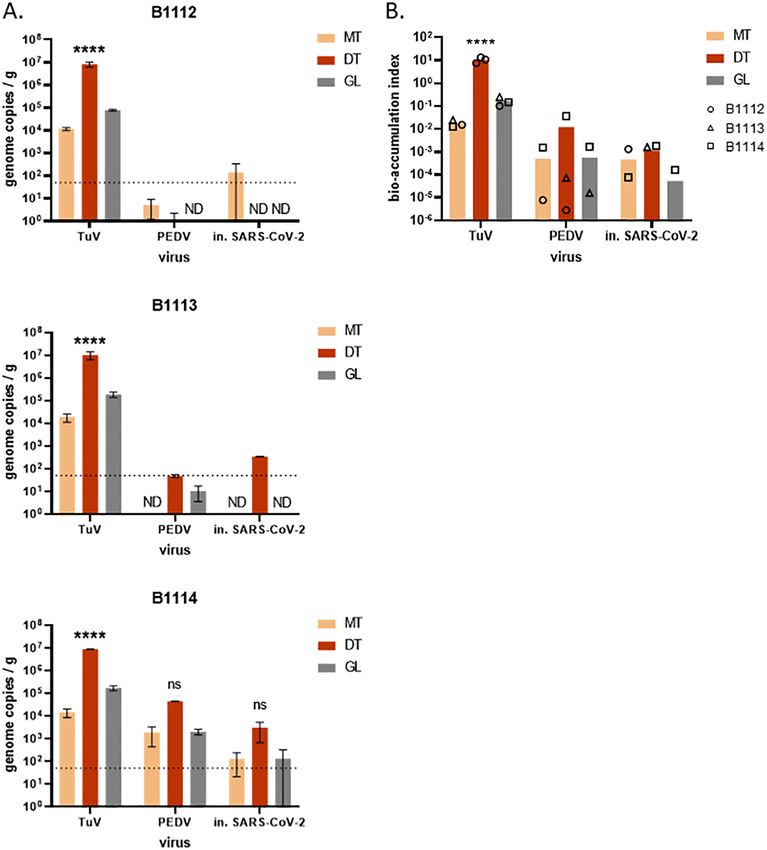
M. Desdouits, J.-C. Piquet, C. Wacrenier et al.                                                                        Science of the Total Environment 778 (2021) 146270

Keywords:                                         Here, we aimed at assessing methods for the detection of CoV, including SARS-CoV-2, in the coastal environment
SARS-CoV-2                                        and testing the possibility that SARS-CoV-2 can contaminate oysters, to monitor the contamination of French
Coastal environment                               shores by SARS-CoV-2 using both seawater and shellfish.
Seawater
                                                  Using the porcine epidemic diarrhea virus (PEDV), a CoV, as surrogate for SARS-CoV-2, and Tulane virus, as sur-
Shellfish
Detection method
                                                  rogate for non-enveloped viruses such as norovirus, we assessed and selected methods to detect CoV in seawater
Genomic detection                                 and shellfish. Seawater-based methods showed variable and low yields for PEDV. In shellfish, the current norm
                                                  for norovirus detection was applicable to CoV detection. Both PEDV and heat-inactivated SARS-CoV-2 could con-
                                                  taminate oysters in laboratory settings, with a lower efficiency than a calicivirus used as control. Finally, we ap-
                                                  plied our methods to seawater and shellfish samples collected from April to August 2020 in France, where we
                                                  could detect the presence of human norovirus, a marker of human fecal contamination, but not SARS-CoV-2.
                                                  Together, our results validate methods for the detection of CoV in the coastal environment, including the use of
                                                  shellfish as sentinels of the microbial quality of their environment, and suggest that SARS-CoV-2 did not contam-
                                                  inate the French shores during the summer season.
                                                                                                                                   © 2021 Published by Elsevier B.V.

1. Introduction                                                                     CoV and SARS-CoV-2 (Kang et al., 2020; McKinney et al., 2006; Yuan
                                                                                    et al., 2020), but foodborne outbreaks were never reported (Jones
    The emergence and global spread of Severe-Acute Respiratory Syn-                et al., 2020). However, SARS-CoV-2 has been detected occasionally in
drome Coronavirus 2 (SARS-CoV-2), responsible for the COVID-19 pan-                 treated sewage (Westhaus et al., 2020; Wurtzer et al., 2020) and in riv-
demics, poses an overwhelming challenge to health policies worldwide                ers (Guerrero-Latorre et al., 2020; Rimoldi et al., 2020), albeit at lower
and has stirred many initiatives to investigate the circulation of this             levels than in raw sewage. This re-inforces the hypothesis that SARS-
virus in the human population. SARS-CoV-2 belongs to the                            CoV-2 can reach the aquatic environment, due to insufficient wastewa-
Coronaviridae family, which is characterized by a 30 kb, positive-sense,            ter treatment (Guerrero-Latorre et al., 2020; Wurtzer et al., 2020) or
single-stranded RNA genome and enveloped virions of around 120-                     sewage spillover before treatment (Rimoldi et al., 2020). Coastal marine
nm in diameter (Gorbalenya et al., 2020). Five genera of CoV have                   waters are also submitted to anthropogenic pollution and sewage con-
been described, among which alpha- and beta- coronavirus (CoV) com-                 tamination, but, to our knowledge, the presence of SARS-CoV-2 in
prise coronaviruses infecting humans (HCoV). SARS-CoV-2 is grouped                  coastal water remains unstudied to date.
among the betaCoV genus with other HCoV, SARS-CoV, MERS-CoV and                         Upon contamination of these waters by sewage containing human
the seasonal HKU1 and OC43 (Gorbalenya et al., 2020). Two other                     pathogens, shellfish can become contaminated in turn and transmit
HCoV, the seasonal NL63 and 229E, belong to the alphaCoV genus                      these pathogens back to human hosts (Iwamoto et al., 2010). Indeed,
(Gorbalenya et al., 2020). Other known CoV infect vertebrates hosts,                filter-feeding bivalve molluscan shellfish are known to concentrate in
and some were used as surrogates for HCoV, such as the alphaCoV Por-                their tissues pollutants or micro-organisms that are present in the sur-
cine Epidemic Diarrhea Virus (PEDV) and Transmissible Gastroenteritis               rounding waters. As such, they can be used as sentinels of the seawater
Virus in pigs; the betaCoV Murine Hepatitis Virus in mice and Bovine co-            quality (Donia et al., 2012; Fiorito et al., 2019; Metcalf et al., 1980;
ronavirus in cattle; and gammaCoV in birds (Ahmed et al., 2020;                     Winterbourn et al., 2016). In the recent years, shellfish have been mon-
Randazzo et al., 2020; Saif, 2004).                                                 itored mainly considering the risk for human consumption as illustrated
    HCoV are respiratory viruses mainly transmitted from person to per-             by the recent study performed in Europe on prevalence of norovirus
son, through exposure to droplets generated by coughing, sneezing or                (NoV) in oysters (EFSA, 2019). Thus, studying the microbiological con-
breathing, either directly in the airways, or through hand-mediated                 tamination of shellfish has a dual purpose: monitoring the presence of
contact (Zhang et al., 2020). Yet, other transmission routes have been              micro-organisms in the aquatic environment, and assessing the sanitary
described for HCoV and especially SARS-CoV-2: aerosol-borne and the                 risks posed to consumers.
fecal-oral route (reviewed in (Arslan et al., 2020)). Indeed, the presence              Many families of human enteric viruses, such as Astroviridae,
of HCoV RNA in feces of infected people has been reported several times             Reoviridae (human rotavirus A), Picornaviridae (aichivirus, enterovirus,
(reviewed in (Jones et al., 2020)). SARS-CoV-2 was detected in stool                hepatovirus) and especially Caliciviridae (human NoV, sapovirus) can
samples from infected individuals, even in the absence of symptoms.                 be detected in sewage-contaminated marine shellfish, leading to
Viral RNA concentration in feces was lower than in saliva or sputum                 human infection upon consumption (Benabbes et al., 2013; Fusco
but could reach 107 genome copies (gc)/mL (Jones et al., 2020).                     et al., 2019; Le Guyader et al., 2008). Conversely, the occurrence of
    Following its shedding in body fluids, SARS-CoV-2 is drained into                Coronaviridae in shellfish has never been reported. This could be due
wastewaters, where its genome has been detected now in many coun-                   to the absence of CoV in the marine environment, to the lack of studies
tries (reviewed in (Kitajima et al., 2020)). Genome concentration of                pertaining to this question, or to the inadequacy of current detection
SARS-CoV-2 in sewage paralleled the number of human cases in the                    methods which were mainly optimized for non-enveloped enteric vi-
corresponding population (Peccia et al., 2020; Wurtzer et al., 2020)                ruses (La Rosa et al., 2020). Following the emergence and spread of
and could reach 106 gc/L (Jones et al., 2020). Thus, wastewater-based               SARS-CoV-2, and its detection in sewage in France, we undertook this
epidemiology (WBE) is now proposed as an efficient strategy to                       study to validate detection methods for Coronaviridae in samples from
monitor SARS-CoV-2 dynamics in the human population (Kitajima                       the coastal environment, assess the ability of bivalve shellfish to accu-
et al., 2020). Yet this promising approach still faces many challenges,             mulate these viruses, and monitor the presence of SARS-CoV-2 on the
especially in areas where wastewater networks are not implemented                   French shores using shellfish and seawater samples.
(Arslan et al., 2020; Street et al., 2020).
    The contamination of aquatic environments by human sewage has                   2. Material and methods
long been recognized as an important transmission route for enteric
pathogens, such as human enteric viruses, either through direct expo-               2.1. Virus stocks and cell lines
sure to contaminated waters, or through their use for food production
and consumption of contaminated foods. (Bosch et al., 2018; Sano                        Tulane virus (TuV) strain M033, kindly provided by T. Farkas (Loui-
et al., 2016). In the case of HCoV, sewage or fecal-borne outbreaks                 siana State University, Baton Rouge, USA) was produced on the LLC-mk2
through aerosols generation were suspected occasionally for SARS-                   cell line as described previously (Polo et al., 2018). Porcine Epidemic

                                                                                2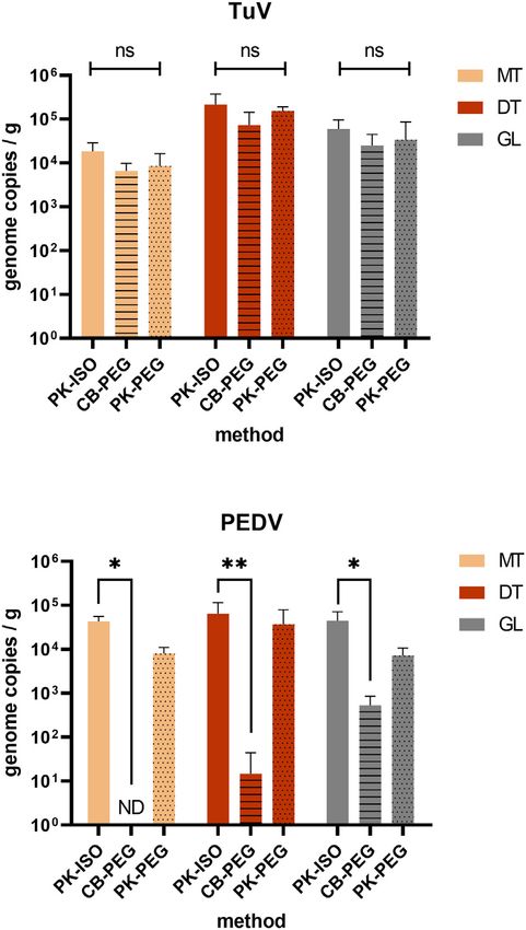
M. Desdouits, J.-C. Piquet, C. Wacrenier et al.                                                                         Science of the Total Environment 778 (2021) 146270

Diarrhea Virus (PEDV) strain CV777 was produced in vero-E6 cells as                        Coastal water (1 L) was sampled together with shellfish from seven
described previously (Bigault et al., 2020). The heat inactivated SARS-                sites (Fig. 1, red dots), sent on ice to the laboratory, where they were
CoV-2 was kindly provided by Dr. C. Bressolette-Bodin (Nantes                          stored at −20 °C until processing.
Université, Centre de Recherche en Transplantation et Immunologie,                         Besides this scheduled sampling, additional shellfish samples were
UMR 1064, ITUN, Nantes, France). Mengovirus (MgV) strain pMC0                          collected on an ad-hoc basis in other coastal sites upon alerts of microbio-
(kindly provided by A. Bosch, University of Barcelona, Spain) was                      logical contamination characterized by increased E. coli concentrations in
propagated in HeLa cells as previously described (Martin et al., 1996).                shellfish flesh (Piquet et al., 2019). A total of 18 shellfish samples linked
   When specified, viruses were inactivated for 15 s. at 60 °C                          to alerts were collected (eleven oyster samples, four mussel samples and
(Abraham et al., 2020). For SARS-CoV-2, inactivation was verified                       three cockle samples), as well as seven water samples.
by TCID50 assay.
                                                                                       2.4. Extraction of viral nucleic acids from coastal water
2.2. Artificial contamination of seawater and oysters (bioaccumulation)
                                                                                           Samples of coastal water (1 L) were analyzed by two methods based
    For protocol validation, 1 L of coastal water sampled in November                  on negative-charged membrane filtration (MF) (Katayama et al., 2002)
2019 and February 2020 were spiked with PEDV and TuV (Table 1).                        and FeCl3 flocculation (FF) (John et al., 2011). For method MF, coastal
This was repeated two or three times to ensure replicate extractions                   water samples were directly filtered on a negative-charged HA-type
for each sample and method.                                                            membrane with a 47 mm diameter and 0.45 μm pores (Millipore, Bur-
    Oysters (Crassostrea gigas) were either purchased live from a pro-                 lington, MA, USA) placed on a vacuum sterile bottle. Filters were rinsed
ducer (commercial oysters), or harvested on the French shore (wild                     with 100 mL of 0.5 mM H2SO4 (pH 3) prior to viral elution with 1 mM
oysters), and kept overnight at 4 °C. Artificial contaminations were car-               NaOH (pH 10.5). After pH neutralization, 10 mL of viral suspension
ried out by bioaccumulation of oysters for 24 h at room temperature                    were concentrated using a 50 kda Centriprep ultrafiltration device
(18–20 °C) in aerated seawater seeded with known concentrations of                     (Millipore) to obtain 2 mL of viral concentrate. In parallel, for method
the viruses (Table 1). The volume of seawater was adjusted to the num-                 FF, 200 μL of 10 g/L FeCL3 solution was added to the filtrate from method
ber of animals in the tank (Table 1), with a ratio of 1 L/6 animals for                MF (kept at 4 °C), and incubated 2 h at 10 °C under gentle agitation, in
commercial oysters, and 1.5 L/6 animals for wild oysters which were                    the dark. A flocculate was then collected on a 0.8 μm pore-size polycar-
twice bigger based on the weight of digestive tissues (DT) recovered.                  bonate filter (Whatman, Maidstone, UK). Virus resuspension was
For each experiment, a fraction of the viral inoculum was titrated in par-             achieved with 2 mL of ascorbate-oxalate–EDTA buffer during a 30 min
allel by qRT-PCR to calculate the total amount of each virus used for bio-             incubation at 4 °C under agitation. Viral suspensions (method FF) and
accumulation. After 24 h of bioaccumulation, oysters were open,                        concentrates (method MF) were extracted using the NucliSens kit
shucked and dissected to collect the DT, the gills and the mantle. Tissues             (bioMérieux, Lyon, France) with 10 mL of lysis buffer and 140 L of mag-
from all oysters were pooled by type, minced, and stored as 2 g-                       netic silica, and eluted in 100 μL of the kit's elution buffer.
aliquotes at −20 °C before analysis.
                                                                                       2.5. Extraction of viral nucleic acids from shellfish
2.3. Environmental sampling
                                                                                           Three methods were tested on 2 g-aliquotes of oyster tissues. The
   Along the French coastline, 21 sites were selected based on exposure                PK-ISO method was applied as described in the norm for Hepatitis A
to human sewage contamination as demonstrated by Escherichia coli                      and NoV detection in shellfish (ISO 15216-1:2017). Briefly, tissues
(Piquet et al., 2019) or NoV contamination (data not shown) (Fig. 1,                   were incubated with 2 mL of a 3000 U/L solution of proteinase K (PK)
black dots). The sites were selected to cover the different French coastal             for 1 h at 37 °C and 15 min at 60 °C, centrifuged for 5 min at 3500 ×g
areas (Fig. 1). From each site, one shellfish sample was collected bi-                  at 4 °C, and 500 μL of supernatant was used for extraction directly
monthly, when possible, from mid-April 2020 to end of August. Only                     using the NucliSens kit (bioMérieux). The remaining supernatant
shellfish present onsite for at least 6 months or from wild populations                 (2.5–3 mL) was used for the PK-PEG extraction method, for which it
were harvested, so that they could reflect the local viral contamination.               was sonicated 3 × 1 min at full power with a Sonopuls sonicator
Most collected samples were cupped oysters (Crassostrea gigas), two                    equipped with a cup-horn (Bandelin, Berlin, Germany), with 1-min
samples were mussels (Mytilus spp.) and one, clams (Ruditapes                          resting on ice between each sonication. Pyrophosphate (100 mM) was
philippinarum). One sample was constituted of at least of 12 oysters,                  added 1:10 in the supernatant, which was then incubated at 4 °C for
20 mussels or 20 clams. Shellfish samples were shipped on ice to the                    40 min with agitation and further treated as described previously
laboratory, where they were dissected and the DT from 10 animals                       (Strubbia et al., 2020) until concentration by poly-ethylene-glycol
pooled, minced, and stored at −20 °C as 2 g-aliquotes.                                 (PEG)-6000 precipitation. For the chloroform:butanol/PEG method

Table 1
Characteristics of artificially contaminated samples.

  Sample               Matrix                          Collection date   Viral inoculum (genome copies)

                                                                         TuV                    PEDV               Inactivated PEDV            Inactivated SARS-CoV-2
                                                                                   9                   9
  E1980                Coastal seawater site O         Oct. 2019         1.8 × 10               2 × 10
  E1982                Coastal seawater site G         Oct. 2019         1.8 × 10⁹              2 × 10⁹
  E1989                Coastal seawater site O         Feb. 2020         2 × 10⁸                3.7 × 10¹⁰
  E1990                Coastal seawater site G         Feb. 2020         2 × 10⁸                3.7 × 10¹⁰
  B1109                36 commercial oysters           Jun. 2020         2 × 10⁹                3.7 × 10¹⁰
  B1112                12 wild oysters                 Jul. 2020         2.3 × 10⁹              2 × 10⁹                                        6.4 × 10⁸
  B1113                18 commercial oysters           Jul. 2020         2.3 × 10⁹              2 × 10⁹                                        6.4 × 10⁸
  B1114                18 commercial oysters           Aug. 2020         3.5 × 10⁹              3.7 × 10⁹                                      5.5 × 10⁹
  B1110                9 commercial oysters            Jul. 2020         2.3 × 10⁹              2 × 10⁹
  B1111                9 commercial oysters            Jul. 2020         2.3 × 10⁹                                 4 × 10⁹
  B1117                9 commercial oysters            Sep. 2020         3.1 × 10⁹              7.9 × 10⁸
  B1118                9 commercial oysters            Sep. 2020         3.1 × 10⁹                                 1.2 × 10⁹

                                                                               3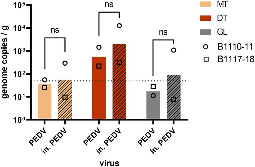
M. Desdouits, J.-C. Piquet, C. Wacrenier et al.                                                                                        Science of the Total Environment 778 (2021) 146270

Fig. 1. Localization of the sampling points for SARS-CoV-2 monitoring along the French coasts. Shellfish (black dots) and coastal seawater (red dots) were sampled bimonthly in 21 sites
distributed along the French coasts and belonging to 4 geographical areas: Normandy (sites A to C), Brittany (sites D to J), Atlantic (sites K to R) and Mediterranean (sites S to U). (For
interpretation of the references to colour in this figure legend, the reader is referred to the web version of this article.)

(CB-PEG), tissues were homogenized with a pestle in a potter with 2 mL                          2.7. Detection of viral genomes by one-step quantitative RT-PCR
glycine buffer (glycine 3.75 g/L, NaCl 9 g/L, pH 9.5). Additional 3 mL of
glycine buffer were used to rinse the pestle and potter, and added to                               The Ultrasens one step quantitative RT-PCR kit (Life technologies,
the tissue homogenate before adding 6 mL of chloroform:butanol                                  Carlsbad, CA, USA) was used for all qRT-PCR reactions, following the man-
(50% vol:vol) solvent and mixing by 30 s on vortex. Cat-Floc T (Calgon,                         ufacturer's indications, using an Aria Mx or MxP3000 real-time PCR sys-
Ellwood City, PA) was added (173 L per tube), the mixture agitated for                          tem (Agilent Technologies, Santa Clara, CA, USA). For SARS-CoV-2, two
5 min at room temperature, before being centrifuged for 15 min at                               sets of primers and probes were used: IP4, targeting the polymerase
13,500 ×g at 4 °C (Atmar et al., 1995). The supernatant was collected,                          gene (Etievant et al., 2020) and E, targeting the envelope gene (Corman
3 mL of PEG-6000 (24%) – NaCl (7%) were added and incubated 1-2 h                               et al., 2020). Cycling were adapted to comply with the qRT-PCR kit re-
at 4 °C with agitation, before a final centrifugation for 20 min at                              quirements: reverse-transcription for 15 min at 55 °C, first denaturation
11,000 ×g at 4 °C. For both the PK-PEG and the CB-PEG methods, the pel-                         and Taq polymerase activation for 5 min at 95 °C, and 45 cycles of denatur-
let was resuspended in 1 mL ddH2O pre-heated at 56 °C, by vortexing                             ation (94 °C, 15 s), annealing (58 °C, 30 s) and extension (65 °C, 30 s)
and pipetting. All viral eluates/concentrates were extracted using the                          followed by fluorescence acquisition. The MgV, TuV and NoV genogroup
NucliSens kit (bioMérieux) following the manufacturer's instruction,                            I (GI) and II (GII) qRT-PCR were carried out as described previously
with 2 mL lysis buffer and 50 μL magnetic silica, and eluted in 100 μL                          (Drouaz et al., 2015; Le Guyader et al., 2009). For PEDV, previously de-
of the kit's elution buffer.                                                                    scribed primers (Bigault et al., 2020) and probe (Kim et al., 2007) were
                                                                                                used based on the same cycling conditions as NoV GII.
2.6. Process control                                                                                For quantification, duplicate 6-points standard curves were made
                                                                                                with TuV synthetic DNA (Drouaz et al., 2015), PEDV in-vitro transcript
    The MgV, a murine picornavirus, was used as a process control for                           T171 (Bigault et al., 2020) and SARS-CoV-2 RNA transcript (CNR des
nucleic acid extraction from shellfish, as described in (ISO15216-                               virus respiratoires, Pasteur Institute), and the synthetic ssRNA-EURM-
1,2017). Briefly, 100 μL of MgV solution were added to each tissue ali-                          019 (European Commission Joint Research Center).
quot just before extraction, and an extraction control was carried out                              Considering the sensitivity of our qRT-PCR assays, the theoretical de-
with 100 μL of pure MgV solution in each series of extraction. MgV con-                         tection limit was set as 1 genome copy per 5 L of nucleic acid that were
centration in nucleic acids extracted from shellfish tissues were com-                           assessed. For shellfish samples, this means 50 gc/g of tissue analyzed
pared to the extraction control to calculate the efficiency of each series                       using the PK-ISO method, 10 gc/g for the CB-PEG method, and 13 gc/g
of extraction. For the environmental screening, samples whose extrac-                           for the PK-PEG method. For seawater, this equals to 20 gc/L for both
tion efficiency was below 1% were not considered for the final analysis,                          methods.
since any absence of virus detection could be due to extraction issues                              For virus detection in shellfish field samples, after verification of
(ISO15216-1,2017). The extraction efficiency was not evaluated for                               extraction efficiency and absence of inhibitors, triplicates of undiluted
water samples collected in the environmental screening.                                         nucleic acid extracts were assessed and for water samples amplifications

                                                                                            4
M. Desdouits, J.-C. Piquet, C. Wacrenier et al.                                                                                   Science of the Total Environment 778 (2021) 146270

were performed on duplicate of undiluted extracts and 1/10 dilutions in                     3.2. Assessment of extraction methods for CoV in shellfish
molecular grade water. For their quantification in seeded or
bioaccumulated contaminated samples, duplicates of undiluted, 1/10                              The current preconized method for the detection of NoV or hepatitis
and 1/100-diluted extracts were used. Good laboratory practices were                        A virus in shellfish relies on a simple protocol based on proteinase K
observed throughout the analysis process, with dedicated separate                           (PK) digestion to release viruses from DT (PK-ISO) (ISO 15216-1). It
rooms for oyster bioaccumulation, shellfish dissection, viral elution                        was compared to the original protocol set up to detect enteric viruses
from shellfish, seawater processing, nucleic acid (NA) extraction,                           in shellfish, which uses chloroform-butanol to elute viruses and PEG
preparation of reaction mixtures, template addition, positive controls ad-                  to concentrate them (CB-PEG) (Atmar et al., 1995). A third protocol,
dition, and amplification. No-template controls were included in all qRT-                    combining PK elution and PEG concentration, able to recover a high di-
PCR assays and proved always negative.                                                      versity of viruses from shellfish (Strubbia et al., 2020) was also tested
                                                                                            here (PK-PEG). We used three tissues dissected from PEDV/TuV-
2.8. Statistics                                                                             bioaccumulated oysters to compare these methods: the mantle (MT),
                                                                                            the digestive tissues (DT) and the gills (GL) (Fig. 2). Three to four series
    GraphPad Prism version 8.4.3 was used for statistical analysis of the                   of extraction were performed. Their efficiencies were calculated for
data by 2-way ANOVA with Tukey's multiple comparisons test. In some                         each method and tissue using the MgV process control, and were com-
instance, the viral concentrations in oyster tissues were below the the-                    prised between 0.4 and 10% for PK-ISO, 0.03 and 4% for CB-PEG, and 0.3
oretical limit of detection, or even non-detected. This was observed be-                    and 5% for PK-PEG. The three methods allowed to recover TuV to similar
fore with other viral targets, and may be due to the complex matrix in                      levels (p > 0.05, Fig. 2) and this virus was more concentrated in the DT
oyster extracts. We chose to keep these values for statistical analysis.                    than in other tissues (p = 0.0002, Fig. 2). PEDV was recovered from the
                                                                                            three shellfish tissues using PK-based methods, when the CB-PEG was
3. Results                                                                                  poorly efficient, allowing PEDV detection only in the gills at a very low
                                                                                            concentration (Fig. 2). Although it used more PK eluate, the PK-PEG
     To validate protocols for the extraction of SARS-CoV-2, we used a                      method was not significantly more efficient at recovering both viruses.
surrogate coronavirus, the porcine epidemic diarrhea virus (PEDV) to
mimic the behavior of SARS-CoV-2 (which requires access to a BSL3 fa-
cility). In addition, we used the TuV, a simian calicivirus often used as a
surrogate for human NoV, as a non-enveloped control virus known to be
bio-accumulated by oyster (Drouaz et al., 2015; Polo et al., 2018).

3.1. Assessment of extraction methods for CoV in seawater

   Several protocols were previously described allowing the concentra-
tion and extraction of viruses from environmental waters, including
seawater. We selected two methods that were found efficient for the re-
covery of enteric viruses (John et al., 2011; Katayama et al., 2002) and
applied them to coastal water samples spiked with PEDV and TuV
(Table 1). The first method (MF) allowed to recover the PEDV and TuV
genomes with a mean yield of 0.981% and 1.33% respectively
(Table 2), but with high inhibition of RT-PCR enzymes necessitating at
least 2-log dilutions of nucleic acid extracts. The second method (FF)
was applied to two samples, where it allowed the recovery of 1.78%
and 0.23% of PEDV and TuV, respectively (Table 2). Both methods
showed a high variability of recovery on both viruses across the differ-
ent samples, and statistical comparison were not significant (Table 2,
p > 0.05). As they present complementary approaches, we chose to
apply both methods on environmental seawater samples for SARS-
CoV-2 monitoring. Besides, given the low viral recovery in seawater
samples, another approach was tested with the use of shellfish to con-
centrate the contamination.

Table 2
Yields in PEDV and TuV using two methods for virus extraction from coastal waters.

  Method                 Method MF                  Method FF              ANOVA

  Virus   Sample N Mean                    SD       Mean           SD      p value
                   recovery (%)            (%)      recovery (%)   (%)

  PEDV E1980         3   0.0754            0.126    3.55           3.38    p = 0.0004
       E1982         3   0.687             0.600    0.0112         0.00899 p = 0.5707
       E1989         2   1.61              0.339    ND
                                                                                            Fig. 2. Assessment of extraction methods for CoV in oysters. Oysters (C. gigas) were
       E1990         2   1.55              0.979    ND
                                                                                            incubated in presence of TuV and PEDV for 24 h, and the concentration of each virus
       Mean              0.981             0.736    1.78           2.50    ns
                                                                                            was measured in three tissues – the mantle (MT, beige), the digestive tissues (DT,
  TuV  E1980         3   0.0777            0.0818   0.471          0.0750  p = 0.2575
                                                                                            brown) and the gills (GL, grey) – by qRT-PCR following repeated extractions by three
       E1982         3   0.471             0.472    0.00513        0.00449 p = 0.0511
                                                                                            different methods – PK-ISO (plain, n = 4), CB-PEG (horizontal lines, n = 3), PK-PEG
       E1989         2   0.948             0.247    ND
                                                                                            (dots, n = 4). *: p < 0.05, **: p < 0.01, ns: non-significant (ANOVA). Theoretical limits of
       E1990         2   3.84              1.09     ND
                                                                                            detection: PK-ISO, 50 gc/g; CB-PEG, 10 cg/g; PK-PEG, 13 cg/g. (For interpretation of the
       Mean              1.33              1.71     0.238          0.329   ns
                                                                                            references to colour in this figure legend, the reader is referred to the web version of
ND: not done.                                                                               this article.)

                                                                                        5
M. Desdouits, J.-C. Piquet, C. Wacrenier et al. Science of the Total Environment 778 (2021) 146270
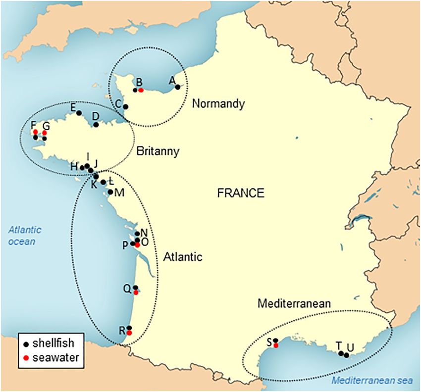
The simpler PK-ISO method was the most efficient on all tissues for three tissues (Fig. 3). TuV was highly concentrated in oyster tissues,
PEDV recovery (p < 0.05 or 0.01), (Fig. 2). Finally, all tissues appeared and most concentrated in the DT (p < 0.0001, Fig. 3, A), as expected,
equally suited for PEDV detection (p > 0.05, Fig. 2). with similar levels of contamination for the three batches. In the two
first batches (B1112 and B1113), PEDV and in. SARS-CoV-2 were de-
3.3. Oysters bioaccumulation with inactivated SARS-CoV-2 tected mainly in the gills and the DT, respectively, at very low levels
(Fig. 3, A). In the third batch, higher quantities of in. SARS-CoV-2
Oysters are known to bio-accumulate very efficiently some enteric (Table 1) were used to contaminate oysters, and CoV were detected in
viruses, such as human NoV (Maalouf et al., 2011), while other viruses the three tissues at intermediate levels, with apparent highest concen-
may be poorly uptaken or kept in their tissues, like bovine NoV tration in the DT that did not reach statistical significance (p > 0.05)
(Zakhour et al., 2010). To test the bio-accumulation of SARS-CoV-2 by (Fig. 3, A). Variability of results across the three oyster batches can be
oysters, and validate the PK-ISO protocol on the target virus, we used explained by a slight inhibition of PCR and lower extraction efficiencies
SARS-CoV-2 from cell culture, heat-inactivated (in.) for safety reasons. for the first batch (2–4%), while the last batch was contaminated with
Three different batches of C. gigas oysters were incubated with in. more inactivated SARS-CoV-2, and also showed the highest extraction
SARS-CoV-2, and with TuV and PEDV as controls. Using the PK-ISO efficiencies (1–21%), which may have resulted in higher amounts of
method, the concentration in viral genomes was then quantified in CoV detected. Importantly, PEDV and in. SARS-CoV-2 displayed very
Fig. 3. Bio-accumulation of heat-inactivated SARS-CoV-2 in oysters. Three batches of C. gigas oysters (B1112, B1113, B1114) were incubated for 24 h in presence of TuV, PEDV and heat-
inactivated (in.) SARS-CoV-2. A. The viral concentration was quantified in three tissues - mantle (MT), digestive tissues (DT) and gills (GL) - by duplicate extractions using the PK-ISO
method. ****: p < 0.0001, ns: non-significant (ANOVA), n = 2 series of extractions. In B1112 and B1113, PEDV or SARS-CoV-2 were not detected (ND) in some tissues. Theoretical limit
of detection: 50 gc/g (dotted line). B. The virus concentration of in each tissue was divided by the initial virus concentration in the seawater to calculate the bio-accumulation index.
Each oyster batch is plotted as a black symbol (circle, B1112; triangle, B1113; square, B1114) when the virus was detected in the corresponding tissue, missing symbols corresponding
to undetected virus. The arithmetic mean values of the three experiments are plotted as columns, for the three tissues. ****: p < 0.0001, ns: non-significant (ANOVA), n = 3
experiments with different oyster batches.
6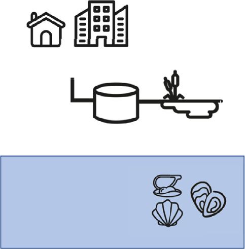
M. Desdouits, J.-C. Piquet, C. Wacrenier et al. Science of the Total Environment 778 (2021) 146270
efficiencies lower than 1% despite repeated extractions, and thus were
excluded of the analysis.
Among the 166 samples collected during the monitoring survey, 141
were oyster samples, 17 mussel samples and 8 clam samples. None of
these samples were found contaminated by SARS-CoV-2 using any of the
two primer sets (Table 3). NoVs searched to confirm human sewage con-
tamination were detected in 35 samples (21%), 69% of these positive sam-
ples being detected at the beginning of the study (from mid-April to end of
May). Four sampling sites (L, J, P, R) were devoid of NoV contamination
and NoV were detected once in nine sites (F to I, O to U). Most of NoV-
contaminated samples were detected in eight sites including three sites
(A, L and N) located close to the mouth of large rivers which displayed
the highest contamination frequency and highest concentrations.
Among the 18 shellfish samples collected following microbiological
alerts suspected to be linked to sewage contaminations events, none
were found contaminated by SARS-CoV-2. They were collected mainly
Fig. 4. Impact of heat inactivation on CoV bioaccumulation in oysters. Oysters (C. gigas) in May and August. Three samples (two collected in May and one in
from two batches (B1110-11 and B1117-18) were incubated in presence of native PEDV June) were found contaminated by NoVs confirming the human fecal
(plain columns) or heat-inactivated (in.) PEDV (hatched columns) for 24 h. The contamination.
concentration of viral genome was quantified in three tissues - the mantle (MT), the
None of the water samples were found contaminated by SARS-CoV-
digestive tissue (DT) and the gills (GL) - following duplicate extractions with the ISO-PK
method and qRT-PCR. Columns represent geometrical means and error bars, geometrical 2, however NoV were detected in 10 samples. Both methods gave posi-
standard deviations. ****: p < 0.0001, ns: nonsignificant (ANOVA), n = 2 experiments tive results with two samples being positives for both methods, two
with different oyster batches. Theoretical limit of detection: 50 gc/g (dotted line). with the MF method and 6 with the FF method. NoV were not detected
in site G, while they were detected twice or three times in all the other
sampling sites (concentrations ranged from 20 to 300 RNAc/L). On one
similar distributions and concentrations in each oyster batch (Fig. 3, A), occasion (site F, sampled on May 5) both water and oyster samples
which supports the use of PEDV as a surrogate for SARS-CoV-2 in were found positive for NoV.
shellfish.
To compare the data more easily regarding the initial amount of
virus used for oyster contamination, the viral concentration in oyster
Table 3
tissues was divided by the initial viral concentration in seawater
Results obtained on water and shellfish samples collected during the monitoring study or
(Fig. 3, B) (Maalouf et al., 2011). TuV bioaccumulation index reached a the microbiological alerts.
mean value of 10.6 in oyster DT and was highly reproducible across
Area Shellfish Water
the three oyster batches. For PEDV and inactivated SARS-CoV-2, the
mean bioaccumulation index was highest in DT (0.012 and 0.0017 re- monitor. Alert Total monitor. Alert Total
spectively), and varied between oyster batches. Together, our data Normandy Nb of sampling sites 3 3 6 1 1 2
show that CoV can contaminate oyster tissues but are not as efficiently Nb of samples 23 3 26 8 1 9
bio-accumulated as a calicivirus like the TuV. collected
SARS-CoV-2 0 0 0 0 0 0
For safety reasons, we could not use native, infectious SARS-CoV-2 to
positive samples
contaminate oysters, and had to rely on heat-inactivated SARS-CoV-2. NoV positive samples 6 0 6 2 0 2
To check that heat inactivation does not impact the bioaccumulation ef- NoV positive sites 2 0 2 1 0 1
ficiency and tissue distribution of CoV, we contaminated oysters with Brittany Nb of sampling sites 7 9 16 2 3 5
TuV and native PEDV or TuV and heat-inactivated PEDV (in. PEDV), in Nb of samples 59 11 70 18 4 22
collected
two separate aquariums with the same batch of oysters at the same SARS-CoV-2 0 0 0 0 0 0
time. Two independent experiments using different batches were con- positive samples
ducted (Fig. 4). For both, TuV displayed the expected distribution and NoV positive samples 8 3 11 3 0 3
was equally concentrated in each tissue between oysters from the two NoV positive sites 6 3 9 1 0 1
Atlantic Nb of sampling sites 8 3 11 3 1 4
aquariums (data not shown). The MgV extraction efficiencies were
Nb of samples 57 3 60 14 2 16
also similar, with respective mean values of 5.5% (range 1–22%) and collected
4,6% (1–11%). In the first experiment (B1110-11), inactivated PEDV ap- SARS-CoV-2 0 0 0 0 0 0
peared more concentrated than native PEDV in the oyster tissues (Fig. 4, positive samples
circles). In the second experiment (B1117-18), native and inactivated NoV positive samples 18 0 18 3 0 3
NoV positive sites 6 0 6 2 0 2
PEDV exhibited the same levels of concentration (Fig. 4, triangles). Con- Mediterranea Nb of sampling sites 3 1 4 1 0 1
sidering both experiments, the mean concentration of native and Nb of samples 27 1 28 9 0 9
inactivated PEDV did not differ significantly (p > 0.05, Fig. 4), and collected
their tissue distribution were similar, suggesting that heat inactivation SARS-CoV-2 0 0 0 0 0 0
positive samples
does not impair CoV bioaccumulation by oysters, and validating our re-
NoV positive samples 3 0 3 2 0 2
sults with in. SARS-CoV-2. NoV positive sites 3 0 3 1 0 1
Total Nb of sampling sites 21 16 37 7 5 12
3.4. Screening of environmental samples for the presence of SARS-CoV-2 Nb of samples 166 18 184 52 7 59
collected
SARS-CoV-2 0 0 0 0 0 0
A total of 187 samples were collected from 37 sites, including 21 positive samples
sites regularly sampled (monitoring, Fig. 1) and 16 sites sampled upon NoV positive samples 35 3 38 10 0 10
alerts on microbiological contamination (alerts). All these samples NoV positive sites 19 3 22 5 0 5
were processed by the PK-ISO method. Among these, three samples monitor.: samples collected during regular monitoring; alert: samples collected following
(one from Normandy, and two from Brittany area) provided extraction alerts of microbiological contamination in additional locations.
7M. Desdouits, J.-C. Piquet, C. Wacrenier et al. Science of the Total Environment 778 (2021) 146270
4. Discussion grab sample of such a small volume is also not representative of the
whole water present in a site. Given these limitations for direct seawa-
Most existing protocols for the detection of viruses in environmental ter analysis, we proposed to use shellfish, which are filter-feeding ani-
samples are optimized for non-enveloped, enteric viruses such as gas- mals known to concentrate chemical and microbial contaminants, as
troenteritis or hepatitis viruses (Bosch et al., 2018). The emergence sentinel for the detection of SARS-CoV-2 in the coastal environment.
and possible environmental spread of the SARS-CoV-2, an enveloped Like was done for seawater, we first evaluated different methods to
virus, raised new challenges to environmental virologists (La Rosa detect CoV in oysters contaminated with TuV and PEDV. Two methods
et al., 2020). Our first aim was to select a method to detect CoV, in sam- used proteinase K (PK) for viral elution from the oyster tissues, and one
ples from the coastal environment, using real-time, quantitative RT- used lipophilic solvents (chloroform/butanol). The latter method was in-
PCR, which is one of the most sensitive and robust techniques available efficient on PEDV, with only traces of this CoV detected in one tissue,
for virus detection in environmental samples (Haramoto et al., 2018). As while the non-enveloped TuV was detected in high concentrations in all
manipulating infectious SARS-CoV-2 required working in a biosafety tissues. Lipophilic solvents disrupt lipid membranes like viral envelopes,
level 3 laboratory, we first selected a surrogate virus allowing to assess and chloroform was already shown to dramatically alter the recovery of
detection methods without this safety considerations. Important points CoV (Conceição-Neto et al., 2015). Contrarily, the PK-based elution
to select a surrogate are the genetic proximity to the target virus, the methods allowed the detection and quantification of both TuV and
physical and chemical characteristics but also the absence of human PEDV in three oyster tissues. We thus chose to apply the current recom-
pathogenicity, and/or easy way of production (Cromeans et al., 2014). mended ISO15216:1–2017 method for NoV and hepatitis A virus detec-
In this study, to use this surrogate with seawater and oysters, the lack tion in shellfish for the next experiments. Indeed, using the ISO method
of natural contamination was another constraint. Phages are good sur- allows for comparisons with more studied viruses (such as NoV). It is
rogate for some eukaryotic viruses but their presence in environmental also a simple protocol, that could be easily implemented in laboratories
samples may complicate their use (Flannery et al., 2012). Usually a virus for routine analysis if this becomes needed for SARS-CoV-2.
from the same family is preferred so that target and surrogate viruses Using PEDV and inactivated SARS-CoV-2, we show that CoV can con-
share a similar size, structure, and other characteristics. For example, taminate oysters. To our knowledge, this is the first demonstration that
the TuV, prototype strain of the genus Recovirus within the Caliciviridae oysters can bioaccumulate a CoV. PEDV and heat-inactivated SARS-CoV-
family, is used to mimic NoV behavior (Drouaz et al., 2015). Among the 2 displayed very similar distributions and levels of contaminations in
Coronaviridae family, we selected PEDV, a porcine enteric CoV which be- three oyster batches. In addition, we show that heat inactivation does
longs to a different group of CoV than SARS-CoV-2 (alpha and beta-CoV, not impair the distribution of PEDV in oyster tissues nor negatively im-
respectively). The first one is an enteric virus while the second is respi- pact its bio-accumulation by oysters. These results validate our observa-
ratory, which could imply differences in environmental stability. Never- tions with inactivated SARS-CoV-2 and reinforce our confidence that
theless, porcine enteric CoV have been used in the past to as surrogates PEDV can be used as a surrogate for SARS-CoV-2 in oysters. The low im-
for HCoV, including SARS-CoV-2 (Randazzo et al., 2020), and in a recent pact of thermal inactivation on CoV bioaccumulation by oysters also
study, all tested CoV (including PEDV) fitted in the same model regard- suggest that partially degraded SARS-CoV-2 present in sewage may
ing their sensitivity to temperature in fomites (Guillier et al., 2020). Al- still be able to contaminate shellfish when reaching the coastal environ-
together with the TuV, it allowed us to control the efficacy of our ment. These observations are encouraging for the use of shellfish as sen-
methods on a target, non-enveloped virus, and to compare with tinel of human contamination. However, given the expected low levels
enveloped coronavirus data. and low stability of CoV in the environment, the persistence of CoV RNA
As the aim of this work was to evaluate the possible coastal contam- in shellfish tissues needs to be investigated to estimate how long after
ination by SARS-CoV-2 shed by infected people, we first evaluated contamination the virus could still be detected.
methods for SARS-CoV-2 detection and quantification in seawater. In Both PEDV and inactivated SARS-CoV-2 were less efficiently bio-
human feces and in sewage, which are the sources of human viruses accumulated by oysters than TuV, a calicivirus, which could indeed be
in the coastal environment, viruses are rarely free but adsorbed onto due to a lower stability in seawater and oysters, and/or to a lower affin-
particles. Thus, we selected a combination of two complementary ity for oyster tissues. The tissue distribution pattern of CoV does not
methods, one recovering large particles (membrane filtration, MF) show a marked concentration in DT, contrarily to TuV, and high concen-
and the other one, smaller aggregates and free viruses (FeCl3 trations of viruses were needed to contaminate oysters, as previously
floculation, FF). When applied on seawater samples spiked with the shown for mengovirus, from the Picornaviridae family (Drouaz et al.,
TuV and the PEDV, these methods allowed to detect both viruses, how- 2015). Bioaccumulation efficiency may vary from one virus to another
ever at low yield and with high variability between water samples. or depend on the shellfish species. If for NoV the impact of ligands and
These very low yields could be explained by the use of coastal marine their seasonal expression has been demonstrated, this is still unclear
waters, which were turbid and contained PCR inhibitors (Hata et al., for other human enteric viruses (Grodzki et al., 2012; Maalouf et al.,
2020). Surprisingly, results were similar for TuV and PEDV for each sam- 2010; Zakhour et al., 2010).
ple, which suggest that the yield of the methods is mostly influenced by In the coastal environment, expected concentrations of enteric vi-
parameters of the seawater matrix (presumably particulate material, ruses are usually much lower than those used for artificial bioaccumula-
PCR inhibitors) and not by the nature of the virus. Considering that tion (Gentry et al., 2009; Keller et al., 2019), and may be even lower for
the two methods showed similar ranges of yields, they were both ap- SARS-CoV-2. Yet, repeated exposures to the virus in the open environ-
plied on naturally contaminated seawater samples during environmen- ment, where larger volumes of seawater are filtered by shellfish, may
tal monitoring, where NoV, but not SARS-CoV-2, were detected. These still lead to their contamination. C. gigas oysters are present on all
results underline that virus detection from environmental waters is French shores and in many countries worldwide (Europe, North
not an easy process. In the ISO15216:1–2017 norm, as low as 1% recov- Africa, China, Japan, Korea, Australia, Pacific coast of USA and Canada)
ery rate is considered an acceptable quality parameter. A recovery of as a farmed animal and/or an invasive species (Herbert et al., 2016),
11% for PEDV and MgV in raw sewage using aluminum hydroxide and is thus suitable for use as sentinel in many settings. As mentioned
adsorption-precipitation was achieved, but the recovery of PEDV was above, other filter-feeding shellfish species may exhibit differences in
down to 3% in treated sewage (Randazzo et al., 2020). Here, the filtra- bioaccumulation efficiency and should be tested in further work, such
tion of one-liter samples was difficult to achieve while still being too as Dreissena polymorpha proposed as a biomonitoring tool in fresh
small for the detection of SARS-CoV-2 that is likely present at very low water (Géba et al., 2020).
concentrations (if present) in the environment. Even if the detection Considering that seawater sampling and analysis is complicated and
of some NoV confirmed the efficacy of these methods in the field, a unlikely to be positive for SARS CoV-2, and our results showing a
8M. Desdouits, J.-C. Piquet, C. Wacrenier et al. Science of the Total Environment 778 (2021) 146270
possible bioaccumulation of SARS-CoV-2 in oysters, we set up a moni- To conclude, we believe that surveying shellfish may help to monitor
toring survey that begun at the end of the first wave of infections in the viral diffusion in seaside communities, and may be especially suited
France to evaluate the possible contamination of coastal areas before for countries lacking centralized sewage collection infrastructures, in
the summer season, using shellfish as sentinels. We used mostly oyster which environmental contamination is also more likely (Guerrero-
samples, as it was the species in which methods were tested, but some Latorre et al., 2020). Further work is needed to evaluate and adapt
samples consisted in mussels or clams in areas where oysters were not existing methods for the detection of SARS-CoV-2 in the environment,
available. Sites known for their sensitivity to human sewage contamina- that may also be suited for other emerging enveloped viruses such as
tion were sampled, hypothesizing that if SARS-CoV-2 could contami- Influenza, Ebola, or Nipah viruses, should we face another emerging
nate the coastal environment, these sites should be positive. Indeed, viral pandemic.
the observed prevalence in NoV (20.5%) was high compared to previous
surveys, especially considering the low epidemic burden of NoV in sum- Declaration of competing interest
mertime (EFSA, 2019; Schaeffer et al., 2013). Several water samples
were also found contaminated with NoV showing that in some instance The authors declare that they have no known competing financial
this approach can be complementary to shellfish sampling, although interests or personal relationships that could have appeared to
technical improvements are necessary to increase the recovery rate. influence the work reported in this paper.
Conversely, all samples (shellfish and seawater) were negative for
SARS-CoV-2. The survey period covered the end of the French lock- Acknowledgements
down (until may 11th, 2020) and the summer season when tourism re-
sults in a larger population on the French coastline. During the first We thank Audrey Rodallec, Virginie Ferré, Berthe-Marie Imbert-
wave of SARS-CoV-2 in France (March to May 2020), most cases oc- Marcille (Service de Virology, Centre Hospitalier Universitaire &
curred in the north-eastern part of France, and viral concentrations Université de Nantes, France) for technical advice and helpful discus-
were likely very low in sewage from the rest of the territory, including sions. We thank the Obepine network for helpful discussions.
western and southern coasts. After the lock-down, although some We are grateful for the help and expertise of our colleagues from
Covid-19 clusters were reported in seaside communities, the overall the Laboratoires Environnement Ressource (LER, Coastal Unit,
prevalence of SARS-CoV-2 remained low in France throughout the sur- Ifremer) who participated in the environmental sampling and sam-
vey period (“Taux d'incidence de 458 l'épidémie de COVID-19 (SI-DEP) ple logistics: Sylviane Boulben and Aourégan Terre-Tillon (LER/BO),
- data.gouv.fr”, n.d.) which was carried out between the two first waves Julien Chevé, Théodore Marie Lepoittevin and Manuel Rouquette
of Covid-19 (Spaccaferri et al., 2020). Although we cannot rule out a (LER/BN), Camille Gianaroli (LER/LR), James Grizon and Jonathan
transient contamination, or contamination outside the study sites, Deborde (LER/PC), Myriam Perrière-Rumebe, Florence d'Amico and
these results suggest that SARS-CoV-2 did not reach the French coastal Elvire Antajan (LER/AR), and all members of the LER/BO, BN, MPL,
environment during summer 2020 at significant levels. Environmental N, LR, PC and PAC.
monitoring should be continued during the winter season, where the
risk of viral spread in the environment is likely to increase due to the Funding
second wave of Covid-19 in the French population, cold temperatures
stabilizing the virus and heavy rainfalls resulting in sewage spillover. This work is supported by the Agence Nationale de la Recherche and
This pandemic raises many questions, including some technical issues the Fondation de France (ANR RA-Covid wave 5, n°00109676), the Ré-
regarding CoV detection in different types of environmental samples. As gion Pays de la Loire (order n°2020-12887), by an internal funding from
mentioned above, environmental virology in the past has tended to con- Ifremer General Direction (SARS-CoV-2 action plan) and the European
sider mainly non-enveloped viruses. After the first emergence of SARS- project VEO (H2020, SC1-2019-874735).
CoV, a study demonstrated the persistence of some strains in environmen-
tal waters (Casanova et al., 2009). Recently, if many papers have been CRediT authorship contribution statement
published regarding sewage contamination by SARS-CoV-2, to our knowl-
edge none report on its detection in seawater and/or shellfish. In devel- MD: Conceptualization; Formal analysis; Funding acquisition; Inves-
oped countries with efficient sewage treatment systems, the risk of tigation; Methodology; Project administration; Visualization; Roles/
coastal contamination may be limited, and linked to accidental contami- Writing - original draft; Writing - review & editing. JCP: Conceptualiza-
nation with untreated sewage. Yet, in some settings, using shellfish as sen- tion; Formal analysis; Funding acquisition; Investigation; Methodology;
tinels for viral diffusion in the environment may be useful, and we show Supervision; Visualization; Writing - review & editing. CW: Investiga-
here that two CoV, including SARS-CoV-2, can contaminate oysters tion; Writing - review & editing. CLM: Investigation; Writing - review
under experimental conditions. The demonstration that a surrogate por- & editing. SP: Formal analysis; Investigation; Methodology; Writing -
cine CoV, PEDV, may be used to mimic SARS-CoV2 in oysters, suggest review & editing. SJ: Investigation; Writing - review & editing. SR: Inves-
that it could be used in other matrices and, to some extent, to evaluate tigation; Methodology; Writing - review & editing. LB: Investigation;
the stability of infectious particles. Infectious SARS-CoV-2 was isolated Methodology; Writing - review & editing. MC: Investigation; Methodol-
from several, but not all, stool or urine samples from Covid-19 patients ogy; Writing - review & editing. PG: Conceptualization; Funding acqui-
(Jones et al., 2020; Sun et al., 2020; Xiao et al., 2020). Although in two out- sition; Project administration; Writing - review & editing. FC:
breaks, sewage was suspected as a SARS-CoV-2 contamination source Investigation; Writing - review & editing. RG: Investigation; Writing
(Kang et al., 2020; Yuan et al., 2020), attempts at isolating infectious - review & editing. LLa: Investigation; Writing - review & editing. LLe:
SARS-CoV-2 from raw or treated sewage, or freshwater, remains unsuc- Investigation; Writing - review & editing. PLG: Investigation; Writing -
cessful to date (Rimoldi et al., 2020; Wang et al., 2020). A recent study re- review & editing. CM: Investigation; Writing - review & editing. AS: In-
ports the infection of non-human primates through gastrointestinal vestigation; Writing - review & editing. JLS, Investigation; Writing
inoculation with a high inoculum of SARS-CoV-2 (Jiao et al., 2020). Yet, - review & editing. OS, Investigation; Writing - review & editing. CP, In-
in humans, the fecal-oral route of transmission has never being observed vestigation; Writing - review & editing. CBB, Investigation; Writing -
for SARS-CoV-2 (Zuber and Brüssow, 2020). The sanitary risk posed by po- review & editing. YB: Funding acquisition; Project administration; Su-
tential contamination of shellfish by SARS-CoV-2 is likely very low but pervision; Writing - review & editing. FLG: Conceptualization; Formal
having a method to detect this virus in a food matrix known to be at analysis; Funding acquisition; Methodology; Project administration;
risk for virus transmission is important to anticipate questions that may Resources; Supervision; Validation; Visualization; Roles/Writing - orig-
raise with environmental or food contamination by this virus. inal draft; Writing - review & editing.
9M. Desdouits, J.-C. Piquet, C. Wacrenier et al. Science of the Total Environment 778 (2021) 146270
References Guerrero-Latorre, L., Ballesteros, I., Villacrés-Granda, I., Granda, M.G., Freire-Paspuel, B., Ríos-
Touma, B., 2020. SARS-CoV-2 in river water: implications in low sanitation countries. Sci.
Total Environ. 743, 140832. https://doi.org/10.1016/j.scitotenv.2020.140832.
Abraham, J.P., Plourde, B.D., Cheng, L., 2020. Using heat to kill SARS-CoV-2. Rev. Med.
Virol. 30. https://doi.org/10.1002/rmv.2115. Guillier, L., Martin-Latil, S., Chaix, E., Thébault, A., Pavio, N., Le Poder, S., Batéjat, C., Biot, F.,
Koch, L., Schaffner, D.W., Sanaa, M., Covid-19 Emergency Collective Expert Appraisal
Ahmed, W., Bertsch, P.M., Bibby, K., Haramoto, E., Hewitt, J., Huygens, F., Gyawali, P.,
Group, 2020. Modeling the inactivation of viruses from the Coronaviridae family in
Korajkic, A., Riddell, S., Sherchan, S.P., Simpson, S.L., Sirikanchana, K., Symonds, E.M.,
response to temperature and relative humidity in suspensions or on surfaces. Appl.
Verhagen, R., Vasan, S.S., Kitajima, M., Bivins, A., 2020. Decay of SARS-CoV-2 and sur-
Environ. Microbiol. 86. https://doi.org/10.1128/AEM.01244-20.
rogate murine hepatitis virus RNA in untreated wastewater to inform application in
wastewater-based epidemiology. Environ. Res. 191, 110092. https://doi.org/ Haramoto, E., Kitajima, M., Hata, A., Torrey, J.R., Masago, Y., Sano, D., Katayama, H., 2018. A
10.1016/j.envres.2020.110092. review on recent progress in the detection methods and prevalence of human enteric vi-
ruses in water. Water Res. 135, 168–186. https://doi.org/10.1016/j.watres.2018.02.004.
Arslan, M., Xu, B., Gamal El-Din, M., 2020. Transmission of SARS-CoV-2 via fecal-oral and
Hata, A., Furumai, H., Katayama, H., 2020. Sequential treatment using a hydrophobic resin
aerosols-borne routes: environmental dynamics and implications for wastewater
and gel fi ltration to improve viral gene quanti fi cation from highly complex environ-
management in underprivileged societies. Sci. Total Environ. 743, 140709. https://
mental concentrates. Water Res. 174, 115652. https://doi.org/10.1016/j.
doi.org/10.1016/j.scitotenv.2020.140709.
watres.2020.115652.
Atmar, R.L., Neill, F.H., Romalde, J.L., Guyader, O.L.E., Woodley, C.M., Metcalf, T.G., Estes,
Herbert, R.J.H., Humphreys, J., Davies, Clare J., Roberts, C., Fletcher, S., Crowe, Tasman P.,
M.K., 1995. Detection of Norwalk virus and hepatitis a virus in shellfish tissues with
2016. Ecological impacts of non-native Pacific oysters (Crassostrea gigas) and man-
the PCR. Appl. Environ. Microbiol. 61, 3014–3018.
agement measures for protected areas in Europe. Biodivers. Conserv. 25,
Benabbes, L., Ollivier, J., Schaeffer, J., Parnaudeau, S., Rhaissi, H., Nourlil, J., Le Guyader, F.S.,
2835–2865. https://doi.org/10.1007/s10531-016-1209-4.
2013. Norovirus and other human enteric viruses in Moroccan shellfish. Food Envi-
Iwamoto, M., Ayers, T., Mahon, B.E., Swerdlow, D.L., 2010. Epidemiology of seafood-
ron. Virol. 5, 35–40. https://doi.org/10.1007/s12560-012-9095-8.
associated infections in the United States. Clin. Microbiol. Rev. 23, 399–411. https://
Bigault, L., Brown, P., Bernard, C., Blanchard, Y., Grasland, B., 2020. Porcine epidemic diarrhea
doi.org/10.1128/CMR.00059-09.
virus: viral RNA detection and quantification using a validated one-step real time RT-
Jiao, L., Li, H., Xu, J., Yang, M., Ma, C., Li, J., Zhao, S., Wang, H., Yang, Y., Yu, W., Wang, J.,
PCR. J. Virol. Methods 283, 113906. https://doi.org/10.1016/j.jviromet.2020.113906.
Yang, J., Long, H., Gao, J., Ding, K., Wu, D., Kuang, D., Zhao, Y., Liu, J., Lu, S., Liu, H.,
Bosch, A., Gkogka, E., Le, F.S., Loisy-Hamon, F., Lee, A., Lieshout, L.V., Marthi, B., Myrmel, M., Peng, X., 2020. The gastrointestinal tract is an alternative route for SARS-CoV-2 infec-
Sansom, A., Schultz, A.C., Winkler, A., Zuber, S., Phister, T., 2018. Foodborne viruses: tion in a nonhuman primate model. Gastroenterology. https://doi.org/10.1053/j.
detection, risk assessment, and control options in food processing. Int. J. Food gastro.2020.12.001.
Microbiol. 285, 110–128. https://doi.org/10.1016/j.ijfoodmicro.2018.06.001.
John, S.G., Mendez, C.B., Deng, L., Poulos, B., Kauffman, A.K.M., Kern, S., Brum, J., Polz, M.F.,
Casanova, L., Rutala, William A., Weber, David J., Sobsey, Mark D., 2009. Survival of surro- Boyle, E.A., Sullivan, M.B., 2011. A Simple and Efficient Method for Concentration of
gate coronaviruses in water. Water Res. 43 (7), 1893–1898. https://doi.org/10.1016/j. Ocean Viruses by Chemical Flocculation. 3, pp. 195–202. https://doi.org/10.1111/
watres.2009.02.002. j.1758-2229.2010.00208.x.
Conceição-Neto, N., Zeller, M., Lefrère, H., De Bruyn, P., Beller, L., Deboutte, W., Yinda, C.K., Jones, D.L., Baluja, M.Q., Graham, D.W., Corbishley, A., McDonald, J.E., Malham, S.K., Hillary,
Lavigne, R., Maes, P., Ranst, M.V., Heylen, E., Matthijnssens, J., 2015. Modular approach L.S., Connor, T.R., Gaze, W.H., Moura, I.B., Wilcox, M.H., Farkas, K., 2020. Shedding of
to customise sample preparation procedures for viral metagenomics: a reproducible SARS-CoV-2 in feces and urine and its potential role in person-to-person transmis-
protocol for virome analysis. Sci. Rep. 5, 16532. https://doi.org/10.1038/srep16532. sion and the environment-based spread of COVID-19. Sci. Total Environ. 749,
Corman, V.M., Landt, O., Kaiser, M., Molenkamp, R., Meijer, A., Chu, D.K., Bleicker, T., 141364. https://doi.org/10.1016/j.scitotenv.2020.141364.
Brünink, S., Schneider, J., Schmidt, M.L., Mulders, D.G., Haagmans, B.L., van der Veer, Kang, M., Wei, J., Yuan, J., Guo, J., Zhang, Y., Hang, J., Qu, Y., Qian, H., Zhuang, Y., Chen, X.,
B., van den Brink, S., Wijsman, L., Goderski, G., Romette, J.-L., Ellis, J., Zambon, M., Peng, X., Shi, T., Wang, J., Wu, J., Song, T., He, J., Li, Y., Zhong, N., 2020. Probable evi-
Peiris, M., Goossens, H., Reusken, C., Koopmans, M.P., Drosten, C., 2020. Detection of dence of fecal aerosol transmission of SARS-CoV-2 in a high-rise building. Ann. Intern.
2019 novel coronavirus (2019-nCoV) by real-time RT-PCR. Euro Surveill. 25. Med. https://doi.org/10.7326/M20-0928.
https://doi.org/10.2807/1560-7917.ES.2020.25.3.2000045. Katayama, H., Shimasaki, A., Ohgaki, S., 2002. Development of a virus concentration
Cromeans, T., Park, G.W., Costantini, V., Lee, D., Wang, Q., Farkas, T., Lee, A., Vinjé, J., 2014. method and its application to detection of enterovirus and Norwalk virus from
Comprehensive comparison of cultivable norovirus surrogates in response to differ- coastal seawater. Appl. Environ. Microbiol. 68, 1033–1039. https://doi.org/10.1128/
ent inactivation and disinfection treatments. Appl. Environ. Microbiol. 80, AEM.68.3.1033.
5743–5751. https://doi.org/10.1128/AEM.01532-14. Keller, R., Pratte-Santos, R., Scarpati, K., Martins, S.A., Loss, S.M., Fumian, T.M.,
Donia, D., Dell’Amico, M.C., Petrinca, A.R., Martinucci, I., Mazzei, M., Tolari, F., Divizia, M., 2012. Miagostovich, M.P., Cassini, S.T., 2019. Surveillance of enteric viruses and
Presence of hepatitis E RNA in mussels used as bio-monitors of viral marine pollution. thermotolerant coliforms in surface water and bivalves from a mangrove estuary in
J. Virol. Methods 186, 198–202. https://doi.org/10.1016/j.jviromet.2012.06.007. southeastern Brazil. Food Environ. Virol. 11, 288–296. https://doi.org/10.1007/
Drouaz, N., Schaeffer, J., Farkas, T., Le Pendu, J., Le Guyader, F.S., 2015. Tulane virus as a po- s12560-019-09391-3.
tential surrogate to mimic norovirus behavior in oysters. Appl. Environ. Microbiol. 81, Kim, S.-H., Kim, I.-J., Pyo, H.-M., Tark, D.-S., Song, J.-Y., Hyun, B.-H., 2007. Multiplex real-
5249–5256. https://doi.org/10.1128/AEM.01067-15. time RT-PCR for the simultaneous detection and quantification of transmissible gas-
EFSA, 2019. Analysis of the European baseline survey of norovirus in oysters. EFSA J. 17, troenteritis virus and porcine epidemic diarrhea virus. J. Virol. Methods 146,
1–99. https://doi.org/10.2903/j.efsa.2019.5762. 172–177. https://doi.org/10.1016/j.jviromet.2007.06.021.
Etievant, S., Bal, A., Escuret, V., Brengel-Pesce, K., Bouscambert, M., Cheynet, V., Generenaz, Kitajima, M., Ahmed, W., Bibby, K., Carducci, A., Gerba, C.P., Hamilton, K.A., Haramoto, E., Rose,
L., Oriol, G., Destras, G., Billaud, G., Josset, L., Frobert, E., Morfin, F., Gaymard, A., 2020. J.B., 2020. SARS-CoV-2 in wastewater: state of the knowledge and research needs. Sci.
Performance assessment of SARS-CoV-2 PCR assays developed by WHO referral lab- Total Environ. 739, 139076. https://doi.org/10.1016/j.scitotenv.2020.139076.
oratories. J. Clin. Med. 9, 1871. https://doi.org/10.3390/jcm9061871. La Rosa, G., Bonadonna, L., Lucentini, L., Kenmoe, S., Suffredini, E., 2020. Coronavirus in
Fiorito, F., Amoroso, M.G., Lambiase, S., Serpe, F.P., Bruno, T., Scaramuzzo, A., Maglio, P., water environments: occurrence, persistence and concentration methods - a scoping
Fusco, G., Esposito, M., 2019. A relationship between environmental pollutants and review. Water Res. 115899. https://doi.org/10.1016/j.watres.2020.115899.
enteric viruses in mussels (Mytilus galloprovincialis). Environ. Res. 169, 156–162. Le Guyader, F.S., Le Saux, J.-C., Ambert-Balay, K., Krol, J., Serais, O., Parnaudeau, S.,
https://doi.org/10.1016/j.envres.2018.11.001. Giraudon, H., Delmas, G., Pommepuy, M., Pothier, P., Atmar, R.L., 2008. Aichi virus,
Flannery, J., Keaveney, S., Rajko-Nenow, P., O’Flaherty, V., Dor??, W., 2012. Concentration Norovirus, Astrovirus, Enterovirus, and Rotavirus involved in clinical cases from a
of norovirus during wastewater treatment and its impact on oyster contamination. French oyster-related gastroenteritis outbreak. J. Clin. Microbiol. 46, 4011–4017.
Appl. Environ. Microbiol. 78, 3400–3406. https://doi.org/10.1128/AEM.07569-11. https://doi.org/10.1128/JCM.01044-08.
Fusco, G., Anastasio, A., Kingsley, D.H., Amoroso, M.G., Pepe, T., Fratamico, P.M., Cio, B., Le Guyader, F.S., Parnaudeau, S., Schaeffer, J., Bosch, A., Loisy, F., Pommepuy, M., Atmar,
Rossi, R., Rosa, G.L., Boccia, F., 2019. Detection of Hepatitis A Virus and Other Enteric R.L., 2009. Detection and quantification of noroviruses in shellfish. Appl. Environ.
Viruses in Shellfish Collected in the Gulf of Naples, Italy 2016. Microbiol. 75, 618–624. https://doi.org/10.1128/AEM.01507-08.
Géba, E., Aubert, D., Durand, L., Escotte, S., La Carbona, S., Cazeaux, C., Bonnard, I., Maalouf, H., Zakhour, M., Pendu, J.L., Le Saux, J.C., Atmar, R.L., Le Guyader, F.S., 2010. Distribu-
Bastien, F., Palos Ladeiro, M., Dubey, J.P., Villena, I., Geffard, A., Bigot-Clivot, A., tion in tissue and seasonal variation of norovirus genogroup I and II ligands in oysters.
2020. Use of the bivalve Dreissena polymorpha as a biomonitoring tool to reflect Appl. Environ. Microbiol. 76, 5621–5630. https://doi.org/10.1128/AEM.00148-10.
the protozoan load in freshwater bodies. Water Res. 170, 115297. https://doi. Maalouf, H., Schaeffer, J., Parnaudeau, S., Le Pendu, J., Atmar, R.L., Crawford, S.E., Le
org/10.1016/j.watres.2019.115297. Guyader, F.S., 2011. Strain-dependent norovirus bioaccumulation in oysters. Appl.
Gentry, J., Vinje, J., Guadagnoli, D., Lipp, E.K., 2009. Norovirus distribution within an estu- Environ. Microbiol. 77, 3189–3196. https://doi.org/10.1128/AEM.03010-10.
arine environment. Appl. Environ. Microbiol. 75, 5474–5480. https://doi.org/10.1128/ Martin, L.R., Duke, G.M., Osorio, J.E., Hall, D.J., Palmenberg, A.C., 1996. Mutational analysis
AEM.00111-09. of the mengovirus poly(C) tract and surrounding heteropolymeric sequences. J. Virol.
Gorbalenya, A.E., Baker, S.C., Baric, R.S., de Groot, R.J., Drosten, C., Gulyaeva, A.A., 70, 2027–2031. https://doi.org/10.1128/JVI.70.3.2027-2031.1996.
Haagmans, B.L., Lauber, C., Leontovich, A.M., Neuman, B.W., Penzar, D., Perlman, S., McKinney, K.R., Gong, Y.Y., Lewis, T.G., 2006. Environmental transmission of SARS at
Poon, L.L.M., Samborskiy, D.V., Sidorov, I.A., Sola, I., Ziebuhr, J., Coronaviridae Study Amoy Gardens. J. Environ. Health 68, 26–30 (quiz 51–52).
Group of the International Committee on Taxonomy of Viruses, 2020. The species se- Metcalf, T.G., Moulton, E., Eckerson, D., 1980. Improved method and test strategy for re-
vere acute respiratory syndrome-related coronavirus classifying 2019-nCoV and covery of enteric viruses from shellfish. Appl. Environ. Microbiol. 39, 141–152.
naming it SARS-CoV-2. Nat. Microbiol. 5, 536–544. https://doi.org/10.1038/s41564- Peccia, J., Zulli, A., Brackney, D.E., Grubaugh, N.D., Kaplan, E.H., Casanovas-Massana, A., Ko,
020-0695-z. A.I., Malik, A.A., Wang, D., Wang, M., Warren, J.L., Weinberger, D.M., Arnold, W., Omer,
Grodzki, M., Ollivier, J., Le Saux, J.C., Piquet, J.C., Noyer, M., Le Guyader, F.S., 2012. Impact of S.B., 2020. Measurement of SARS-CoV-2 RNA in wastewater tracks community infec-
Xynthia tempest on viral contamination of shellfish. Appl. Environ. Microbiol. 78, tion dynamics. Nat. Biotechnol. 38, 1164–1167. https://doi.org/10.1038/s41587-020-
3508–3511. https://doi.org/10.1128/AEM.07604-11. 0684-z.
10You can also read