Detecting Borrelia Spirochetes: A Case Study With Validation Among Autopsy Specimens - Frontiers
←
→
Page content transcription
If your browser does not render page correctly, please read the page content below
ORIGINAL RESEARCH
                                                                                                                                                   published: 10 May 2021
                                                                                                                                          doi: 10.3389/fneur.2021.628045

                                               Detecting Borrelia Spirochetes: A
                                               Case Study With Validation Among
                                               Autopsy Specimens
                                               Shiva Kumar Goud Gadila 1 , Gorazd Rosoklija 2,3 , Andrew J. Dwork 2,3,4,5 , Brian A. Fallon 2*
                                               and Monica E. Embers 1*
                                               1
                                                Division of Immunology, Tulane National Primate Research Center, Tulane University Health Sciences, Covington, LA,
                                               United States, 2 Department of Psychiatry, Columbia University, New York, NY, United States, 3 Division of Molecular Imaging
                                               and Neuropathology, New York State Psychiatric Institute, New York, NY, United States, 4 Macedonian Academy of Sciences
                                               and Arts, Skopje, Macedonia, 5 Department of Pathology and Cell Biology, Columbia University, New York, NY, United States

                                               The complex etiology of neurodegenerative disease has prompted studies on
                                               multiple mechanisms including genetic predisposition, brain biochemistry, immunological
                                               responses, and microbial insult. In particular, Lyme disease is often associated with
                                               neurocognitive impairment with variable manifestations between patients. We sought
                                               to develop methods to reliably detect Borrelia burgdorferi, the spirochete bacteria
                          Edited by:
                       Peter Kraiczy,          responsible for Lyme disease, in autopsy specimens of patients with a history of
Goethe University Frankfurt, Germany           neurocognitive disease. In this report, we describe the use of multiple molecular detection
                        Reviewed by:           techniques for this pathogen and its application to a case study of a Lyme disease patient.
             Catherine Ayn Brissette,
           University of North Dakota,
                                               The patient had a history of Lyme disease, was treated with antibiotics, and years later
                         United States         developed chronic symptoms including dementia. The patient’s pathology and clinical
                       Paul Auwaerter,
                                               case description was consistent with Lewy body dementia. B. burgdorferi was identified
            Johns Hopkins University,
                         United States         by PCR in several CNS tissues and by immunofluorescent staining in the spinal cord.
                 *Correspondence:              These studies offer proof of the principle that persistent infection with the Lyme disease
                      Brian A. Fallon          spirochete may have lingering consequences on the CNS.
            baf1@cumc.columbia.edu
                  Monica E. Embers             Keywords: Lyme, Borrelia, dementia, immunofluorescent, PCR, RNA in situ hybridization, Lewy body
                members@tulane.edu

                    Specialty section:         INTRODUCTION
          This article was submitted to
            Neuroinfectious Diseases,          Neuroborreliosis can occur in up to 15% of patients with Lyme disease, affecting both the central
                a section of the journal       nervous system (CNS) and peripheral nervous system (PNS). The disease of the nervous system can
                 Frontiers in Neurology
                                               become chronic and debilitating. Prior studies of persistent post-treatment Lyme encephalopathy
       Received: 10 November 2020              demonstrated both immune activation in CSF and serum and metabolic and blood flow deficits
           Accepted: 13 April 2021             in the CNS (1–3). While the persistence of the pathogen after antibiotic treatment in humans
           Published: 10 May 2021
                                               remains controversial, animal studies have clearly demonstrated its occurrence (4–8). Evidence
                             Citation:         from experiments performed in mice, dogs and primates have shown that intact spirochetes can
  Gadila SKG, Rosoklija G, Dwork AJ,           persist in the mammalian host after the administration of antimicrobial drugs, and that they can
    Fallon BA and Embers ME (2021)
                                               be metabolically viable (9). Studies in vitro have demonstrated that persister Borrelia develop
    Detecting Borrelia Spirochetes: A
   Case Study With Validation Among
                                               stochastically in the presence of microbiostatic antibiotics and that tolerance is enabled by slowed
                 Autopsy Specimens.            growth (10, 11).
            Front. Neurol. 12:628045.             We have recently demonstrated both inflammation and persistence of Borrelia in the CNS and
     doi: 10.3389/fneur.2021.628045            PNS of doxycycline-treated rhesus macaques that were infected with the Lyme disease pathogen

Frontiers in Neurology | www.frontiersin.org                                          1                                             May 2021 | Volume 12 | Article 628045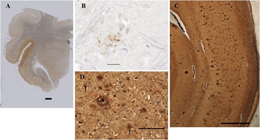
Gadila et al.                                                                                                  Borrelia Spirochetes in Autopsy Specimens

(9, 12). In humans, persistence has been studied early after              sucrose and twice with 30% exchanges. The tissue was then
treatment and in Post-Treatment Lyme Disease (PTLD) patients.             wiped with Kimwipes to remove excess solution and transferred
In one study, skin biopsies were taken from the erythema migrans          into a cryomold (Usplastic, 75642) containing optimal cutting
(EM) lesion and after treatment (∼2 mo later). Approximately              temperature (OCT) compound (VWR avantor, 25608-930) and
1.7% of these were culture-positive and confirmed as the same             were snap-frozen in a dry ice/ethanol bath, then stored at −80◦ C
strain (13, 14). Human xenodiagnoses were also performed in a             until further use. For paraffin-embedded blocks, three infected
more recent study. Larval ticks were placed on patients who had           (A1–A3) and one uninfected (A4) tissue slices were embedded
EM (early stage) or PTLDS (15). Tick samples were evaluated               in paraffin after the PFA washing step. The rest of the three
by PCR and culture; of the 23 patients on whom ticks fed and              infected (C1–C3) and one uninfected (C4) tissue slices that were
were recovered, 19 were negative, 2 were indeterminate, and 2             not treated by any fixative were directly embedded in OCT
were positive by PCR (1 patient with EM and 1 with PTLDS).                compound and were snap-frozen, serving as fresh-frozen control
Two other studies have indicated that the spirochetes could be            blocks. Fixation and cryoprotection limit the extent of ice crystal
cultured from late stage Lyme patients, yet the cultures took many        damage to fine histological detail (20).
weeks and rounds of subculturing without active growth (16, 17).
Thus, in the absence of a reliable detection system, persistent
infection in humans remains difficult to assess. One means to             Human Sample Preparation and Assay Design
address this issue is to interrogate patient tissue for persistent        Brains removed at autopsy were sent, cold but not frozen, to the
pathogen through the analysis of post-mortem specimens.                   Neuropathology Laboratory at the New York State Psychiatric
    In this report, we describe the use of multiple overlapping           Institute. Brain stem and cerebellum were removed from the
techniques, including immunofluorescence assay (IFA), RNA in              fresh brain and midsagittal sectioning was performed to separate
situ hybridization (RNAscope), and PCR for detection of Borrelia          cerebral hemispheres and cut into 2 cm coronal slices. The right
spirochetes in post-mortem tissues. As example, we describe the           hemisphere slices, brain stem, cerebellum, spinal cord, and the
detection of B. burgdorferi in the brain tissue of a post-mortem          rostral 2 cm slices of the left hemisphere were rapidly frozen,
donor from the brain repository of the Lyme and Tick-Borne                either in 1,1,1,2- tetrafluoroethane or in dry ice/acetone. Frozen
Diseases Research Center at the Columbia University Irving                slices were placed in individual plastic bags and stored at −80◦ C.
Medical Center. This individual had a history of Lyme disease             The remaining left hemispheric 2 cm slices were fixed in 10%
that appeared to have been successfully treated with antibiotics;
4 years later developed a neurodegenerative disorder leading
to dementia.

MATERIALS AND METHODS
Preparation of Tissues
NHP Controls
Ex vivo experiments were carried out initially on fresh
brain tissues collected from non-human primates (NHPs) of
Indian origin immediately after euthanasia. These tissues were
collected in phosphate-buffered saline (PBS) of pH 7.2 at room
temperature. These tissues were then sliced into 0.6 cm sections
using a brain tissue slicer and tissue slicing blades and placed in
individual wells of a 12-well plate (Figure 1), containing 2 ml of
RPMI 1640 medium (18). Nine wells (A1–A3, B1–B3, and C1–
C3) out of 12 wells containing the NHP tissue were incubated
with Borrelia burgdorferi strain B31 clone 5A19 spirochetes (19)
at a concentration of 1.0 × 10⁷ /ml, grown in respective culture
medium (18) serving as positive controls. The 12-well plate was
then placed in a 37◦ C incubator with 5% CO2 overnight. Negative
control wells (A4, B4, and C4) with NHP tissue received culture            FIGURE 1 | Non-human primate (NHP) control tissue block preparation
medium alone without any spirochetes.                                      design. 0.6 cm thick sections of NHP control brain tissues were placed in
   On the next day, three of the infected slices from wells B1–            individual wells of 12-well plate containing 2 ml of RPMI 1640 medium. Tissues
                                                                           in wells A1–A3, B1–B3, and C1–C3 were incubated with 1.0 × 10⁷ /ml of
B3 and one uninfected slice from the well B4 were transferred
                                                                           spirochetes per well. Tissues in wells A4, B4, and C4 received culture medium
into a fresh 12-well plate containing 4% paraformaldehyde (PFA)            alone. After overnight incubation, tissues from wells C1–C4 were washed in
in PBS (Fisher scientific, AAJ19943K2) and incubated for 24 h              PBS and excess buffer was removed using kim wipes and tissues were
at 4◦ C. Tissue slices were then washed in 1X PBS three times              embedded in OCT and flash-frozen in dry-ice ethanol bath and stored at
for 1 h each. Later, these fixed tissues were immersed in 10%              −80◦ C. Rest of the tissues in wells A1–A4 and B1–B4 were fixed. Tissues
                                                                           from wells A1–A4 were processed for paraffin-embedded blocks and tissues
sucrose solution made in 1X PBS and let to sink to the bottom              from wells B1–B4 were processed for Fixed-frozen blocks.
of the well, which took ∼24 h. This step was repeated with 15%

Frontiers in Neurology | www.frontiersin.org                          2                                             May 2021 | Volume 12 | Article 628045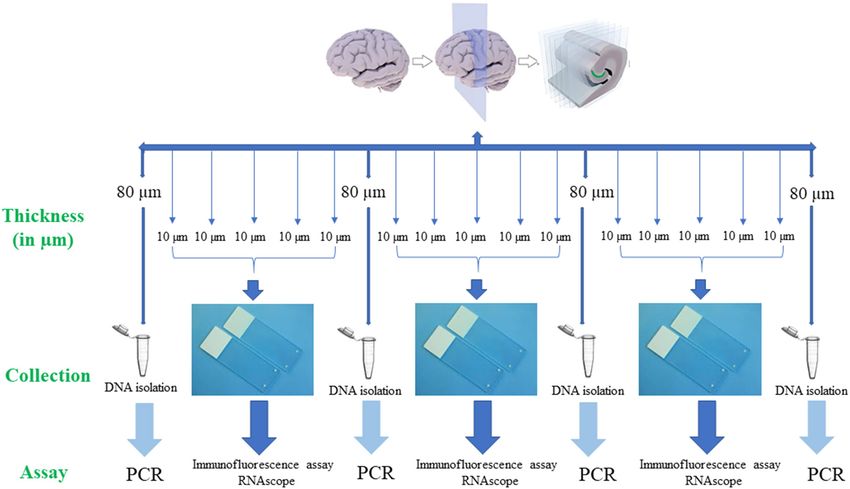
Gadila et al.                                                                                                                Borrelia Spirochetes in Autopsy Specimens

 FIGURE 2 | Human brain tissue samples assay design. Serial sections of fresh-frozen samples/fixed tissue samples were analyzed according to the above design.
 Firstly, 80 µm thickness of the tissue sample was collected into a 1.5 ml microcentrifuge tube for the extraction of the DNA. Next, 5 serial sections each of 10 µm in
 thickness that were adhered to a positively charged glass slide were used for immunofluorescence (IFA) and RNAscope assays. If these tissue slices were negative for
 all the assays, subsequent 6 sections, one of 80 µm and five of 10 µm were used for the analysis. This entire process is repeated to a depth of ∼500 µm−1 mm of the
 tissue. Once the Borrelial DNA was detected using PCR, the rest of the tissue slices were used for both IFA and RNAscope assays.

buffered formalin at room temperature for 5 days, then rinsed                          to dry at room temperature. After drying, slides were kept
and stored at 4◦ C in PBS with 0.02% sodium azide.                                     in slide boxes and stored in a desiccator that was placed at
    For this study, selected regions were dissected from the frozen                    4◦ C until further use. Just before the experiment, sections were
slices over dry ice with a dental drill and shipped from New York                      deparaffinized using a deparaffinizing station (Leica Autostainer
to New Orleans on dry ice. Immediately after receipt, the frozen                       XL) that goes through a series of xylenes followed by graded
slices were embedded in OCT cryomolds as described above                               alcohols. The slides were washed three times with 1X PBS
and stored at −80◦ C until further use. Pieces of formalin fixed                       pH 7.4 for 10 min each while gently rocking in a Coplin jar.
tissue were sent with cold packs. Paraffin blocks were shipped at                      Deparaffinization was followed by a heat-mediated citrate buffer
ambient temperature.                                                                   antigen retrieval (HC-AR) step. For this, slides were immersed
    For PCR, 80 µm thick sections (Figure 2) were used to extract                      in a Coplin jar containing 1X citrate buffer (Vector Labs, H-
DNA from the tissue sample. Subsequent 4 serial sections of                            3300) that was exposed to 5–6 (20 s) cycles of microwave heating
10 µm each were used for immunofluorescence assay to detect                            until the buffer reached 99◦ C. Then, the jar with slides was
B. burgdorferi. Additionally, one section of 10 µm was stained for                     tightly closed and immersed in a water bath maintained at
RNAscope to determine the integrity of the RNA in the tissue                           90◦ C for 15 min. Later, the jar was left at room temperature
sample. If the tissue sample doesn’t express 4–9 dots of PPIB,                         for additional 20 min before proceeding to the next step. Slides
which is considered a standard to determine the RNA quality of                         were then washed and incubated in permeabilization solution
the brain sample, degradation of the RNA in the tissue sample is                       containing 0.2% fish skin gelatin (FSG) (Sigma, G704) and
probable. A total of ∼500–1,000 µm thickness of the tissue was                         0.1% Triton X-100 (TX-100) made in 1X PBS for 1 h at room
analyzed with these multiple methodologies to evaluate the brain                       temperature under gentle shaking. TX-100 was washed from the
tissue samples for borrelia spirochetes.                                               slides using PBS/FSG buffer. Slides were then placed in a dark
                                                                                       humidifying staining chamber, the tissue sections were circled
Immunofluorescence Protocol                                                            with a hydrophobic pen and the tissue was covered with blocking
PFA-treated tissues that were embedded in paraffin were                                buffer (10% Normal Goat Serum-Gibco 16-210-072, in PBS
sectioned at 5 µm in thickness and were mounted on positively                          containing 0.2% FSG) for a minimum of 1 h at room temperature.
charged slides (Fisher Scientific). The slides were then allowed                       After blocking, tissue sections were incubated with primary

Frontiers in Neurology | www.frontiersin.org                                       3                                             May 2021 | Volume 12 | Article 628045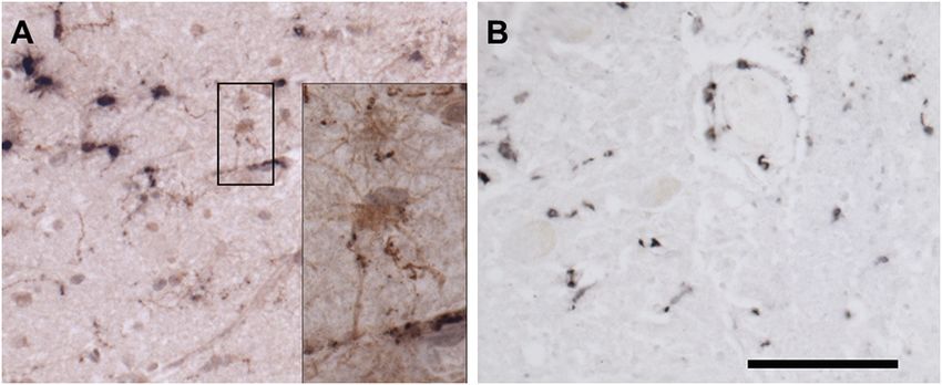
Gadila et al.                                                                                              Borrelia Spirochetes in Autopsy Specimens

antibody rabbit polyclonal anti–B. burgdorferi [derived from a             together possess a 28-base hybridization site for the preamplifier,
hyperimmunized rabbit 6 weeks after inoculation with in vitro              which in turn contains 20 binding sites for the amplifier.
propagated B. burgdorferi strain B31 (21)] for 1 h followed by             These amplifiers contain 20 binding sites for the label probe,
TX-100 buffer and PBS/FSG washes. Slides were then incubated               yielding a total of 8,000 labels for a target RNA molecule of
with secondary antibody Goat Anti-rabbit IgG-Alexa Fluor 488               1,000 bp (23). The single Z probe binding doesn’t affect the
(Thermo Fisher Scientific, A-11034) diluted 1:500 in blocking              signal amplification. The pre-amplifiers won’t bind to the single
buffer. Slides were washed as after incubation with primary                Z probe, preventing the amplification of non-specific signals,
antibody, then partially dried by tapping the glass edge onto a            contributing to the specificity.
paper towel. Finally, to enhance the specificity of the fluorescence           Tissue samples that were positive for both PCR and
signal, a final incubation step with TrueBlack Lipofuscin                  Immunofluorescence were tested with RNAscope. Three serial
Autofluorescence Quencher (Biotium, 23007) was performed                   tissue 10 µm sections adjacent to the section that was positive
(22). For this, slides were incubated with 1X TrueBlack solution           for immunofluorescence, were used. These three sections were
for 30 s. Only one slide at a time was processed for this step.            incubated with hydrogen peroxide at room temperature for
After 30 s, the quencher was rinsed off of the slides with PBS.            10 min and washed three times with distilled water for 1 min
Under gentle rocking, slides were then washed three times in               each. Next, slides were immersed in the antigen retrieval solution
PBS (22). Slides were then counterstained with DAPI for 10 min             that was pre-boiled to a temperature of 99◦ C. slides were left
to stain nuclei (EMD millipore, 2160) and mounted with anti-               in the solution for 15 min. After the treatment, slides were
quenching solution (Thermofisher, P36934) and coverslipped.                washed in distilled water for 15 s and were transferred to 100%
To prevent the decay of fluorescent signal intensity, slides were          alcohol and let sit for 3 min. Next, slides were placed at 60◦ C
examined and photographed within 1 week of completion of the               oven until dried and a hydrophobic barrier was created around
staining procedure.                                                        the tissue using a pap pen. Once the barrier was completely
    For Fixed-frozen samples, tissues were sectioned at a thickness        dried, tissue sections were covered with protease III solution
of 10 µm on a cryostat and mounted onto positively charged                 and incubated at 40◦ C for 30 min in the EZ Hybridization oven
slides. These sections were then dried in −20◦ C freezer                   (ACD, 321710) using the humidity control tray (ACD, 310012)
overnight. Slides were then individually wrapped in aluminum               and slide rack (ACD, 310017). A wet humidifying paper was
foil and stored at −80◦ C until further use. On the day of staining,       placed in the humidity control tray to maintain the humidity.
slides were thawed at room temperature for 20 min followed by              After the protease treatment, slides were washed 3 times with
three 1X PBS washes each for 10 min each. Then slides were                 distilled water 1 min each. Later, we used a 3-plex positive
introduced directly into the antigen retrieval solution as just            control probe, a mixture of three endogenous housekeeping
described, and similar steps were followed for the entire staining         probes-UBC (ubiquitin C), PPIB (cyclophilin B), and Polr2A
process. For fresh-frozen tissue blocks, sections were cut at a            (DNA-directed RNA polymerase II RPB1), each assigned to an
thickness of 10 µm and stored similarly to fixed-frozen slides. On         individual detection channel, to determine the RNA integrity
the day of staining, after PBS washes, slides were immersed in             of the tissue sample on one slide. dapB was used as a negative
4% paraformaldehyde for 20 min at 4◦ C. Slides were then washed            control probe (ACD, Bacillus subtilis dapB, 320871) on a slide and
three times with 1X PBS followed by permeabilization step and              a probe against Borrelia burgdorferi (ACD, 23S rRNA transcript,
remaining staining process is as described above.                          468211) was added to an individual slide and incubated in the
    Imaging was performed using a Nikon Ti2-E motorized                    hybridization over as mentioned above for 2 hrs. After probe
fluorescence microscope equipped with pco.edge SN:61005789                 hybridization, amplification and detection steps were performed.
camera, Plan Apo λ 40× objective with a numerical aperture 0.95            Signal amplification is a branched hybridization of amplifiers that
and refractive index 1.000, and a final image with a resolution            are unique to each individual channel. Amplifier 1 was initially
of 2,048 × 2,044 pixels. Control NHP slides and experimental               incubated on slides for 30 min at 40◦ C and washed with wash
autopsy tissue slides were imaged during the same session                  buffer three times each for 2 min. Slides were then incubated with
with identical acquisition parameters. Fluorescence intensity was          Amplifier 2 for 30 min as before, washed, and then incubated with
optimized on control tissues to eliminate the tissue background            a final layer of amplifier, Amplifier 3, and incubated for 15 min at
and remained constant for all the experimental slides. When                40◦ C and washed as before. For development of the signal, HRP
made, adjustments to brightness, contrast, or color balance were           (horseradish peroxidase)-C1 was added to the tissue to target the
applied to the whole image.                                                probe assigned to channel C1 and incubated in the hybridization
                                                                           oven for 15 min at 40◦ C. Finally, slides were incubated with
RNAscope (RNA in-situ Hybridization)                                       fluorescent molecules from PerkinElmer [Tyramide Signal
RNAscope is commercially available from Advanced Cell                      Amplification (TSA) plus fluorescein: NEL745001KT or TSA
Diagnostics, Inc. (ACDBio) (23). RNAscope was performed                    plus Cyanine 3 system: NEL744001KT or TSA plus Cyanine
according to the manufacturer’s guidelines from RNAscope                   5 system: NEL745001KT]. These fluorescent molecules bind to
Multiplex Fluorescent Detection Kit version 2 (323100-USM).                the cascade of signal amplification molecules resulting in the
Typically, the probes provided in the kit hybridize to the                 signal detection. Similarly, HRP-C2 and HRP-C3 were incubated
specific target RNA molecule. These probes contain 20 ZZ                   on the tissue for the signal development of probes targeted on
probe pairs (50 bp/pair), each pair hybridizes contiguously to             channel 2 and channel 3. Individual channels were assigned
the target region, covering ∼1,000 bp. Each “ZZ” tail sequence             different fluorophores.

Frontiers in Neurology | www.frontiersin.org                           4                                       May 2021 | Volume 12 | Article 628045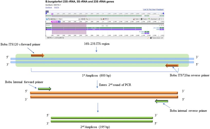
Gadila et al.                                                                                             Borrelia Spirochetes in Autopsy Specimens

Conventional and Nested Polymerase Chain                                   Case Study Description
Reaction (PCR)                                                             This 69 year old woman (Patient 12,577) contracted Lyme
Genomic DNA from the tissue samples was extracted using                    disease at age 54 with a well-documented erythema migrans
QIAamp DNA FFPE kit (Qiagen, 56404). Initially, slides were                rash accompanied by a severe headache, joint pains and a fever
deparaffinized using Leica Autostainer X and tissue sections were          of 104; convalescent serologies were positive on ELISA and
scrapped into a sterile microcentrifuge tube using sterile blades.         both IgM and IgG Western blots. Treatment with doxycycline
For each preparation, tissue sections with a thickness of 80 µm            for 10 days led to symptom resolution. Two years later, a
and a surface area of up to 250 mm2 was combined in a tube.                sleep behavior disorder emerged. Four years later, cognitive
Usage of the tissue section beyond this recommendation leads               problems (processing speed, mental tracking, and word-finding)
to clogging of the purification column. The tissue sections in             emerged and gradually worsened. Other symptoms included
the tube were then resuspended in a mixture containing 180                 photophobia, paresthesias, fasciculations, and myoclonic jerks.
µl of ATL and 20 µl of proteinase K and incubated at 56◦ C                 Neurocognitive testing revealed deficits in visuospatial skills and
for 1 h to overnight until the liquid in the tube appeared clear.          executive functions with preservation of verbal skills, suggesting
The microcentrifuge tube was then incubated at 90◦ C for 1 h               a neurodegenerative process. Brain Magnetic Resonance Imaging
to reverse the aldehyde cross-linking. After this step, 200 µl             with and without contrast showed mild atrophy and non-specific
of AL buffer was added and mixed thoroughly by vortexing,                  scattered white matter hyperintensities without enhancement.
and an additional 200 µl of (96–100%) ethanol was added and                Brain Single Photon Emission Computed Tomography scans
mixed. The lysed samples were then loaded onto a QIAamp                    showed decreased perfusion in the right posterior parietal and
minElute column and centrifuged for 1 min. The columns were                temporal lobes. Serum was negative or normal for erythrocyte
then placed in a new collection tube. Next, two column wash                sedimentation rate, c-reactive protein, antinuclear antibody, and
steps were performed to remove any unwanted material. The                  thyroid stimulating hormone. PCR assays of blood for Bartonella
column was then placed in a new collection tube and centrifuged            henselae, Babesia microti, and Borrelia burgdorferi were negative.
at 14,000 rpm for 2 min to remove any residual buffer present              Serum C6 ELISA was negative but Lyme IgG Western blot
in the column and then was dried. Finally, the dry column                  was positive with 9/10 bands. Treatment with IV ceftriaxone at
was placed in a new sterile microcentrifuge tube and incubated             age 60 for 8 weeks led to 60% improvement in cognition and
with 50 µl of pre-warmed (37◦ C) nuclease-free water for 5 min             interpersonal engagement; oral amoxicillin 500 mg three times
and centrifuged at full speed for the elution. The isolated pure           daily was continued for 6 months after the IV treatment. The
DNA was quantified using a Nanodrop 2000 spectrophotometer                 initial improvement was not sustained and subsequent antibiotic
(Thermo scientific).                                                       therapy with minocycline was of no clear benefit; gradually her
   The DNA samples were initially tested with standard PCR                 visual spatial skills and executive functions deteriorated further,
using primers designed to amplify Borrelia burgdorferi 16S−23S             and anxiety worsened. Serum IgG Western blot continued to be
ITS region of ribosomal DNA (24). For this, Q5 Hot start High-             positive. At age 62, a cerebrospinal fluid study demonstrated 4
Fidelity DNA polymerase (NEB, M0493L) was used. Initial PCR                CSF IgG bands on Lyme Western blot; unfortunately, because
was performed using a total of 300 ng of isolated DNA as a                 CSF and serum ELISA studies were not conducted, intrathecal
template. Negative control included the DNA extracted from                 Bb specific antibody production could not be assessed. Other
uninfected NHP brain tissue and DNA from B. burgdorferi                    CSF studies were unremarkable including absence of pleocytosis
incubated NHP brain tissue was used as positive control. The               or elevated protein, absence of P-tau elevation, Venereal
primers used for the initial conventional PCR were F: Bobu                 Disease Research Laboratory assay, Acid-Fast bacteria, fungi, and
ITS120 s 5′ AGGTCATTTTGGGGGTTTAGCTCAGTTGGCT                                negative Herpes Simplex Virus and Epstein-Barr Virus PCRs.
3′ and R: Bobu ITS720 as 5′ AGTGTCGGGCAAATCCAAACT                          A second brain MRI showed periventricular and subcortical
GAAATCTG 3′ (24). The thermocycling conditions were: Initial               T2 hyperintensities possibly due to “small vessel ischemia or
denaturation 98◦ C for 30 sec, followed by 55 cycles of 98◦ C/10 s,        demyelinating disorders like Lyme disease.” Fluorodeoxyglucose-
65◦ C/15 s, 72◦ C/18 s and a final extension of 72 C for 2 min.            Positron Emission Tomography scan showed “diffuse cortical
The second round of PCR was performed similar to the first                 hypometabolism, worse in the posterior parietal and temporal
reaction except that, two different primers that are internal to the       lobes, with sparing of the sensory motor cortex and visual cortex
Borrelia burgdorferi 16S−23S ITS region were used to increase              bilaterally—findings consistent with Alzheimer’s disease.” The
the sensitivity and specificity of the PCR (Figure 3). For this, 2         extensive workup at that time led to the diagnoses of both a
µl of the first PCR product was used as template DNA. Primers              REM behavioral disorder with verbalizations and movements
used were F: 5′ ATTAAGAAAAATGTCTAGA AGCAAAAGC                              and a neurodegenerative dementia characterized by expressive
AAGCT3′ and R: 5′ TACAATACTTGTCCTTCTCTCAGACAT                              aphasia, visual agnosia, anomia, deficits in executive function
CA 3′ . Reactions were setup as before. The thermocycling                  and calculation, and mild memory problems. Eventually, she
condition were initial denaturation 55 cycles of denaturation              developed severe oral dystonia, making feeding progressively
98◦ C for 30 s, followed by 55 cycles of 98◦ C/10 s, 55◦ C/15 s,           more difficult; she died 15 years after the initial infection with
and 7 s @ 72◦ C/7 s and a final extension of 72◦ C for 2 min.              B. burgdorferi. Early and severe movement disorders, REM
For negative and positive controls, 2 µl of the initial reaction           behavioral disorder, paranoia, and personality changes all favored
was used.                                                                  a clinical diagnosis of dementia with Lewy bodies.

Frontiers in Neurology | www.frontiersin.org                           5                                      May 2021 | Volume 12 | Article 628045
Gadila et al.                                                                                                                 Borrelia Spirochetes in Autopsy Specimens

 FIGURE 3 | DNA Primers targeting the 16S−23S internal transcribed spacer (ITS) region of Borrelia burgdorferi B31 strain. Bobu ITS120 s forward and Bobu
 ITS720as reverse primers were targeting the region of ITS of the Borrelia genomic sequence with the accession number X85745.1 from 120 to 720 bp making the first
 amplicon of 600 bp. These primers were designed and tested previously on Ixodes affinis and Ixodes scapularies tick species that are widely found in coastal plain
 region of North Carolina. To increase the specificity and sensitivity, nested primers were designed. Bobu Internal forward and Bobu internal reverse primers amplify the
 initial amplicon to generate a second amplicon size of 195 bp.

Human Control Tissues                                                                   numerous immunoreactive Lewy bodies and fibers in substantia
Tissue blocks from various regions of seven specimens from                              nigra, hippocampal formation and neocortex, Figures 4C–E).
brains of deceased Macedonian residents that were housed                                IHC for hyperphosphorylated tau (monoclonal antibody AT8;
in the Macedonian/New York State Psychiatric Institute Brain                            ThermoFisher) revealed intense staining of many limbic
Collection were used as controls. Though none had a clinical                            neurofibrillary tangles and neuropil threads (Braak stage 2–3,
history of Lyme disease based on interview with the surviving                           Figure 5), and of occasional neurofibrillary tangles in neocortex,
family members, Borrelia is endemic in Macedonia. These brain                           but senile plaques were extremely rare, and each contained
tissues were probed in the same manner as the human case study                          only a few fibrils (Figure 5). H&E showed prominent thickening
with IFA and PCR-based detection methods.                                               of small blood vessels in gray and white matter, extensive
                                                                                        mineralization of pallidal vessels, and rare microglial nodules
                                                                                        in the hippocampal formation. Immunohistochemistry for Iba-
RESULTS                                                                                 1 (Wako), CD68 (clone KP1, Dako), and CD163 (clone EDHu-1;
                                                                                        Bio-Rad) showed moderate numbers of activated microglia and
The Case Study Pathology Is Characteristic                                              large numbers of macrophages in hippocampal formation and
of Dementia With Lewy Bodies (DLB)                                                      spinal cord (Figure 6). In summary, we see DLB accompanied by
The fresh brain weighed 996 g and appeared atrophic Coronal                             features of Alzheimer’s disease, a common presentation.
sections through the left cerebral hemisphere and brain
stem revealed mild enlargement of the lateral ventricle,
particularly the temporal horn. The substantia nigra was                                Nested PCR Provides Sensitive Detection
normally pigmented or nearly so. Microscopically, nigral and                            for Borrelia sp. DNA
cortical Lewy bodies, were seen with hematoxylin and eosin                              To determine the presence of Borrelia burgdorferi, we first
stain (H&E, Figures 4A,B). Immunohistochemistry (IHC) for                               investigated the presence of DNA in the brain samples. To
α-synuclein (clone 42, BD Transduction Laboratories) showed                             this end, nested PCR was adapted to increase the sensitivity

Frontiers in Neurology | www.frontiersin.org                                        6                                              May 2021 | Volume 12 | Article 628045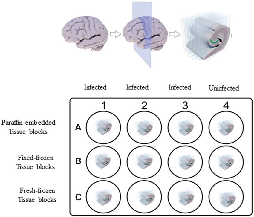
Gadila et al. Borrelia Spirochetes in Autopsy Specimens
FIGURE 6 | Immunohistochemical staining for microglia. (A) Frontal cortex
stained for Iba-1 (brown) and CD68 (black). Most microglia in this field contain
large deposits of CD68 immunoreactivity and shirt, thick processes, indicating
activation. By contrast, occasional surveilling microglia, are also present. The
black box surrounds two such cells, whose somas and long, fine processes
are apparent in the inset. (B) Spinal cord stained for CD163. CD163 is
expressed by resident perivascular macrophages, which are normally present,
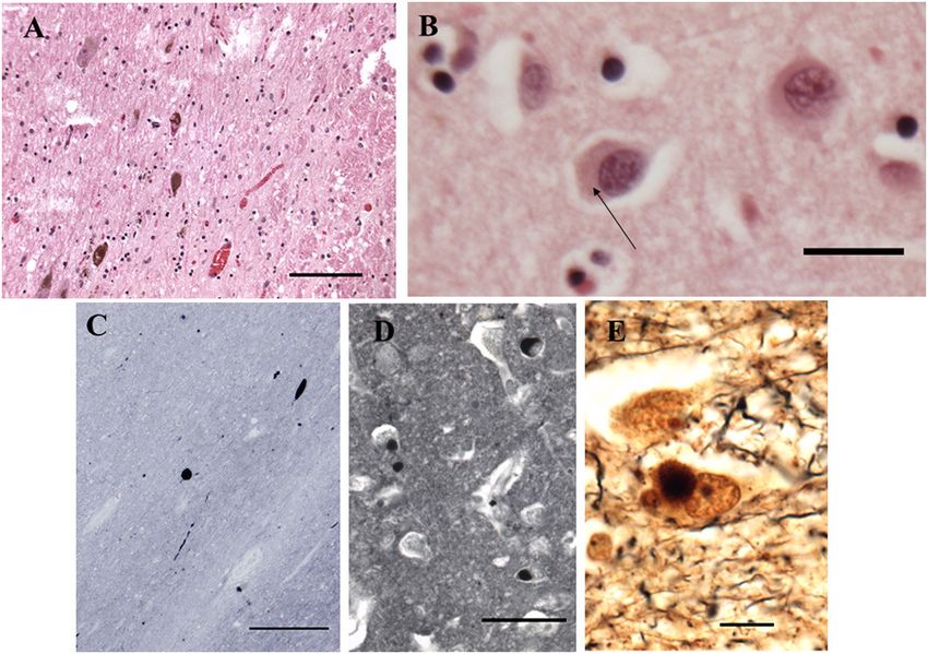
FIGURE 4 | Histopathological findings from the case study. (A) Substantia but also by macrophages derived from circulating monocytes. Bar = 100
Nigra. H&E. Disintegration of pigmented neurons, with Lewy bodies in two of microns for (A,B). Inset is photographed at three times greater magnification in
those remaining (center and lower left). Bar = 100 microns. (B) Frontal cortex. a stack spanning the thickness of the tissue at 0.25-micron intervals. The
H&E. A Lewy body is seen in the central neuron as a perinuclear region of “Deep Focus” feature of Neurolucida 360 (MBF Bioscience) was used to
increased eosinophilia (arrow). Bar = 20 microns. (C–E) combine the best focused objects from each layer of the stack into a
Immunohistochemistry for α-synuclein. (C) Substantia nigra pars compacta. single layer.
Inclusions are seen in cell bodies and neurites. Bar= 200 microns. (D) Hilus
(CA3) of hippocampus. Bar = 50 microns. (E) Bielschowsky stain,
hippocampus. Central neuron contains a Lewy body. Bar = 10 microns.
FIGURE 7 | Non-human primate (MJ 61) frontal cortex tissue was incubated
with Borrelia burgdorferi ex-vivo. These ex-vivo tissues were fixed, sectioned
at 10 µm, and stained against Borrelial spirochetes. (A,B) Are representative
images of positive staining of Borrelial spirochetes. White arrows were pointed
toward Borrelial spirochetes that were stained with primary rabbit polyclonal
FIGURE 5 | Detection of amyloid plaques in the patient’s CNS. (A) Amygdala anti-Borrelia antibody and secondary Goat anti-rabbit IgG antibody conjugated
and rostral hippocampus, entorhinal cortex, and transentorhinal cortex. AT8 to Alexafluor 488. To increase the signal intensity, (B) was incubated with
immunohistochemistry with Verhoeff myelin counterstain. BAR = 2 mm. (B) Autofluorescence quencher after completion of immunofluorescence that
Rare cortical neuritic plaque in middle frontal gyrus. AT8 with Verhoeff myelin caused a significant decrease in the autofluorescence caused by lipofuscin
counterstain. BAR = 20 microns. (C) Bielschowsky stain of CA1 and and aldehyde fixation. Scale bars−10 µm. Image was acquired with a Nikon
subiculum demonstrates numerous amyloid plaques. Bar = 1 mm. (D) fluorescence microscope using a 40x objective.
Bielschowsky stain of frontal cortex. Amyloid plaques predominate; rare
neuritic plaques are also present (arrow). Bar = 100 microns.
Immunofluorescent Detection Is the Most
and specificity of the assay (Supplementary Table 1). DNA Reliable Method to Detect Spirochetes in
isolated from frontal cortex of an uninfected non-human Fixed Tissues
primate (NHP) was used as a negative control and as a To strengthen the results of our PCR, we next explored
positive control, DNA isolated from NHP frontal cortex tissue immunofluorescence assay (IFA) to see if these tissue specimens
that was incubated with B. burgdorferi spirochetes was used contain morphologically intact spirochetes. Initially, we tested
(Supplementary Figure 1). To prevent cross-contamination our rabbit polyclonal antibody on NHP frontal cortex tissue
between positive control and autopsy specimens, DNA from that was incubated with borrelial spirochetes to see the
autopsy tissue samples was isolated in laboratories that were specificity of the antibody (Figure 7A). The image cleanliness
never exposed to B. burgdorferi DNA. was increased after incubating the tissue with the lipofuscin
Frontiers in Neurology | www.frontiersin.org 7 May 2021 | Volume 12 | Article 628045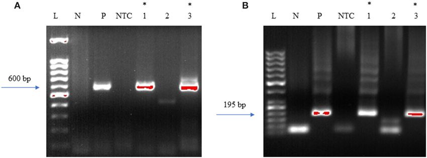
Gadila et al. Borrelia Spirochetes in Autopsy Specimens
quencher that quenches the fluorescence of lipofuscin aggregates, fixed; under-fixation leads to tissue deformation, shrinkage and
which are formed during normal aging process (Figure 7B). autolysis (25), whereas over-fixation may permanently mask
Secondary antibodies were tested with isotype controls for the antigen, abolishing its detection even after antigen-retrieval
specificity on human (Supplementary Figure 2A) and macaque steps (26).
(Supplementary Figure 2B). Optimal fixation time is very
crucial for the success of the IFA. Tissues must be properly RNAscope Is a Sensitive Technique for
Finding Viable Pathogen but Requires
Freshly Prepared Tissue
Next, to determine if the persistent Borrelia spirochete was
actively dividing, we probed the tissue with RNA probes from
FIGURE 8 | Validation of RNA integrity on Fresh frozen NHP (MJ 61) frontal
cortex brain tissue using RNAscope assay. NHP tissue was stained with a
3-plex positive control probe POLR2A targeting DNA-directed RNA
polymerase II RPB1, PPIB targeting cyclophilin B, and UBC that targets
Ubiquitin C. In terms of relative expression levels, UBC is highest, PPIB is
considered a moderate-high, POLR2A is moderate-to low expressor target.
Successful staining has 4–9 dots per cell of PPIB/POLR2A and UBC of 10–15 FIGURE 10 | Validation of RNA integrity on a fresh-frozen tissue sample from
dots per cell. PPIB is considered a standard to determine the RNA quality of patient 12,577 autopsy tissue using RNAscope assay; (A) = merged. Tissue
brain samples. (C) Represents red fluorescence, indicating the specificity of was stained with a 3-plex positive control probe POLR2A targeting
PPIB, whereas, (D) displays a far-red puncta targeting UBC gene. Absence of DNA-directed RNA polymerase II RPB1, PPIB targeting cyclophilin B, and
POLR2A signal in (B) represents, quality of RNA being partially compromised. UBC that targets Ubiquitin C (D). Absence of signal in POLR2A (B) and only 2
A merge image of all the channels can be seen in (A). White arrows were puncta of PPIB per cell (C) indicates that the quality of the RNA has been
pointing toward the fluorescence puncta and orange arrows toward compromised. White arrows were pointing toward the fluorescence puncta
autofluorescence of the tissue. and orange arrows toward autofluorescence of the tissue.
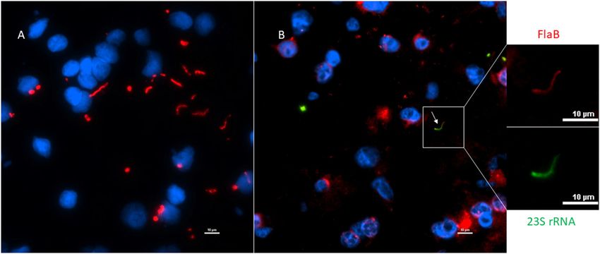
FIGURE 9 | Technical validation of RNAscope assay performed on NHP (MJ 61) frontal cortex brain tissue cultured with Borrelia burgdorferi. (A) Fixed-frozen tissue:
Spirochetes are highlighted by an intense red (cy3, PerkinElmer catalog number NEL744001KT), indicating strong specificity of B. burgdorferi probe (B. burgdorferi
probe targeting the 23S rRNA transcript; Advanced Cell Diagnostics catalog number 468211). (B) Fresh-frozen tissue: RNAscope assay combined with
Immunofluorescence (IFA). Initially, spirochetes were probed with 23S rRNA and detected using fluorescein fluorophore (PerkinElmer catalog number NEL741001KT).
To counterstain the spirochetes with IFA, tissue was incubated with primary anti-FlaB mouse monoclonal antibody H9724 and goat anti-mouse IgG2a Alexa Fluor 594
(Thermofisher catalog number A-21135). White arrow indicates an intact spirochete showing the colocalization of RNAscope probe with monoclonal antibody with
subsequent green and red fluorescence.
Frontiers in Neurology | www.frontiersin.org 8 May 2021 | Volume 12 | Article 628045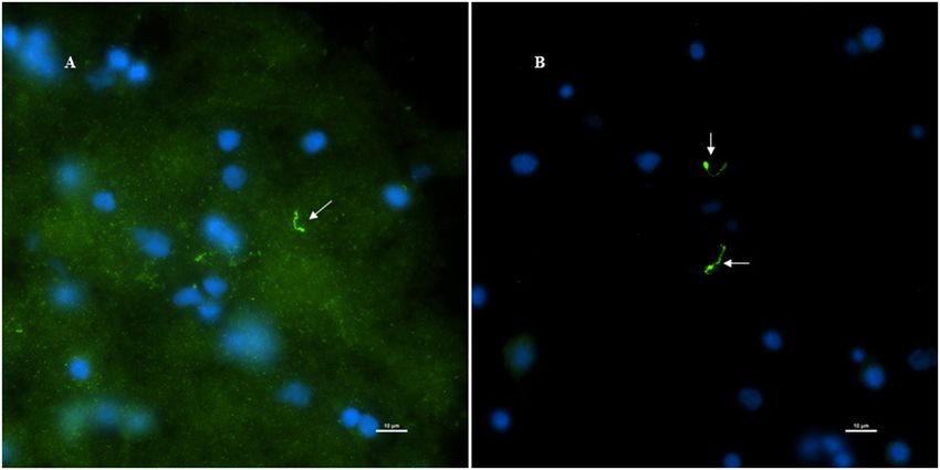
Gadila et al.                                                                                                              Borrelia Spirochetes in Autopsy Specimens

 FIGURE 11 | Primers amplify Borrelia 16S−23S internal transcribed spacer (ITS) region. Bobu primers generate an amplicon size of 600 bp. To increase the specificity
 Bobu internal primers were designed to generate an amplicon size of 195 bp. (A) External PCR: L-molecular weight marker of 100 bp, N-negative control, P-positive
 control, NTC-No Template Control, 1–12,577 amygdala, 2–12,577 pons, 3–12,577 spinal cord. (B) Internal PCR: L-molecular weight marker of 50 bp, N-negative
 control, P-positive control, NTC-No Template Control, 1–12,577 amygdala, 2–12,577 pons, 3–12,577 spinal cord. *Samples that were positive to Borrelial DNA.

ACDBio. First, to optimize the protocol, the probes were tested                       internal transcribed spacer (ITS) DNA region of Borrelia found
on NHP control tissue (Supplementary Figure 3). To determine                          in Ixodes affinis and Ixodes scapularis (24). Out of the three
the RNA integrity of the NHP tissue, 3-plex positive control                          tissue regions that were tested for PCR, amygdala and spinal
RNA probe was used on NHP tissue. Figure 8 shows the staining                         cord were PCR positive for Borrelia DNA (Figure 11A). This
of frontal cortex region of NHP fresh frozen tissue, where,                           initial amplicon of 600 bp was used for the second round of
white arrows in Figures 8C,D were pointed toward the positively                       PCR using primers that were designed and tested in-house. For
stained PPIB dots and UBC dots, respectively. PPIB is considered                      this, 2 µl of the initial reaction was used as a template DNA.
a standard to determine the RNA quality of the brain samples.                         As expected, amygdala and spinal cord tissues were positive
However, the low expresser target POLR2A was not detected in                          with the nested PCR (Figure 11B). To avoid cross-contamination
these tissues indicating partial degradation of RNA. The reason                       during reaction setup, a no template control was included.
behind this could be that the tissues were processed after 16 h                       Amplicons from all three of the positive PCR reactions were
of incubation with the Borrelia spirochetes. Next, to test the 23S                    sequenced and the results are shown in Supplementary Table 3.
rRNA probe that targets the Borrelial RNA, the frontal cortex                         According to the BLAST, these sequences were aligned with 98–
region of NHP fresh frozen tissue and fixed tissue that were                          99% identity with a query coverage in between 48 and 62%
incubated with Borrelia spirochetes was used. Red fluorescence                        to different isolates of B. burgdorferi. Our results demonstrated
in Figure 9A represents Borrelia spirochetes in NHP fixed tissue.                     specificity with the nested primers and some amplification
Whereas, tissue in Figure 9B is a representative image obtained                       from human control tissue. Other than the case study, one
by combining RNAscope with the immunofluorescence assay.                              tissue from seven Macedonian control autopsy specimens was
Initially, the tissue was probed with the 23S rRNA probe and                          positive by both primer sets (Supplementary Figure 4 and
detected using fluorescein fluorophore. Next, the same tissue was                     Supplementary Table 2). As mentioned, Borrelia infection is
counterstained for IFA assay using the borrelia specific anti-FlaB                    endemic in the region, so some of those individuals may have
monoclonal antibody H9724 (27, 28) and Goat anti-mouse IgG2a                          harbored an asymptomatic or unrecognized persistent infection.
secondary antibody conjugated to Alexa Fluor 594. The inset                           Immunofluorescent staining of the control patient tissue did not
in Figure 9B showing an intact spirochete that is labeled green                       reveal obvious spirochetes.
with RNAscope assay and red with IFA assay. After optimizing                              Tissue slices adjacent to the autopsy tissue specimens that were
the positive control probes and Borrelia specific RNA probes on                       positive for DNA were stained for the presence of persisters. For
NHP tissues, the positive control probe was used to determine                         this, slides were stained with the primary rabbit polyclonal anti–
the integrity of RNA in human autopsy samples. For this, fresh-                       B. burgdorferi antibody and a goat anti-rabbit IgG-Alexa Fluor
frozen tissue was used. Figure 10 is a representative image of the                    488 secondary antibody. An intact Borrelia spirochete with the
positive control probe showing only 5–6 dots of UBC and one dot                       expected morphology was identified within fixed tissue of the
of PPIB per cell indicating that the RNA integrity of the sample                      spinal cord (Figure 12). The spirochete appeared to be adjacent
has been declined and failed QC for RNAscope.                                         to vasculature.
                                                                                          Next, RNAscope was tested on serial sections of the tissue
The Patient Harbored Borrelia in the Amygdala and                                     where the persistent spirochete was detected by IFA. However,
Spinal Cord                                                                           positive staining of the Borrelia with 23S rRNA probe was
Initially, the first round of PCR was done on the DNA extracted                       not detected. Absence of the concurrent RNA staining could
from fresh-frozen tissues of amygdala, pons, and spinal cord,                         be due to multiple factors, the most prominent of which
using primers that were previously used to target the 16S−23S                         being delayed sample collection after the death. According to

Frontiers in Neurology | www.frontiersin.org                                      9                                             May 2021 | Volume 12 | Article 628045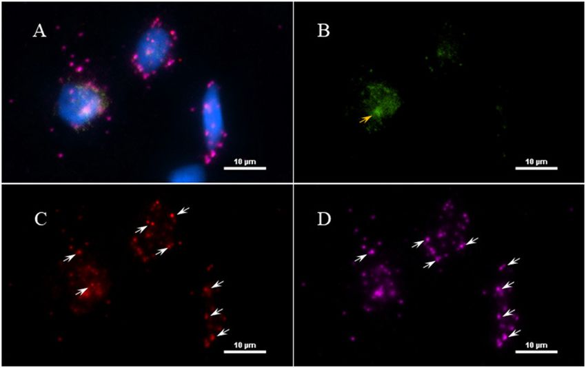
Gadila et al.                                                                                                      Borrelia Spirochetes in Autopsy Specimens

                                                                                   availability of tissue may be a challenge, the role of Borrelia
                                                                                   burgdorferi in the etiology of chronic neurological disease, can
                                                                                   be studied as a “proof of principle.”
                                                                                       Our study confirms that Borrelia burgdorferi was detected in
                                                                                   the brain and spinal cord tissue of this patient with a history of
                                                                                   previously treated Lyme disease. These results however do not
                                                                                   clarify whether the Borrelia infection had anything to do with
                                                                                   her progressive neurodegenerative disorder. It is possible this
                                                                                   is an unrelated incidental finding or that there is a relationship
                                                                                   between CNS infection with Bb and the development of a
                                                                                   neurodegenerative dementing disorder.
                                                                                       Previous studies suggest that Borrelial spirochetes can start
                                                                                   invading the nervous system during early stages of the infection
                                                                                   resulting in meningeal seeding (29), and this later leads to
                                                                                   neuroborreliosis. To define the pivotal neurological deficits, a
                                                                                   study in Europe examined the clinical manifestations of 68
                                                                                   patients hospitalized for neuroborreliosis. Meningitis was found
                                                                                   to be one of the least frequent conditions, present in 6% of the
                                                                                   patients (30), whereas cranial neuritis was the most frequent
                                                                                   (25%). The clinical Lyme case presented here was documented
                                                                                   with meningismus at the time of the EM rash, supporting
 FIGURE 12 | Detection of an intact Borrelia spirochete in a fixed autopsy
                                                                                   the possibility of mild meningitis at early infection. Bacterial
 tissue using Immunofluorescence assay. 12,577-spinal cord autopsy tissue          meningitis leading to cognitive impairment was well-studied in
 was immunostained with rabbit B. burgdorferi hyperimmunized polyclonal            Treponema pallidum in relation to dementia (31). B. burgdorferi
 (primary) and goat anti-rabbit IgG Alexa Fluor 488 (secondary) antibodies.        infection has also been associated with mild (32) to severe (33)
 White arrow points toward a green fluorescence of morphologically intact
                                                                                   cognitive deficits. In the endemic areas of Lyme disease, Borrelia
 spirochete. Scale bar−10 µm. Image was acquired with a Nikon fluorescence
 microscope using a 40x objective.                                                 infections as a possible cause of cognitive impairment has to be
                                                                                   carefully considered.
                                                                                       Neurotropic viruses have been associated with
                                                                                   neurodegenerative syndromes, as have spirochetal infections
the manufacturer’s instructions, the post-mortem tissue sample                     (34–38). Precedence for an association between B. burgdorferi
should be collected within 12 h after the death to preserve                        infection, specifically, and dementia exist (38–42), however there
the RNA integrity and tissues collected after 24 h would lend                      are also reports that have failed to link B. burgdorferi to AD (43).
decreased sensitivity. Overfixation of the tissue may also decrease                Evidence that amyloid plaques may have a functional protective
detection sensitivity. The optimal fixation of the autopsy tissue                  role in combatting microbial infection has also come to the fore
specimen would be between 16 and 32 h in freshly prepared                          (44). Evidence that Borrelia can induce amyloid production
10% neutral buffered formalin solution or 4% PFA at room                           is suggestive of a possible mechanism for development of
temperature. However, thick brain slices require a longer period                   AD (45–47).
of fixation. Considering the low copy number of the Borrelia                           To comprehensively evaluate the possible role of Borrelia
transcripts present in the autopsy specimens, this assay would                     in dementia (Alzheimer’s and LB), 20 patients were identified
be highly dependent on tissue preparation techniques to preserve                   from a total of 1,594 patients who were seen for dementia,
RNA integrity.                                                                     who had positive intrathecal anti-Borrelia antibody index (AI),
                                                                                   indicative of past or present Lyme disease (48). Among these
DISCUSSION                                                                         20 patients, 7 patients with neuroborreliosis dementia showed
                                                                                   stability or mild improvement in their cognitive functions after
Two reasons exist for the interrogation of autopsy specimens                       treatment with ceftriaxone, and the others showed progressive
for the Lyme disease spirochete. First, in patients with a known                   worsening despite antibiotic treatment (48). The individual
history of Lyme disease and a record of antibiotic treatment,                      in our clinical case reported 60% cognitive improvement
the potential for treatment to fail in eradicating the infection                   after the antibiotic treatment. However, this improvement was
can be evaluated. Notably, a detailed patient history, including                   not sustained and cognition gradually worsened, a finding
history of possible second B. burgdorferi infection and treatment                  consistent with a previous study demonstrating cognitive
non-compliance, is necessary. Given the difficulty in recovering                   functional deficits in treated Lyme neuroborreliosis patients (49).
organisms from living people, looking at post-mortem tissue can                    The possible anti-inflammatory effects of antibiotic cannot be
provide some resolution on the issue of persistence. Secondly,                     discounted (50).
patients such as the one presented here, can manifest neurological                     A recent study aimed at testing the hypothesis that
disease that may or may not be related to infection. Here, the                     polymicrobial infections contribute to Alzheimer’s disease was
patient developed dementia with Lewy body pathology. While                         conducted. Brain sample tissues were probed for B. burgdorferi

Frontiers in Neurology | www.frontiersin.org                                  10                                       May 2021 | Volume 12 | Article 628045
Gadila et al. Borrelia Spirochetes in Autopsy Specimens
using a commercially available monoclonal antibody (43). not one of them (59). Most recently, a group that analyzed the
However, this study was unable to demonstrate the presence microbiomes of ticks collected from the states of New York and
of Borrelia spirochetes in the tissue samples. The possibility Connecticut identified only two Borrelia species, B. burgdorferi
exists that this could be due to the selection of antibody. and B. miyamotoi, in adult Ixodes scapularis ticks (60). Out of
The polyclonal used exhibited some cross-reactivity to fungal 197 ticks that were analyzed, B. burgdorferi was detected in 111
structures and the monoclonal antibody may have targeted an (56.3%) of the individual ticks and B. miyamotoi in 10 (5.07%)
antigen (OspA) that is downregulated as spirochetes migrate ticks. Among these 10 ticks, seven ticks harbored both species
from tick to mammalian host. Studies have shown that the (60). Considering the geographical location and the environment
expression of the OspA is abundant on the surface of bacteria of the Lyme case used in this study and the tick microbiome
when residing in tick midguts, but its expression is repressed study, designing primers that are sensitive and specifically detect
during host infections (51). However, there are studies showing B. burgdorferi was of utmost importance.
the expression of OspA in one-third of the spirochetes inoculated Given the disparity in findings over multiple studies, having
in mice and in cerebrospinal fluid of early neurologic Lyme multiple methodologies to evaluate specimens for Bb should
disease (52, 53), suggesting that OspA might not be an ideal significantly strengthen any results. Studies suggesting a role
choice in the interpretation of the analysis of a study. In a for Bb in dementia have been published previously by (38, 46,
recent study from our laboratory, we were able to identify 47, 61, 62), but negative findings for Borrelia spirochetes have
B. burgdorferi with a monoclonal antibody to OspA in some also been reported by others as mentioned above (43, 63). Our
tissues (e.g., heart) but not others, where they were positively studies here represent a major improvement in methodology–
identified with polyclonal antibodies instead (12). Anti-OspA both in terms of microbial probing techniques and in numbers
in combination with anti-Flagellin may be an exemplary choice of brain samples.
in the analysis of either nucleic acid data or IFA, as these In this report, we provide methodology which succeeded
two proteins constitute one-third of the total protein content in identifying persistent Borrelia in the CNS of a deceased
of a spirochete during early Lyme disease (54, 55). The gene woman with a history of Lyme disease. This patient did not
expression profile of long-term persisters within a host is as meet full diagnostic criteria for neuroborreliosis, as it was never
yet unknown. demonstrated that she had B. burgdorferi- specific intrathecal
Recently, another study was published in which Borrelia antibody production, nor did her CSF show lymphocytosis.
spirochetes appeared to be present in the form of biofilms in While she did have 4 IgG Bb-specific IgG bands in her CSF
human brain specimens of a chronic Lyme disease case. This when assessed by Western blot, specific intrathecal production
study refers to the usage of a monoclonal antibody that is specific which requires a comparison of serum and CSF by a diagnostic
for B. burgdorferi sensu stricto (56), yet there was no reference to ELISA was never assessed. The lack of CSF lymphocytosis may
a commercial source or a research laboratory. The methodology reflect the prior extensive antibiotic therapy. Our molecular
section of the paper cites articles that used a conjugated version results however confirm B. burgdorferi invasion of the central
of rabbit-polyclonal antibodies which target Borrelia spirochetes. nervous system. An earlier lumbar puncture at the time of the
The study neglected to include controls testing cross-reactivity of initial cognitive decline and prior to the intravenous antibiotic
the antibodies used, so it is difficult to determine the validity of therapy may have confirmed the diagnosis of neuroborreliosis;
the IFA and to repeat the assay. The authors, however, indicated this case highlights the clinical importance of CSF studies
that Borrelia sequences were identified from the tissues through before initiating antibiotic therapy for presumed neurologic
metagenomics sequencing. Lyme disease. Her initial good response to the IV ceftriaxone
In the study reported here, we used primers that target internal suggests a microbial infection was being treated, or that
transcribed spacer region (ITS) of the bacterial ribosomal inflammation was dampened. The decline thereafter suggests
RNA. Although the protein coding regions often have a higher either that persister Borrelia were present that are now known
specificity compared to ribosomal markers (57), low PCR not to remit with standard antibiotic therapy (6, 12), that
amplification, integrity of the tissue sample, and low copy an irreversible neurodegenerative process had been triggered
number eliminated them as candidates for the PCR assay of by the prior B. burgdorferi infection, or that an unrelated
our human autopsy specimens. Previously, 16S rRNA gene was neurodegenerative disorder was present at the same time as the
utilized for rapid detection and identification of Borrelia species presumed B. burgdorferi CNS infection.
considering its ubiquity among all the members of the Borrelial A prior case series of patients who developed chronic
genus and almost all bacteria (58). However, this 16S rRNA neurologic Lyme disease in the United States (64) noted that
gene would be very difficult to differentiate between species of encephalopathy may emerge months to many years after treated
Borrelia because of its high sequence similarity. To differentiate erythema migrans and that about 22% of these patients with
Borrelia burgdorferi from other species, we utilized nested PCR. late neurologic manifestations show an initial improvement in
According to a BLAST search, these primers matched 100% with cognition after intravenous ceftriaxone therapy that is followed
different isolates of B. burgdorferi and didn’t align with any other months later by relapse. Our patient demonstrated severe
bacteria or host species except, the Borrelia species finlandensis. headache at the time of the EM rash which suggests meningeal
According to a recent study in which 7,292 clinical specimens inflammation, a symptom profile also reported by 41% of the
were tested for Borrelia species in US patients, five different patients at initial infection in the case series of patients who later
species of Borrelia were identified and the species finlandensis was developed chronic neurologic Lyme disease. Notably, our patient
Frontiers in Neurology | www.frontiersin.org 11 May 2021 | Volume 12 | Article 628045Gadila et al. Borrelia Spirochetes in Autopsy Specimens
did have a good response to the antibiotic treatment only to AUTHOR CONTRIBUTIONS
develop a sleep disorder 2 years later and a cognitive disorder 4
years later. SG developed methodology, performed all Borrelia detection
This patient’s neurodegenerative disorder demonstrated techniques, and contributed significantly to writing the
clinical (REM behavior disorder, visuospatial, and report. GR provided clinical assessment of the patient. AD
attention problems) and neuropathologic features of performed pathology and consultation. BF provided clinical
a Lewy Body Dementia. The case report raises the data, procurement of samples, and consultation. ME provided
question of whether B. burgdorferi may play a role in the resources, consultation, and manuscript preparation. All authors
development of Lewy body dementia. Future studies will contributed to the article and approved the submitted version.
be directed at testing more affected subjects and more
control subjects in order to substantiate or refute this FUNDING
possible link.
This work was supported by the Steven and Alexandra Cohen
DATA AVAILABILITY STATEMENT Foundation, the TNPRC base grant (NIH) 5 P51 OD 011104-
56, and the Lyme and Tick-borne Diseases Research Center at
The original contributions presented in the study are included Columbia University established by Global Lyme Alliance and
in the article/Supplementary Material, further inquiries can be the Lyme Disease Association.
directed to the corresponding author/s.
SUPPLEMENTARY MATERIAL
ETHICS STATEMENT
The Supplementary Material for this article can be found
The animal study was reviewed and approved by Tulane online at: https://www.frontiersin.org/articles/10.3389/fneur.
University IACUC. 2021.628045/full#supplementary-material
REFERENCES 11. Sharma B, Brown AV, Matluck NE, Hu LT, Lewis K. Borrelia
burgdorferi, the causative agent of Lyme disease, forms drug-tolerant
1. Schutzer SE, Angel TE, Liu T, Schepmoes AA, Clauss TR, Adkins JN, persister cells. Antimicrob Agents Chemother. (2015) 59:4616–24.
et al. Distinct cerebrospinal fluid proteomes differentiate post-treatment doi: 10.1128/AAC.00864-15
Lyme disease from chronic fatigue syndrome. PLoS ONE. (2011) 6:e17287. 12. Crossland NA, Alvarez X, Embers ME. Late disseminated Lyme disease:
doi: 10.1371/journal.pone.0017287 associated pathology and spirochete persistence posttreatment in rhesus
2. Jacek E, Fallon BA, Chandra A, Crow MK, Wormser GP, Alaedini A. Increased macaques. Am J Pathol. (2018) 188:672–82. doi: 10.1016/j.ajpath.2017.
IFNα activity and differential antibody response in patients with a history 11.005
of Lyme disease and persistent cognitive deficits. J Neuroimmunol. (2013) 13. Hunfeld K-P, Ruzic-Sabljic E, Norris DE, Kraiczy P, Strle F. In vitro
255:85–91. doi: 10.1016/j.jneuroim.2012.10.011 susceptibility testing of Borrelia burgdorferi sensu lato isolates cultured
3. Fallon BA, Lipkin RB, Corbera KM, Yu S, Nobler MS, Keilp JG, from patients with erythema migrans before and after antimicrobial
et al. Regional cerebral blood flow and metabolic rate in persistent chemotherapy. Antimicrob Agents Chemother. (2005) 49:1294–301.
Lyme encephalopathy. Arch Gen Psychiatry. (2009) 66:554–63. doi: 10.1128/AAC.49.4.1294-1301.2005
doi: 10.1001/archgenpsychiatry.2009.29 14. Hunfeld K-P, Ruzić-Sabljić E, Norris DE, Kraiczy P, Strle F. Risk of culture-
4. Barthold SW, Hodzic E, Imai DM, Feng S, Yang X, Luft BJ. Ineffectiveness confirmed borrelial persistence in patients treated for erythema migrans
of Tigecycline against Persistent Borrelia burgdorferi. Antimicrob Agents and possible mechanisms of resistance. Int J Med Microbiol IJMM. (2006)
Chemother. (2010) 54:643–51. doi: 10.1128/AAC.00788-09 296(Suppl. 40):233–41. doi: 10.1016/j.ijmm.2006.01.028
5. Hodzic E, Feng S, Holden K, Freet KJ, Barthold SW. Persistence of 15. Marques A, Telford SR, Turk S-P, Chung E, Williams C, Dardick K,
Borrelia burgdorferi following antibiotic treatment in mice. Antimicrob Agents et al. Xenodiagnosis to detect Borrelia burgdorferi infection: a first-in-
Chemother. (2008) 52:1728–36. doi: 10.1128/AAC.01050-07 human study. Clin Infect Dis Off Publ Infect Dis Soc Am. (2014) 58:937–45.
6. Hodzic E, Imai D, Feng S, Barthold SW. Resurgence of persisting non- doi: 10.1093/cid/cit939
cultivable Borrelia burgdorferi following antibiotic treatment in mice. PLoS 16. Rudenko N, Golovchenko M, Vancova M, Clark K, Grubhoffer L, Oliver JH.
ONE. (2014) 9:e86907. doi: 10.1371/journal.pone.0086907 Isolation of live Borrelia burgdorferi sensu lato spirochaetes from patients
7. Straubinger RK, Summers BA, Chang YF, Appel MJ. Persistence of Borrelia with undefined disorders and symptoms not typical for Lyme borreliosis.
burgdorferi in experimentally infected dogs after antibiotic treatment. J Clin Clin Microbiol Infect Off Publ Eur Soc Clin Microbiol Infect Dis. (2016)
Microbiol. (1997) 35:111–6. 22:267.e9–15. doi: 10.1016/j.cmi.2015.11.009
8. Embers ME, Barthold SW, Borda JT, Bowers L, Doyle L, Hodzic E, 17. Middelveen MJ, Sapi E, Burke J, Filush KR, Franco A, Fesler MC,
et al. Persistence of Borrelia burgdorferi in rhesus macaques following et al. Persistent Borrelia infection in patients with ongoing symptoms
antibiotic treatment of disseminated infection. PLoS ONE. (2012) 7:e29914. of Lyme disease. Healthcare. (2018) 6:33. doi: 10.3390/healthcare60
doi: 10.1371/journal.pone.0029914 20033
9. Embers ME, Hasenkampf NR, Jacobs MB, Tardo AC, Doyle-Meyers 18. Ramesh G, Borda JT, Dufour J, Kaushal D, Ramamoorthy R, Lackner
LA, Philipp MT, et al. Variable manifestations, diverse seroreactivity AA, et al. Interaction of the Lyme disease spirochete Borrelia burgdorferi
and post-treatment persistence in non-human primates exposed to with brain parenchyma elicits inflammatory mediators from glial cells
Borrelia burgdorferi by tick feeding. PLoS ONE. (2017) 12:e0189071. as well as glial and neuronal apoptosis. Am J Pathol. (2008) 173:1415.
doi: 10.1371/journal.pone.0189071 doi: 10.2353/ajpath.2008.080483
10. Caskey JR, Embers ME. Persister development by Borrelia burgdorferi 19. Purser JE, Norris SJ. Correlation between plasmid content and infectivity
populations in vitro. Antimicrob Agents Chemother. (2015) 59:6288–95. in Borrelia burgdorferi. Proc Natl Acad Sci USA. (2000) 97:13865–13870.
doi: 10.1128/AAC.00883-15 doi: 10.1073/pnas.97.25.13865
Frontiers in Neurology | www.frontiersin.org 12 May 2021 | Volume 12 | Article 628045You can also read