RNA sequencing-based analysis of the magnum tissues revealed the novel genes and biological pathways involved in the egg-white formation in the ...
←
→
Page content transcription
If your browser does not render page correctly, please read the page content below
Sah et al. BMC Genomics      (2021) 22:318
https://doi.org/10.1186/s12864-021-07634-x

 RESEARCH ARTICLE                                                                                                                                  Open Access

RNA sequencing-based analysis of the
magnum tissues revealed the novel genes
and biological pathways involved in the
egg-white formation in the laying hen
Nirvay Sah1, Donna Lee Kuehu2, Vedbar Singh Khadka3, Youping Deng3, Rajesh Jha1, Sanjeev Wasti1 and
Birendra Mishra1*

  Abstract
  Background: The mechanism of egg formation in the oviduct of laying hens is tightly controlled; each segment of
  the oviduct contributes a unique component of the egg. Several genes/proteins are involved in the synthesis of a
  completely healthy egg. This implies a time- and tissue-specific expression of genes and proteins in the different
  oviductal segments. We used hens at different physiological stages and time points to understand the
  transcriptional regulation of egg-white (albumen) synthesis and secretion onto the eggs in the magnum of laying
  hens. This study used Next-Generation Sequencing and quantitative real-time PCR (qPCR) to detect the novel genes
  and the cognate biological pathways that regulate the major events during the albumen formation.
  Results: Magnum tissues collected from laying (n = 5 each at 3 h post-ovulation, p.o. and 15–20 h p.o.), non-laying
  (n = 4), and molting (n = 5) hens were used for differential gene expression analyses. A total of 540 genes (152
  upregulated and 388 down-regulated) were differentially expressed at 3 h p.o. in the magnum of laying hens. Kyoto
  Encyclopedia of Genes and Genomes pathways analysis of the 152 upregulated genes revealed that glycine, serine,
  and threonine metabolism was the most-enriched biological pathway. Furthermore, the top two most enriched
  keywords for the upregulated genes were amino-acid biosynthesis and proteases. Nine candidate genes associated
  with albumen formation were validated with qPCR to have differential expression in laying, non-laying, and molting
  hens. Proteases such as TMPRSS9, CAPN2, MMP1, and MMP9 (protein maturation, ECM degradation, and
  angiogenesis); enzymes such as PSPH, PHGDH, and PSAT1 (amino-acid biosynthesis); RLN3, ACE, and REN (albumen
  synthesis, secretion and egg transport); and AVD, AvBD11, and GPX3 (antimicrobial and antioxidants) were
  recognized as essential molecules linked to albumen deposition in the magnum.
  Conclusions: This study revealed some novel genes that participate in the signaling pathways for egg-white
  synthesis and secretion along with some well-known functional genes. These findings help to understand the
  mechanisms involved in albumen biosynthesis.
  Keywords: Egg formation, Egg-white, Laying hens, Magnum, RNA-Seq, Transcriptome

* Correspondence: bmishra@hawaii.edu
1
 Department of Human Nutrition, Food and Animal Sciences, University of
Hawaii at Manoa, HI 96822 Honolulu, USA
Full list of author information is available at the end of the article

                                       © The Author(s). 2021 Open Access This article is licensed under a Creative Commons Attribution 4.0 International License,
                                       which permits use, sharing, adaptation, distribution and reproduction in any medium or format, as long as you give
                                       appropriate credit to the original author(s) and the source, provide a link to the Creative Commons licence, and indicate if
                                       changes were made. The images or other third party material in this article are included in the article's Creative Commons
                                       licence, unless indicated otherwise in a credit line to the material. If material is not included in the article's Creative Commons
                                       licence and your intended use is not permitted by statutory regulation or exceeds the permitted use, you will need to obtain
                                       permission directly from the copyright holder. To view a copy of this licence, visit http://creativecommons.org/licenses/by/4.0/.
                                       The Creative Commons Public Domain Dedication waiver (http://creativecommons.org/publicdomain/zero/1.0/) applies to the
                                       data made available in this article, unless otherwise stated in a credit line to the data.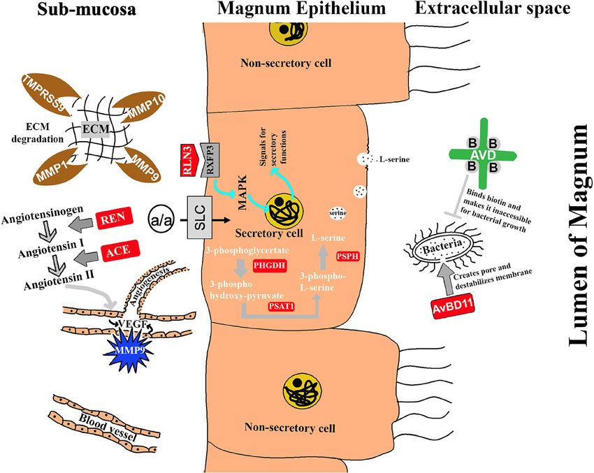
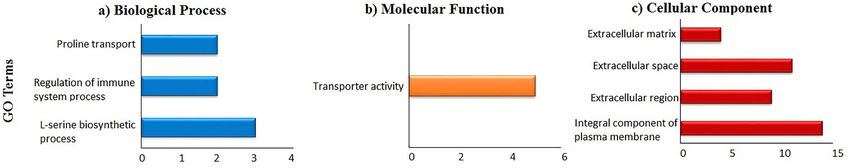
Sah et al. BMC Genomics   (2021) 22:318                                                                      Page 2 of 16

Background                                                   synthesis are very intricate, and their regulation in the
The chicken oviduct is a long tubular organ with histo-      oviduct is not clearly understood. Therefore, we hypoth-
logically and functionally five distinct segments (infun-    esized that the transcriptomic analyses, using RNA-
dibulum, magnum, isthmus, shell gland, and vagina)           Sequencing (RNA-Seq), of the magnum of laying hens in
having specific functions in egg formation. The ovulated     contrast to a magnum of non-laying hens can reveal the
egg-yolk traverses through the magnum in about 2–3 h,        novel genes and biological pathways involved in the
during which the egg-white (albumen) is continuously         regulation of egg-white synthesis and secretion.
deposited around it. Though 88% of the albumen is              In this study, we analyzed the genes and cognate path-
water, this component of the egg contributes more than       ways active in the magnum of laying hens whose expres-
60% to the total egg weight and determines the quality       sions are directly influenced by the presence of an egg.
of an egg. Smaller eggs cannot make it to the hatcheries     We further validated the expression profiles of novel
and also have low market-value as table eggs. Con-           genes in the laying (3 h, and 15–20 h post-ovulation,
sumers consider egg as a “functional food” because of        p.o.), molting, and non-laying hens.
several proteins incorporated in the albumen [1]. Funda-
mentally, the albumen is the primary source of nutrients     Results
and a barrier to the pathogenic infections of the devel-     Identification of differentially expressed genes (DEGs)
oping embryo [2]. The food processing industry uses          from RNA sequence
only the albumen portion of the egg for its foaming and      Raw sequencing reads in FASTQ format from replicated
gelling properties [3]; These perspectives necessitate an    RNA-Seq libraries were obtained, and their qualities
egg with qualitative and proportionate albumen in it.        were checked using FastQC. There was an average of
   The synthesis and storage of the principle egg-white      30.5 M and 33.4 M original raw reads in laying and non-
proteins (i.e., ovalbumin, ovotransferrin, ovomucoid, and    laying hens, respectively. After trimming and filtration,
lysozyme), constituting approximately 90% of the total       more than 97% of input reads from both laying and
albumen protein, occur exclusively in the tubular gland      non-laying hens were found as excellent quality se-
cells of the magnum epithelium [4, 5]. Following the exit    quences (Supplementary Table S1). Mapping results to
of an egg from the magnum, the epithelial cells of the       the chicken genome database showed that an average of
magnum begin the synthesis and storage of the egg-           93.42% of the retained reads from layers and 87.88%
white proteins to be deposited around the next egg,          from non-layers were uniquely mapped (Supplementary
which continues for 20–23 h [6, 7]. Each egg-white pro-      Table S2). A total of 19,152 transcripts were annotated
tein is synthesized at a rate proportional to its compos-    from Ensembl alignment (release 94), representing
ition in the egg-white [5]. After synthesis, the essential   50.24% of the chicken genome assembly. The DESeq2
proteins are packaged in secretory granules and secreted     analysis showed that 540 genes were differentially
from the tubular glands into the lumen of the magnum,        expressed between laying and non-laying hens (compre-
where they are deposited over the egg yolk [5, 8]. The       hensive gene list in Supplementary Table S3). Among
cellular signaling for the biosynthesis of albumen is reg-   the differentially expressed genes (DEGs), 457 genes
ulated by estrogen, progesterone, and testosterone [5, 8].   were officially characterized, while the rest were novel
The existence of an egg in the magnum causing a mech-        transcripts without any annotation. There were 152 up-
anical distention of the magnum wall, which stimulates       regulated and 388 downregulated genes in the magnum
the secretion of the stored egg-white proteins [8, 9].       of laying hens (at 3 h p.o.) as compared to the non-
Some transcriptomic studies and some gene-specific           laying hens. The top 30 upregulated and downregulated
studies have highlighted the importance of several           genes in the magnum of laying hens are presented in Ta-
genes/proteins in albumen synthesis and secretion [10–       bles 1 and 2, respectively. A visual representation of the
13]. The solute carriers, a large family of membrane         30 most upregulated and downregulated genes in layers
transporters, transport glucose, amino acids, and electro-   is shown as a heatmap image (Fig. 1).
lytes across the magnum epithelium, are upregulated
during the egg-white formation [14]. The matrix metal-       Functional annotation and pathways enrichment analysis
loproteases rapidly degrade the collagens and other          of DEGs
matrix proteins underlying the cells of the magnum for       The Database for Annotation, Visualization, and Inte-
continuous cellular growth and development [15]. Also,       grated Discovery (DAVID) bioinformatics resource was
proteins incorporated during the egg-white formation in      used to gain insight into various Gene Ontology (GO)
the magnum further determine the structures of calcium       terms of the upregulated genes in layers. Only the anno-
crystals being formed on the eggshell during                 tated 121 genes that were upregulated in laying hens
mineralization in the shell gland [16]. The egg-white is a   were uploaded for functional annotation in the DAVID
composite of several proteins whose secretion and            system, and results showed 119 genes were annotated
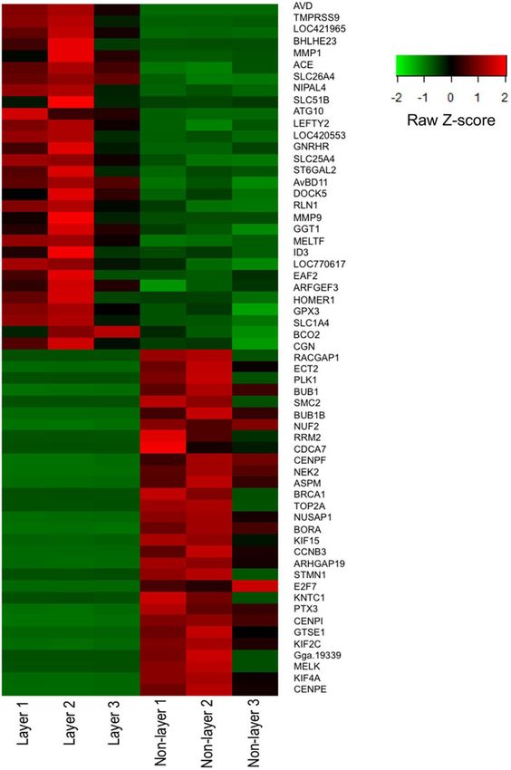
Sah et al. BMC Genomics        (2021) 22:318                                                                                                 Page 3 of 16

Table 1 The 30 most up-regulated DEGs in the magnum of laying compared to non-laying hens
Gene Name                                 Gene Description                                                                                  Fold Change
AVD                                       Avidin                                                                                            250.6824
TMPRSS9                                   Transmembrane protease, serine 9                                                                  33.0611
SLC7A9                                    Solute carrier family 7 member 9                                                                  29.9439
BHLHE23                                   Basic helix-loop-helix family member e23                                                          17.2940
MMP1                                      Matrix metallopeptidase 1                                                                         16.0790
ACE                                       Angiotensin converting enzyme                                                                     14.8313
SLC26A4                                   Solute carrier family 26 member 4                                                                 9.7876
NIPAL4                                    NIPA like domain containing 4                                                                     8.8105
SLC51B                                    Solute carrier family 51 beta subunit                                                             8.7190
ATG10                                     Autophagy related 10                                                                              8.6403
LEFTY2                                    Left-right determination factor 1                                                                 8.5433
ACCSL                                     1-Aminocyclopropane-1-carboxylate synthase homolog (inactive) like                                8.4146
GNRHR                                     Gonadotropin releasing hormone receptor                                                           7.7644
SLC25A4                                   Solute carrier family 25 member 4                                                                 7.5906
ST6GAL2                                   ST6 beta-galactoside alpha-2,6-sialyltransferase 2                                                7.5662
AvBD11                                    Avian beta defensin 11                                                                            7.5066
DOCK5                                     Dedicator of cytokinesis 5                                                                        7.4964
RLN1                                      Relaxin                                                                                           7.4939
MMP9                                      Matrix metallopeptidase 9                                                                         7.4666
GGT1                                      Gamma-glutamyltransferase 1                                                                       7.0938
MELTF                                     Melanotransferrin                                                                                 6.9221
ID3                                       Inhibitor of DNA binding 1, HLH protein                                                           6.5950
LOC770617                                 Transmembrane protein 100-like                                                                    6.4296
KIAA1109                                  Fragile site-associated protein                                                                   6.4112
EAF2                                      ELL associated factor 2                                                                           6.0836
ARFGEF3                                   ARFGEF family member 3                                                                            5.8841
HOMER1                                    Homer scaffolding protein 1                                                                       5.6404
GPX3                                      Glutathione peroxidase 3                                                                          5.6046
SLC1A4                                    Solute carrier family 1 member 4                                                                  5.5812
LOC419409                                 Golgi integral membrane protein 4-like                                                            5.4493
Transcripts from the magnum of layers and non-layers were aligned to the chicken genome and mapped genes with at least a 3-fold change difference and
Benjamini Hochberg q-value < 0.05 were considered differentially expressed. DEGs, differentially expressed genes

into the three GO terms; biological process, cellular                             Canonical pathways
component, and molecular function. Altogether 85 genes                            After submitting the DEGs to the ingenuity pathway
were recognized in the biological process, among which                            analysis (IPA), 417 molecules were recognized in its
three processes; L-serine biosynthetic process, regulation                        database that belonged to 34 significant canonical path-
of immune system process, and proline transport were                              ways (Table 3). Cell cycle control of chromosomal repli-
enriched (Fig. 2a). The molecular function had only one                           cation, the role of BRCA1 in DNA damage response,
enriched GO term, i.e., transporter activity, with 83                             mitotic roles of polo-like kinase, cell cycle: DNA damage
genes recognized (Fig. 2b), while the cellular component                          checkpoint regulation, and role of CHK proteins in cell
contained 4 enriched GO terms of the 90 identified                                cycle checkpoint control were the 5 most-significant ca-
genes (Fig. 2c). We also analyzed pathway enrichment                              nonical pathways. Among the significant canonical path-
for the upregulated genes in laying hens using the KEGG                           ways, 2 pathways (Cell cycle: G2/M DNA damage
pathways as incorporated in the DAVID system. Glycine,                            checkpoint regulation and regulation of cellular mecha-
serine, and threonine metabolism was the only pathway                             nisms by calpain protease) were predicted to be acti-
to be enriched for upregulated genes.                                             vated, while 7 pathways were predicted to be inhibited;
Sah et al. BMC Genomics (2021) 22:318 Page 4 of 16
Table 2 The 30 most down-regulated DEGs in the magnum of laying compared to non-laying hens
Gene Name Gene Description Fold Change
RACGAP1 Rac GTPase activating protein 1 16.6262
ECT2 Epithelial cell transforming 2 14.9228
PLK1 Polo like kinase 1 14.0534
BUB1 BUB1 mitotic checkpoint serine/threonine kinase 13.9712
SMC2 Structural maintenance of chromosomes 2 13.8266
BUB1B BUB1 mitotic checkpoint serine/threonine kinase B 13.7985
NUF2 NUF2, NDC80 kinetochore complex component 13.3569
RRM2 Ribonucleotide reductase regulatory subunit M2 12.9268
CDCA7 Cell division cycle associated 7 12.8785
CENPF Centromere protein F 12.7394
NEK2 NIMA related kinase 2 12.5734
ASPM Abnormal spindle microtubule assembly 12.2945
BRCA1 BRCA1(Breast cancer) 12.2056
TOP2A Topoisomerase (DNA) II alpha 11.5891
NUSAP1 Nucleolar and spindle associated protein 1 11.1474
BORA Bora, aurora kinase A activator 11.0740
KIF15 Kinesin family member 15 10.6809
CCNB3 CCNB3 (cyclin B3) 10.6117
ARHGAP19 ARHGAP19 10.4602
STMN1 Stathmin 1 10.4540
E2F7 E2F transcription factor 7 10.4150
KNTC1 Kinetochore associated 1 10.0559
PTX3 Pentraxin 3 9.9658
CENPI Centromere protein I 9.8662
GTSE1 G2 and S-phase expressed 1 9.7306
KIF2C Kinesin family member 2C 9.6465
Gga.19339 Family with sequence similarity 72, member A 9.5760
MELK Maternal embryonic leucine zipper kinase 9.4771
KIF4A Kinesin family member 4A 9.2900
CENPE Centromere protein E 9.2869
Transcripts from the magnum of layers and non-layers were aligned to the chicken genome and mapped genes with at least a 3-fold change difference and
Benjamini Hochberg q-value < 0.05 were considered differentially expressed. DEGs, differentially expressed genes
the rest lacked sufficient literature to be predicted. We and secretion, were determined in laying, molting, and
were particularly interested in the most significant meta- non-laying hens by real-time PCR (qPCR) assay. The se-
bolic pathways such as serine biosynthesis, superpath- lected candidate genes were avidin (AVD), transmem-
ways of serine and glycine biosynthesis I, inhibition of brane protease serine 9 (TMPRSS9), matrix
matrix metalloproteases, asparagine biosynthesis I, as- metallopeptidase 1 (MMP1), angiotensin-converting en-
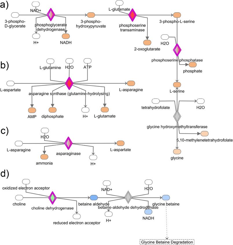
paragine degradation I, and choline degradation I (Fig. 3) zyme (ACE), autophagy-related 10 (ATG10), avian beta-
because of their prominent role in albumen synthesis defensin 11 (AvD11), relaxin (RLN3), matrix metallopep-
and secretion. tidase 9 (MMP9), melanotransferrin (MELTF), glutathi-
one peroxidase 3 (GPX3), cingulin (CGN), protein C
Validation of the expression profiles of selected candidate (PROC), phosphoserine phosphatase (PSPH), phospho-
genes glycerate dehydrogenase (PHGDH), asparagine synthe-
Following the identification of DEGs, the expression tase (ASNS), phosphoserine aminotransferase 1 (PSAT1),
profiles of the 19 most relevant upregulated genes, spec- matrix metallopeptidase 10 (MMP10), calpain 2
ulated to be related to the event of albumen synthesis (CAPN2), and renin (REN).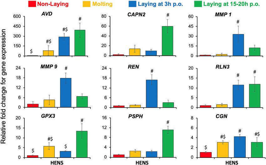
Sah et al. BMC Genomics      (2021) 22:318                                                                                            Page 5 of 16

 Fig. 1 Heat map of top thirty DEGs in the magnum of laying and non-laying hens. The raw z-score depicts the standard deviation of the gene
 expression value from the mean after normalization. A gene having a negative z-score is represented by green color while a positive z-score is
 represented by red color. RNA-Seq was performed on magnum from three laying (3 h p.o.) and three non-laying hens. Transcripts were aligned
 to the chicken genome and mapped genes with at least a 3-fold change difference and Benjamini Hochberg q-value < 0.05 were considered
 differentially expressed

   The gene networks showing the interactions of some                      calculate the relative fold change of the candidate genes
selected candidate genes using the IPA network analysis                    after normalization with the house-keeping gene TATA-
are shown in Fig. 4. For the qPCR gene expression pro-                     Box Binding Protein (TBP). A total of nine genes,
files, the double delta Ct (2-ΔΔCt) method was used to                     amongst the 19 candidate genes, showed significant
Sah et al. BMC Genomics     (2021) 22:318                                                                                         Page 6 of 16

 Fig. 2 Gene Ontology enrichment analysis of upregulated genes in the magnum of laying and non-laying hens. a Biological Process, b Molecular
 Function, c Cellular Component. The up-regulated genes in the laying hens (3 h p.o.) were subjected to the DAVID database for Gene Ontology
 (GO) enrichment analysis. All the GO terms with a modified Fisher Exact p-value < 0.05 and a threshold gene count of 2 were
 considered enriched

changes (p-value < 0.05) in expression profiles between                  p.o.) and non-laying hens. As previously reported, we
the experimental groups (Fig. 5). The mRNA expression                    observed increased expression of genes encoding the
of CAPN2 and PSPH were highest in laying hens at 15–                     common egg-white proteins such as ovalbumin, lyso-
20 h p.o. compared to either laying hens at 3 h p.o.,                    zyme, and avidin in the magnum when the egg was
molting, or non-laying hens. Expressions of REN,                         present in this segment of the oviduct [11, 17, 18].
MMP1, and MMP9 mRNAs were upregulated only in                            Amongst the DEGs, several proteases (TMPRSS9, ACE,
laying hens at 3 h p.o. compared to either laying hens at                REN, MMP1, MMP9, MMP10, CAPN2, and PROC) and
15–20 h p.o., molting, or non-laying hens. Expression of                 enzymes for biosynthesis (PHGDH, PSPH, PSAT1, and
RLN3 gene was increased in laying hens, both at 3 h p.o.                 ASNS) were of particular interest. Some of these genes
and 15–20 h p.o. relative to molting and non-laying                      were detected in the microarray and RNA-Seq studies in
hens. AVD mRNA was highest in the laying hens at 15–                     the magnum [11, 19]. However, their potential role in
20 h p.o. followed by 3 h p.o. and molting hens, while                   the formation of egg-white was not reported. In this
lowest in the non-laying hens. The expression of GPX3                    study, we validated and assessed the specificity of these
mRNA was higher in laying hens at 15–20 h p.o. com-                      identified novel genes and pathways in the laying (3 h
pared to both non-laying and laying hens at 3 h p.o. The                 and 15–20 h p.o), molting, and non-laying hens using
CGN mRNA had increased expression in laying hens at                      qPCR. Then, we used their expression profile to extrapo-
3 h p.o. relative to 15–20 p.o. and molting hens, while                  late their novel role in the synthesis and/or secretion of
significantly higher than non-laying hens. The results                   egg-white proteins based on existing literature. The
obtained from RNA-Seq and qPCR were highly corre-                        newly identified genes were involved in antimicrobial
lated (R2 = 0.94; Supplementary Table S4), showing                       defense, matrix remodeling, albumen synthesis and/or
consistency between RNA-Seq and qPCR data for fold                       secretion, and egg transport (Fig. 6).
change of gene expression.
                                                                         Proteases associated with the albumen synthesis and
Discussion                                                               secretions
The magnum is highly glandular tissue, and molecules                     Proteases are enzymes having catalytic activity on pro-
secreted and/or transported from the luminal and glan-                   teins. There are seven different classes (based on cata-
dular epithelium contribute to the egg albumen. The egg                  lytic residue) of proteases, including serine proteases and
remains in the magnum for 1–3 h to complete the de-                      metalloproteases [20]. Both the serine- and metallo-
position of albumen around the yolk. In the first few                    proteases actively regulate the protein turnover of the
hours of the ovulation cycle (1–3 h p.o.), the egg is in                 extracellular matrix (ECM), influencing various cellular
the magnum, during which the stored proteins from the                    functions [21]. Our RNA-Seq data showed that TMPR
magnum epithelium are secreted in the lumen [6]. In                      SS9 mRNA was the second most upregulated DEG (FC =
the later period of the ovulation cycle (4–23 h p.o.), im-                33) in laying hens when the egg was inside the magnum.
mediately after the egg has left the magnum, the protein                 TMPRSS9, also known as polyserase-I, is a transmem-
synthesis process begins and continues until the next                    brane type II serine protease that uniquely produces
egg reaches the magnum [6–8]. Using RNA-Sequencing                       three other proteases, including 2 active ones [22].
and qPCR, we identified several novel genes and bio-                     TMPRSS9 facilitates the formation of urokinase plas-
logical pathways associated with egg albumen formation.                  minogen activator that converts plasminogen to plasmin
  In the present study, RNA-Seq data revealed a total of                 responsible for the degradation of ECM components
540 genes differentially expressed between laying (at 3 h                [23]. Higher expression of TMPRSS9 uniquely in laying
Sah et al. BMC Genomics (2021) 22:318 Page 7 of 16 Table 3 Significant canonical pathways identified by IPA involved in the egg-white formation Ingenuity Canonical Pathways -log (p-value) Cell Cycle Control of Chromosomal Replication 11.2 Role of BRCA1 in DNA Damage Response 7.57 Mitotic Roles of Polo-Like Kinase 6.34 Cell Cycle: G2/M DNA Damage Checkpoint Regulation 5.43 Role of CHK Proteins in Cell Cycle Checkpoint Control 5.05 Hereditary Breast Cancer Signaling 4.68 Pyrimidine Deoxyribonucleotides De Novo Biosynthesis I 4.21 Serine Biosynthesis 4.18 Estrogen-mediated S-phase Entry 4.11 DNA Double-Strand Break Repair by Homologous Recombination 3.95 ATM Signaling 3.85 Superpathways of Serine and Glycine Biosynthesis I 3.64 GADD45 Signaling 3.40 DNA damage-induced 14–3-3σ Signaling 3.40 Regulation of Cellular Mechanics by Calpain Protease 3.13 Antiproliferative Role of TOB in T Cell Signaling 2.86 Neuroprotective Role of THOP1 in Alzheimer’s Disease 2.69 Salvage Pathways of Pyrimidine Ribonucleotides 2.64 Atherosclerosis Signaling 2.52 Cyclins and Cell Cycle Regulation 2.43 Dopamine-DARPP32 Feedback in cAMP Signaling 2.38 Complement System 2.28 Inhibition of Matrix Metalloproteases 2.20 Pyridoxal 5′-phosphate Salvage Pathway 2.10 Hepatic Fibrosis / Hepatic Stellate Cell Activation 2.06 Airway Pathology in Chronic Obstructive Pulmonary Disease 2.03 Breast Cancer Regulation by Stathmin1 1.79 Asparagine Biosynthesis I 1.72 Cell Cycle Regulation by BTG Family Proteins 1.54 Mismatch Repair in Eukaryotes 1.44 Asparagine Degradation I 1.42 Choline Degradation I 1.42 eNOS Signaling 1.37 Cardiac β-adrenergic Signaling 1.32 All the differentially expressed genes in the layers were used in Ingenuity Pathway Analysis and significant canonical pathways based on IPA scores were identified. IPA, ingenuity pathway analysis hens suggests that it potentially participates in the deg- the MMPs family in vertebrates, including MMP-1, − 9, radation of the ECM to release the stored proteins mak- and − 10, which are secreted proteins involved in a wide ing them available for receptor binding and signaling range of physiological activities such as cellular migra- action, as proposed by ten Dijke et al. [24]. This process tion and angiogenesis, and inflammation [25, 26]. In this is indeed relevant in laying hens when the egg is present study, the expressions of MMP-1, MMP-9, and MMP-10 in the magnum or the shell gland to keep up with the were upregulated (FC = 16, 7.5, and 3.5, respectively) in enormous amount of protein synthesis and secretion. the laying hens with the presence of egg in the magnum, Matrix metalloproteases (MMPs) are the primary reg- compared to the non-laying hens. MMP1 is down- ulators of ECM remodeling. There are 24 members of regulated in magnum when the hen transitions from
Sah et al. BMC Genomics       (2021) 22:318                                                                                                Page 8 of 16

 Fig. 3 Results of most significant and relevant canonical pathways associated with albumen formation in laying hens. a Superpathways of serine
 and glycine biosynthesis I, b asparagine biosynthesis I, c asparagine degradation I, and d choline degradation. The canonical pathways were
 analyzed using QIAGEN’s Ingenuity Pathway Analysis (IPA; QIAGEN Inc., https://www.qiagenbioinformatics.com/products/ingenuity-pathway-
 analysis). Differentially expressed genes in the layers were subjected to IPA analysis, and significant canonical pathways were identified at p-value
 < 0.05. The above-identified canonical pathways demonstrate how the candidate molecules (genes) are involved in amino acid synthesis
 and degradation

laying to molting stage [11]. MMP1, also known as colla-                      MMP-1, MMP-9, and MMP-10 in the magnum of laying
genase, can degrade the most highly abundant ECM; col-                        hens are associated with the tissue remodeling and for-
lagen, in several tissues, including the chicken ovary                        mation of new vasculatures to support the expeditious
[27]. MMP-9 (gelatinase) is known to degrade the gelatin                      conveyance of precursor molecules for the biosynthesis
matrix [28], provokes angiogenesis [29], and also regu-                       of egg-white proteins.
lates the laying process in hen [15]. MMP10 breaks                              Calpains, on the other hand, are ubiquitous intracellu-
down several collagen-related connective tissues [30].                        lar cysteine proteases having very low specificity for rec-
There is no report of MMP10 mRNA expression in the                            ognition of amino acid sequence. Calpains have a wide
chicken oviduct; however, a metastatic study has con-                         range of functions in various tissues, including mem-
firmed its association in angiogenesis [31]. Several pro-                     brane repair, cell adhesion and motility, cell death, pro-
teins need to be synthesized and transported into the                         tein cleavage, and activation [32]. Our study reports an
lumen for deposition around the egg yolk for albumen                          increased expression of CAPN2 in the laying hens during
formation. The required proteins are synthesized in the                       15–20 h p.o. compared to either molting or non-laying
tubular gland cells of the magnum, which require the                          hens. Similarly, the expression of CAPN2 is higher in
rapid transport of amino acids from the blood circula-                        hens at the laying stage than in the molting stage [11].
tion [6]. We speculate that the higher expression of                          Therefore, we posit that CAPN2 is responsible for the
Sah et al. BMC Genomics (2021) 22:318 Page 9 of 16
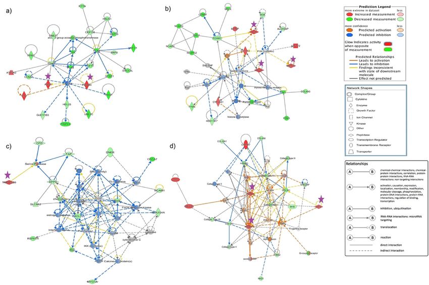
Fig. 4 Gene network highlighting some of the candidate genes involved in albumen formation. The candidate genes and their interaction in
potentially regulating the synthesis, secretion, and transport of molecules during egg-white formation is shown. This was derived from QIAGEN’s
Ingenuity Pathway Analysis (IPA; QIAGEN Inc., https://www.qiagenbioinformatics.com/products/ingenuity-pathway-analysis). a PHGDH, ASNS; b
ACE, MMP10, PROC; c TMPRSS9; and d MMP1, MMP9, REN. Differentially expressed genes in the layers were used in Ingenuity Pathway Analysis,
and significant gene networks based on IPA scores were identified
maturation and activation of the synthesized egg-white hens. Cingulin is involved in the organization of the tight
proteins. junctions, but simultaneously, it inhibits RhoA (Ras
Also, we observed that serine protease inhibitor family homolog gene family member A) activation and sup-
B member 2 (SERPINB2) was higher (> 3-fold) in the presses epithelial cell proliferation and gene expression
magnum, and similar up-regulation of SERPINB3 ex- [36]. However, it is also implicated that CGN regulates cell
pression in the magnum of laying hens was reported by growth and morphology and creates a single layer of
Jeong et al. [11]. Recently, Zhang et al. [33] also reported small, tightly packed cells [37]. To the best of the available
the upregulation of SERPINF1 and SEPRINH1 when an literature on CGN function, we postulate that CGN
egg was present in the magnum of duck. This suggests mRNA is involved in the cellular organization and integ-
that the SERPIN family of protease inhibitors has an im- rity of the magnum epithelium in laying hens regulating
portant role in regulating the secretory activity of mag- molecular transport across the epithelial barrier.
num for egg-white formation. Indeed, proteomic analysis The solute carriers (SLCs) are exclusive membrane
of the egg white has shown that the SERPIN proteins transporters that carry several solutes such as amino
are incorporated in the egg-white [34]. acids, organic and inorganic ions, and sugars. Several
SLC members, including SLC7A9, SLC1A4, SLC7A11,
Transporters of proteins in the magnum epithelium SLC7A7, and SLC6A17, were increased by 29.9, 5.6, 5.3,
Cingulin is a protein localized at the tight junction of epi- 4.5, and 4.4-folds, respectively in the laying hens. The
thelial and endothelial cells, first discovered in the chicken upregulation of these genes in the magnum of laying
intestine, and creates a barrier for molecular transport hens suggests that they actively participate in the trans-
across cells [35]. In the present study, CGN mRNA was porter of precursor molecules to synthesize egg-white
5.4 fold higher in laying as compared to the non-laying proteins.Sah et al. BMC Genomics (2021) 22:318 Page 10 of 16 Fig. 5 Validation of the gene expression in the magnum of non-laying, molting, and laying hens. Data represented as the mean ± SE. The x-axis represents the physiological status of hens used in the experiment; Y-axis represents relative fold change for gene expression. #, $ denotes significance at p-value < 0.05 Molecules involved in the biosynthesis since cells of the magnum are involved in the production Several enzymes such as PHDGH, PSPH, PSAT1, ASNS, of a massive amount of proteins, and concurrently ROS ASPG, GALNT6, PDE3A, and PHYKPL were increased as by-products. in laying hens as shown by our RNA-Seq data. GO en- richment analysis revealed that PHDGH, PSPH, PSAT1, Genes involved in albumen secretion and/or oviductal and ASNS were involved in amino-acid biosynthesis. transport of egg The biosynthesis of L-serine from 3- Relaxin hormone produced from the ovary and placenta phosphoglyceraldehyde is mediated by three enzymes in mammals helps to ease the parturition process by PHGDH, PSAT1, and PSPH at each successive step, re- relaxing the ligaments and dilating the cervix. The spectively [38]. Interestingly, the mRNA of PHGDH had relaxin-like family peptide has seven peptides, including higher expression in laying hens at 15–20 h p.o. (during relaxin-3, which belong to the insulin superfamily. How- the albumen synthesis period), and PSAT1 and PSPH ever, a phylogenetic study showed that the chicken gen- mRNAs were also relatively higher in those hens. The ome had lost all the relaxin family peptides, but relaxin- upregulated expression of PHGDH, PSAT1, and PSPH in 3 having high homology to the human analog [40]. The laying hens in this study strongly indicates the biosyn- relaxin-like peptide is produced in granulosa cells of the thesis of serine in magnum, which may be required to post-ovulatory follicles, localized in the uterus of laying synthesize egg-white proteins. Besides, microarray ana- hens, and influences the oviduct and uterus to aid in ovi- lysis of the magnum has shown that the expression of position [41]. Also, loss in functionality of this avian re- ASNS and PSPH is higher at the laying stage compared laxin has been shown to cause a drastic delay in to the molting stage [11]. A report by Li et al. [39] sug- oviposition timing [42, 43]. Studies of Brackett [41] and gests an additional role of these enzymes (PHGDH, Wilkinson [40] suggest that the hormonal action of PSAT1, and PSPH) in protection from reactive oxygen relaxin-3 from ovaries help in egg-laying. This study also species (ROS) by providing the substrate-serine for detected a significant expression of RLN3 mRNA in the glutathione synthesis. The antioxidative function of magnum of laying hens (7.5-fold higher) both during al- serine biosynthesis enzymes in the magnum is plausible bumen synthesis and secretion period. This is a novel
Sah et al. BMC Genomics      (2021) 22:318                                                                                          Page 11 of 16

 Fig. 6 The hypothetical model showing the identified genes and their predicted roles associated with egg-white formation. Solute carriers such
 as SLC1A4, SLC7A11, SLC7A7, and SLC6A17 may expedite the transport of precursor molecules for protein synthesis. Proteases such as CAPN2,
 TMPRSS9, MMP1, and MMP9 may be involved in protein maturation and activation, ECM degradation, and angiogenesis for the delivery of
 molecules from blood circulation so that the magnum epithelium can utilize them for the synthesis of egg-white proteins. Upregulated PHGDH,
 PSPH, and PSAT1 suggest their active role in the synthesis of amino acids that are basic units of the complex albumen proteins. Increased
 expression of Relaxin-3 and renin-angiotensin system (REN and ACE) may be linked to their participation in the transport of egg through the
 oviduct controlling how long the egg stays in the magnum for efficient protein deposition around the yolk. In addition to those genes involved
 in biosynthesis, some other genes which have a protective function to the egg such as, avidin, avian-beta-defensin 11, and glutathione
 peroxidase are also incorporated in the egg albumen

report on RLN3 expression suggesting its synthesis in                     [44]. Renin found in ovarian theca cells [45, 46] and
the magnum, and we hypothesize that its over-                             angiotensin-converting enzyme (ACE) localized in the
expression at 3 h p.o. in the oviduct may be related to                   granulosa cells and blood vessels of the ovary [47] are
the mechanical distention of the magnum to ease the                       the principal components of the RAS system. Apart from
passage of the developing egg and/or secretion of the                     the endocrine function of RAS, the localized action of
stored egg-white proteins. Since the mechanical pressure                  RAS in the ovary is towards follicular development and
on the walls of the magnum provokes the secretion of                      ovulation [48]. In this study, REN mRNA had signifi-
the synthesized albumen proteins [9], RLN3 potentially                    cantly increased expression in the magnum of laying
is one of the markers of mechanical stimulus for the se-                  hens during the albumen secretion period as compared
cretion of albumen from the goblet cells of the magnum.                   to molting and non-laying hens. The ACE mRNA was
  The renin-angiotensin system (RAS), besides its well-                   also higher (14.8 folds) in laying hens relative to non-
known endocrine role in maintaining extracellular fluid                   laying hens. There are some reports on the activity of
in the body, also regulates ovarian growth dynamics                       RAS in the uterus of humans [49], rats [50], rabbits [51],
Sah et al. BMC Genomics   (2021) 22:318                                                                      Page 12 of 16

and quail [52]. So far, there is no report on RAS in the      localized in plasma and extracellular spaces [58]. We ob-
chicken oviduct. Verma and Panda [52] reported that           served a 5.6 folds higher expression of GPX3 mRNA in
ACE is expressed in immature and mature (with exogen-         the magnum of laying hens during the albumen synthe-
ous estrogen) quails with the highest expression in mag-      sis period. These findings are indeed concurrent with
num, amongst the other oviductal parts. REN and ACE,          the underlying physiological activities in laying hens. In
fundamental molecules of the RAS, are predominantly           the magnum of laying hens, rapid protein synthesis oc-
found in the glandular epithelium of the human uterus,        curs at 4–23 h p.o. indicating that the cells of magnum
where the RAS had different roles during the menstrual        have increased metabolism. As a result, simultaneous
cycle [49]. Collectively, the RAS controls the blood sup-     with protein synthesis, there is the release of ROS and
ply to the magnum by altering the vascular smooth             other free radicals. So, the increased GPX3 expression in
muscle tone (through bradykinin), and forming new             the magnum is indicative of the protective response
blood vessels [53]. Also, the RAS system, specifically in     against oxidative damage. Also, several other genes dif-
the magnum, might aid in relaxing the magnum to re-           ferentially expressed in laying hens, such as urotensin 2
tain the egg for sufficient time, allowing optimum depos-     and spermine oxidase involved in the production of ROS
ition of albumen. Concurrently, the expression of the         and hydrogen peroxide [59, 60], respectively, support the
ACE gene in the magnum of pigeon decreases by more            fact that oxidative stress is evident in the magnum.
than four-fold when the egg has passed through the
magnum during the egg-laying cycle [19]. The previous         Conclusion
studies in association with the findings of this study sug-   We have identified a substantial number of novel genes
gest that the expression of REN and ACE in the magnum         and biological pathways that decipher the cascade of
of laying hens is strong evidence that the RAS system is      events associated with the albumen formation and de-
also involved in the oviductal transport of egg in the        position in the magnum (Fig. 6). The series of events
chicken.                                                      that occurs in the magnum contributing to the albumen
                                                              formation include transport of precursor molecules
Antimicrobials for the egg defense                            (amino acids, proteins, solutes, and ions), synthesis of
Antimicrobial agents are crucial for the livability of the    proteins (such as ovalbumin, avidin, lysozyme), and se-
hen’s embryo. The albumen holds the yolk (with ovum)          cretion or transport of the synthesized proteins to be de-
in the center of the egg, without any contact with the        posited around the egg yolk. This study revealed the
eggshell. Albumen acts as a thick protective layer con-       upregulation of several genes in laying hens that are po-
sisting of several antibacterial proteins. One such estab-    tentially involved in the aforementioned events for egg-
lished protein is avidin, and interestingly in our study,     white formation (Fig. 6). Solute carriers such as SLC1A4,
AVD was the most overly expressed (250.7 folds) mRNA          SLC7A11, SLC7A7, and SLC6A17 are upregulated in lay-
in laying hens. Avidin is also abundant in the egg white      ing hens for expeditious convey of precursor molecules
[1] and has a very high affinity for biotin required for      for protein synthesis. Also, the upregulated status of pro-
bacterial growth and proliferation, thus preventing the       teases such as CAPN2, TMPRSS9, MMP1, and MMP9 in
invasion by microbial pathogens [54].                         laying hens advocates their involvement in protein mat-
   Another newly discovered and widely studied chicken        uration and activation, ECM degradation, and angiogen-
antimicrobial protein is avian beta-defensins (AvBDs).        esis for the transport of molecules from the blood
AvBD11 is among the 14 members of the AvBDs whose             circulation so that the magnum epithelium can utilize
mRNA expression was increased by 7.5 folds in the mag-        them for the synthesis of egg-white proteins. Increased
num of laying hens in our study. Previous studies have        expression of enzymes such as PHGDH, PSPH, and
also revealed the expression of AvBD11 in the egg vitel-      PSAT1 only in laying hens suggests their active role in
line membrane, eggshell membrane, eggshells, and mag-         synthesizing amino acids that are basic units of the com-
num, suggesting that the AvBD11 is an important               plex albumen proteins. During egg formation, laying
molecule for innate immunity in hens [17, 55–57].             hens have increased expression of relaxin-3, and renin-
Taken together, AvBD11 incorporation in the albumen           angiotensin system (REN and ACE), which posits their
protects the developing embryo and might increase the         participation in the transport of egg through the oviduct
hatchability of the eggs.                                     controlling how long the egg stays in the magnum for
                                                              efficient protein deposition around the yolk. They also
Antioxidant for protection of the magnum epithelium           ease the secretion of albumen from the granular cells for
Glutathione peroxidase (GPX) is a well-known enzyme           deposition around the egg. In addition to those genes in-
capable of protecting the cells and tissues from ROS,         volved in biosynthesis, some other genes have a protect-
such as hydrogen peroxidases and other lipid hydroper-        ive function on the egg, such as avidin, avian-beta-
oxides. GPX3 is an isoform of the enzyme GPX class,           defensin 11, and glutathione peroxidase are also
Sah et al. BMC Genomics (2021) 22:318 Page 13 of 16
incorporated in the egg albumen. Thus, the findings of segment. Magnum tissues were collected from the seg-
this study advanced the knowledge of genes and bio- ments immediately before the site where the egg was
logical pathways involved in albumen biosynthesis and present, to prevent any contamination with an excess of
can potentially be used as markers for formulating strat- albumen from the developing egg. The albumen secre-
egies to improve the size and quality of the eggs. tion and deposition from magnum epithelium around
the egg yolk starts when the egg is in the magnum, while
Methods the secretion of egg-white proteins for the next egg be-
Animal husbandry and tissue collection gins once the egg leaves the magnum. Therefore, the ex-
Hy-Line white (laying, non-laying, and molting) hens pressions of the genes involved in the secretion and
were brought from a commercial layer farm (Mikilua synthesis processes are supposed to be upregulated dur-
Poultry Farm Inc., Hawaii). Before sampling, hens were ing 3 h p.o. and 4–23 h p.o., respectively. Pieces of mag-
acclimatized for 2 weeks in the Small Animal Facility of num tissues were collected, snap-frozen, and stored at −
College of Tropical Agriculture and Human Resources, 80 °C until further analysis.
University of Hawaii at Manoa. Hens used for this study
were at three different physiological stages; i) laying hens RNA library preparation and sequencing
of 35 weeks (n = 12), ii) molting hens (n = 6) of 60 weeks, Total RNA from the frozen tissues was isolated using
and iii) non-laying hens (n = 6) between 35 and 60 weeks TRIzol reagent (Invitrogen, Carlsbad, CA) following
of age. standard protocol. The concentrations and quality of the
The laying hens were in their peak egg production extracted RNA samples were measured using NanoPhot-
period, while the molting hens were in their first week ometer® P330 (IMPLEN, Los Angeles, CA) and Agilent
of programmed molting procedure. The physiological 2100 Bioanalyzer (Agilent Technologies, Massy, France),
status of molting hens was further verified based on the respectively. High-quality RNA samples (RNA integrity
history of the absence of any laying activity during the number > 8.5) were used for library preparation and
experimental period. The molting hens had matured ovi- sequencing.
duct and ovarian follicular dynamics was evident, but RNA-Seq libraries from the magnum tissues of laying
without any follicular clutches or ovulation. The non- (n = 3) at 3 h p.o. and non-laying (n = 3) hens were pre-
laying hens were selected initially based on speculation pared using a TruSeq Stranded mRNA kit (Illumina, San
with the help of flock attendants at the commercial farm. Diego, CA) as described previously [62]. Following li-
Non-laying hens were identified with meticulous obser- brary preparation, a high sensitivity DNA Bioanalyzer
vations and physical assessments such as the shallow ab- assay (Agilent Technologies, Massy, France) was used to
domen, stiff pubic bones, and dry and puckered cloaca assess the size and quality of the libraries, while KAPA
[61]. Therefore, the non-laying hens used for this experi- Library Quantification Kit (KAPA Biosystems, Boston,
ment were identified and selected from different flocks MA) was used to quantify the libraries by qPCR. The se-
and thus belonged to a range of ages between 35 and 60 quencing run was executed with a single-end mode with
weeks. Such non-laying hens were further confirmed a read length of 1x76bp on a NextSeq 500 (Illumina, San
based on their atrophied oviduct and absence of any fol- Diego, CA) platform.
licular recruitment or maturation in the ovary, examined
during necropsy. Each hen was housed in individual RNA-sequencing analysis
pens, reared under a standard light regimen and, fed ad Illumina BaseSpace-created FASTQ files with single-end
libitum. During the acclimatization period, the egg- reads were explored using FastQC (Babraham Institute,
laying pattern and time of lay were monitored three Cambridge, UK). Prinseq, a perl script [63] was used to
times (8 am, 12 pm, and 4 pm) daily for each bird to clean the raw reads as mentioned previously [62]. Then,
keep track of its laying performance. To know the exact Array Studio (version10; OmicSoft, Cary, NC [64];) was
time of ovulation (~ 30 min after oviposition) for the used to align the cleaned against the chicken reference
ease of sampling time points, the hens were monitored genome Galgal 5.0. The DESeq2 algorithm [65], as im-
hourly from 6 am till 4 pm on the day before sampling. plemented in the Array Studio, was used to analyze the
Hens were euthanized by carbon dioxide asphyxiation. differential gene expression in layers with respect to
Magnum tissues were collected from laying hens (n = 5/ non-layers’ groups. The genes are having a fold change
group) when the egg was in the magnum (3 h post- (FC) greater than 3 and Benjamini and Hochberg q-
ovulation; p.o.) or the uterus (15–20 h p.o.), molting value < 0.05 were categorized as differentially expressed.
(n = 5), and non-laying (n = 4) hens. Egg in the magnum/
uterus of laying hens was presumed by laying history Biological pathways and molecular function analyses
and confirmed with post-mortem analysis of the oviduct Enriched pathways and molecular function of the upreg-
to determine the exact location of egg in the oviductal ulated genes in laying hens were determined by usingSah et al. BMC Genomics (2021) 22:318 Page 14 of 16
public databases such as the Database for Annotation, Supplementary Information
Visualization and Integrated Discovery (DAVID [66],) The online version contains supplementary material available at https://doi.
org/10.1186/s12864-021-07634-x.
and Kyoto Encyclopedia of Genes and Genomes (KEGG
[67],) Pathway as described previously [62]. A list of the Additional file 1: Table S1. Filtration and alignment summary of RNA-
upregulated genes was uploaded to the functional anno- Seq Reads from magnum in laying and non-laying hens. Table S2. Sum-
tation tool in the DAVID system, and the chicken was mary of magnum RNA-Seq data mapping to the chicken genome (Gal-
gal5.0). Table S3. Differentially expressed genes at FDR_BH < 0.05 and
selected as the reference genome for Gene Ontology FC > 3 in the magnum of laying and non-laying hens. Table S4. Correl-
(GO) enrichment analysis to obtain the enriched bio- ation between RNA-seq and qPCR data of relative gene expression in
logical pathways, molecular function, cellular compo- magnum of laying and non-laying hens. Table S5. List of primers for the
candidate genes used in qPCR assay. Primers for the candidate genes
nent, and the pathways. The GO terms with a modified were designed using Primer Blast tool of NCBI with filters of amplicon
Fisher Exact p-value < 0.05 and a threshold gene count size between 100 and 250 bp, primers must span an exon-exon junction,
of 2 were considered enriched. The Ingenuity Pathway melting point between 55 and 60 °C with other filters set at default.
Analysis (QIAGEN Inc. [68],) tool was also employed to
gain insights into the molecular networks and canonical Acknowledgments
pathways of the DE genes. The DE genes were fed to the We sincerely thank Mikilua Poultry Farm for providing hens for this
experiment, and Amit Singh for sample collection.
IPA software, and significant differential analyses were
made at a p-value < 0.05. Since the IPA is based on the Authors’ contributions
human genome mapping, we tried to derive only cred- N.S., D.L.K., and B. M conducted the experiment and collected samples; N.S.
ible information as applicable to the hen’s physiology. and S. W performed sample analyses; V.S.K. and Y.D. did the bioinformatics
analyses, N.S. and B. M analyzed the data; and N. S, D.L.K, R. J, S.W., and B. M
contributed in drafting this manuscript. All the authors read and approved
the manuscript.
Quantitative real-time RT-PCR (qPCR)
To confirm the accuracy of the results obtained by Funding
RNA-Seq, nineteen genes having a predicted function in This work was supported by a Start-up grant from CTAHR University of Ha-
albumen synthesis and/or secretion were selected for waii at Manoa, and USDA Multistate (2052R) to B.M. Apart from providing
funds, these organizations were not involved in any experimental procedure
qPCR validation. Primers for qPCR were designed using and manuscript preparation.
the NCBI primer blast tool (shown in Supplementary
Table S5). Standard qPCR protocols were followed as Availability of data and materials
described by Sah et al. [62] in a reaction mixture of The datasets generated and/or analyzed for this study are available in the
Gene Expression Omnibus (GEO) repository and can be accessed with the
10 μl. TATA-Box Binding Protein (TBP) was used as a
Accession Number GSE123588 at https://www.ncbi.nlm.nih.gov/geo/query/
reference gene after analyzing it along with glyceralde- acc.cgi?acc=GSE123588.
hyde 3-phosphate dehydrogenase (GAPDH), beta-actin The datasets from Jeong et al. [11] were obtained from https://doi.org/10.
1371/journal.pone.0076784.s001
(B-actin) for stable expression in all the samples. All tar-
The datasets from Yin et al. [18] are available at PRJNA492958 in NCBI
get genes were analyzed in duplicates, and the expres- (https://www.ncbi.nlm.nih.gov/bioproject/?term=prjna492958), and was
sion level was determined using the normalized cycle obtained from https://www.sciencedirect.com/science/article/pii/S088875431
8305810?via%3Dihub#bi0005
threshold (Ct) values following the standard curve
The datasets from Lu et al. [19] does not contain a repository name;
method. The relative fold change for genes was calcu- however, the data were obtained from mrd23428-sup-0001-APPENDICES.xlsx.
lated using the 2-ΔΔCt method and presented as mean ± The datasets from Zhang et al. [33] are available at GenBank Short Read
Archive (SRA: PRJNA493510) and were obtained from https://www.
standard error. Statistical analyses were performed using
sciencedirect.com/science/article/pii/S0888754320300033#s0075
SAS software (SAS Institute Inc., NC) using a one-way
analysis of variance followed by the Tukey-Kramer test Declarations
to determine significance at p-value < 0.05.
Ethics approval and consent to participate
All the animal care and use protocols were approved by the Institutional
Abbreviations Animal Care and Use Committee of the University of Hawaii at Manoa.
ACE: Angiotensin-converting enzyme; AvD11: Avian beta-defensin 11;
ANOVA: One-way analysis of variance; ASNS: Asparagine synthetase;
AVD: Avidin; ATG10: Autophagy-related 10; B-actin: Beta-actin; Consent for publication
CAPN2: Calpain 2; CGN: Cingulin; DAVID: Database for Annotation, Not applicable.
Visualization and Integrated Discovery; DEGs: Differentially expressed genes;
ECM: Extracellular matrix; GAPDH: Glyceraldehyde 3-phosphate dehydrogen- Competing interests
ase; GO: Gene Ontology; GPX3: Glutathione peroxidase 3; IPA: Ingenuity The authors declare that they have no competing interests.
pathway analysis; KEGG: Kyoto Encyclopedia of Genes and Genomes;
MMP1: Matrix metallopeptidase 1; MMP9: Matrix metallopeptidase 9; Author details
1
MMP10: Matrix metallopeptidase 10; MELTF: Melanotransferrin; PHGD Department of Human Nutrition, Food and Animal Sciences, University of
H: Phosphoglycerate dehydrogenase; PROC: Protein C; PSAT1: Phosphoserine Hawaii at Manoa, HI 96822 Honolulu, USA. 2Department of Molecular
aminotransferase 1; PSPH: Phosphoserine phosphatase; RLN3: Relaxin; Biosciences and Bioengineering, University of Hawaii at Manoa, Honolulu, HI
REN: Renin; SLCs: Solute carriers; TBP: TATA-Box Binding Protein; TMPR 96822, USA. 3Department of Quantitative Health Sciences, John A. Burns
SS9: Transmembrane protease serine 9 School of Medicine, University of Hawaii at Manoa, Honolulu, HI 96813, USA.Sah et al. BMC Genomics (2021) 22:318 Page 15 of 16
Received: 30 May 2020 Accepted: 21 April 2021 egg-laying cycle. Mol Reprod Dev. 2020;87(11):1141–51. https://doi.org/10.1
002/mrd.23428.
20. Oda K. New families of carboxyl peptidases: serine-carboxyl peptidases and
glutamic peptidases. J Biochem. 2012;151(1):13–25. https://doi.org/10.1093/
References jb/mvr129.
1. Mann K. The chicken egg white proteome. Proteomics. 2007;7(19):3558–68. 21. Wilkins-Port CE, Higgins SP, Higgins CE, Kobori-Hotchkiss I, Higgins PJ.
https://doi.org/10.1002/pmic.200700397. Complex regulation of the Pericellular Proteolytic microenvironment during
2. Stevens L. Egg proteins: what are their functions? Sci Prog. 1996;79:65–87 tumor progression and wound repair: functional interactions between the
http://www.ncbi.nlm.nih.gov/pubmed/8693328. serine protease and matrix metalloproteinase cascades. Biochem Res Int.
3. Alleoni ACC. Albumen protein and functional properties of gelation and 2012;2012:454368. https://doi.org/10.1155/2012/454368 https://www.hinda
foaming. Sci Agric. 2006;63(3):291–8. https://doi.org/10.1590/S0103-90162 wi.com/journals/bri/2012/454368/.
006000300013. 22. Cal S, Quesada V, Garabaya C, López-Otín C. Polyserase-I, a human
4. Kohler PO, Grimley PM, O’Malley BW. Protein synthesis: differential polyprotease with the ability to generate independent serine protease
stimulation of cell-specific proteins in epithelial cells of chick oviduct. domains from a single translation product. Proc Natl Acad Sci U S A. 2003;
Science. 1968;160(3823):86–7. http://www.ncbi.nlm.nih.gov/pubmed/4 100(16):9185–90. https://doi.org/10.1073/pnas.1633392100.
868141. Accessed 19 Nov 2018. https://doi.org/10.1126/science.160.3823.86. 23. Fontanil T, Mohamedi Y. Esteban mm, Obaya Aj, Cal S. Polyserase-1/
5. Palmiter RD. Regulation of protein synthesis in Chick oviduct. I. Independent TMPRSS9 induces pro-tumor effects in pancreatic cancer cells by activation
regulation of ovalbumin, conalbumin, ovomucoid, and lysozyme induction. of pro-uPA. Oncol Rep. 2014;31(6):2792–6. https://doi.org/10.3892/or.2
J Biol Chem. 1972;247(20):6450–61. https://doi.org/10.1016/S0021-9258(19)44 014.3146.
713-3. 24. ten Dijke P, Arthur HM. Extracellular control of TGFβ signaling in vascular
6. Edwards NA, Luttrell V. The protein content of the magnum of the development and disease. Nat Rev Mol Cell Biol. 2007;8(11):857–69. https://
domestic fowl in relation to the secretion and synthesis of albumin. doi.org/10.1038/nrm2262.
Biochem Soc Trans. 1976;4(6):1103–5. http://www.ncbi.nlm.nih.gov/ 25. Page-McCaw A, Ewald AJ, Werb Z. Matrix metalloproteinases and the
pubmed/1022570. Accessed 7 Aug 2017. https://doi.org/10.1042/bst00411 regulation of tissue remodeling. Nat Rev Mol Cell Biol. 2007;8(3):221–33.
03. https://doi.org/10.1038/nrm2125.
7. Muramatsu T, Hiramoto K, Okumura J. Changes in ovalbumin and protein 26. Mishra B, Kizaki K, Koshi K, Ushizawa K, Takahashi T, Hosoe M, et al.
synthesis in vivo in the magnum of laying hens during the egg formation Expression of extracellular matrix metalloproteinase inducer (EMMPRIN) and
cycle. Comp Biochem Physiol Part B Comp Biochem. 1991;99(1):141–6. its related extracellular matrix degrading enzymes in the endometrium
https://doi.org/10.1016/0305-0491(91)90019-A. during estrous cycle and early gestation in cattle. Reprod Biol Endocrinol.
8. Mishra B, Sah N, Wasti S. Genetic and hormonal regulation of egg formation 2010;8(1):60. https://doi.org/10.1186/1477-7827-8-60.2016.1161620.
in the oviduct of laying hens. In poultry. London: IntechOpen; 2019. 27. Zhu G, Kang L, Wei Q, Cui X, Wang S, Chen Y, et al. Expression and
9. Wasti S, Sah N, Kuehu DL, Kim YS, Jha R, Mishra B. Expression of follistatin is regulation of MMP1, MMP3, and MMP9 in the chicken ovary in response to
associated with egg formation in the oviduct of laying hens. Anim Sci J. gonadotropins, sex hormones, and TGFB1. Biol Reprod. 2014;90(3):57.
2020;91(1):e13396. https://doi.org/10.1111/asj.13396. https://doi.org/10.1095/biolreprod.113.114249.
10. Jeong W, Lim W, Kim J, Ahn SE, Lee HC, Jeong J-W, et al. Cell-specific and 28. Sternlicht MD, Werb Z. How matrix metalloproteinases regulate cell
temporal aspects of gene expression in the chicken oviduct at different behavior. Annu Rev Cell Dev Biol. 2001;17(1):463–516. https://doi.org/10.114
stages of the laying cycle. Biol Reprod. 2012;86(6):172. https://doi.org/10.1 6/annurev.cellbio.17.1.463.
095/biolreprod.111.098186. 29. Bergers G, Brekken R, McMahon G, Vu TH, Itoh T, Tamaki K, et al. Matrix
11. Jeong W, Lim W, Ahn SE, Lim C-H, Lee J-Y, Bae S-M, et al. Recrudescence metalloproteinase-9 triggers the angiogenic switch during carcinogenesis.
mechanisms and gene expression profile of the reproductive tracts from Nat Cell Biol. 2000;2(10):737–44. https://doi.org/10.1038/35036374.
chickens during the molting period. PLoS One. 2013;8(10):e76784. https:// 30. Muller D, Quantin B, Gesnel MC, Millon-Collard R, Abecassis J, Breathnach R.
doi.org/10.1371/journal.pone.0076784. The collagenase gene family in humans consists of at least four members.
12. Wan Y, Jin S, Ma C, Wang Z, Fang Q, Jiang R. RNA-Seq reveals seven Biochem J. 1988;253(1):187–92. http://www.ncbi.nlm.nih.gov/pubmed/28441
promising candidate genes affecting the proportion of thick egg albumen 64. Accessed 10 Oct 2018. https://doi.org/10.1042/bj2530187.
in layer-type chickens. Sci Rep. 2017;7(1):18083. https://doi.org/10.1038/s41 31. Zhang G, Miyake M, Lawton A, Goodison S, Rosser CJ. Matrix
598-017-18389-5. metalloproteinase-10 promotes tumor progression through regulation of
13. Sah N, Mishra B. Regulation of egg formation in the oviduct of laying hen. angiogenic and apoptotic pathways in cervical tumors. BMC Cancer. 2014;
Worlds Poult Sci J. 2018;74(3):509–22. https://doi.org/10.1017/S004393391 14(1):310. https://doi.org/10.1186/1471-2407-14-310.
8000442. 32. Ono Y, Sorimachi H. Calpains — an elaborate proteolytic system. Biochim
14. Lim C-H, Jeong W, Lim W, Kim J, Song G, Bazer FW. Differential expression Biophys Acta, Proteins Proteomics. 1824;2012(1):224–36. https://doi.org/10.1
of select members of the SLC family of genes and regulation of expression 016/J.BBAPAP.2011.08.005.
by microRNAs in the chicken oviduct. Biol Reprod. 2012;87(6):145. https:// 33. Zhang F, Yin Z-T, Zhang J-F, Zhu F, Hincke M, Yang N, et al. Integrating
doi.org/10.1095/biolreprod.112.101444. transcriptome, proteome and QTL data to discover functionally important
15. Leśniak-Walentyn A, Hrabia A. Expression and localization of matrix genes for duck eggshell and albumen formation. Genomics. 2020;112(5):
metalloproteinases (MMP-2, −7, −9) and their tissue inhibitors (TIMP-2, −3) 3687–95. https://doi.org/10.1016/j.ygeno.2020.04.015.
in the chicken oviduct during maturation. Cell Tissue Res. 2016;364(1):185– 34. Hu S, Qiu N, Liu Y, Zhao H, Gao D, Song R, et al. Identification and
97. https://doi.org/10.1007/s00441-015-2290-9. comparative proteomic study of quail and duck egg white protein using 2-
16. Nys Y, Gautron J, Garcia-Ruiz JM, Hincke MT. Avian eggshell mineralization: dimensional gel electrophoresis and matrix-assisted laser desorption/
biochemical and functional characterization of matrix proteins. Comptes ionization time-of-flight tandem mass spectrometry analysis. Poult Sci. 2016;
Rendus Palevol. 2004;3(6-7):549–62. https://doi.org/10.1016/J.CRPV.2004.08. 95(5):1137–44. https://doi.org/10.3382/ps/pew033.
002. 35. Citi S, Sabanay H, Jakes R, Geiger B, Kendrick-Jones J. Cingulin, a new
17. Yin L, Yu L, Zhang L, Ran J, Li J, Yang C, et al. Transcriptome analysis reveals peripheral component of tight junctions. Nature. 1988;333(6170):272–6.
differentially expressed genes and pathways for oviduct development and https://doi.org/10.1038/333272a0.
defense in prelaying and laying hens. Am J Reprod Immunol. 2019;82(3): 36. Balda MS, Matter K. Tight junctions and the regulation of gene expression.
e13159. https://doi.org/10.1111/aji.13159. Biochim Biophys Acta Biomembr. 2009;1788(4):761–7. https://doi.org/10.101
18. Yin ZT, Lian L, Zhu F, Zhang ZH, Hincke M, Yang N, et al. The transcriptome 6/j.bbamem.2008.11.024.
landscapes of ovary and three oviduct segments during chicken (Gallus 37. Guillemot L, Citi S. Cingulin regulates claudin-2 expression and cell
gallus) egg formation. Genomics. 2020;112(1):243–51. https://doi.org/10.101 proliferation through the small GTPase RhoA. Mol Biol Cell. 2006;17(8):3569–
6/j.ygeno.2019.02.003. 77. https://doi.org/10.1091/mbc.e06-02-0122.
19. Lu L, Xu X, Du X, Zeng T, Yang T, Chen Y, et al. Transcriptome analyses to 38. Chen J, Chung F, Yang G, Pu M, Gao H, Jiang W, et al. Phosphoglycerate
reveal the dynamic change mechanism of pigeon magnum during one dehydrogenase is dispensable for breast tumor maintenance and growth.You can also read