The Changing Landscape in the Genetic Etiology of Human Tooth Agenesis
←
→
Page content transcription
If your browser does not render page correctly, please read the page content below
G C A T
T A C G
G C A T
           genes
Review
The Changing Landscape in the Genetic Etiology of
Human Tooth Agenesis
Meredith A. Williams 1 and Ariadne Letra 2,3,4, *      ID

 1   University of Texas Health Science Center at Houston School of Dentistry, Houston, TX 77054, USA;
     Meredith.A.Whitman@uth.tmc.edu
 2   Department of Diagnostic and Biomedical Sciences, University of Texas Health Science Center at Houston
     School of Dentistry, Houston, TX 77054, USA
 3   Center for Craniofacial Research, University of Texas Health Science Center at Houston School of Dentistry,
     Houston, TX 77054, USA
 4   Pediatric Research Center, University of Texas Health Science Center at Houston McGovern Medical School,
     Houston, TX 77030, USA
 *   Correspondence: Ariadne.M.Letra@uth.tmc.edu; Tel.: +1-713-486-4228

 Received: 3 April 2018; Accepted: 9 May 2018; Published: 16 May 2018

 Abstract: Despite much progress in understanding the genetics of syndromic tooth agenesis (TA),
 the causes of the most common, isolated TA remain elusive. Recent studies have identified novel
 genes and variants contributing to the etiology of TA, and revealed new pathways in which tooth
 development genes belong. Further, the use of new research approaches including next-generation
 sequencing has provided increased evidence supporting an oligogenic inheritance model for TA, and
 may explain the phenotypic variability of the condition. In this review, we present current knowledge
 about the genetic mechanisms underlying syndromic and isolated TA in humans, and highlight the
 value of incorporating next-generation sequencing approaches to identify causative and/or modifier
 genes that contribute to the etiology of TA.

 Keywords: tooth agenesis; etiology; gene; inheritance

1. Introduction
     Tooth development requires a sequential and reciprocal series of signaling interactions between
the oral epithelium and the neural crest-derived mesenchyme, which are under strict genetic control
by a number of signaling molecules and their downstream signaling pathways. During these stages,
the continuous interplay of inductive signals between epithelia and mesenchyme in a precisely
organized manner results in the formation of distinct and highly specialized structures, such as incisor,
canine, premolar and molar teeth [1]. Disturbances during any step of tooth development may affect
growth, differentiation and pattern formation [2].
     Tooth agenesis (TA) is the congenital absence of one or more permanent teeth, and results
from disturbances during early stages of tooth development. TA is recognized as the most common
abnormality of dental development and may appear as a characteristic feature of ≈150 syndromes,
although it is more frequent as an isolated trait that may appear sporadically or segregate in
families [3,4]. Autosomal dominant, autosomal recessive, or X-linked patterns of inheritance have been
described for TA, with considerable variation in penetrance and expressivity. Based on the number of
missing teeth other than third molars, TA is referred to as hypodontia (≤5 teeth missing) or oligodontia
(≥6 teeth missing). Anodontia refers to the absence of all permanent teeth and is associated with
syndromic forms of TA [5].
     There are large differences in the prevalence of TA, whether it refers to hypodontia or oligodontia,
and also among genders and different racial populations. In general, population studies have revealed

Genes 2018, 9, 255; doi:10.3390/genes9050255                                       www.mdpi.com/journal/genes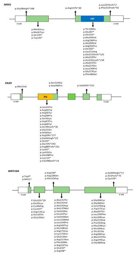
Genes 2018, 9, 255                                                                                2 of 24

that the prevalence of isolated hypodontia ranges from 3–10%, while oligodontia is less common
with a reported prevalence of 0.1–0.5%, excluding third molars. Upon inclusion of third molars,
the prevalence rises to 25% [4,6]. The most commonly missing teeth are permanent mandibular second
premolars, maxillary lateral incisors, and maxillary second premolars [3]. Females are more affected
than males in a 3:2 ratio [5].
     TA may be occasionally caused by exogenous factors, such as infections, trauma, chemotherapy,
or radiotherapy, however, the majority of cases are due to genetic factors [7]. Studies of odontogenesis
in mice have elegantly shown that tooth development is under strict genetic control, which determines
the positions, numbers and shapes of different teeth [8]. Over many years, studies using transgenic
animals provided functional data showing that disruption of genes in the bone morphogenetic protein
(BMP), fibroblast growth factor (FGF), sonic hedgehog (SHH), and wingless-related integration site
(WNT) signaling pathways resulted in severe abnormalities of tooth development ranging from defects
in tooth patterning to complete arrest of tooth development [1,4].
     In humans, mutations in several genes have been reported as causal for TA. However, known
mutations explain a restricted number of cases and much remains to be learned about the underlying
molecular mechanisms in TA [4]. Epigenetic regulation affecting a network of interconnected signaling
pathways that included tooth development was also suggested to play a role in the etiology of TA [9].
Recently, new technologies such as next-generation sequencing have proven to be valuable tools in the
identification of novel TA genes and variants, and begun to elucidate the genetic defects responsible
for this condition [10,11].
     The purpose of this review is to present an overview of genes and pathways identified as having
a role in human TA, and discuss recent findings from next-generation sequencing studies. We also
present new perspectives on the potential molecular mechanisms underlying human TA that may
bring new possibilities for future prevention and treatment.

2. Genetic Basis of Tooth Agenesis

2.1. Syndromes Associated with Tooth Agenesis
     More than 150 syndromes include TA as a phenotype. These include mostly oral-facial cleft
syndromes and ectodermal dysplasia syndromes [12,13]. Teeth share developmental mechanisms
with other ectodermal organs in terms of timing and the cellular processes involved, therefore genes
involved in odontogenesis may play a role in other developmental processes [12,14]. A summary of
the syndromic forms of TA and their etiologic genes is presented in Table 1.
Genes 2018, 9, 255 3 of 24
Table 1. Genes involved in syndromic tooth agenesis and associated phenotypes.
Gene/Locus OMIM Chromosome Syndrome Inheritance Dental/Oral Phenotypes Animal Model Animal Model Phenotype Reference
Hypodontia, microdontia, tooth Dermatosparatic phenotype
ADAMTS2 604539 5q35.3 Ehlers–Danlos syndrome AR Yes Bovine [15–17]
discoloration resembling EDS type VII C
Growth delay, bone loss,
Growth retardation, alopecia,
shortened skulls with frontal
ANTXR1 606410 2p13.3 pseudoanodontia, and optic AR Hypodontia, delayed eruption Yes [18–20]
bossing, and midfacial
atrophy (GAPO) syndrome
hypoplasia
Oligodontia-colorectal
AXIN2 604025 17q24.1 AD Oligodontia Yes Abnormal cranium morphology [21,22]
cancer syndrome
Osteogenesis imperfecta
COL1A1/2 120150 17q21.33 AD Hypodontia, oligodontia Yes Lethal, bone fractures [23]
type 1
Hypodontia, retrognathia,
micrognathia, arched/narrow
CREBBP 600140 16p13.3 Rubinstein–Taybi syndrome AD palate, talon cusps, dental Yes Skeletal malformations [24]
crowding, screwdriver incisors,
cross bite, and enamel hypoplasia
Ectodermal dysplasia, Anodontia, hypodontia, Incomplete set of conically
EDA 300451 Xq13.1 XLR Yes Canine [25–28]
hypohidrotic misshapen teeth, microdontia shaped teeth
Ectodermal dysplasia, Decreased molar number, small
Anodontia, hypodontia,
EDAR 604095 2q13 hypohidrotic/hair/ AR Yes Mouse incisor, small molars, abnormal [29–33]
oligodontia
tooth type enamel knot morphology
Abnormal tooth morphology,
Ectodermal dysplasia,
Anodontia, hypodontia, decreased molar number,
EDARADD 606603 1q42-q43 hypohidrotic/hair/ AD Yes Mouse [34–36]
taurodontism, microdontia small molars, abnormal enamel
tooth type
morphology
Ellis–van Creveld syndrome
Natal teeth, enamel abnormalities, Enamel defects, abnormal tooth
EVC 604831 4p16.2 and Weyers acrofacial AR/AD Yes Mouse [37–39]
hypodontia, microdontia morphology
dysostosis
Ellis–van Creveld syndrome Natal teeth, enamel abnormalities,
Microdontia, small upper
EVC2 607261 4p16.2 and Weyers acrofacial AR/AD hypodontia, oligodontia, Yes Mouse [37–40]
incisors, small cranium
dysostosis microdontia
Abnormal tooth morphology,
Hypodontia (maxillary incisors),
Lacrimoauriculodentodigital short incisors, small molars,
FGF10 602115 5p12 AD microdontia, delayed eruption, Yes Mouse [41–43]
syndrome abnormal palatal development,
enamel dysplasia
abnormal tongue morphologyGenes 2018, 9, 255 4 of 24
Table 1. Cont.
Gene/Locus OMIM Chromosome Syndrome Inheritance Dental/Oral Phenotypes Animal Model Animal Model Phenotype Reference
Abnormal cranium morphology,
FGFR1 136350 8p11.23 Kallmann syndrome XLR Hypodontia, cleft lip/palate Yes Mouse [44]
facial asymmetry, long incisors
Hypodontia (maxillary incisors), Arrest of tooth development,
Lacrimoauriculodentodigital
AD microdontia peg laterals, delayed Yes Mouse long incisors, decreased molar [43]
syndrome
eruption, enamel dysplasia number, micrognathia
FGFR2 176943 10q26.13
Hypodontia (maxillary canines), Arrest of tooth development,
Apert syndrome AD enamel opacities, ectopic Yes Mouse long incisors, decreased molar [45–47]
eruptions, gingival hyperplasia number, micrognathia
Tooth misalignment, long
Hypodontia, malocclusion,
Crouzon syndrome with incisors, malocclusion,
FGFR3 134934 4p16.3 AD cementomas, delayed eruption, Yes Mouse [48–50]
acanthosis nigricans prognathia, maxillary
midface hypoplasia
retrognathia
Hypodontia, delayed dental
FLNB 603381 3p14.3 Larsen syndrome AD development, class 3 occlusion, Yes Mouse Abnormal cranium morphology [51]
morphological anomalies
Axenfeld–Rieger Hypodontia, microdontia,
FOXC1 601090 6p25.3 AD Yes, Mouse Short mandible [52,53]
syndrome type 3 taurodontism
Abnormal tooth morphology,
Microdontia, enamel hypoplasia, microdontia, small mandible
GJA1 121014 6q22.31 Oculodentodigital dysplasia AD, AR Yes Mouse [54,55]
hypodontia, delayed eruption and maxilla, reduced enamel
thickness
Ectodermal dysplasia/short Delayed eruption, hypodontia, Abnormal cranium morphology,
GRHL2 608576 8q22.3 AR Yes, Mouse [56]
stature syndrome enamel hypoplasia facial and midline clefts
Abnormal tooth morphology,
IRF6 607199 1q32.2 van der Woude syndrome AD Hypodontia, cleft lip/palate Yes Mouse abnormal palatal development, [57,58]
small mandible
Hypodontia, enamel hypoplasia Short maxilla, malocclusion,
JAG1 601920 20p12.2 Alagille Syndrome AD Yes mouse [59]
and opacities, hypomineralization abnormal palate morphology
High-arched palate, malocclusion,
microdontia, a small dental arch,
KDM6A 300128 Xp11.3 Kabuki syndrome 2 XLD Yes Mouse Cranioschisis [60,61]
hypodontia, severe maxillary
recession, conical teethGenes 2018, 9, 255 5 of 24
Table 1. Cont.
Gene/Locus OMIM Chromosome Syndrome Inheritance Dental/Oral Phenotypes Animal Model Animal Model Phenotype Reference
High-arched palate, malocclusion,
microdontia, a small dental arch,
KMT2D 602113 12q13.12 Kabuki syndrome 1 AD Yes Mouse Short maxilla, flattened snout [62,63]
hypodontia, severe maxillary
retrognathia, conical teeth
Oligodontia, hypodontia,
Ectodermal dysplasia,
KREMEN1 609898 22q12.1 AR alveolar ridge deficiency, Yes No craniofacial phenotype [64]
hair/tooth type
increased palatal depth
Dental crowding, high-arched
MKKS 604896 20p12.2 Bardet-Biedl syndrome AR palate, hypodontia, malocclusion, Yes Mouse Abnormal olfactory epithelium [65]
enamel hypoplasia, retrognathia
Arrest of tooth development,
MSX1 142983 4p16.1 Witkop syndrome AD Hypodontia, oligodontia Yes Mouse nail bed and nail plates defective, [66]
cleft palate
Hypodontia, anodontia,
NEMO 300248 Xq28 Incontinentia pigmenti XLD Yes Mouse No craniofacial phenotype [67,68]
microdontia
Hypodontia, enamel defects,
NSD1 606681 5q35.3 Sotos syndrome I AD Yes Mouse No craniofacial phenotypes [69,70]
malocclusion
Hypodontia, missing lateral
Primary cilia formed then
OFD1 300170 Xp22.2 Orofaciodigital syndrome I XLD incisors, canine malposition, Yes [71,72]
disappeared, renal cysts
micrognathia
Hypodontia, enamel hypoplasia,
extensive dental caries,
Orofacial cleft 8, hypodontia of the mandibular
Arrest of tooth development,
Rapp-Hodgkin, and canines, generalized microdontia,
small mandible and maxilla,
P63 603273 3q28 Ectrodactyly, ectodermal AD prominent marginal ridges of Yes Mouse [73–75]
abnormal craniofacial
dysplasia, and cleft permanent maxillary incisors,
development, cleft palate
lip/palate syndrome 3 round-shaped permanent molars,
and barrel-shaped permanent
maxillary central incisors
Abnormal maxilla and
Axenfeld–Rieger syndrome, Hypodontia, microdontia,
PITX2 601542 4q25 AD Yes Mouse mandible morphology, [53,76–78]
type 1 enamel hypoplasia
arrested tooth development
Hypodontia, cleft lip and palate,
Cleft lip/palate-ectodermal
PVRL1 600644 11q23.3 AR abnormal dental morphology, Yes Mouse Abnormal tooth morphology [79,80]
dysplasia
microdontiaGenes 2018, 9, 255 6 of 24
Table 1. Cont.
Gene/Locus OMIM Chromosome Syndrome Inheritance Dental/Oral Phenotypes Animal Model Animal Model Phenotype Reference
Rothmund–Thomson Hypodontia, microdontia, Delayed tooth eruption,
RECQL4 603780 8q24.3 AR Yes Mouse [81]
syndrome hypoplastic teeth cleft palate
High narrow palate, midline
Abnormal tooth morphology,
RSK2 300075 Xp22.12 Coffin–Lowry syndrome XLD lingual furrow, hypodontia, Yes Mouse [82]
supernumerary teeth
and microdontia
Cleft lip and palate, single central Abnormal tooth morphology,
SHH 600725 7q36.3 Holoprosencephaly AD Yes [83,84]
incisor, micrognathia microdontia
Hypodontia, ectopic and
TBX3 601621 12q24.21 Ulnar-mammary syndrome AD Yes Secondary palate clefting [85]
hypoplastic canines
Hypodontia, micrognathia,
TCOF1 606847 5q32-q33 Treacher Collins syndrome AD Yes Short mandible and maxilla [86,87]
malocclusion, spaced teeth
Oligodontia, hypodontia, thick
TFAP2B 601601 6p12.3 Char syndrome AD Yes No craniofacial phenotype [88]
lips, retention of primary teeth
General hypoplasia and
Hypodontia, delayed eruption,
developmental delay,
Trisomy 21 190685 21q22.13 Down syndrome IC barrel-shaped permanent Yes [89,90]
hydronephrosis, heart and
maxillary central incisors
neurologic defects
TSPEAR 612920 21q22.3 Ectodermal dysplasia AR Hypodontia, microdontia Yes No craniofacial phenotypes [91]
UBR1 605981 15q15.2 Johanson–Blizzard syndrome AR Oligodontia Yes No craniofacial phenotypes [92]
Arrested tooth development of
Odontoonychodermal Oligodontia, hypodontia,
AR Yes molars, supernumerary molars, [93–95]
dysplasia microdontia
abnormal tooth morphology
WNT10A 606268 2q35
Arrested tooth development of
Schopf–Schulz–Passarge Oligodontia, hypodontia,
AR Yes molars, supernumerary molars, [94,96,97]
syndrome microdontia
abnormal tooth morphology
OMIM, Online Mendelian Inheritance in Man (https://www.ncbi.nlm.nih.gov/omim/) AR, autosomal recessive; AD, autosomal dominant, XLD, x-linked dominant, XLR, x-linked
recessive; IC, isolated cases.Genes 2018, 9, 255 7 of 24
2.1.1. Ectodermal Dysplasia Syndromes
Ectodermal dysplasias (ED) comprise a group of disorders characterized by a combination of
findings involving defects in the skin, hair, nails, teeth, exocrine and sebaceous glands. Distinct types of
ED have been reported, caused by mutations in different genes. X-linked recessive hypohidrotic (HED)
is the most common ED and caused by mutations in the EDA (ectodysplasin A) gene. Currently, more
than 200 mutations in EDA have been found in individuals with HED. The EDA protein is a type-II
trimeric transmembrane protein belonging to the tumor necrosis factor (TNF) ligand superfamily
which functions as a signaling molecule during epithelial morphogenesis [98]. Upon stimulation, EDA
binds to its receptor, EDAR (ectodysplasin A receptor), which in turn binds to the adaptor EDARADD
(EDAR-associated death domain) for activation of downstream target proteins. Mutations in EDAR
and EDARADD cause autosomal dominant and recessive forms of HED [4,7]. In addition to variable
involvement of teeth in distinct forms of ED, minor ectodermal anomalies are often observed.
Witkop syndrome is a rare autosomal dominant ED involving the teeth and nails, caused by
mutations in the MSX1 (muscle segment homeobox, homolog 1) gene. Although a few reported cases
have sparse or fine hair, most affected individuals have normal hair and sweat glands [99]. MSX1 is
a homeobox gene that belongs to a family of transcription factors that are expressed in overlapping
patterns at multiple sites of tissue interactions during vertebrate development [2]. In mice, MSX1 is
essential for initiation of the tooth germ during the bud stage and then promotes the odontogenic
potential of the dental mesenchyme [2]. Mice lacking MSX1 protein function present cleft palate,
deficient mandibular and maxillary alveolar bones, and arrest of tooth development [100]. Further,
MSX1 is a direct downstream target of WNT/ß-catenin signaling during craniofacial development and
regulates the expression of additional genes within this pathway, including BMP2 (bone morphogenetic
protein 2) and BMP4 (bone morphogenetic protein 4) [4]. Over 20 MSX1 mutations have been reported
in association with both syndromic and nonsyndromic TA phenotypes in humans.
Odontoonychodermal dysplasia (OODD) and Schopf–Schulz–Passarge syndrome (SSPS) are
autosomal recessive disorders caused by homozygous or compound heterozygous missense mutations
in WNT10A (wingless-type MMTV integration site family, member 10A). Affected individuals have
dry hair, severe TA, smooth tongue, palmoplantar keratosis, and dystrophic nails [93,94]. WNT10A
is thought to function through the canonical Wnt-ß catenin signaling pathway and activates target
genes in the nucleus through binding to LEF1 transcription factor [101]. During tooth development,
continuous Wnt-β catenin signaling in the dental epithelium and mesenchyme is required for tooth
formation and morphogenesis [102]. During mouse tooth development, WNT10A is first detected
in the enamel knot and then shifting to the secondary enamel knot to the underlying mesenchyme
and developing odontoblasts [103,104]. In WNT10A null mice, however, supernumerary teeth and
altered molar crown morphology are observed, in contrast to the TA phenotype in humans [105].
Numerous reports have shown the involvement of WNT10A mutations in OODD and SSPS, and in a
wide spectrum of autosomal recessive ectodermal dysplasias [93,94,106,107] as well as in isolated TA
cases [11,108–110].
A homozygous missense mutation (c.626T > C, p.Phe209Ser) in KREMEN1 (kringle containing
transmembrane protein 1), a negative regulator of the Wnt pathway, was identified in four
consanguineous Palestinian families with ED. This variant in its heterozygous state was also identified
in 6 out of 39 unaffected control individuals [64]. Recently, two additional variants in KREMEN1
(c.146C > G and c.773_778del) were identified as pathogenic in two Turkish families with suspected
ED [11]. KREMEN1 encodes a kringle domain-containing transmembrane protein that binds to DKK1,
creating a DKK1-Kremen-LRP6 ligand-receptor complex critical for Wnt signaling [111]. While in this
complex, Kremen triggers the internalization of LRP6 inhibiting Wnt signaling. In the absence of
DKK1, however, Kremen can increase Wnt signaling through LRP6 binding. Targeted disruption of
the Wnt regulator Kremen in mice induces limb defects and high bone density but no other obvious
phenotypes [111].Genes 2018, 9, 255 8 of 24
2.1.2. Oral-Facial Cleft Syndromes
Van der Woude syndrome (VWS) is an autosomal dominant disorder and one of the most
common clefting syndromes. Affected individuals present with cleft lip with or without cleft palate
(CL/P) and a range of associated features including lower lip pits and TA, which is present in ≈70%
of the cases. VWS is caused by mutations in the IRF6 (interferon regulatory factor 6) gene that
encodes a transcription factor highly expressed during craniofacial development and a regulator
of keratinocyte proliferation and differentiation [57]. Mice deficient for IRF6 have abnormal skin,
limb and craniofacial development, resultant from a primary defect in keratinocyte differentiation
and proliferation. Furthermore, mice homozygous for the IRF6 null allele have a cleft palate which
seems to be caused by a defect in elevation, either as a primary defect or secondary to crowding
of the craniofacial structures owing to the constrictive action of the skin or oral adhesions [112].
IRF6 mutations are recognized as primary genetic causes of isolated and syndromic CL/P [58,113,114].
Cleft lip/palate-ectodermal dysplasia syndrome is a rare, autosomal recessive disorder caused
by homozygous loss-of-function mutations of the PVRL1 (poliovirus receptor-like 1) gene encoding
nectin-1 [79]. Nectin-1 is a cell–cell adhesion molecule that is important for the initial step in the
formation of adherens junctions and tight junctions; it is expressed in keratinocytes, neurons, and the
developing face and palate. Clinical manifestations comprise a unique facial appearance with cleft
lip/palate, ectodermal dysplasia, cutaneous syndactyly of the fingers and/or toes, hypodontia and in
some cases, mental retardation [80].
2.1.3. Axenfeld–Rieger Syndrome
Axenfeld–Rieger syndrome (MIM #180500) is an autosomal dominant disorder characterized by
abnormal development of the anterior segment of the eye, and results in blindness from glaucoma in
approximately 50% of affected individuals [115]. Systemic anomalies are associated and include failure
of involution of the umbilicus, hypospadias and dental anomalies ranging from microdontia, TA, and
tooth malformations [116]. The cause of Axenfeld–Rieger syndrome has been attributed to mutations
in the PITX2 (paired like homeodomain 2) gene, which encodes the earliest transcription marker of
tooth development and is expressed in the oral epithelium and dental placode [117]. PITX2 null mice
have tooth development arrested at E12.5 [118]. Additionally, PITX2 has been shown to play a critical
role in establishing left–right asymmetry in vertebrates [119].
2.1.4. Familial Adenomatous Polyposis Syndrome
Familial adenomatous polyposis (FAP) is an autosomal dominant condition characterized
by the development of multiple adenomatous polyps in the colon and rectum with high risk of
subsequent malignant transformation. In addition, extracolonic changes occur in many affected
subjects. These include epidermoid cysts, desmoid tumours, congenital hypertrophy of retinal pigment
epithelium (CHRPE), osseous changes in the jaws and skeleton, and dental anomalies. FAP results
from germline mutations in the APC (adenomatous polyposis coli) gene on chromosome 5q21 [120].
Approximately 17% of individuals with APC gene mutations have dental anomalies, particularly
supernumerary teeth and compound odontomas, although cases of TA as well as impacted teeth
have also been reported. Importantly, in addition to the established association between certain
dental anomalies and FAP, an association between TA and genetic predisposition to colon cancer was
suggested [22,121,122].
2.1.5. Oligodontia–Colorectal Cancer Syndrome
A germline nonsense mutation in AXIN2 (c.1996C > T, p.Arg656*) was identified as the likely
cause of autosomal dominant oligodontia (severe TA) and colorectal cancer segregating in a large,
four-generation Finnish family. Eleven members of the family lacked at least eight permanent teeth,
two of whom developed only three permanent teeth. Colorectal cancer or precancerous lesions ofGenes 2018, 9, 255 9 of 24
variable types were found in eight of the patients with oligodontia [22]. In this same study, a second
frameshift mutation leading to a heterozygous 1-bp insertion (c.1994_1995insG, p.706*) in exon 7
of the AXIN2 gene was identified in an unrelated individual with oligodontia, suggesting that this
gene may contribute to both tooth development and tumor development later in life [22]. A recent
study showed that variations in colorectal cancer-related genes (ATF1 and DUSP10) were significantly
associated with TA (albeit in isolated cases); further, Atf1 and Dusp10 expression was detected in the
mouse developing teeth from early bud stages to the formation of the complete tooth, suggesting
a potential role for these genes and their encoded proteins in toothdevelopment [123]. Taken together,
these findings continue to support a potential overlap in the molecular etiology of TA and colorectal
cancer, although no cause-effect relationship can yet be established and more research is warranted in
this area.
2.2. Isolated (Nonsyndromic) Tooth Agenesis
Syndromic forms of TA and genes implicated during normal tooth development in animal models
have provided important clues to identify candidate genes for isolated TA in humans. To date,
numerous genes have been proposed as etiologic for isolated TA (Table 2).
Table 2. Genes involved in isolated tooth agenesis.
Gene OMIM Chromosome Dental Phenotypes Reference(s)
AXIN2 604025 17q24.1 Oligodontia, hypodontia [22,98,124,125]
ANTXR1 606410 2p13.3 Oligodontia, hypodontia [10]
COL17A1 113811 10q25.1 Hypodontia [11]
DKK1 605189 10q21.1 Hypodontia [11,126]
EDA 300451 Xq13.1 Oligodontia, hypodontia [33,127–130]
EDAR 604095 2q13 Oligodontia, hypodontia [33]
EDARADD 606603 1q42-q43 Oligodontia, hypodontia [33]
FGFR1 136350 8p11.23 Hypodontia [131]
GREM2 608832 1q43 Hypodontia, microdontia, taurodontia [132]
IRF6 607199 1q32.2 Hypodontia, lip pits [131,133]
MSX1 142983 4p16.2 Oligodontia, hypodontia [98,134,135]
LAMA3 600805 18q11.2 Hypodontia [11]
LRP6 603507 12p13.2 Oligodontia [11,136,137]
LTBP3 602090 11q13.1 Oligodontia, hypodontia [138,139]
PAX9 167416 14q13.3 Oligodontia, hypodontia, microdontia [98,140–145]
SMOC2 607223 6q27 Oligodontia, microdontia, abnormal morphology [146,147]
WNT10A 606268 2q35 Oligodontia, hypodontia [11,108–110,148,149]
WNT10B 601906 12q13.12 Oligodontia, microdontia [150,151]
2.2.1. MSX1
Mutations in MSX1 were the first to be described in individuals with isolated TA [152]. Since
then, over 20 mutations in MSX1 have been reported in association with isolated TA, most of which
are nonsense or missense mutations located in the homeobox domain, and which suggest that
haploinsufficiency of MSX1 underlies TA phenotypes [153] (Figure 1). Mutations in the homeobox
domain disrupt DNA-binding and preferentially cause isolated TA, meanwhile variants in the
natively unfolded N-terminal part of the protein generally cause oral-facial clefts. These observations
suggest that the effect of MSX1 mutations are directly related to the affected protein domain.
MSX1-associated TA typically includes missing maxillary and mandibular second premolars and
maxillary first premolars.Genes 2018, 9, 255 10 of 24
Figure 1. Location of predicted missense, frameshift, and nonsense mutations in MSX1, PAX9, and
WNT10A genes. Green boxes represent exons, horizontal lines between exons represent introns.
HD, corresponds to homeodomain in MSX1. PD, corresponds to paired domain in PAX9. UTR:
Untranslated region.
2.2.2. PAX9
PAX9 belongs to the paired box (PAX) family of transcription factors that are essential for normal
development in several multicellular organisms. In addition to MSX1, PAX9 has also long been
implicated in isolated TA phenotypes and is one of the most widely studied genes in odontogenesis [4].Genes 2018, 9, 255 11 of 24
PAX9 is expressed in the presumptive dental mesenchyme to activate signals and initiate tooth
development. Absence of PAX9 in mice results in arrest of tooth development at the bud stage [154,155].
To date, more than 30 variations in PAX9 have been described in association with TA, most of
which are insertions/deletions or missense mutations located in exon 2 of the gene and affecting the
paired domain of the PAX9 protein [156] (Figure 1). The presence of PAX9 variation has primarily been
associated with agenesis of permanent second molars, followed by second premolars; a few reports of
agenesis of anterior teeth also exist [7,156]. In general, the severity of the TA phenotype is associated
with the type of mutation and its impact on PAX9 function. Individuals with nonsense/frameshift
mutations present with a more severe phenotype when compared to those with missense mutations.
In TA, known PAX9 mutations are heterozygous and show autosomal dominant inheritance, indicating
that haploinsufficiency is likely contributing to the phenotype. Smaller tooth crown dimensions
throughout the dentition have also been reported in TA patients with PAX9 mutations [157].
2.2.3. AXIN2
Rare and common variants in AXIN2 have been found in association with isolated TA, presenting
a mixed pattern of affected teeth [98,124,125]. Agenesis of molars, lower incisors and upper lateral
incisors have been described in TA individuals, and the absence of at least one incisor is frequently
reported [4,98]. Five AXIN2 mutations have been widely reported in the literature, including four
missense (c.956 + 16A > G; p.Pro50Ser, c.2051C > T; p.Ala684Val, c.2062C > T; p.Leu688Leu, and
c.2272G > A; p.Ala758Thr), and one frameshift (c.1994insG; p.Asn666GlyfsX41). The presence of this
frameshift mutation was associated with more missing teeth than missense mutations in all affected
individuals [98,158].
2.2.4. WNT10A
WNT10A has been the focus of many genetic studies of TA. Over 50 heterozygous, homozygous as
well as compound heterozygous variants in WNT10A have been identified in 15.8% of TA patients with
1 to 3 missing teeth, and in ≈52% of patients with >4 missing teeth [109]. Recent genotype–phenotype
correlations have provided insights for the role of WNT10A in TA. Overall, WNT10A compound
heterozygous mutations have been found in association with severe TA and a larger number of
missing teeth in comparison to individuals with a single variant [108–110,148]. While there are no
preferential patterns of missing teeth in individuals with WNT10A variants, the absence of maxillary
and mandibular molars, as well as mandibular incisors is often reported [149]. Of note, heterozygous
WNT10A variants have also been identified in unaffected individuals in TA families, as well as in
unrelated control individuals with no TA or family history of TA [108,109]. It has been estimated that
approximately 41% of individuals showing a single heterozygous variant in WNT10A will not have
TA [148].
Figure 1 shows WNT10A variants identified in TA patients. A few WNT10A variants have
been suggested to be common ‘hotspots’ for mutations in specific populations. For example,
the c.637G > A (p.Gly213Ser) variant has been found more frequently in Asian populations [108,149],
meanwhile the c.682T > A (p.Phe228Ile) variant has been widely reported in homozygous or
heterozygous forms in Caucasian individuals with TA, but also in normal controls at a frequency
of 2.3% [109]. The Phe228Ile variant is the most commonly found variant, and often described in
combination with additional variants in WNT10A or in other genes [159,160]. These findings support
an oligogenic inheritance model for TA as discussed later in this review.
2.2.5. LRP6
LRP6 (LDL receptor related protein 6) is a co-receptor in the Wnt/β-catenin pathway and has been
recently reported to contribute to isolated TA in different studies [11,136,137]. Six variants, including
a nonsense variant (c.1779dupT, p.Glu594*), two insertion (c.2224_2225dupTT, p.Leu742Phefs*7 and
c.1144_1145dupAG, p.Ala383Glyfs*8) and a splice-site (c.3607 + 3_6del, p.?) variant resulting inGenes 2018, 9, 255 12 of 24
a truncated mRNA product, as well as a missense variant (c.56C > T, p.Ala19Val) were found in
individuals with sporadic TA and/or segregating with TA in families [11,136,137]. In mice, LRP6
expression was noted in the tooth follicle and inner enamel epithelium [137], while homozygous
deletion of LRP6 led to severe skeletal abnormalities and lethality [161].
2.2.6. Other Genes Recently Implicated in TA
Mutations in GREM2, which encodes GREMLIN2, were identified in 7 patients with TA
(hypodontia) and additional malformations, including taurodontism, sparse and slow-growing hair
and dry and itchy skin [132]. GREMLIN2 is known to regulate BMPs in embryonic development.
Specifically, BMP4 has an important role in tooth development and its knockdown resulted in the arrest
of tooth development in mice [8]. Interestingly, GREM2 knockout mice have small and malformed
teeth but do not have tooth development arrested. However, these findings suggest a potential
role for GREM2 during tooth development [162]. Three missense mutations in GREM2 (p.Ala13Val,
p.Glu136Asp and p.Gln76Glu) were identified as pathogenic in individuals with isolated TA and
have not been reported in association with other structural malformations. GREM2 mutations exhibit
variable expressivity even within the same families [132].
EDA, EDAR and EDARADD have also been suggested to contribute to isolated TA [98,129,163].
In a genotype–phenotype correlation study, all EDA mutations in individuals with isolated TA were
missense mutations and most likely to be located in the TNF domain [163].
Another WNT pathway gene, WNT10B (wingless-type MMTV integration site family, member
10B), has also been implicated in isolated TA, albeit mostly in families from China and Thailand.
Three heterozygous missense mutations (c.632G > A, p.Arg211Gln; c.569C > G, p.Pro190Arg; and
c.851T > G, p.Phe284Cys) and one nonsense mutation (c.786G > A, p.Trp262*) in WNT10B were
identified in Chinese individuals with TA, especially those missing the upper lateral incisors [151].
More recently, two additional heterozygous missense mutations (c.475G > C, p.Ala159Pro and
c.1052G > A, p.Arg351His) were identified in five Thai families, and associated with isolated TA
and other dental anomalies including microdontia and taurodontism [150].
A homozygous missense variant c.1312C > T (p.Arg438Cys) in ANTXR1 (anthrax toxin receptor 1)
was identified in association with TA (oligodontia) in a Turkish family [10]. Homozygous and biallelic
variants in ANTXR1 have been associated with Growth retardation, Alopecia, Pseudoanodontia,
and Optic atrophy (GAPO) syndrome, characterized by delayed growth, alopecia, failure of tooth
eruption, and optic atrophy segregating as an AR trait [18,19,164]. Targeted disruption of Antxr1 in
mice resulted in viable mice without major structural defects, although dental overgrowth, incisor
misalignment, and dental dysplasia were observed, due to an accumulation of extracellular matrix
in various tissues [165]. Antxr1 expression was detected in the epithelium of developing tongue,
maxillary and mandibular processes, as well as in the dental epithelium and mesenchyme at early
stages of tooth development. At later stages, Antxr1 expression was noted in the epithelium of the
enamel organ and in the dental papilla, and then shifted to the polarized layer of ameloblasts and
differentiating odontoblasts. ANTXR1 is a tumor-specific endothelial marker implicated in colorectal
cancer, and upregulated in tumor angiogenesis [166–168]. Previously, Lammi et al. [22] showed that
variants in the tumor suppressor gene AXIN2 segregated in a family with severe TA (oligodontia)
and colorectal cancer, and suggested that TA and colorectal cancer may have a common genetic
etiology. Numerous studies have since reported on variations in cancer-related genes in association
with TA [123,169,170]. These findings highlight the complex nature of TA and emphasize the need to
consider modifier genes and/or gene-gene interactions in studies of this condition.
A recent genome-wide association study (GWAS) that included over 1900 TA cases and
330,000 controls of European ancestry identified 4 novel risk variants that associate with TA, and 5 that
associate with a combined phenotype of TA plus oral-facial clefts [171]. Dental anomalies are frequent
findings in children with oral-facial clefts, and cleft subphenotypes have been proposed based on the
pattern of the associated dental anomalies [172]. Of the 9 variants found, 5 were located in or close toGenes 2018, 9, 255 13 of 24
Wnt pathway genes that have been implicated in tooth development and/or development of other
ectodermal structures (EDA, EDAR, FOXI3, FORXP1 and LEF1), and 4 were located in or close to genes
that have not been implicated in TA or tooth development (ASCL5/CACNA1S, ARHGAP15, NOL11
and FAM49A). In addition, two known variants in WNT10A (p.Phe228Ile and p.Cys107*) were also
found to be significantly associated with TA in this GWAS [171].
3. Monogenic vs. Oligogenic Inheritance Models
In recent years, oligogenic inheritance and multi-locus variation models have been proposed for a
number of Mendelian diseases, further establishing the concept of mutational load in human genetic
disease [173]. For TA, evidence for oligogenic inheritance is emerging, supported by the findings
of recent whole exome sequencing studies and/or direct sequencing studies with more than one
candidate gene [10,11].
Digenic mutations in MSX1 and PAX9 had been reported as associated with a more severe
TA phenotype (15–17 missing teeth) [142], and the interactions between these genes had begun to
be elucidated. Studies have shown that PAX9 interacts with MSX1 to synergistically activate the
expression of downstream tooth development genes, i.e., BMP4, which is essential for proper tooth
morphogenesis [2]. The presence of digenic mutations in these genes might abolish their interactions
and thus lead to more severe TA phenotypes [142].
More recently, biallelic or heterozygous genotypes of WNT10A were found in TA patients who
also presented homozygous or heterozygous genotypes of EDA, EDAR or EDARADD, suggesting
the combined phenotypic effects of alleles in distinct genes as contributing to TA [174]. Additionally,
compound heterozygous mutations in WNT10A (IVS2 + 1G > A and c.637G > A) were identified
segregating together with a missense heterozygous variant in GREM2 (c.38C > T) in a patient with TA
of maxillary permanent canines [132].
Additional heterozygous splicing mutations in DKK1 (dickkopf WNT signaling pathway inhibitor
1; c.548-4G > T) and in COL17A1 (collagen type XVII alpha 1 chain; c.3277 + 3G > C), and a heterozygous
missense variant in LAMA3 (laminin subunit alpha 3; c.2798G > T) were identified segregating with
TA in one consanguineous Turkish family [11]. Pathogenic mutations in these genes had not yet been
identified in individuals with TA, although they can be considered biologically plausible candidate
genes due to their biological roles and/or disease-associated phenotypes. DKK1 encodes a high-affinity
dickkopf homolog 1 transmembrane receptor that cooperates with LRP6 to block Wnt signaling
during development and other cellular processes [175]. In mice, DKK1 is expressed in the dental
mesenchyme, odontoblasts and osteoblasts, and its ectopic expression in the oral epithelia of transgenic
mouse embryos resulted in blocked epithelial and mesenchymal signaling leading to arrest of tooth
development at the early bud stage [176]. A common single-nucleotide polymorphism in DKK1
(rs11001553) was previously associated with isolated TA in a Chinese Han population [126].
Mutations in LAMA3 cause junctional epidermolysis bullosa (OMIM #226650), an autosomal
recessive skin disorder characterized by the presence of multiple blisters and erosions, dystrophic nails,
enamel hypoplasia and hypodontia [177]. Further, targeted disruption of LAMA3 in mice resulted
in defects of ameloblast differentiation [178]. Variations in COL17A1 have also been described in
epidermolysis bullosa patients with enamel defects [179].
A homozygous variant (c.-387delC > G) in the 5’ UTR of the PITX2 gene, described above as
etiologic for Axenfeld–Rieger syndrome, and a homozygous missense variant in BMP4 (c.T455C,
p.Val152Ala) were identified segregating with isolated TA in two siblings from an Italian family [180].
The finding of likely pathogenic alleles in more than one locus suggests the potential for oligogenic
inheritance and multilocus variation models in isolated TA, likely contributing to the variable
phenotypes. With advances in genome-wide sequencing studies of well-characterized TA individuals
and families, and careful genotype-phenotype correlations, new TA genes acting individually or
interactively with other genes are likely to be identified.Genes 2018, 9, 255 14 of 24
4. Genetic Pathways as the Focus of Future Studies
Over the years, studies using transgenic animals demonstrated that defects in genes belonging
to BMP, FGF and WNT signaling pathways resulted in severe abnormalities of tooth development
ranging from defects in tooth patterning to complete arrest of tooth development [1,4,8]. Meanwhile,
mutations in genes belonging to the FGF family have not yet been described in association with TA,
whereas a single variant in BMP4 (see above) was found in one TA family.
The current available evidence supports a significant role for WNT pathway genes in isolated
TA, mostly supported by the higher frequency of pathogenic mutations in AXIN2, WNT10A, WNT10B
and LRP6 in TA individuals [11,22,150,171]. WNT signaling molecules are essential for patterning,
proliferation and differentiation of multiple cell types during embryonic development. Secretion of
WNTs, particularly WNT4, WNT6 and WNT10, from the dental epithelium has been reported as critical
for tooth development, as the absence of WNT signaling leads to a dysfunctional enamel knot and
subsequently in arrest of tooth development [103,181].
EDA, EDAR and EDARADD, with roles in both syndromic and isolated TA, belong to the NF-kB
signaling pathway. Additional genes in the NF-kB pathway include NEMO (inhibitor of nuclear
factor kappa B kinase subunit gamma), an important pathway modulator, and TRAF6 (TNF receptor
associated factor 6), although little is known about the exact roles of these genes in tooth development
and variations in these two genes were reported in individuals with syndromic TA [4].
Based on the aforementioned observations of multilocus variation as a potential explanation for
some TA cases, with candidate genes belonging to the same or different signaling pathways, it is
presumable to hypothesize that isolated TA may be the result of variation in more than one gene,
acting individually or in combination with other genes and contributing to the variable expressivity of
the condition [182]. Determining the full spectrum of putative defective genes in TA, the pathways in
which they belong (Figure 2), their functions and interactive partners, will allow for improving our
understanding of the underlying mechanisms in TA and may be the basis of future prevention and
tooth replacement strategies.
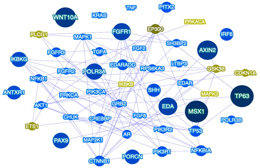
Figure 2. Tooth agenesis gene network as predicted by Phenolyzer [183]. The network shown includes
the top 50 prioritized genes, and their predicted relations with seed genes. Larger dark blue nodes
indicate seed genes, medium royal blue nodes indicate interacting genes. Green font indicates predicted
genes. Blue lines indicate protein–protein interactions.Genes 2018, 9, 255 15 of 24
5. Conclusions
Isolated TA is a heterogeneous condition with variable expressivity. While variations in numerous
genes have been attributed as causal for TA, the etiology of TA in many individuals is still unsolved
and may reflect mutations in genes yet unknown to tooth development, or the presence of multilocus
variation. Moreover, environmental and epigenetic factors may also be considered likely contributors
to TA phenotypes and should be explored in future studies. Next-generation sequencing studies
of well-characterized individuals and families present the unique ability to identify all of the
TA-predisposing variants throughout the genome while revealing important genetic and network
interactions that may be critical for tooth development. Further genetic and functional studies focusing
on newly identified genes and pathways have the potential to elucidate the genetic landscape of
isolated TA and provide insights into preventive and treatment strategies. Targeted therapeutics for
TA-relevant genes and/or pathways may represent future tooth replacement therapies.
Author Contributions: M.A.W. contributed with literature search, provided analysis and tables, critically revised
the paper; A.L. conceived and wrote the paper; critically revised the paper.
Acknowledgments: This study was supported by NIH/NIDCR R03-DE024596 to A.L.
Conflicts of Interest: The authors declare no conflict of interest.
References
1. Bei, M. Molecular genetics of tooth development. Curr. Opin. Genet. Dev. 2009, 19, 504–510. [CrossRef]
[PubMed]
2. Thesleff, I.; Vaahtokari, A.; Vainio, S.; Jowett, A. Molecular mechanisms of cell and tissue interactions during
early tooth development. Anat. Rec. 1996, 245, 151–161. [CrossRef]
3. Gorlin, R.J.; Cohen, M.M., Jr.; Levin, S.L. Syndromes of the Head and Neck, 3rd ed.; Oxford University Press:
New York, NY, USA, 1990.
4. Yin, W.; Bian, Z. The gene network underlying hypodontia. J. Dent. Res. 2015, 94, 878–885. [CrossRef]
[PubMed]
5. Polder, B.J.; Van’t Hof, M.A.; Van der Linden, F.P.; Kuijpers-Jagtman, A.M. A meta-analysis of the prevalence
of dental agenesis of permanent teeth. Commun. Dent. Oral Epidemiol. 2004, 32, 217–226. [CrossRef] [PubMed]
6. Carter, K.; Worthington, S. Morphologic and demographic predictors of third molar agenesis: A systematic
review and meta-analysis. J. Dent. Res. 2015, 94, 886–894. [CrossRef] [PubMed]
7. Ye, X.; Attaie, A.B. genetic basis of nonsyndromic and syndromic tooth agenesis. J. Pediatr. Genet. 2016, 5,
198–208. [CrossRef] [PubMed]
8. Lan, Y.; Jia, S.; Jiang, R. Molecular patterning of the mammalian dentition. Semin. Cell Dev. Biol. 2014, 25,
61–70. [CrossRef] [PubMed]
9. Wang, J.; Sun, K.; Shen, Y.; Xu, Y.; Xie, J.; Huang, R.; Zhang, Y.; Xu, C.; Zhang, X.; Wang, R.; et al.
DNA methylation is critical for tooth agenesis: Implications for sporadic non-syndromic anodontia and
hypodontia. Sci. Rep. 2016, 6, 19162. [CrossRef] [PubMed]
10. Dinckan, N.; Du, R.; Akdemir, Z.C.; Bayram, Y.; Jhangiani, S.N.; Doddapaneni, H.; Hu, J.; Muzny, D.M.;
Guven, Y.; Aktoren, O.; et al. A biallelic ANTXR1 variant expands the anthrax toxin receptor associated
phenotype to tooth agenesis. Am. J. Med. Genet. A 2018, 176, 1015–1022. [CrossRef] [PubMed]
11. Dinckan, N.; Du, R.; Petty, L.E.; Coban-Akdemir, Z.; Jhangiani, S.N.; Paine, I.; Baugh, E.H.; Erdem, A.P.;
Kayserili, H.; Doddapaneni, H.; et al. Whole-exome sequencing identifies novel variants for tooth agenesis.
J. Dent. Res. 2018, 97, 49–59. [CrossRef] [PubMed]
12. Nieminen, P. Genetic basis of tooth agenesis. J. Exp. Zool. B Mol. Dev. Evol. 2009, 312, 320–342. [CrossRef]
[PubMed]
13. Howe, B.J.; Cooper, M.E.; Vieira, A.R.; Weinberg, S.M.; Resick, J.M.; Nidey, N.L.; Wehby, G.L.; Marazita, M.L.;
Moreno Uribe, L.M. Spectrum of dental phenotypes in nonsyndromic orofacial clefting. J. Dent. Res. 2015,
94, 905–912. [CrossRef] [PubMed]
14. Vieira, A.R. Oral clefts and syndromic forms of tooth agenesis as models for genetics of isolated tooth
agenesis. J. Dent. Res. 2003, 82, 162–165. [CrossRef] [PubMed]Genes 2018, 9, 255 16 of 24
15. Colige, A.; Nuytinick, L.; Hausser, I.; van Esse, A.J.; Thiry, M.; Herens, C.; Adès, L.C.; Malfait, F.; Paepe, A.D.;
Franck, P.; et al. Novel types of mutation responsible for the dermatosparactic type of Ehlers-Danlos
syndrome (type VIIC) and common polymorphisms in the ADAMTS2 gene. J. Invest. Dermatol. 2004, 123,
656–663. [CrossRef] [PubMed]
16. Colige, A.; Sieron, A.L.; Li, S.W.; Schwarze, U.; Petty, E.; Wertelecki, W.; Wilcox, W.; Krakow, D.; Cohn, D.H.;
Reardon, W.; et al. Human Ehlers-Danlos syndrome type VIIC and bovine dermatosparaxis are caused
by mutations in the procollagen I N-proteinase gene. Am. J. Hum. Genet. 1999, 65, 308–317. [CrossRef]
[PubMed]
17. Bekhouche, M.; Colige, A. The procollagen N-proteinases ADAMTS2, 3 and 14 in pathophysiology.
Matrix Biol. 2015, 44, 46–53. [CrossRef] [PubMed]
18. Bayram, Y.; Pehlivan, D.; Karaca, E.; Gambin, T.; Jhangiani, S.N.; Erdin, S.; Elcioglu, N.H. Whole exome
sequencing identifies three novel mutations in ANTXR1 in families with GAPO syndrome. Am. J. Med.
Genet. A 2014, 164, 2328–2334. [CrossRef] [PubMed]
19. Stranecky, V.; Hoischen, A.; Hartmannová, H.; Zaki, M.S.; Chaudhary, A.; Zudaie, E.; Nosková, L.;
Baresová, V.; Pristoupilová, A.; Hodanová, K.; et al. Mutations in ANTXR1 cause GAPO syndrome. Am. J.
Hum. Genet. 2013, 92, 792–799. [CrossRef] [PubMed]
20. Salas-Alanis, J.C.; Scott, C.A.; Fajardo-Ramírez, O.R.; Duran, C.; Moreno-Treviño, M.G.; Kelsell, D.P. New
ANTXR1 gene mutation for GAPO syndrome: A case report. Mol. Syndromol. 2016, 7, 160–163. [CrossRef]
[PubMed]
21. Marvin, M.L.; Mazzoni, S.M.; Herron, C.M.; Edwards, S.; Gruber, S.B.; Petty, E.M. AXIN2-associated
autosomal dominant ectodermal dysplasia and neoplastic syndrome. Am. J. Med. Genet. A 2011, 155, 898–902.
[CrossRef] [PubMed]
22. Lammi, L.; Arte, S.; Somer, M.; Järvinen, H.; Lahermo, P.; Thesleff, I.; Pirinen, S.; Nieminen, P. Mutations
in AXIN2 cause familial tooth agenesis and predispose to colorectal cancer. Am. J. Hum. Genet. 2004, 74,
1043–1050. [CrossRef] [PubMed]
23. Malmgren, B.; Andersson, K.; Lindahl, K.; Kindmark, A.; Grigelioniene, G.; Zachariadis, V.; Dahllöf, G.;
Aström, E. Tooth agenesis in osteogenesis imperfecta related to mutations in the collagen type I genes.
Oral Dis. 2017, 23, 42–49. [CrossRef] [PubMed]
24. Bloch-Zupan, A.; Stachtou, J.; Emmanouil, D.; Arveiler, B.; Griffiths, D.; Lacombe, D. Oro-dental features as
useful diagnostic tool in Rubinstein-Taybi syndrome. Am. J. Med. Genet. A 2007, 143, 570–573. [CrossRef]
[PubMed]
25. Monreal, A.W.; Zonana, J.; Ferguson, B. Identification of a new splice form of the EDA1 gene permits
detection of nearly all X-linked hypohidrotic ectodermal dysplasia mutations. Am. J. Hum. Genet. 1998, 63,
380–389. [CrossRef] [PubMed]
26. Han, D.; Gong, Y.; Wu, H.; Zhang, X.; Yan, M.; Wang, X.; Qu, H.; Feng, H.; Song, S. Novel EDA mutation
resulting in X-linked non-syndromic hypodontia and the pattern of EDA-associated isolated tooth agenesis.
Eur. J. Med. Genet. 2008, 51, 536–546. [CrossRef] [PubMed]
27. Lexner, M.O.; Bardow, A.; Juncker, I.; Jensen, L.G.; Almer, L.; Kreiborg, S.; Hertz, J.M. X-linked hypohidrotic
ectodermal dysplasia. Genetic and dental findings in 67 Danish patients from 19 families. Clin. Genet. 2008,
74, 252–259. [CrossRef] [PubMed]
28. Tao, R.; Jin, B.; Guo, S.Z.; Qing, W.; Feng, G.Y.; Brooks, D.G.; Liu, L.; Xu, J.; Li, T.; Yan, Y. A novel missense
mutation of the EDA gene in a Mongolian family with congenital hypodontia. J. Hum. Genet. 2006, 51,
498–502. [CrossRef] [PubMed]
29. Henningsen, E.; Svendsen, M.T.; Lidballe, D.L.; Jensen, P.K. A novel mutation in the EDAR gene causes
severe autosomal recessive hypohidrotic ectodermal dysplasia. Am. J. Med. Genet. A 2014, 164, 2059–2061.
[CrossRef] [PubMed]
30. Naeem, M.; Muhammad, D.; Ahmad, W. Novel mutations in the EDAR gene in two Pakistani consanguineous
families with autosomal recessive hypohidrotic ectodermal dysplasia. Br. J. Dermatol. 2005, 153, 46–50.
[CrossRef] [PubMed]
31. Naqvi, S.K.; Wasif, N.; Javaid, H.; Ahmad, W. Two novel mutations in the gene EDAR causing autosomal
recessive hypohidrotic ectodermal dysplasia. Orthod. Craniofac. Res. 2011, 14, 156–159. [CrossRef] [PubMed]Genes 2018, 9, 255 17 of 24
32. Shimomura, Y.; Sato, N.; Miyashita, A.; Hashimoto, T.; Ito, M.; Kuwano, R. A rare case of hypohidrotic
ectodermal dysplasia caused by compound heterozygous mutations in the EDAR gene. J. Invest. Dermatol.
2004, 123, 649–655. [CrossRef] [PubMed]
33. Zeng, B.; Zhao, Q.; Li, S.; Lu, H.; Lu, J.; Ma, L.; Zhao, W.; Yu, D. Novel EDA or EDAR Mutations identified in
patients with X-linked hypohidrotic ectodermal dysplasia or non-syndromic tooth agenesis. Genes 2017, 8,
259. [CrossRef] [PubMed]
34. Bal, E.; Baala, L.; Cluzeau, C.; El Kerch, F.; Ouldim, K.; Hadj-Rabia, S.; Bodemer, C.; Munnich, A.; Courtois, G.;
Sefiani, A. Autosomal dominant anhidrotic ectodermal dysplasias at the EDARADD locus. Hum. Mutat.
2007, 28, 703–709. [CrossRef] [PubMed]
35. Headon, D.J.; Emmal, S.A.; Ferguson, B.M.; Tucker, A.S.; Justice, M.J.; Sharpe, P.T.; Zonana, J.; Overbeek, P.A.
Gene defect in ectodermal dysplasia implicates a death domain adapter in development. Nature 2001, 414,
913–916. [CrossRef] [PubMed]
36. Wohlfart, S.; Söder, S.; Smahi, A.; Schneider, H. A novel missense mutation in the gene EDARADD associated
with an unusual phenotype of hypohidrotic ectodermal dysplasia. Am. J. Med. Genet. A 2016, 170, 249–253.
[CrossRef] [PubMed]
37. Ruiz-Perez, V.L.; Ide, S.E.; Strom, T.M.; Lorenz, B.; Wilson, D.; Woods, K.; King, L.; Francomano, C.;
Freisinger, P.; Spranger, S.; et al. Mutations in a new gene in Ellis-van Creveld syndrome and Weyers
acrodental dysostosis. Nat. Genet. 2000, 24, 283–286. [CrossRef] [PubMed]
38. D’Asdia, M.C.; Torrent, I.; Consoli, F.; Ferese, R.; Magliozzi, M.; Bernardini, L.; Guida, V.; Diglio, M.C.;
Marino, B.; Dallapiccola, B.; et al. Novel and recurrent EVC and EVC2 mutations in Ellis-van Creveld
syndrome and Weyers acrofacial dyostosis. Eur. J. Med. Genet. 2013, 56, 80–87. [CrossRef] [PubMed]
39. Ye, X.; Song, G.; Fan, M.; Shi, L.; Jabs, E.W.; Huang, S.; Guo, R.; Bian, Z. A novel heterozygous deletion in the
EVC2 gene causes Weyers acrofacial dysostosis. Hum Genet 2006, 119, 199–205. [CrossRef] [PubMed]
40. Shen, W.; Han, D.; Zhang, J.; Zhao, H.; Feng, H. Two novel heterozygous mutations of EVC2 cause a mild
phenotype of Ellis-van Creveld syndrome in a Chinese family. Am. J. Med. Genet. A 2011, 155, 2131–2136.
[CrossRef] [PubMed]
41. Entesarian, M.; Matsson, H.; Klar, J.; Bergendal, B.; Olson, L.; Arakaki, R.; Hayashi, Y.; Ohuchi, H.; Falahat, B.;
Bolstad, A.I.; et al. Mutations in the gene encoding fibroblast growth factor 10 are associated with aplasia of
lacrimal and salivary glands. Nat Genet 2005, 37, 125–127. [CrossRef] [PubMed]
42. Milunsky, J.M.; Zhao, G.; Maher, T.A.; Colby, R.; Everman, D.B. LADD syndrome is caused by FGF10
mutations. Clin. Genet. 2006, 69, 349–354. [CrossRef] [PubMed]
43. Rohmann, E.; Brunner, H.G.; Kayserili, H.; Uyguner, O.; Nürnberg, G.; Lew, E.D.; Dobbie, A.;
Eswarakumar, V.P.; Uzumcu, A.; Ulubil-Emeroglu, A.; et al. Mutations in different components of FGF
signaling in LADD syndrome. Nat. Genet. 2006, 38, 414–417. [CrossRef] [PubMed]
44. Bailleul-Forestier, I.; Gros, C.; Zenaty, D.; Bennaceur, S.; Leger, J.; de Roux, N. Dental agenesis in Kallmann
syndrome individuals with FGFR1 mutations. Int. J. Paediatr. Dent. 2010, 20, 305–312. [CrossRef] [PubMed]
45. Letra, A.; de Almeida, A.L.; Kaizer, R.; Esper, L.A.; Sgarbosa, S.; Granjeiro, J.M. Intraoral features of Apert’s
syndrome. Oral Surg. Oral Med. Oral Pathol. Oral Radiol. Endod. 2007, 103, e38–e41. [CrossRef] [PubMed]
46. Stavropoulos, D.; Bartzela, T.; Bronkhorst, E.; Mohlin, B.; Hagberg, C. Dental agenesis patterns of permanent
teeth in Apert syndrome. Eur. J. Oral Sci. 2011, 119, 198–203. [CrossRef] [PubMed]
47. Ibrahimi, O.A.; Chiu, E.S.; McCarthy, J.G.; Mohammadi, M. Understanding the molecular basis of Apert
syndrome. Plast. Reconstr. Surg. 2005, 115, 264–270. [PubMed]
48. Meyers, G.A.; Orlow, S.J.; Munro, I.R.; Przylepa, K.A.; Jabs, E.W. Fibroblast growth factor receptor 3 (FGFR3)
transmembrane mutation in Crouzon syndrome with acanthosis nigricans. Nat. Genet. 1995, 11, 462–464.
[CrossRef] [PubMed]
49. Reardon, W.; Wilkes, D.; Rutland, P.; Pulleyn, L.J.; Malcolm, S.; Dean, J.C.; Evans, R.D.; Jones, B.M.;
Hayward, R.; Hall, C.M. Craniosynostosis associated with FGFR3 pro250arg mutation results in a range
of clinical presentations including unisutural sporadic craniosynostosis. J. Med. Genet. 1997, 34, 632–636.
[CrossRef] [PubMed]
50. Wilkes, D.; Rutland, P.; Pulleyn, L.J.; Reardon, W.; Moss, C.; Ellis, J.P.; Winter, R.M.; Malcolm, S. A recurrent
mutation, ala391glu, in the transmembrane region of FGFR3 causes Crouzon syndrome and acanthosis
nigricans. J. Med. Genet. 1996, 33, 744–748. [CrossRef] [PubMed]You can also read