Mitochondrial Retrograde Signalling and Metabolic Alterations in the Tumour Microenvironment - MDPI
←
→
Page content transcription
If your browser does not render page correctly, please read the page content below
cells
Review
Mitochondrial Retrograde Signalling and Metabolic
Alterations in the Tumour Microenvironment
Dongki Yang 1 and Jaehong Kim 2,3, *
 1    Department of Physiology, College of Medicine, Gachon University, Incheon 21999, Korea;
      dkyang@gachon.ac.kr
 2    Department of Biochemistry, College of Medicine, Gachon University, Incheon 21999, Korea
 3    Department of Health Sciences and Technology, Gachon Advanced Institute for Health Science and
      Technology, Gachon University, Incheon 21999, Korea
 *    Correspondence: geretics@gachon.ac.kr; Tel.: +82-32-899-6441

 Received: 27 February 2019; Accepted: 19 March 2019; Published: 22 March 2019

 Abstract: This review explores the molecular mechanisms that may be responsible for mitochondrial
 retrograde signalling related metabolic reprogramming in cancer and host cells in the tumour
 microenvironment and provides a summary of recent updates with regard to the functional
 modulation of diverse cells in the tumour microenvironment.

 Keywords: mitochondria; retrograde signalling; metabolic reprogramming; tumour microenvironment;
 EMT

1. Introduction
     The precise role of the mitochondria in the pathogenesis of specific chronic diseases such as
diabetes, neurodegenerative diseases and cancer is still uncertain. Advances in molecular biology
and in the field of metabolic research have shown that the metabolic alterations in cancer not only
are a simple secondary effect from the aberrant signalling regulation for growth and proliferation but
can also act as a primary cause for tumorigenic [1], metastatic [2] and stem cell-like characteristics [3]
and can cause therapeutic resistance in cancer [4]. For ATP production, healthy cells commonly use
glycolysis in the absence of oxygen and OXPHOS in the presence of oxygen [5]. Despite enhanced
aerobic glycolysis (Warburg effect), most cancer cells also maintain mitochondrial respiratory capacity
to produce a significant amount of ATP [6–8] and functionally competent mitochondria are essential for
the survival of cancer cells [9–11]. Although cancer cells in general may maintain OXPHOS function,
it does not necessarily mean that cancer cells have no defects in mitochondrial respiration. Enhanced
glycolysis in certain cancers is clearly due to a functional abnormality of the mitochondria [12,13]
from decreased expression of oxidative enzymes and transporters, a truncated TCA cycle, a lowering
in the number of mitochondria and defective respiratory chain, a higher sensitivity of mtDNA to
oxidative stress such as ROS injury and an increase in natural inhibitors of the mitochondrial ATP
synthase [14,15]. Indeed, certain mtDNA mutations compromise ETC functions and result in a shift to
aerobic glycolysis, a metabolic phenotype typical for cancer progression. However, dominant OXPHOS,
rather than aerobic glycolysis or mixed phenotypes, can also be commonly observed in various types
of cancers and is known to be responsible for the metastatic progression of cancer [2,16]. These findings
indicate that cancers maintain functional mitochondria, rather than ‘the defective mitochondria’ that
Otto Warburg’s colleagues hypothesized and that metabolic flexibility is common in the progression of
cancer [2]. The basic components of mitochondrial function, genetics and epigenomic regulation are
discussed in detail here [17].

Cells 2019, 8, 275; doi:10.3390/cells8030275                                     www.mdpi.com/journal/cells
Cells 2019, 8, 275                                                                                2 of 19

      Although cancer research has focused exclusively on cancer cells, the role of stromal and
immune cells in cancer progression has become a new centre of focus. Non-transformed stromal,
endothelial and immune cells outnumber their neoplastic counterparts in cancer [18,19]. From early
carcinogenesis to progression and metastasis, cancer cells interact with various types of stromal
cells such as cancer-associated fibroblasts (CAFs), endothelial cells and immune cells in the tumour
microenvironment (TME). Indeed, pleiotropic interactions between various cells are responsible for the
maintenance and disturbance of homeostasis in the TME [20]. Cancer-associated metabolic changes,
including metabolic flexibility, are not a strictly uniform feature of malignant cells. They also differ
across distinct cancers and are found even in non-transformed cells in the TME [21,22], indicating
that metabolic flexibility can occur not only from genetic changes in genomic nDNA of cancer cells
but also from modulation of metabolism by cells in the TME depending on the requirements of
these cells to adapt. Since rapid cell proliferation requires accelerated production of the basic
cellular building blocks for assembling new cells, differences in metabolism between cancer cells
and non-transformed stromal and endothelial cells together can fuel cancer growth by lactate shuttling,
maximally producing substrates for biosynthesis [23–25]. However, the mechanism responsible for
the pleiotropic metabolic flexibility observed in various cells in the TME remains unclear. It has been
speculated that mitochondria retrograde signalling can be responsible for the metabolic flexibility and
progression of cancer [26–28].
      The significance of mitochondria in the regulation of metabolism is reflected by their involvement
in multiple signalling pathways. Altered energy metabolism with a diverse range of metabolic
profiles is commonly observed in cancer cells [26], involving genetic alterations not only in nDNA
but also in mtDNA and changes in mtDNA copy number, a phenotype recently speculated to
originate from the ‘mitochondria to nucleus crosstalk.’ Mitochondrial retrograde signalling is a
major form of mitochondria to nucleus crosstalk, which enables extensive communication between the
mitochondria and the nucleus, influencing many cellular and cancer phenotypes including changes in
metabolism, stemness, survival, drug resistance and metastasis. Mitochondrial retrograde response in
response to environmental clues was discovered in S. cerevisiae [29], a direct mitochondrial retrograde
response pathway was first described in response to mtDNA depletion in S. cerevisiae [30] and
elegant studies established that the retrograde signalling is conserved in yeast and mammals [31–34].
In yeast, Rtg1p and Rtg3p are transcription factors, forming a dimer that translocates from the
cytosol to the nucleus to regulate gene expression, while Rtg2p functions as a sensor of mitochondrial
stress (Figure 1) [35]. Although mammalian orthologs of these proteins have not been found [36],
similar signalling pathways, to be discussed in the next section, definitely function in mammals.
      This review explores the molecular mechanisms responsible for metabolic reprogramming related
to mitochondrial retrograde signalling and provides a summary of recent updates regarding the
functional modulation of cancer and host cells in the TME by mitochondrial retrograde signalling.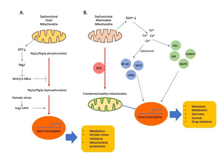
Cells 2019, 8, x                                                                                                            3 of 20
      Cells 2019, 8, 275                                                                                                     3 of 19

              Figure 1. Retrograde signalling and cellular adaptations. (A). In yeast, Rtg dependent signalling is
              the first found major pathway through which mitochondria communicates with nucleus. Rtg1p and
        Figure   1. Retrograde
              Rtg3p              signalling and cellular adaptations.
                      are basic helix-loop-helix/leucine                  (A). In yeast,transcription
                                                            zipper (bHLH/LeuZip)           Rtg dependent     signalling
                                                                                                         factors         is the
                                                                                                                 and nuclear
        first translocation
              found major of   pathway
                                 Rtg1/3pthrough      whichonmitochondria
                                            is dependent                     communicates
                                                              partial dephosphorylation      of with
                                                                                                Rtg3p.nucleus.    Rtg1p and
                                                                                                          Rtg2 promotes     the
        Rtg3p    are basic helix-loop-helix/leucine
              dephosphorylation                          zippertranslocation
                                    of Rtg3p and its nuclear       (bHLH/LeuZip)      transcription
                                                                              [37]. Osmotic            factors and
                                                                                               stress activates  Hog1nuclear
                                                                                                                        SAPK.
              Hog1 SAPK
        translocation     of binds to the
                             Rtg1/3p       Rtg1/3p transcription
                                       is dependent    on partial factor  and allows its translocation
                                                                     dephosphorylation      of Rtg3p. Rtg2to the nucleus [38].
                                                                                                               promotes     the
              (B). Mitochondrial
        dephosphorylation       of dysfunction,
                                   Rtg3p and its such as mitochondrial
                                                   nuclear  translocationDNA   (mtDNA)
                                                                            [37]. Osmotic  depletion or OXPHOS
                                                                                             stress activates      inhibition,
                                                                                                                Hog1   SAPK.
        Hog1triggers
                SAPK mitochondrial      retrograde
                        binds to the Rtg1/3p       signalling infactor
                                                transcription     mammalian   cells.its
                                                                       and allows    Fortranslocation
                                                                                          example, mitochondrial    stress[38].
                                                                                                        to the nucleus      can
              activate a Ca2+ -dependent retrograde signalling that is comprised of two branches: one mediated by
        (B). Mitochondrial dysfunction, such as mitochondrial DNA (mtDNA) depletion or OXPHOS
              calcineurin for the nuclear translocation of NF-κB or CREB or NFAT and the other directly dependent
        inhibition, triggers mitochondrial           retrograde signalling in mammalian cells. For example,
              on activation of Ca2+ -dependent protein kinases, such as PKC, JNK, MAPK and CAMKIV. Horizontal
        mitochondrial stress can activate a Ca2+-dependent retrograde signalling that is comprised of two
              transfer of healthy mitochondria from stromal cells in the TME can restore mitochondrial functions and
        branches: one mediated by calcineurin for the nuclear translocation of NF-κB or CREB or NFAT and
              is suggested to participate in the retrograde signalling. Emerging evidences show that ROS stimulates
        the other    directly dependent on activation of Ca2+-dependent protein kinases, such as PKC, JNK,
              horizontal transfer of mitochondria [39,40].
        MAPK and CAMKIV. Horizontal transfer of healthy mitochondria from stromal cells in the TME can
        2. Mitochondria
        restore          to Nucleus
                mitochondrial         Crosstalk:
                              functions           Mitochondria
                                        and is suggested          Regulates
                                                         to participate in the Nuclear  Events
                                                                               retrograde signalling. Emerging
        evidences
            Due toshow
                    the that ROSregulatory
                        pivotal  stimulatesfunction
                                            horizontal
                                                    of transfer of mitochondria
                                                        the mitochondria        [39,40]. cells have regulatory
                                                                          in homeostasis,
        systems to maintain the integrity, mass and metabolic functions of the mitochondria. The majority
2. Mitochondria
    (1500–2000) oftomitochondrial
                     Nucleus Crosstalk:
                                  proteinsMitochondria
                                          are encoded byRegulates
                                                         the nuclearNuclear
                                                                     genome,Events
                                                                             so communication among
     cytoplasmic,  nuclear and
     Due to the pivotal         mitochondrial
                           regulatory  functioncompartments    is essentialin
                                                  of the mitochondria       forhomeostasis,
                                                                                maintaining mitochondrial  function
                                                                                             cells have regulatory
     and to
systems   cellular homeostasis
             maintain           [41,42].mass
                        the integrity,   The two
                                               andmain   signalling
                                                    metabolic         systemsofthat
                                                                 functions       thelink the mitochondria
                                                                                      mitochondria.         and the
                                                                                                     The majority
     nucleus  are  nucleus to  mitochondria   anterograde   signalling   and  mitochondria   to
(1500–2000) of mitochondrial proteins are encoded by the nuclear genome, so communication among nucleus retrograde
     signalling,nuclear
cytoplasmic,     which comprise    bi-directional
                         and mitochondrial        communication
                                                compartments       is between
                                                                      essentialtheformitochondria
                                                                                      maintainingand   the nucleus.
                                                                                                    mitochondrial
     Through control of transcription and translation of genes, which directly regulate mitochondrial biogenesis,
function and cellular homeostasis [41,42]. The two main signalling systems that link the mitochondria
     nuclear signalling to the mitochondria (anterograde signalling) regulates OXPHOS and mitochondrial
and the nucleus are nucleus to mitochondria anterograde signalling and mitochondria to nucleus
     biogenesis in response to environmental signals received by the nucleus [43].
retrograde signalling, which comprise bi-directional communication between the mitochondria and the
          Mitochondria endlessly and extensively crosstalk with the nucleus and the cytosol, activating
nucleus. Through control of transcription and translation of genes, which directly regulate
     all of transcriptional, translational and post-translational processes aimed at restoring proper
mitochondrial biogenesis, nuclear signalling to the mitochondria (anterograde signalling) regulates
     mitochondrial function. Mitochondrial retrograde signalling, an emerging area of mitochondrial
OXPHOS     andcomprises
     research,   mitochondrial   biogenesis
                            signalling        in response
                                       pathways    by which to dysfunctional
                                                               environmentalmitochondria
                                                                                  signals received  by the nucleus
                                                                                                communicate   with
[43].
    Mitochondria endlessly and extensively crosstalk with the nucleus and the cytosol, activating all
of transcriptional, translational and post-translational processes aimed at restoring proper
Cells 2019, 8, 275 4 of 19
the nuclear genetic compartment to relay metabolic, oxidative and respiratory stressful conditions
prevailing in the mitochondria as cellular adaptation [37,41]. Retrograde signalling is very pleiotropic
with respect to its origin, functional mediators involved and the resulting phenotypes. Since it is
a cellular adaptation process, retrograde signalling does not necessarily result in full restoration of
mitochondrial homeostasis.
Mitochondrial dysfunction can be induced by various stress factors including low mtDNA copy
number, mtDNA mutations, nDNA mutations that affect mitochondrial function, mitochondrial
respiratory defects that result in changes in the mtROS levels and functional competence of respiratory
chain complexes. Unfortunately, we do not have clear understanding about the identity of specific
signalling molecules that can trigger the retrograde signalling so far either in yeast or mammals.
Mitochondrial dysfunction can affect a complex cytosolic and mitochondrial network of protein
homeostasis pathways, found and mostly studied in yeast or C. elegans [36,44,45]. By inhibiting protein
synthesis and by activation of the proteasome, unfolded protein response activated by mistargeting
of proteins (UPRam) is beneficial for the cells, providing a means for buffering the consequences of
physiological slowdown in mitochondrial protein import and for counteracting pathologies that are
caused or contributed by mitochondrial dysfunction [46]. Mitochondrial unfolded protein response
(UPRmt) is a transcriptional response to increase mitochondrial localized molecular chaperones
and proteases to promote the recovery of mitochondrial proteostasis [47]. UPR mt also results in a
reduction of nuclear and mtDNA-encoded OXPHOS transcripts to reduce the substrate burden on the
overwhelmed proteostasis in stressed mitochondria. In C. elegans, ATFS-1, a transcriptional factor
participating in retrograde signalling, was found to limit the accumulation of OXPHOS transcripts
during mitochondrial stress and also to stimulate OXPHOS recovery by matching the expression of
OXPHOS genes to the proteostatic capacity of mitochondria [48]. However, to our knowledge, it is
currently unknown whether direct regulators of retrograde signalling other than G-Protein pathway
suppressor 2 (GPS2), yet to be identified, exist in mammals [49].
From studies regarding metabolic diseases, inflammation and cancer, we have a lengthy list of
key small molecules, participating in retrograde signalling, including but not limited to, ROS [50,51],
NAD+/NADH ratio [52,53], acetyl-CoA [54,55], ATP [56], Ca2+ [57,58] and oncometabolites [59,60].
Depending on cell type and conditions, there are essentially two branches in Ca2+ mediated retrograde
signalling pathway: (1) Ca2+ /calcineurin-mediated retrograde signalling for the nuclear translocations
of transcription factors, NF-κB [61], NFAT, CREB and HnRNPA2 [41,62,63]; and (2) direct activation
of Ca2+ -dependent protein kinases, such as PKC, JNK, MAPK and CAMKIV [61,64]. Activation
of these signalling pathways in epithelial cells converges on the upregulation of genes affecting
several cellular functions, including apoptosis resistance, multi-drug resistance, invasion and EMT
(Figure 1). Oncometabolites are metabolites whose abundance is significantly increased in cancer
cells compared to normal cells. Increasing evidence shows that oncometabolites contribute to
cancer progression. In addition, we advise our readers to refer to several excellent articles with
more detailed information about retrograde signalling involved in epigenetic changes [37,41,65] and
posttranslational modifications of proteins including c-Src, MAPK, AMPK, PARPs, SIRT1 [26,66,67],
in diverse pathological conditions.
Considerable attention is given to these mitochondrial stresses because they drive both beneficial
and pathogenic adaptive responses [68]. Retrograde signalling appears to be capable of affecting
a wide range of processes in cancer progression including activating signalling pathways that
regulate metabolic adaptation, antioxidant systems, cellular proliferation, apoptosis-resistance,
chemo-resistance and cellular migration and invasion [69,70]. Emerging evidence suggests that
it is responsible for metabolic flexibility observed between different cancers and even between cancer
cells in the same cancer tissue. Indeed, mitochondria-to-nucleus retrograde signalling in cancer
may be a highly plausible mechanism by which altered mitochondrial function modulates adaptive
changes in nuclear gene expression and metabolism mediated by specific transcription factors [62,64]
towards enhanced tumorigenesis and invasiveness. In addition, we are beginning to gather seeminglyCells 2019, 8, 275 5 of 19
still fragmentary but highly important evidence suggesting that mitochondria can regulate several
important nuclear events including genetic and epigenetic changes in cancer cells [17,41,71].
Reduced mtDNA content has been associated with aggressive features including a metabolic shift
to glycolysis, apoptosis and increased invasiveness in multiple cancer types, such as prostate [72–74]
and colorectal [75] cancers. However, caution must be taken in interpreting these findings because
we do not have enough convincing data to show that retrograde signalling actually engages in
the whole stages of cancer progression. Cancer cell proliferation in primary sites, intravasation,
survival, migration in blood vessels, extravasation and colonization of tumour cells in distant sites
are distinct steps in cancer progression. It has been suggested that retrograde signalling actually
counts in the late stage of gene expression reprogramming that alters the metabolic phenotype during
malignant transformation [76]. Mitochondrial dysfunction induced by mtDNA depletion promotes
EMT in breast epithelial cells through a Ca2+ /calcineurin-mediated mitochondrial retrograde signalling
that triggers transcriptional activation of SLUG, SNAIL and TWIST, MMP-9 and the mesenchymal
markers, vimentin, fibronectin and N-cadherin, with a corresponding decrease in the epithelial marker,
E-cadherin [77]. Of note, reduced mtDNA content has been directly correlated with induction of EMT
through activation of mitochondria-to-nucleus signalling [27,70] and revitalization of OXPHOS is
commonly observed in cancer cells undergoing EMT [16,78–81]. The changes in mitochondrial genome
are likely metastasis modifiers rather than drivers [28].
There is accumulating evidence that mtDNA can be transferred between cells or
species [79,80,82–84]. Mitochondria can move from one cell to another by various intercellular
structures such as tunnelling nanotubes (TNTs) or cytoplasmic bridges [84,85]. Mesenchymal stem
cells (MSCs) sense mitochondria released from damaged cells as danger signals to activate their
rescue properties for the damaged cells [86]. Foreign mitochondria from damaged cells were engulfed
and degraded by MSC, leading to induction of the cytoprotective enzyme heme oxygenase-1 and
stimulation of mitochondrial biogenesis in MSC. As a result, the rescue capacity of MSC to transfer
their mitochondria to injured cells to combat oxidative stress injury was enhanced. Horizontal transfer
of mtDNA from the host cells in the TME to cancer cells with compromised respiratory function
to re-establish respiration and metastatic efficacy was also recently shown [80,84,87]. Transfer of
mitochondria between leukemic cells and bone marrow mesenchymal stem cells is increased by
chemotherapy [39]. It has been shown that uptake of mitochondria by leukemic cells increases
oxidative phosphorylation and favours survival, indicating new insights into a novel mechanism of
drug resistance [88,89]. Another report also showed that cancer cells uptaking mitochondria displayed
chemoresistance, indicating functional aspects of mitochondrial acquisition beyond respiration
recovery [90]. It may be feasible that interfering with transfer of mtDNA together with targeting
OXPHOS metabolism would be an efficient adjuvant strategy to affect the intrinsic crucial metabolic
function of cancer cells and the supportive function of TME. Some of the outstanding questions would
be (1) what are mechanisms that are responsible for the transfer of host mtDNA to cancer cells in
TME? (2) can mtDNA from dysfunctional mitochondria in cancer cells move to the host cells in turn?
(3) would the metabolic profiles of the host cells in the TME be affected from the mtDNA transferred
from cancer cells? and (4) does the transfer of mtDNA extend the spectrum of retrograde signalling as
communication between different cells?
3. Oncometabolites from Dysfunctional Mitochondria
Increasing evidence shows that oncometabolites, participating in the retrograde signalling,
contribute to cancer progression. For now, the list of established oncometabolites is very short and
consists of 2-hydroxyglutarate (2HG), succinate and fumarate, that result from oncogenic mutations
in isocitrate dehydrogenase (IDH), succinate dehydrogenase (SDH) or fumarate hydratase (FH),
respectively [60]. Since their function is often regulated by both their substrate and the product,
metabolic enzymes can directly sense the supply and demand of nutrients. Thus, the capability
of metabolic enzymes to both sense metabolic stress and also regulate nuclear gene transcriptionCells 2019, 8, 275 6 of 19
is an efficient way by which an adaptive response can be achieved. A major functional
mechanism of 2HG is their structural similarity to α-KG, which allows 2HG to act as a
competitive inhibitor of α-KG-dependent enzymes including the Jumonji-C domain containing
histone demethylase (JMJD/JHDM) and the 10-11-translocation methylcytosine dioxygenase (TET)
families of chromatin-modifying enzymes and the prolyl hydroxylases (PHD) family [91]. The TCA
cycle intermediate, α-KG, is a co-substrate for many enzymes in the cytoplasm and the nucleus,
including JMJD/JHDM and TET families of chromatin-modifying enzymes and the PHD family.
Glutamine-derived α-KG contributes to TET-dependent demethylation reactions [92]. Mutant versions
of cytoplasmic and mitochondrial IDH isoforms, IDH1 and IDH2, respectively, reduce α-KG to generate
2HG [93]. Therefore, through the production of oncometabolite 2HG, mitochondria exert a strong
influence on chromatin structure associated with DNA hypermethylation and cause a broad epigenetic
change to promote cancer progression [59,94]. 2HG also inhibits the enzymatic activity of cytochrome
c oxidase and ATP synthase [70,95] and alters the gene expression of TCA cycle enzymes in cancer
cells [96]. These findings suggest that the accumulation of 2HG contributes to the changes in energy
metabolism in IDH-mutant cancer cells.
Succinate and fumarate accumulation resulting from mutations in SDH and FH, respectively,
stabilize HIF-1α via PHD inhibition, reinforcing the Warburg effect [91,97]. Accumulation of fumarate
and succinate also inhibits the α-KG dependent histone and DNA methylases, the JMJD/JHDM
and TET family of proteins, respectively [98–101]. The accumulation of intracellular fumarate
can result in the succination modification of cysteine residues within the Keap1 and results in
an increase in levels of NRF2 from abrogation of Keap1-mediated degradation of NRF2 [102].
NRF2, acetylated by transcription coactivator and acetyltransferase, p300, activates several antioxidant
genes and supports cancer formation. Decreased level of FH can increase the levels of both
NRF1 and NRF2 [103] and NRF1 also promotes mesenchymal transition and spheroid survival in
mammary epithelial cells by stimulating OXPHOS [81]. Activating mutations of NRF2 or treatment
of cancer cells with antioxidants can not only reduce level of ROS but also turn on oncogenic
activities [104]. Additionally, the accumulation of fumarate might promote tumorigenesis by inhibiting
α-KG-dependent genome-wide histones and DNA methylations, resulting in epigenetic alterations
in gene expression [101] or by increasing ROS dependent signalling via generation of succinated
glutathione [105]. Fumarate can increase histone H3 methylation by inhibiting KDM2B demethylase
and promote binding of DNA-dependent protein kinase and the recruitment of end-processing
enzymes for DNA repair [101,106,107]. In conclusion, mutations in mitochondrial metabolic
enzymes, IDH2, SDH or FH, results in abundant oncometabolites and leads to epigenetic changes,
further dysfunction of mitochondria, production of ROS, cancer progression and notably, increased
EMT. Table 1 summarizes functions and producers of oncometabolites.
Table 1. Summary of oncometabolites.
Metabolites Producer Specific Function Common Function
Produces acetyl-CoA and increases histone
Acetyl CoA PDC acetylation. Increases the expression of genes that
promote cell cycle progression and cell proliferation
2-hydroxybutyrate Mutated IDH Inhibits cytochrome c oxidase and ATP synthase.
Inhibits Keap1-mediated degradation of NRF.
Increases ROS signalling via generation of
succinated glutathione. Inhibits the demethylase Inhibits the JMJD family, TET family and
Fumarate WT SDH, mutated FH
KDM2B, increases H3 methylation and promotes PHD family. Increased methylation
binding of DNA-dependent protein kinase and the promotes the expression of genes increasing
recruitment of end-processing DNA repair enzymes. proliferation and inhibiting differentiation.
Succinate Mutated SDH
Phosphorylation of H3 with PEP facilitates H3
acetylation, promotes expression of c-Myc and
Phosphoenolpyruvate (PEP) PKM2
cyclin D1. STAT3 phosphorylation promotes
MEK5 activation.Cells 2019, 8, 275 7 of 19
4. Nuclear Metabolic Enzymes as New Regulators of Retrograde Signalling
Recent updates indicate that many metabolic enzymes found in the cytosol or mitochondria can
move to the nucleus and have non-canonical functions [71,108–111], directly linking metabolism with
gene transcription, particularly epigenetic mechanisms such as methylation of DNA and histones or
acetylation of histones. These findings suggest that metabolic enzymes themselves can participate in
the retrograde signalling from their non-canonical function in the nucleus. Both cytoplasmic pyruvate
kinase (PKM2) and mitochondrial pyruvate dehydrogenase complex (PDC) can translocate to the
nucleus and form a complex with p300, which locally produces acetyl-CoA to acetylate histones and can
facilitate locally confined specific gene transcription [111,112]. Nuclear PKM2 produces pyruvate from
phosphoenolpyruvate (PEP), which is used by PDC for the production of acetyl-CoA, which in turn
phosphorylates histone H3 [110] and STAT3 [109], as a novel non-canonical kinase using PEP instead
of ATP as the phosphate donor. Like acetylation, phosphorylation of H3 is also an important histone
modification, which promotes cell-cycle progression and cancer growth by increasing the expression of
cyclin D1 and c-Myc [110]. Independent of its kinase activity, PKM2 can function as a co-transcription
factor, promoting HIF-1α binding activity to DNA and thereby participating in a positive feedback loop
with HIF-1α, which upregulates several glycolytic enzymes including PKM2 itself [113,114]. Nuclear
PDC levels are increased in a cell-cycle-dependent manner and in response to serum or mitochondrial
stress, with a concomitant decrease in mitochondrial PDC levels, suggesting a translocation of PDC
from the mitochondria to the nucleus [112]. Indeed, whole-transcriptome analysis revealed that
mitochondria have the ability to regulate the expression of more than 66% of genes within the human
genome, including the epigenetic/chromatin remodelling machineries [115,116]. Acetyl-CoA can
influence posttranslational acetylation on histone tails and change the nuclear epigenome both globally
throughout the nucleus and locally for specific histones and proteins. Translocation of mitochondrial
PDC to the nucleus promotes acetyl-CoA synthesis in the nucleus, which is required for histone
acetylation and epigenetic regulation [112]. Nuclear PDC increases acetylation of specific lysine
residues on histones, upregulates expression of phosphorylated Rb, E2F, Cdk2 and cyclin A and
promotes G1-S phase progression and expression of S phase markers [112,117]. Mitochondrial stress
activates Akt1 and Akt1 mediates transcription activation via phosphorylation and activation of
p300 [118]. Acetylation of PKM2 by p300 promotes its nuclear translocation and its non-canonical
function as a transcription regulator and a kinase, respectively [119]. Acetylation of the c-Myc
oncoprotein by p300 increases its stability and the transcription of its target genes [120], also suggesting
a link between mitochondrial stress, acetylation events in the nucleus and cancer progression. Of note,
mitochondrial stress can promote nuclear translocation of PDC [112], in addition to the nuclear
translocation of cytosolic PKM2, indicating that nuclear translocation of mitochondrial enzyme can be
triggered by mitochondrial dysfunction. Our Figure 2 shows a functional summary of nuclear PKM2
and PDC.
Other glycolytic enzymes such as hexokinase 2 (HK2), lactate dehydrogenase (LDH) and
3-phosphoglycerate kinase (PGK) can also move to the nucleus and perform their non-canonical
functions. HK2 is an enzyme catalysing the first committed step of glycolysis and found overexpressed
in many cancer cells. In the nucleus of HeLa cells, HK2 is found in nucleus and low glucose
environment or Akt inhibition augments their nuclear localization [121,122]. In yeast, HK2 regulates
its incorporation into the repressor complex of the Mig1-dependent gene promoters in response to
cytoplasmic glucose level, indicating its role as a fuel sensor regulating expression of other metabolic
enzymes [123]. Y238 phosphorylation of LDH is important for the nuclear translocation, although
upstream molecule responsible for the phosphorylation is not clear [124,125]. Nuclear LDH is
increased under oxidative stress [126] and binds DNA, stimulates UV-induced DNA repair [127].
Nuclear LDH is also increased by E7-induced ROS accumulation in cervical cancer cells, performs a
non-canonical enzyme activity to produce α-hydroxybutyrate and triggers DOT1L mediated histone
H3K79 hypermethylation, resulting in the activation of antioxidant responses and Wnt signalling
pathway in cervical cancer cells [128]. PGK stimulates DNA synthesis catalysed by DNA polymeraseinto the repressor complex of the Mig1-dependent gene promoters in response to cytoplasmic glucose
level, indicating its role as a fuel sensor regulating expression of other metabolic enzymes [123]. Y238
phosphorylation of LDH is important for the nuclear translocation, although upstream molecule
responsible for the phosphorylation is not clear [124,125]. Nuclear LDH is increased under oxidative
stress
Cells 2019,[126]
8, 275and binds DNA, stimulates UV-induced DNA repair [127]. Nuclear LDH is also increased 8 of 19
by E7-induced ROS accumulation in cervical cancer cells, performs a non-canonical enzyme activity to
produce α-hydroxybutyrate and triggers DOT1L mediated histone H3K79 hypermethylation, resulting
and
α in on single-stranded
theε activation DNA
of antioxidant [127]. LDH
responses in nucleus
and Wnt canpathway
signalling also interact with SIRT1
in cervical cancer and
cellsregulate
[128].
PGK stimulates
epigenetic DNA synthesis
modifications catalysed by
by manipulating DNAindicating
NAD+, polymeraseanαintricate
and ε onlink
single-stranded DNA [127].
between metabolism and
LDH
the in nucleus
processing can alsoinformation
of genetic interact with[126].
SIRT1 and regulate epigenetic modifications by manipulating
NAD+, indicating an
We anticipate intricate
that furtherlink between
studies metabolism
on the and theofprocessing
identification metabolicoftriggers
genetic information
that enable [126].
nuclear
We anticipate
translocation that further
of metabolic studies
enzymes onreveal
will the identification of nuclear
that specific metabolic triggers that
metabolic enableare
enzymes nuclear
potent
translocation of metabolic enzymes
participants of the retrograde signalling. will reveal that specific nuclear metabolic enzymes are potent
participants of the retrograde signalling.
Figure2.2.Translocation
Figure Translocation ofof PKM2
PKM2ororPDC PDCinto
into nucleus,
nucleus, where
where it serves
it serves as transcriptional
as transcriptional coactivator
coactivator and
and
as aasprotein
a protein
kinasekinase
or as aorproducer
as a producer
of acetyl of
CoAacetyl CoA totranscriptional
to modulate modulate transcriptional program.
program. In nucleus,
Indimeric
nucleus,PKM2dimeric PKM2
becomes becomes
a protein kinasea using
protein
PEPkinase using PEP
as a phosphate as PKM2
donor. a phosphate
is able todonor. PKM2 is
phosphorylate
able to phosphorylate STAT3 at Y705 and promotes transcription of MEK5. PKM2
STAT3 at Y705 and promotes transcription of MEK5. PKM2 directly binds to and phosphorylates histone directly binds
toH3and phosphorylates
at threonine 11 uponhistone
epidermalH3 growth
at threonine 11 upon
factor (EGF) epidermal
receptor growth
activation [109]factor
. PKM2(EGF) receptor
can function
activation [109]. PKM2
as a transcriptional can function
coactivator, as a transcriptional
promoting HIF-1α binding coactivator,
activity topromoting
DNA and HIF-1α
therebybinding activity
participating
toinDNA and thereby
a positive feedback participating in a positive
loop with HIF-1α feedback
[114]. Grey circleloop withmitochondrial
denotes HIF-1α [114].events
Grey circle denotes
and orange
mitochondrial
circle indicatesevents and
nuclear orange
events. circle indicates
Subcellular nuclear
localization events. Subcellular
of glycolytic metaboliteslocalization of glycolytic
and tetrameric PKM2
metabolites and tetrameric PKM2 is in cytoplasm. PKM2 and PDC are shown
is in cytoplasm. PKM2 and PDC are shown in red. Canonical cytosolic functions of HK2, LDH and in red. Canonical
cytosolic
PGK are functions
shownof here
HK2, inLDH and PGK
blue. are shown here
Abbreviations: G3P,inglyceraldehyde
blue. Abbreviations: G3P, glyceraldehyde
3-phosphate; 3PG, 3-
phosphoglyceraste;
3-phosphate; 2PG, 2-phosphoglyceraste;
3PG, 3-phosphoglyceraste; PEP, phosphoenolpyruvate;
2PG, 2-phosphoglyceraste; PDC, pyruvate
PEP, phosphoenolpyruvate; PDC,
dehydrogenase
pyruvate complex;complex;
dehydrogenase PS, pyruvate symporter.
PS, pyruvate symporter.
5. Retrograde Signalling and Metabolic Switching in the Tumour Microenvironment
Cancer-related non-resolving inflammation in the TME is a hallmark of cancer. TME is generally
hypoxic, ROS rich and an acidic environment. Recent findings clearly show that both, a shift to Warburg
effect or OXPHOS or combined metabolic phenotypes exist in rapidly proliferating cells, including
various types of immune cells, most notably in macrophages and T cells and determine the function of
the immune cell subsets in disease conditions such as those in inflamed tissue, obese adipose tissue or
cancer [129–132]. For example, due to metabolic needs to maintain a higher level of ATP, tolerogenic
dendritic cells show the highest OXPHOS activity and production of ROS, increased spare respiratoryCells 2019, 8, 275 9 of 19
capacity and more pronounced glycolytic capacity and reserve compared to immunogenic mature
dendritic cells [133].
M1-associated inhibition of mitochondrial OXPHOS is the factor responsible for preventing
M1-like to M2-like activation [134] and glycolytic stimulation is not required for M2-like activation
when OXPHOS is intact [135], indicating that metabolism determines macrophage activation. M2-like
activated macrophages exploit FAO to fuel OXPHOS, rather than aerobic glycolysis that M1-like
proinflammatory activated macrophages exploit for ATP production [136–138]. The reports that
inhibition of ROS from ETC with metformin [139] or activation of AMPK with AICAR promotes M1
like to M2 like activation [140] indicate that boosting FAO or OXPHOS can be suggested to switch
macrophage class from M1-like to M2-like [141]. A “shift” from OXPHOS to aerobic glycolysis is a
hallmark of T cell activation [130]. T cells, if not activated, show low levels of metabolic requirements,
use OXPHOS to maximize production of ATP as an energy source and engage scarcely in biosynthesis,
whereas activated T cells use aerobic glycolysis to produce effector molecules for rapid cellular
proliferation [138]. Mitochondrial oxidative metabolism supports immunosuppression and lineage
commitment of Tregs [142–144]. Cancer aggressiveness is promoted by metabolic synergy between
cancer cells and stromal cells [145]. Metabolic synergy induces efficient utilization of catabolites
by cancer cells in TME [24] and cancer cells induce aerobic glycolysis in neighbouring fibroblasts
by providing a hypoxic ROS-rich microenvironment [24,146]. Induced fibroblasts differentiate to
myofibroblasts and upregulate MCT4 to secrete lactate and pyruvate that transform the normal stroma
to ultimately help cancer cells grow (the Reverse Warburg effect). All these findings indicate that cells
in the TME show mixed metabolic phenotypes of aerobic glycolysis and OXPHOS, resulting from
various interactions between different cells or metabolic stress (Figure 3).
Therefore, with metabolic regulation via suppression of aerobic glycolysis and concomitant
promotion of OXPHOS, would it be possible that mitochondrial retrograde signalling, an adaptive
mechanism to restore mitochondrial homeostasis in response to mitochondrial stress, can also
participate in activation of immune cells? If yes, is it responsible for the metabolic flexibility in
the TME? It appears that we have some answers suggesting that retrograde signalling can suppress
glycolysis and promote OXPHOS in myeloid cells. Mitochondrial ROS is important both for M1-like
and M2-like activation [147,148] and can induce retrograde signalling by alterations in mitochondrial
membrane potential (∆ψm) and activation of Ca2+ /calcineurin dependent factors, suggesting an
involvement of the retrograde signalling pathway in M1-like to M2-like activation. Indeed, in murine
macrophages, mitochondrial dysfunction induced by hypoxic insult or ATP synthase inhibitor,
activates Ca2+ /calcineurin or mediated retrograde signalling pathway with activation of AMPK
and NF-κB, in which ROS induced mitochondrial membrane damage is a component of the signalling
pathway [149,150]. Of note, the regulators involved in macrophage M2-like activation are also shared
by the retrograde signalling pathway. Depletion of glucose or a glucose-rich hypoxic ROS environment
favours M2-like activation or M1-like activation of macrophages, respectively and depletion of glucose
can disarm T cells in the TME [132,151]. Low levels of ATP due to dietary restrictions or energy
consumption, induce expression of nicotinamide phosphoribosyl transferase that generates NAD+ ,
which is a key factor for SIRT1 activation. SIRT1, also a regulator of retrograde signalling, acetylates
and activates PGC1ß to increase OXPHOS in macrophage [152–154].
In addition, Akt activation in response to mitochondrial respiratory stress has been found
in different tumour cell systems [63,72,155] and PI3K/Akt signalling also regulates macrophage
activation in the TME. The PI3K regulator lipid phosphatase, phosphatase and tensin homolog
(PTEN), contributes to macrophage polarization because deletion of PTEN and resultant activation
of PI3K/Akt signalling results in increased M2-like activation [156,157]. Interestingly, deletion of
Akt1 promotes upregulation of inducible NO synthase and IL-12β (M1-like activation) and Akt2
deficiency in macrophages highlights the opposing roles of Akt isoforms in macrophage polarization,
because Akt2−/− macrophages possess an M2-like phenotype [158]. These findings suggest that
mitochondrial stress induced retrograde signalling can determine the activation of myeloid cells.Cells 2019, 8, 275 10 of 19
Tumour suppressor p53 is critically important in preventing oncogenesis but its role in inflammation
in general and in the function of inflammatory macrophages in particular is not certain. Mitochondrial
stress induced by doxorubicin or partial depletion of mtDNA (~70%) activates the Ca2+/calcineurin
retrograde signalling pathway, inducing expression of p53, which in turn attenuates HIF-1α activity
in multiple types of cells [159–161]. p53 mediated suppression of HIF-1α controls regulation of
glycolysis and ‘loss of function mutation’ of p53 is partially responsible for enhanced glycolysis in
cancer cells [162]. Notably, deletion or activation of p53 in myeloid cells represses or induces the
M2-like phenotype, respectively, in a chronic inflammatory venous thrombus model [163]. However,
conflicting reports that p53 drives M1-like phenotype in tumour-associated macrophages (TAMs) of
several cancer models [164–166] do not support this speculation, possibly indicating pathological
differences in different microenvironments or in different model systems.
Figure 3. Summary of metabolic switching in the TME. Red arrows indicate immune suppressive
response favourable to growth and survival of cancer cells. Blue arrows indicate immune stimulatory
response detrimental to cancer cells. Abbreviations: EndMT, endothelial to mesenchymal transition;
Gly, aerobic glycolysis; MDSC, myeloid derived suppressor cell; M-MDSC, monocytic myeloid derived
suppressor cell; OxPh, oxidative phosphorylation; TAM, tumour-associated macrophage.
6. Conclusions
Many important questions about the involvement of retrograde signalling pathways in cancer
biology, for example, the specific sensors of different types of mitochondrial stress and the cell
and tissue specificity of the signalling responses, are yet to be explored. Importantly, with a list
of small molecules and potential protein regulators involved in retrograde signalling, it appears
that we still do not clearly understand the identity of specific signalling molecules that can trigger
the retrograde signalling so far either in yeast or mammals. In addition, what determines whether
retrograde signalling results in beneficial or maladaptive responses, its significance in determination
of cancer progression and which cell type in the TME is mostly affected, should be identified. For now,
relevant studies are largely limited by the difficulty of experimentally manipulating mtDNA in cancer
cells and the lack of animal models with mtDNA mutations. It is not technically feasible at thisCells 2019, 8, 275 11 of 19 moment to differentiate mtDNA abnormalities in host cells from those of the cancer cells in the TME. Would it be possible for mtDNA from cancer cells to move to and affect the non-transformed host stromal cells that have no genetic or epigenetic changes? Indeed, intercellular structures such as TNTs have been involved in the transfer of mitochondria between different cells. Thus, are such structures involved in the retrograde signalling and responsible for the communication between cells in the TME? Further studies on the horizontal transmission of mitochondrial retrograde signalling between diverse cells in the TME may also extend our understanding of cancer progression. As stated, retrograde signalling may suppress glycolysis and promote OXPHOS in myeloid cells, indicating a possibility that the retrograde signalling may regulate glycolysis in some type of cancer cells whose metabolism and genetic or epigenetic events are rewired. If it actually happens, this will also extend our understanding about functional implications of retrograde signalling. Finally, we expect the feasibility of future approaches, targeting retrograde signalling for successful cancer therapy from suppression of metastasis or resistance to drugs, to become an exciting topic of study in the near future. Funding: This work was supported by the Basic Science Research Program, through the National Research Foundation of Korea (NRF), funded by the Ministry of Science, ICT & Future Planning (NRF-2017R1D1A1B03029063) and by the Korean Government (NRF-2018R1D1A1B07041381). Conflicts of Interest: The authors declare no conflict of interest. Abbreviations AICAR 5-Aminoimidazole-4-carboxamide ribonucleotide AMPK 50 AMP-activated protein kinase ATFS-1 Activating transcription factor associated with stress-1 CAMKIV Calcium/calmodulin-dependent protein kinase type IV CREB cAMP response element-binding protein DOT1L Disruptor of telomeric silencing 1-like EMT Epithelial to mesenchymal transition ETC Electron transfer chain FAO Fatty acid oxidation HIF-1α Hypoxia-inducible factor-1α JNK c-Jun N-terminal kinase Keap1 Kelch-like ECH-associated protein 1 α-KG α-Ketoglutarate MAPK Mitogen-activated protein kinas MCT4 Monocarboxylate transporter 4 MMP-9 Matrix metalloproteinases-9 mtDNA Mitochondrial DNA nDNA Nuclear DNA NF-κB Nuclear factor kappa-light-chain-enhancer of activated B cell NFAT Nuclear factor of activated T-cells NO Nitric oxide NRF2 Nuclear factor erythroid 2-related factor 2 OXPHOS Oxidative phosphorylation PARP Poly (ADP-ribose) polymerase PGC1ß Peroxisome proliferator-activated receptor gamma coactivator 1-alpha PI3K Phosphoinositide 3-kinase PKC Protein kinase C ROS Reactive oxygen species SAPK Stress-activated protein kinase SIRT1 Silent mating type information regulation 2 homolog 1/Sirtuin STAT3 Signal transducer and activator of transcription 3 TCA cycle Tricarboxylic acid cycle/Krebs cycle Treg regulatory T cell
Cells 2019, 8, 275                                                                                              12 of 19

References
1.    Rose, G.; Santoro, A.; Salvioli, S. Mitochondria and mitochondria-induced signalling molecules as longevity
      determinants. Mech. Ageing Dev. 2017, 165, 115–128. [CrossRef]
2.    Potter, M.; Newport, E.; Morten, K.J. The Warburg effect: 80 years on. Biochem. Soc. Trans. 2016, 44, 1499–1505.
      [CrossRef] [PubMed]
3.    Weinhouse, C. Mitochondrial-epigenetic crosstalk in environmental toxicology. Toxicology 2017, 391, 5–17.
      [CrossRef] [PubMed]
4.    Wei, Y.; Chen, L.; Xu, H.; Xie, C.; Zhou, Y.; Zhou, F. Mitochondrial Dysfunctions Regulated Radioresistance
      through Mitochondria-to-Nucleus Retrograde Signaling Pathway of NF-κB/PI3K/AKT2/mTOR. Radiat. Res.
      2018, 190, 204–215. [CrossRef] [PubMed]
5.    Vander Heiden, M.G.; Cantley, L.C.; Thompson, C.B. Understanding the Warburg effect: The metabolic
      requirements of cell proliferation. Science 2009, 324, 1029–1033. [CrossRef]
6.    Ward, P.S.; Thompson, C.B. Metabolic reprogramming: A cancer hallmark even warburg did not anticipate.
      Cancer Cell 2012, 21, 297–308. [CrossRef] [PubMed]
7.    Schulze, A.; Harris, A.L. How cancer metabolism is tuned for proliferation and vulnerable to disruption.
      Nature 2012, 491, 364–373. [CrossRef]
8.    Koppenol, W.H.; Bounds, P.L.; Dang, C.V. Otto Warburg’s contributions to current concepts of cancer
      metabolism. Nat. Rev. Cancer 2011, 11, 325–337. [CrossRef]
9.    Wallace, D.C. Mitochondria and cancer. Nat. Rev. Cancer 2012, 12, 685–698. [CrossRef]
10.   Lu, J.; Tan, M.; Cai, Q. The Warburg effect in tumor progression: Mitochondrial oxidative metabolism as an
      anti-metastasis mechanism. Cancer Lett. 2015, 356, 156–164. [CrossRef]
11.   Zheng, J. Energy metabolism of cancer: Glycolysis versus oxidative phosphorylation (Review). Oncol. Lett.
      2012, 4, 1151–1157. [CrossRef] [PubMed]
12.   Chandra, D.; Singh, K.K. Genetic insights into OXPHOS defect and its role in cancer. Biochim. Biophys. Acta
      2011, 1807, 620–625. [CrossRef] [PubMed]
13.   Owens, K.M.; Kulawiec, M.; Desouki, M.M.; Vanniarajan, A.; Singh, K.K. Impaired OXPHOS complex III in
      breast cancer. PLoS ONE 2011, 6, e23846. [CrossRef]
14.   Lopez-Rios, F.; Sanchez-Arago, M.; Garcia-Garcia, E.; Ortega, A.D.; Berrendero, J.R.; Pozo-Rodriguez, F.;
      Lopez-Encuentra, A.; Ballestin, C.; Cuezva, J.M. Loss of the mitochondrial bioenergetic capacity underlies
      the glucose avidity of carcinomas. Cancer Res. 2007, 67, 9013–9017. [CrossRef] [PubMed]
15.   Moreno-Sanchez, R.; Rodriguez-Enriquez, S.; Marin-Hernandez, A.; Saavedra, E. Energy metabolism in
      tumor cells. FEBS J. 2007, 274, 1393–1418. [CrossRef] [PubMed]
16.   LeBleu, V.S.; O’Connell, J.T.; Gonzalez Herrera, K.N.; Wikman, H.; Pantel, K.; Haigis, M.C.; de Carvalho, F.M.;
      Damascena, A.; Domingos Chinen, L.T.; Rocha, R.M.; et al. PGC-1alpha mediates mitochondrial biogenesis
      and oxidative phosphorylation in cancer cells to promote metastasis. Nat. Cell Biol. 2014, 16, 992–1003,
      1001–1015. [CrossRef] [PubMed]
17.   Wallace, D.C.; Fan, W. Energetics, epigenetics, mitochondrial genetics. Mitochondrion 2010, 10, 12–31.
      [CrossRef]
18.   Galluzzi, L.; Kepp, O.; Vander Heiden, M.G.; Kroemer, G. Metabolic targets for cancer therapy. Nat. Rev.
      Drug Discov. 2013, 12, 829–846. [CrossRef] [PubMed]
19.   Pietras, K.; Ostman, A. Hallmarks of cancer: Interactions with the tumor stroma. Exp. Cell Res. 2010, 316,
      1324–1331. [CrossRef] [PubMed]
20.   Kim, J. Regulation of Immune Cell Functions by Metabolic Reprogramming. J. Immunol. Res. 2018, 2018,
      8605471. [CrossRef] [PubMed]
21.   Michalek, R.D.; Rathmell, J.C. The metabolic life and times of a T-cell. Immunol. Rev. 2010, 236, 190–202.
      [CrossRef] [PubMed]
22.   Altman, B.J.; Dang, C.V. Normal and cancer cell metabolism: Lymphocytes and lymphoma. FEBS J. 2012,
      279, 2598–2609. [CrossRef]
23.   Doherty, J.R.; Cleveland, J.L. Targeting lactate metabolism for cancer therapeutics. J. Clin. Investig. 2013, 123,
      3685–3692. [CrossRef] [PubMed]
24.   Martinez-Outschoorn, U.; Sotgia, F.; Lisanti, M.P. Tumor microenvironment and metabolic synergy in breast
      cancers: Critical importance of mitochondrial fuels and function. Semin. Oncol. 2014, 41, 195–216. [CrossRef]
Cells 2019, 8, 275 13 of 19
25. Sonveaux, P.; Vegran, F.; Schroeder, T.; Wergin, M.C.; Verrax, J.; Rabbani, Z.N.; De Saedeleer, C.J.;
Kennedy, K.M.; Diepart, C.; Jordan, B.F.; et al. Targeting lactate-fueled respiration selectively kills hypoxic
tumor cells in mice. J. Clin. Investig. 2008, 118, 3930–3942. [CrossRef] [PubMed]
26. Jia, D.; Park, J.H.; Jung, K.H.; Levine, H.; Kaipparettu, B.A. Elucidating the Metabolic Plasticity of Cancer:
Mitochondrial Reprogramming and Hybrid Metabolic States. Cells 2018, 7, 21. [CrossRef]
27. Guerra, F.; Guaragnella, N.; Arbini, A.A.; Bucci, C.; Giannattasio, S.; Moro, L. Mitochondrial Dysfunction:
A Novel Potential Driver of Epithelial-to-Mesenchymal Transition in Cancer. Front. Oncol. 2017, 7, 295.
[CrossRef] [PubMed]
28. Beadnell, T.C.; Scheid, A.D.; Vivian, C.J.; Welch, D.R. Roles of the mitochondrial genetics in cancer metastasis:
Not to be ignored any longer. Cancer Metastasis Rev. 2018, 37, 615–632. [CrossRef]
29. Parikh, V.S.; Morgan, M.M.; Scott, R.; Clements, L.S.; Butow, R.A. The mitochondrial genotype can influence
nuclear gene expression in yeast. Science 1987, 235, 576–580. [CrossRef]
30. Jazwinski, S.M.; Kriete, A. The yeast retrograde response as a model of intracellular signaling of
mitochondrial dysfunction. Front. Physiol. 2012, 3, 139. [CrossRef]
31. Guaragnella, N.; Zdralevic, M.; Lattanzio, P.; Marzulli, D.; Pracheil, T.; Liu, Z.; Passarella, S.; Marra, E.;
Giannattasio, S. Yeast growth in raffinose results in resistance to acetic-acid induced programmed cell death
mostly due to the activation of the mitochondrial retrograde pathway. Biochim. Biophys. Acta 2013, 1833,
2765–2774. [CrossRef] [PubMed]
32. Laera, L.; Guaragnella, N.; Zdralevic, M.; Marzulli, D.; Liu, Z.; Giannattasio, S. The transcription factors ADR1
or CAT8 are required for RTG pathway activation and evasion from yeast acetic acid-induced programmed
cell death in raffinose. Microb. Cell 2016, 3, 621–631. [CrossRef] [PubMed]
33. Jazwinski, S.M. The retrograde response: A conserved compensatory reaction to damage from within and
from without. Prog. Mol. Biol. Transl. Sci. 2014, 127, 133–154. [CrossRef] [PubMed]
34. Guaragnella, N.; Coyne, L.P.; Chen, X.J.; Giannattasio, S. Mitochondria-cytosol-nucleus crosstalk: Learning
from Saccharomyces cerevisiae. FEMS Yeast Res. 2018, 18, 88. [CrossRef]
35. Liao, X.; Butow, R.A. RTG1 and RTG2: Two yeast genes required for a novel path of communication from
mitochondria to the nucleus. Cell 1993, 72, 61–71. [CrossRef]
36. Arnould, T.; Michel, S.; Renard, P. Mitochondria Retrograde Signaling and the UPR mt: Where Are We in
Mammals? Int. J. Mol. Sci. 2015, 16, 18224–18251. [CrossRef] [PubMed]
37. da Cunha, F.M.; Torelli, N.Q.; Kowaltowski, A.J. Mitochondrial Retrograde Signaling: Triggers, Pathways
and Outcomes. Oxid. Med. Cell. Longev. 2015, 2015, 482582. [CrossRef]
38. Ruiz-Roig, C.; Noriega, N.; Duch, A.; Posas, F.; de Nadal, E. The Hog1 SAPK controls the Rtg1/Rtg3
transcriptional complex activity by multiple regulatory mechanisms. Mol. Biol. Cell 2012, 23, 4286–4296.
[CrossRef]
39. Griessinger, E.; Moschoi, R.; Biondani, G.; Peyron, J.F. Mitochondrial Transfer in the Leukemia
Microenvironment. Trends Cancer 2017, 3, 828–839. [CrossRef]
40. Herst, P.M.; Dawson, R.H.; Berridge, M.V. Intercellular Communication in Tumor Biology: A Role for
Mitochondrial Transfer. Front. Oncol. 2018, 8, 344. [CrossRef]
41. Guha, M.; Avadhani, N.G. Mitochondrial retrograde signaling at the crossroads of tumor bioenergetics,
genetics and epigenetics. Mitochondrion 2013, 13, 577–591. [CrossRef]
42. Aon, M.A.; Cortassa, S.; Juhaszova, M.; Sollott, S.J. Mitochondrial health, the epigenome and healthspan.
Clin. Sci. 2016, 130, 1285–1305. [CrossRef] [PubMed]
43. Quiros, P.M.; Mottis, A.; Auwerx, J. Mitonuclear communication in homeostasis and stress. Nat. Rev. Mol.
Cell Biol. 2016, 17, 213–226. [CrossRef] [PubMed]
44. D’Amico, D.; Sorrentino, V.; Auwerx, J. Cytosolic Proteostasis Networks of the Mitochondrial Stress Response.
Trends Biochem. Sci. 2017, 42, 712–725. [CrossRef] [PubMed]
45. Topf, U.; Wrobel, L.; Chacinska, A. Chatty Mitochondria: Keeping Balance in Cellular Protein Homeostasis.
Trends Cell Biol. 2016, 26, 577–586. [CrossRef]
46. Wrobel, L.; Topf, U.; Bragoszewski, P.; Wiese, S.; Sztolsztener, M.E.; Oeljeklaus, S.; Varabyova, A.; Lirski, M.;
Chroscicki, P.; Mroczek, S.; et al. Mistargeted mitochondrial proteins activate a proteostatic response in the
cytosol. Nature 2015, 524, 485–488. [CrossRef]
47. Lin, Y.F.; Haynes, C.M. Metabolism and the UPR(mt). Mol. Cell 2016, 61, 677–682. [CrossRef]Cells 2019, 8, 275 14 of 19
48. Nargund, A.M.; Fiorese, C.J.; Pellegrino, M.W.; Deng, P.; Haynes, C.M. Mitochondrial and nuclear
accumulation of the transcription factor ATFS-1 promotes OXPHOS recovery during the UPR(mt). Mol. Cell
2015, 58, 123–133. [CrossRef]
49. Cardamone, M.D.; Tanasa, B.; Cederquist, C.T.; Huang, J.; Mahdaviani, K.; Li, W.; Rosenfeld, M.G.; Liesa, M.;
Perissi, V. Mitochondrial Retrograde Signaling in Mammals Is Mediated by the Transcriptional Cofactor
GPS2 via Direct Mitochondria-to-Nucleus Translocation. Mol. Cell 2018, 69, 757–772. [CrossRef]
50. Droge, W. Free radicals in the physiological control of cell function. Physiol. Rev. 2002, 82, 47–95. [CrossRef]
51. Chen, Y.; Azad, M.B.; Gibson, S.B. Superoxide is the major reactive oxygen species regulating autophagy.
Cell Death Differ. 2009, 16, 1040–1052. [CrossRef]
52. Canto, C.; Gerhart-Hines, Z.; Feige, J.N.; Lagouge, M.; Noriega, L.; Milne, J.C.; Elliott, P.J.; Puigserver, P.;
Auwerx, J. AMPK regulates energy expenditure by modulating NAD+ metabolism and SIRT1 activity. Nature
2009, 458, 1056–1060. [CrossRef] [PubMed]
53. Houtkooper, R.H.; Canto, C.; Wanders, R.J.; Auwerx, J. The secret life of NAD+: An old metabolite controlling
new metabolic signaling pathways. Endocr. Rev. 2010, 31, 194–223. [CrossRef]
54. Spange, S.; Wagner, T.; Heinzel, T.; Kramer, O.H. Acetylation of non-histone proteins modulates cellular
signalling at multiple levels. Int. J. Biochem. Cell Biol. 2009, 41, 185–198. [CrossRef]
55. Park, J.H.; Vithayathil, S.; Kumar, S.; Sung, P.L.; Dobrolecki, L.E.; Putluri, V.; Bhat, V.B.; Bhowmik, S.K.;
Gupta, V.; Arora, K.; et al. Fatty Acid Oxidation-Driven Src Links Mitochondrial Energy Reprogramming and
Oncogenic Properties in Triple-Negative Breast Cancer. Cell Rep. 2016, 14, 2154–2165. [CrossRef] [PubMed]
56. Acin-Perez, R.; Gatti, D.L.; Bai, Y.; Manfredi, G. Protein phosphorylation and prevention of cytochrome
oxidase inhibition by ATP: Coupled mechanisms of energy metabolism regulation. Cell Metab. 2011, 13,
712–719. [CrossRef] [PubMed]
57. Rasola, A.; Bernardi, P. Mitochondrial permeability transition in Ca(2+)-dependent apoptosis and necrosis.
Cell Calc. 2011, 50, 222–233. [CrossRef]
58. Csordas, G.; Hajnoczky, G. SR/ER-mitochondrial local communication: Calcium and ROS.
Biochim. Biophys. Acta 2009, 1787, 1352–1362. [CrossRef]
59. Chowdhury, R.; Yeoh, K.K.; Tian, Y.M.; Hillringhaus, L.; Bagg, E.A.; Rose, N.R.; Leung, I.K.; Li, X.S.;
Woon, E.C.; Yang, M.; et al. The oncometabolite 2-hydroxyglutarate inhibits histone lysine demethylases.
EMBO Rep. 2011, 12, 463–469. [CrossRef]
60. Yang, M.; Soga, T.; Pollard, P.J. Oncometabolites: Linking altered metabolism with cancer. J. Clin. Investig.
2013, 123, 3652–3658. [CrossRef]
61. Guha, M.; Tang, W.; Sondheimer, N.; Avadhani, N.G. Role of calcineurin, hnRNPA2 and Akt in mitochondrial
respiratory stress-mediated transcription activation of nuclear gene targets. Biochim. Biophys. Acta 2010, 1797,
1055–1065. [CrossRef] [PubMed]
62. Guha, M.; Srinivasan, S.; Guja, K.; Mejia, E.; Garcia-Diaz, M.; Johnson, F.B.; Ruthel, G.; Kaufman, B.A.;
Rappaport, E.F.; Glineburg, M.R.; et al. HnRNPA2 is a novel histone acetyltransferase that mediates
mitochondrial stress-induced nuclear gene expression. Cell Discov. 2016, 2, 16045. [CrossRef]
63. Guha, M.; Fang, J.K.; Monks, R.; Birnbaum, M.J.; Avadhani, N.G. Activation of Akt is essential for the
propagation of mitochondrial respiratory stress signaling and activation of the transcriptional coactivator
heterogeneous ribonucleoprotein A2. Mol. Biol. Cell 2010, 21, 3578–3589. [CrossRef] [PubMed]
64. Butow, R.A.; Avadhani, N.G. Mitochondrial signaling: The retrograde response. Mol. Cell 2004, 14, 1–15.
[CrossRef]
65. Castegna, A.; Iacobazzi, V.; Infantino, V. The mitochondrial side of epigenetics. Physiol. Genom. 2015, 47,
299–307. [CrossRef] [PubMed]
66. Chandel, N.S. Evolution of Mitochondria as Signaling Organelles. Cell Metab. 2015, 22, 204–206. [CrossRef]
67. Frezza, C. Mitochondrial metabolites: Undercover signalling molecules. Interface Focus 2017, 7, 20160100.
[CrossRef] [PubMed]
68. Olsen, R.K.; Cornelius, N.; Gregersen, N. Redox signalling and mitochondrial stress responses; lessons from
inborn errors of metabolism. J. Inherit. Metab. Dis. 2015, 38, 703–719. [CrossRef]
69. Sullivan, L.B.; Chandel, N.S. Mitochondrial reactive oxygen species and cancer. Cancer Metab. 2014, 2, 17.
[CrossRef]
70. Hsu, C.C.; Tseng, L.M.; Lee, H.C. Role of mitochondrial dysfunction in cancer progression. Exp. Biol. Med.
2016, 241, 1281–1295. [CrossRef]You can also read