Astrocyte-specific expression of interleukin 23 leads to an aggravated phenotype and enhanced inflammatory response with B cell accumulation in ...
←
→
Page content transcription
If your browser does not render page correctly, please read the page content below
Nitsch et al. Journal of Neuroinflammation          (2021) 18:101
https://doi.org/10.1186/s12974-021-02140-z

 RESEARCH                                                                                                                                           Open Access

Astrocyte-specific expression of interleukin
23 leads to an aggravated phenotype and
enhanced inflammatory response with B
cell accumulation in the EAE model
Louisa Nitsch1*†, Simon Petzinna1†, Julian Zimmermann1, Linda Schneider1,2, Marius Krauthausen1,
Michael T. Heneka3, Daniel R. Getts4, Albert Becker5 and Marcus Müller1,6

  Abstract
  Background: Interleukin 23 is a critical cytokine in the pathogenesis of multiple sclerosis. But the local impact of
  interleukin 23 on the course of neuroinflammation is still not well defined. To further characterize the effect of
  interleukin 23 on CNS inflammation, we recently described a transgenic mouse model with astrocyte-specific
  expression of interleukin 23 (GF-IL23 mice). The GF-IL23 mice spontaneously develop a progressive ataxic
  phenotype with cerebellar tissue destruction and inflammatory infiltrates with high amounts of B cells most
  prominent in the subarachnoid and perivascular space.
  Methods: To further elucidate the local impact of the CNS-specific interleukin 23 synthesis in autoimmune
  neuroinflammation, we induced a MOG35-55 experimental autoimmune encephalomyelitis (EAE) in GF-IL23 mice
  and WT mice and analyzed the mice by histology, flow cytometry, and transcriptome analysis.
  Results: We were able to demonstrate that local interleukin 23 production in the CNS leads to aggravation and
  chronification of the EAE course with a severe paraparesis and an ataxic phenotype. Moreover, enhanced
  multilocular neuroinflammation was present not only in the spinal cord, but also in the forebrain, brainstem, and
  predominantly in the cerebellum accompanied by persisting demyelination. Thereby, interleukin 23 creates a
  pronounced proinflammatory response with accumulation of leukocytes, in particular B cells, CD4+ cells, but also γδ
  T cells and activated microglia/macrophages. Furthermore, transcriptome analysis revealed an enhanced
  proinflammatory cytokine milieu with upregulation of lymphocyte activation markers, co-stimulatory markers,
  chemokines, and components of the complement system.
  Conclusion: Taken together, the GF-IL23 model allowed a further breakdown of the different mechanisms how IL-
  23 drives neuroinflammation in the EAE model and proved to be a useful tool to further dissect the impact of
  interleukin 23 on neuroinflammatory models.
  Keywords: EAE, IL-23, Neuroinflammation, B cells, CNS

* Correspondence: Louisa.Nitsch@ukb.uni-bonn.de
Louisa Nitsch and Simon Petzinna have shared authorship in this article.
1
 Department of Neurology, University Clinic Bonn, Campus Venusberg 1,
D-53127 Bonn, Germany
Full list of author information is available at the end of the article

                                        © The Author(s). 2021 Open Access This article is licensed under a Creative Commons Attribution 4.0 International License,
                                        which permits use, sharing, adaptation, distribution and reproduction in any medium or format, as long as you give
                                        appropriate credit to the original author(s) and the source, provide a link to the Creative Commons licence, and indicate if
                                        changes were made. The images or other third party material in this article are included in the article's Creative Commons
                                        licence, unless indicated otherwise in a credit line to the material. If material is not included in the article's Creative Commons
                                        licence and your intended use is not permitted by statutory regulation or exceeds the permitted use, you will need to obtain
                                        permission directly from the copyright holder. To view a copy of this licence, visit http://creativecommons.org/licenses/by/4.0/.
                                        The Creative Commons Public Domain Dedication waiver (http://creativecommons.org/publicdomain/zero/1.0/) applies to the
                                        data made available in this article, unless otherwise stated in a credit line to the data.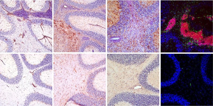
Nitsch et al. Journal of Neuroinflammation   (2021) 18:101                                                  Page 2 of 16

Background                                                   tissues pointing to the possible local effect of IL-23 in
Multiple sclerosis (MS) is the most common auto-             MS [15]. In addition, in some MS patients, a single nu-
immune disease of the central nervous system (CNS)           cleotide polymorphism of the IL-23 subunit p40 in-
and the most common disabling neurological disease of        creases the p40 expression and is associated with a
the young adulthood. The key features of MS are infil-       younger onset of the disease and single nucleotid poly-
tration of leukocytes, demyelinating plaques, and axonal     morphisms of the p19 subunit or the IL23R gene were
damage [1]. Triggers and mechanisms leading to the de-       associated with a higher risk of MS [9, 16]. Animal
velopment of the disease are not fully decoded, but it is    models emphasize the non-redundant role of IL-23 in
widely accepted to be an autoimmune disorder with            the development of the EAE, as EAE cannot be induced
known genetic and environmental risk factors. Besides        in mice lacking IL-23 or the IL-23 receptor complex
autoreactive T cells [2], B cells and plasma cells are es-   [17–19] and IL-23 produced by CNS-resident cells con-
sential components of MS lesions. B cell and plasma cell     trols T cell encephalitogenicity during the EAE [17].
accumulation in perivascular lesions and in the sub-            IL-23 is produced by classical antigen-presenting cells
arachnoid space as well as leptomeningeal inflammation       (APC) such as macrophages, dendritic cells, but can also
with ectopic lymphoid follicles were described in several    be produced by astrocytes [12, 20, 21]. Activation of the
studies [3–5]. The characteristic findings in the cerebro-   IL-23 receptor complex leads to stimulation and activa-
spinal fluid (CSF) of many MS patients are oligoclonal       tion of T helper 17 cells (Th17) lymphocytes. This sub-
immunogobulin bands, which indicate intrathecal anti-        type of CD4+ lymphocytes is crucial in chronic
body synthesis. Thus, immunotherapies targeting B cells      inflammation, autoimmune diseases and is detected in
have been successful and approved in the treatment not       MS lesions [10].
only for relapsing-remitting MS patients but also for           Since naïve CD4+ cells lack the IL-23R, their differen-
progressive MS patients [6].                                 tiation into Th17 cells is initiated by the transformation
  Data of the pathogenesis and derived therapeutic ap-       of the growth factor β (TGFβ), IL-6 or IL-21, which con-
proaches frequently arise from the experimental auto-        secutively stimulate the expression of the IL-23 receptor
immune encephalomyelitis (EAE) model as the animal           [22–24]. Upon IL-23 receptor signaling, Th17 cells pro-
model of MS. Although the EAE model was already dis-         duce various cytokines including their signature cyto-
covered in the 1930s [7], it is still used to decipher the   kines IL-17A, IL-17F, and others such as IL-21, IL-22,
pathogenesis of autoimmune neuroinflammation until           tumor necrosis factor a (TNFα), and colony stimulation
now. EAE can be induced with different kinds of myelin-      factor 2 (GM-CSF).
derived antigens. One widely used is the myelin oligo-          To conclude, the critical role of IL-23 in neuroinflam-
dendrocyte glycoprotein (MOG) 35-55 peptide. It is a         mation is striking. Nevertheless, the local impact of IL-
CNS- specific myelin glycoprotein expressed on the           23 in inflammatory CNS disorders such as MS remains
outer surface of the myelin sheaths [8]. After               elusive to some extent. To further characterize the im-
immunization with the MOG35-55 peptide along with            pact of IL-23 on neuroinflammatory processes, we gen-
immune-activating adjuvants, the animals usually de-         erated a transgenic mouse model with astrocyte-specific
velop a monophasic course with an ascending paresis          expression of IL-23 (GF-IL23). Our previous study
and subsequent regression of the symptoms over time.         showed that CNS-restricted IL-23-expression enables
  With the help of the EAE model, it was possible to         the spontaneous formation of infiltrates in the brain, es-
further decipher the role of cytokines in the develop-       pecially in the cerebellum [24]. After several months,
ment of MS. Cytokines are essential in the pathogenesis      GF-IL23 mice develop an ataxic phenotype and cerebel-
of various autoimmune disorders. Among the cytokines,        lar infiltrates with high amounts of lymphocytes particu-
a large number of studies have identified Interleukin 23     larly B cells. To further study the local influence of IL-
(IL-23) to play a critical role in various neuroinflamma-    23 in autoimmune CNS inflammation, we induced an
tory conditions, including MS [9–11]. It is part of the      EAE with MOG35-55 in the transgenic GF-IL23 mice.
so-called Interleukin 12 (IL-12) cytokine family and con-    The aim of this project was to further clarify the effect
sists of a unique p19 subunit and a common p40 subunit       of CNS-specific IL-23 expression on autoimmune neuro-
shared with IL-12 [12]. The IL-23 receptor complex con-      inflammation, especially concerning the impact of IL-23
sists of the IL-12Rβ1 component that binds to the p40        on the disease course and the development of infiltrates,
subunit and the specific IL-23 receptor (IL23R) subunit      cell accumulation, and the proinflammatory cytokine mi-
that binds to p19.                                           lieu in EAE. We demonstrate that the local IL-23 pro-
  IL-23 is increased in serum and cerebrospinal fluid of     duction in the CNS leads to aggravation and
MS patients [13, 14]. A significant increase of mRNA ex-     chronification of the disease course with severe parapar-
pression and protein production of both subunits of IL-      esis and an ataxic phenotype. Local synthesis of IL-23
23 was found in lesional compared to non-lesional            enhances gliosis and neuroinflammation not only in the
Nitsch et al. Journal of Neuroinflammation   (2021) 18:101                                                  Page 3 of 16

spinal cord but also in the cerebellum, brainstem, and        body lean, and 5 moribund. Moribund mice with a score
forebrain. The infiltrates mainly consist of lymphocytes      of 5 points were assigned to the score with the dominant
with pronounced B cell accumulation. It leads to demye-       clinical signs in the days before.
lination and induces a proinflammatory milieu with up-
regulation of cytokines and several complement factors.       Routine histology and immunohistochemistry
The data underline the importance of IL-23 in enhan-          Mice were sacrificed by deep isoflurane anesthesia and
cing and maintaining autoimmune inflammation in the           transcardially perfused with phosphate-buffered saline
CNS.                                                          (PBS). The brain was dissected and cut along the sagittal
                                                              midline. After fixation with PBS-buffered 4% paraformal-
Methods                                                       dehyde, half brains were embedded in paraffin or, for
Animals                                                       the cryosections, in Tissue Tek (Sakura Finetek, Staufen,
The generation of transgenic mice with astrocyte-             Germany). The 10-μm paraffin-embedded sections were
specific expression of particular genes was described in      deparaffinized with xylene and rehydrated in graded
detail previously [24]. Genotyping of the transgenic off-     ethanol series. Sections were stained with hematoxylin
springs was performed by polymerase chain reaction            and eosin (HE) (Sigma-Aldrich, Munich, Germany) and
(PCR) of tail DNA with the primer previously published        Luxol fast blue (LFB) for routine histology and analysis
[24]. All mice were kept under standardized pathogen-         of myelination, respectively. For immunohistochemistry,
free conditions at the animal facility of the University      slides were incubated over night at 4°C with the primary
Hospital of Bonn, Germany. Animal experiments were            antibodies (polyclonal rabbit anti-Laminin, Sigma-
approved by the Animal Care Commission of                     Aldrich, 1:50, polyclonal rabbit anti-GFAP 1:250) after
Nordrhein-Westfalen, Germany.                                 incubation with Proteinase K (ThermoScientific, Wal-
                                                              tham, USA; 1:1000) or incubated with biotin-conjugated
Induction of EAE                                              tomato lectin (Axxora, 1:50) and washed with PBS. A
Starting at the age of 6 months, GF-IL23 mice spontan-        corresponding biotinylated secondary antibody was used
eously develop an ataxia with cerebellar infiltrates [24],    for 45min (Jackson ImmunoResearch, Newmarket, UK,
but neither clinical abnormalities nor histological           1:200) and horseradish peroxidase-coupled streptavidin
changes occur before that age. To investigate the local       for another 45 min (Vector Labs, Burlingame, USA; 1:
effect of IL-23 in an EAE model, we immunized 2-              200). For the immunoperoxidase, Nova Red (Vector
month-old WT controls and transgenic animals at an            Labs) as the substrate was used according to the manu-
age well before clinical symptoms and spontaneous neu-        facturer’s instruction. Sections were counterstained with
roinflammation of the transgenic GF-IL23 mice occur.          hematoxylin (Sigma-Aldrich, Munich, Germany). For
On day 0, mice were immunized subcutaneously into             fluorescent immunohistochemistry, 10-μm cryosections
the rear flanks with a 1:1 emulsion of 100 μl MOG35–          were incubated with the primary antibodies (anti-CD3e
55 or bovine serum albumin (BSA) (3 mg/ml) in 100 μl          BD Bioscience 1:500; anti-B220 BD Bioscience 1:200)
complete Freund’s adjuvant (CFA) (Sigma-Aldrich,              over night at 4° C, washed with PBS, incubated with an
Munich, Germany) supplemented with 4 mg/ml Myco-              A594 or A488 fluorescence-conjugated secondary anti-
bacterium tuberculosis H37RA (Difco, Detroit, MI) as          body (Invitrogen, Darmstadt, Germany; 1:400) for 45
described before [25]. In addition, each mouse received       min, and counterstained with DAPI (Sigma-Aldrich,
an intraperitoneal injection of 500 ng pertussis toxin        Munich, Germany).
(Sigma-Aldrich, Munich, Germany) on days 0 and 2.                A Nikon eclipse 800 bright-field and fluorescence
                                                              microscope (Nikon, Duesseldorf, Germany) was used.
Clinical assessment                                           Brightfield and monochrome fluorescent images were
Animals were weighed and examined for 60 days on a            captured with a SPOT flex camera and SPOT advanced
daily basis with two different scores: the typical EAE        4.5 software (Diagnostic instruments, Sterling, USA).
score [25] and the atypical score [26], reflecting an as-     Myelinated and demyelinated areas of the spinal cord
cending paresis and an ataxic phenotype, respectively.        were measured on cross-sections with ImageJ image
The typical EAE score was assessed as follows: 0 no clin-     analysis software to determine the proportion of the
ical symptoms, 1 limp tail, 2 hind limb weakness, 3 hind      demyelinated spinal cord area.
limb paralysis, 4 hind and forelimb paralysis, and 5 mori-
bund. The atypical EAE score was assessed as follows: 0       Flow cytometry
no clinical symptoms, 1 hunched appearance, stiff tail, 2     Isolation of microglia and infiltrating leukocytes from
staggered walking, scruffy coat, 3 head tilt, ataxia, obvi-   the cerebellum was described in detail before [27]. Mice
ous impaired balance/ambulation, body lean, 4 inability       were sacrificed by deep isoflurane anesthesia and trans-
to maintain upright posture, severe axial rotation, severe    cardially perfused with 4°C cold 1x PBS for removal of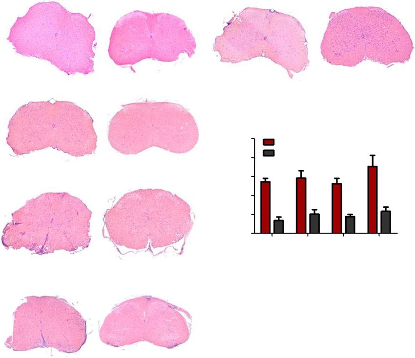
Nitsch et al. Journal of Neuroinflammation   (2021) 18:101                                                     Page 4 of 16

intravascular leukocytes until the fluid was clear. The      met the log2 fold change >1 cutoff and an p value < 0.05
dissected cerebellum and myelon were homogenized in          were uploaded to the Database for Annotation,
Hank’s Balanced Salt Solution (HBSS, Gibco, Eggenstein,      Visualization and Integrated Discovery (DAVID). Rele-
Germany) by a tissue homogenizer (glass potter, Braun,       vant categories of cellular functions and pathways were
Melsungen, Germany) before passing through a 70-μm           depicted [28, 29]. The number of targets from the data
cell strainer (BD biosciences, Heidelberg, Germany).         set mapping to the category is displayed. A modified
After centrifugation, the pellets of the homogenates were    Fisher’s exact test was used to calculate a p value deter-
resuspended in 75% isotonic 4°C Percoll solution (GE-        mining the probability that the association to the cat-
Healthcare, Uppsala, Sweden) and overlayed with 25%          egory is by chance alone with p
Nitsch et al. Journal of Neuroinflammation      (2021) 18:101                                                                         Page 5 of 16

  Fig. 1 Exacerbation of EAE in GF-IL23 mice. a, b GF-IL23 mice develop a more severe EAE with an ascending paralysis and in addition an ataxic
  phenotype compared to WT mice. The characteristic findings are also summarized in Table 1. c Consistent with the clinical phenotype the GF-
  IL23 mice show a considerable weight loss compared to the WT mice. The data represent 9-12 GF-IL23 mice and 9 WT mice

3.3 (± 1.0) points. In contrast to the WT controls, the                    clinical course of the EAE. Thereby, day 15 represents
transgenic mice did not recover from the deficits, but ra-                 the analysis at the beginning of the disease, day 22 the
ther exhibited a chronic disease with an outcome score                     experiments shortly after the maximum score of the as-
of still 3.3 (± 1.0) points.                                               cending paresis and before onset of the ataxia, day 33
   Beside the progressive paraparesis, GF-IL23 mice devel-                 the period around the onset of the ataxia, and day 60 the
oped a progressive ataxic phenotype, which prompted us to                  results in the chronic stage and at the end of the obser-
use an additional score for ataxia to monitor these mice                   vation period.
more accurately. The ataxia started at an average at day 36.0                The myelon of the EAE transgenic animals displayed
(± 14.5) after immunization with a maximum score of 2.9 (±                 pronounced infiltrates in transverse sections around the
1.8) on day 44.5 (± 10.0) without a relevant recovery until                period of the maximum score on day 22, but also up to
day 60 (Tbl. 1, Fig. 1b). In comparison, none of the WT con-               day 33 compared to WT mice (Fig. 2a–h). In the chronic
trols developed an ataxia. Correlating with the more severe                stage, at day 60, the EAE GF-IL23 mice show only few
clinical manifestation, GF-IL23 mice showed a pronounced                   infiltrates, but still pronounced demyelination compared
weight loss compared to WT mice (Fig. 1c). In contrast, WT                 to WT mice (Fig. 2i–k).
and transgenic controls immunized with only BSA showed
neither clinical deficits (data not shown) nor histological                Immunization of GF-IL23 mice with MOG35-55 peptide
signs of infiltration in cerebellum, myelon, or upregulation of            results in prominent infiltration of the cerebellum, brain
proinflammatory marker (Fig. S1).                                          stem, and forebrain
   To summarize, CNS-specific expression of IL-23 led to                   Congruent with the ataxic phenotype, the cerebellum of
a chronic progressive EAE with a severe form of the as-                    EAE GF-IL23 mice displayed pronounced infiltrates in
cending paresis and, in contrast to WT controls, a severe                  the histology. Naïve mice show no inflammatory infil-
ataxia without a regression of the deficits.                               trates (Fig. 3a, d, g, j). On day 15, even before onset of
                                                                           the ataxia, smaller infiltrates became manifest, especially
Enhanced spinal cord infiltration in GF-IL23 mice and                      intraparenchymal and perivascular in transgenic mice
persisting demyelination                                                   (Fig. 3b, e). On days 22, 33, 60 along with the clinical
For the following experiments, 5 different time points,                    manifestation of the cerebellar ataxia, there is a stronger
days 0, 15, 22, 33, and 60, were depicted based on the                     infiltration including also subarachnoidal infiltrates at

Table 1 Clinical features of the EAE in GF-IL23 and WT mice
                                              Myelitis                                                       Ataxia
                                              GF-IL23                         WT                             GF-IL23                              WT
Incidence                                     12/12                           8/9                            7/9                                  0/9
Disease (days) onset                          11.5 ± 1.5*                     15.5 ± 7.5                     36.0 ± 14.5***                       -
Time (days) to maximum                        21.5 ± 6.0                      23.5 ± 14.0                    44.5 ± 10.0***                       -
Maximum score                                 3.3 ± 1.0                       2.5 ± 0.4                      2.9 ± 1.8**                          -
Outcome (d60)                                 3.3 ± 1.0***                    1.1 ± 0.3                      2.9 ± 1.7**                          -
*p < 0.05, **p < 0.01, ***p < 0.001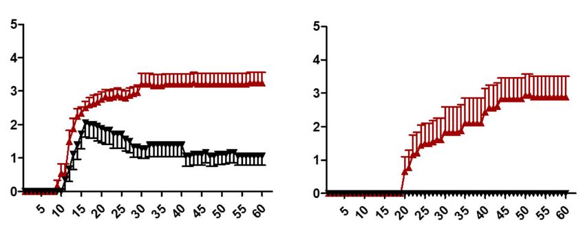
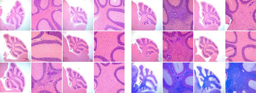
Nitsch et al. Journal of Neuroinflammation      (2021) 18:101                                                                             Page 6 of 16

 Fig. 2 Increased myelon infiltration in the cerebellum of EAE-induced GF-IL23 mice. HE-staining of the myelon (transverse sections) at different
 time points (d0, d15, d22, d33, d60). GF-IL23 naïve mice show no infiltration (a WT control E). b–d I GF-IL23 mice show enhanced infiltration over
 the time, especially at d22, d33 compared to WT mice (f, g, h, j). Arrows point to infiltrates. Scale bar: 100 μm. The images are representative of
 those from at least different 8 transgenic/WT mice d0, d15, d22, d33, and 8 transgenic, 6 WT mice d60. k Demyelinated area relative to the white
 matter area was quantified in cross-sections of the spinal cord from GF-IL23 and WT mice (n = 4–5 for each group) with *p < 0.05 and **p< 0.01
 and showed a significant enhanced demyelination in GF-IL23 mice

 Fig. 3 Subarachnoidal and intraparenchymal infiltration in cerebellum of EAE-induced GF-IL23 mice. HE- and LFB-staining of the cerebellum at
 different time points (d0, d15, d22, d33, d60) after induction of the EAE. Naïve GF-IL23 mice show no infiltration (a higher magnification (d), WT
 controlls (g, j)). GF-IL23 mice (b, c, m, n, o higher magnification (e, f, p, q, r)) show increasing subarachnoidal (arrow) and intraparenchymal
 (arrow head) infiltrates over the time and persistent demyelination on day 60 whereas WT mice (h, i, s, t higher magnification (k, l, v, w)) show
 only little and few infiltrates without persisting demyelination on day 60. Scale bar: 100 μm. The images are representative of those from at least
 8 different transgenic/WT mice d0, d15, d22, d33, and 8 transgenic, 6 WT mice d60
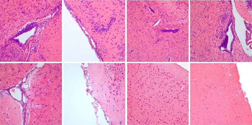
Nitsch et al. Journal of Neuroinflammation       (2021) 18:101                                                                             Page 7 of 16

this stage (Fig. 3c, f, m, n, p, q). WT showed only                           Infiltrates consist mainly of lymphocytes with a high
mild and sporadic infiltrates (Fig. 3h, I, k, l, s, t, v w).                  proportion of B cells
Accompanying the infiltrates, the GF-IL23 mice                                The infiltrates were further characterized by immuno-
showed pronounced demyelination in the tissue sur-                            histochemistry and flow cytometry analysis. Immuno-
rounding the infiltrates (Fig. 3 o, r, u, x). Beside infil-                   fluorescence histology of the cerebellum illustrated
tration of the myelon and cerebellum, GF-IL23 mice                            that the infiltrates of the transgenic mice predomin-
developed a widespread CNS infiltration with ven-                             antly consist of lymphocytes with a high proportion
tricular and meningeal involvement, smaller intrapar-                         of B cells (Fig. 5g, h for d60, Fig. S2 for d0, d15, d22,
enchymal infiltrates in the forebrain and in the brain                        d33), which was confirmed by flow cytometry data as
stem (Fig. 4a, b, d, f, h). WT mice, on the other                             well.
hand, showed no or only few small infiltrates in these                          In the cerebellum of immunized GF-IL23 mice, there
regions (Fig. 4a, c, e, g, i). Immunohistochemistry of                        was an increase of CD4+ cells on day 22 and d60 and of
the cerebellum revealed microglia and astrocyte acti-                         CD8+ cells on day 60 (Fig. 6a). Beside the T cell infiltra-
vation in the transgenic immunized animals compared                           tion, vast amounts of CD19+ B cells were detectable in
to WT mice (Fig. 5c–f for d60, Fig. S2 for d0, d15,                           the cerebellum of GF-IL23 mice at day 22, d33, d60.
d22, d33). In addition, hypervascularization was de-                          Non-immunized transgenic mice did not show lympho-
tected on day 60 (Fig. 5a, b).                                                cyte infiltration. Regarding other immune cell

 Fig. 4 Infiltration of the ventricle, meninges, and parenchyma of the forebrain and brain stem in GF-IL23 mice. a Fraction of GF-IL23 and WT EAE
 mice with cellular infiltrates in ventricles, meninges, parenchyma, or brain stem. Naïve mice did not show these infiltrates. Transgenic mice show
 infiltrates from day 15 in these locations, whereas only sporadic infiltrates were found in WT mice. b–i Exemplary histology findings at d33. b, d, f,
 h Beside the cerebellum, infiltrates (arrow heads) in other locations such as ventricle, meninges, and parenchyma of the forebrain and brain stem
 were found in GF-IL23 mice. WT mice (c, e, g, i) display no infiltrates in these locations. Scale bar: 100 μm. The images are representative of those
 from at least 8 different transgenic/WT mice d33
Nitsch et al. Journal of Neuroinflammation      (2021) 18:101                                                                             Page 8 of 16

 Fig. 5 Hypervascularization, microglia, astrocyte activation, and B cell accumulation in GF-IL23 cerebellum. Histology findings at d60. a–f GF-IL23
 mice display hypervascularization (a), microglia, and astrocyte activation (c, e) compared to WT mice (b, d, f). g, h Fluorescent
 immunohistochemistry of GF-IL23 cerebellum reveals that the infiltrates consist of large amounts of B cells. Scale bar: 100 μm. The images are
 representative of those from at least 8 different transgenic/6 WT mice

 Fig. 6 Infiltrates consist of large amounts of B cells. a Flow cytometry data of the cerebellum (absolute numbers) on days 0, 22, 33, and 60 show
 consistent findings with predominant CD19+ cells in the cerebellum of MOG-immunized GF-IL23 mice. In addition, CD4+ cells (d22, d60), CD8+
 cells (d60), CD11bhighCD45high (d33, d60), and also γδ TCR+ cells (d33) were elevated compared to WT mice. b Flow cytometry data of the
 myelon on day 22 and day 33 show elevation of CD4+ and CD19+ cells on d22 and CD19+ cells compared to WT mice. The mean value,
 standard deviation are given with *p < 0.05, **p< 0.01, ***p< 0.001 with n=5 transgenic/WT mice d0, n= 8–12 transgenic/WT mice d22, d33, and 6
 transgenic/4 WT mice d60
Nitsch et al. Journal of Neuroinflammation (2021) 18:101 Page 9 of 16
populations, an increase of CD11bhigh CD45high cells IL-23 induces cell activation, a proinflammatory cytokine
could be detected on d33 and d60. γδ T cells were ele- profile, and upregulation of complement components
vated on day 33. NK 1.1+ and Ly6G+ cells were not ele- For the analysis of surface cell markers, activation
vated (data not shown). markers, inflammation mediators, and complement fac-
In the myelon, CD19+ B cells were elevated on day tors, a transcriptome analysis of the cerebellum of the
22 and day 33 and CD4+ cells on day 22 compared transgenic and WT animals was performed (Fig. 7a, for
to WT mice (Fig. 6b). Other cell populations, such as the statistical parameters Fig. S3). The findings with ele-
NK 1.1 and γδ T cells were not elevated (data not vated mRNA levels of the surface cell marker CD19 at
shown). day 22 and day 33 were in line with the flow cytometry
Taken together, the flow cytometry data demon- results. Furthermore, the expression of CD4 on d22 and
strated a pronounced infiltration of lymphocytes, par- d33, CD8 on day 33, and CD11b on day 33 was also sig-
ticular B cells next to CD4+, CD8+ cells in the nificantly increased in the transgenic mice as was the ex-
cerebellum and CD4+ cells in the myelon accompan- pression of CD11c.
ied by γδ T cells on day 33 and CD11bhigh CD45high The expression of other surface cell markers, among
cells on day 33 and 60 in the cerebellum of the trans- them markers for T and B cell activation and co-
genic mice. stimulatory molecules, such as CD20, CD25, CD40,
A GF-IL23 naive WT d22 WT d33 GF-IL23 d22 GF-IL23 d33
CD3e
B
30 *
GF-IL23/ WT mice
CD4
X fold change-
CD8a
CD19
CD11b 20
CD11c * * *
CCL5
CCL7 10
CCL8 d22 tg/WT
CCR1 *
CCR6 0 d33 tg/WT
CCR7 Stat 3 Rorc IL-17a Stat 4 Tbet IFN
CXCL9
CXCL10
CXCL13
CXCR3
CD20
C 45 Cerebellum EAE d33/ upregulation in GF-IL23 mice
# upregulated genes
CD25 40
CD27
CD40 35
CD44
CD68 30
CD69 25
CD80
CD86 20 P< 0.005
H2-Eb1
Icam1 15
IFN 10
IL-1a
IL-1b 5
IL-2
IL-5 0
chemotaxis
T cell activation
cytokine activity
Complement pathway
immunological synapse
B cell activation
B cell homeostasis
innate immune response
inflammatory response
B cell differentiation
T cell differentiation
Antigen processing and presentation
Immunoglobulin
TNF signaling pathway
positive regulation of cell migration
adaptive immune response
Chemokine signaling pathway
T cell receptor signaling pathway
negative regulation of T cell proliferation
Cell adhesion molecules (CAMs)
Cytokine-cytokine receptor interaction
positive regulation of angiogenesis
regulation of B cell differentiation
B cell receptor signaling pathway
positive regulation of T cell proliferation
IL-6
IL-7
IL-10
IL-17a
IL-17f
IL-21
IL-22
Foxp3
TGF
TNF
C6
C7
C8a
C8b
C8g
C1qb
C1qc
C2
C4a
C4b
C3
CHF
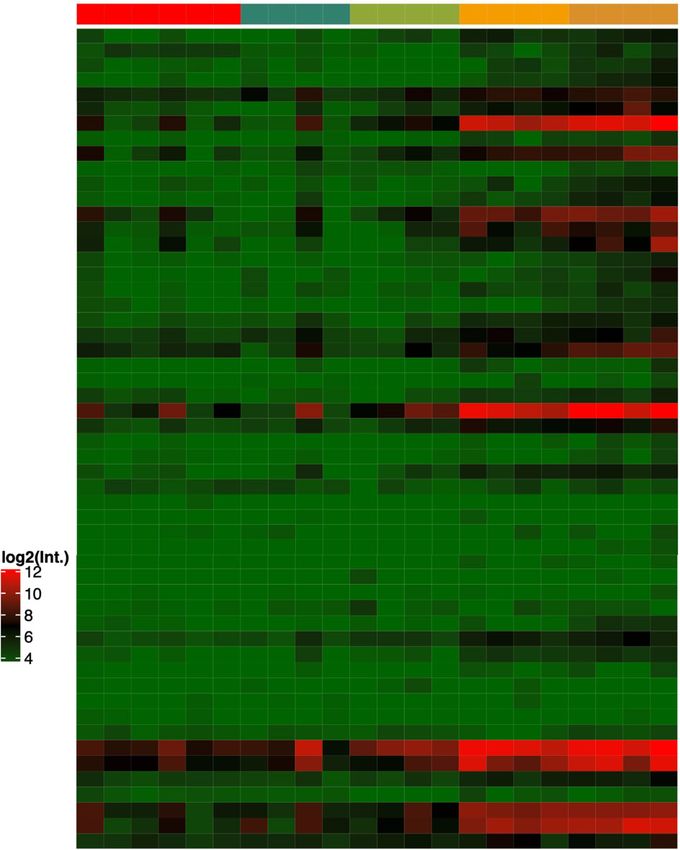
Fig. 7 mRNA-profile of the cerebellum. a The transcriptome analysis illustrates the mRNA levels of several surface cell markers, pro-/
antiinflammatory markers, and complement components of the cerebellum of GF-IL23 and WT mice at d22 and d33 after induction of the EAE. b
The mRNA levels of targets involved in Th17 and Th1 cell signaling are listed from MOG-immunized mice at d22 and d33 in relation to WT
immunized with *p < 0.05. c To generate functional categories of upregulated mRNA levels, targets from the data set that met the log2 fold
change >1 cutoff and an p value < 0.05 were uploaded to the Database for Annotation, Visualization and Integrated Discovery (DAVID) and
relevant categories of cellular functions and pathways were depicted. The number of targets from the data set that map to the category is
displayed. Only significant categories with pNitsch et al. Journal of Neuroinflammation (2021) 18:101 Page 10 of 16
CD27, CD44, CD68, CD69, CD86, ICAM-1, and H2- tissue destruction and inflammatory infiltrates most
Eb1, was increased. prominent in the subarachnoid and perivascular space.
In addition, the mRNA levels of various proinflamma- The CNS-cytokine milieu was characterized by elevation
tory cytokines and cytokine receptors were elevated in of numerous inflammatory mediators such as IL-17a and
the transgenic cerebellum such as CCL5, CCL7, CCL8, IFNγ. However, the leukocytic infiltrates were surpris-
CCR6, CCR7, CXCL9, CXCL10, CXCL13, CXCR3, IFNγ, ingly predominated by B cells. In the current study, GF-
IL-1a, IL-1b, TNFα, and TGFβ, but not significantly the IL23 mice and WT controls were immunized with MOG
Th17 cell signature cytokines IL-17a, IL-17f, IL-21, and 35-55 to evaluate the impact of locally synthesized IL-23
IL-22. Foxp3, playing an important role in regulatory T on the course of EAE as the animal model of MS.
cell function, was also elevated. After immunization with the peptide MOG 35-55, the
Furthermore, analysis of the complement system indi- transgenic mice showed a slightly earlier onset of the
cates increased expression of C1qb, C1qc, C2, C3, and progressive paraparesis compared to WT mice. Further-
C4b on day 22 and 33 as well as C4a on day 33 and more, the GF-IL23 mice developed an ataxia and most
complement component factor H (CFH) on day 22. essentially no remission of both paraparesis and ataxia.
Figure 7b lists the expression of the typical down- This indicates that CNS-specific synthesis of IL-23 is not
stream targets involved in Th17 and Th1 cell signaling only crucial for the development of the paraparesis and
in GF-IL23 compared to WT mice. Regarding Th17 cell ataxia, but also leads to a rather chronic course of the
signaling, signal transducer and activator of transcription EAE.
(Stat) 3 was elevated in d33 transgenic mice, but not IL- In addition, the GF-IL23 mice developed a pro-
17a or RAR-related orphan receptor C (Rorc), which en- nounced stronger infiltration in the spinal cord with
codes RAR-related orphan receptor gamma (Rorγ). In marked reduction of infiltration at the end of the obser-
d22 and d33 mice, T box expressed in T cells (T bet), in vation period. In contrast, severe clinical deficits per-
d33 mice Stat 4 and IFNγ, all involved in Th1 cell signal- sisted in the presence of IL-23. The ongoing para- or
ing, was upregulated in GF-IL23 mice. tetraparesis appeared to be related to the stronger de-
Furthermore, we generated functional categories of myelination on day 60, which was more pronounced in
upregulated mRNA targets in immunized GF-IL23 mice GF-IL23 mice. The clinical and histological findings of
compared to WT immunized mice and relevant categor- the spinal cord point toward a direct or indirect role of
ies of cellular functions and pathways were depicted and IL-23 in demyelination and interfering with remyelina-
illustrated in Fig. 7c. We found categories of the innate tion after EAE induction. Demyelinated lesions are one
and adaptive immune response are important in our of the hallmarks of MS and are present in the white as
model. But also upregulation of cell adhesion molecules, well as in the gray matter [30]. Remyelination is a crucial
targets involved in chemotaxis, antigen processing and step during the remission phase of an MS relapse as it
presentation, and B and T cell activation/proliferation/ preserves the axon from dissection and degeneration. In
differentiation are relevant in the EAE GF-IL-23 model. chronic MS, incomplete spinal cord remyelination corre-
In summary, the transcriptome analysis also illustrates lates with higher disease-related disability [31].
a B and T cell accumulation in the cerebellum. But it Moreover, besides the clinical and histological signs of
also points toward T and B cell activation, upregulation spinal cord affection, the cerebellum of the GF-IL23
of co-stimulatory molecules, proinflammatory cytokines, mice showed a chronic EAE course with manifest and
and components of the complement system. here persisting infiltration and again pronounced demye-
lination. In addition, infiltrates were detected at various
Discussion other CNS locations such as ventricles, meninges, in the
MS is an autoimmune disorder resulting in focal neu- forebrain, and brain stem.
roinflammatory lesions in the gray and white matter. Be- The inflammatory response of GF-IL23 mice during
sides neuroinflammation, demyelination, and axonal EAE is a specific response to the immunization with
damage throughout the brain are the hallmarks of MS. MOG-peptide, because BSA-immunized GF-IL23 mice
IL-23 has been established as one of the crucial cyto- did not show any of the described pathological features.
kines in autoimmune disorders and especially in the In our previous study, although we saw infiltrates in the
pathogenesis of MS and corresponding animal models long-term LPS experiment in GF-IL23, these animals did
[9, 13, 14, 17]. not show an ataxia, the extent of infiltrates that we see
Our previous study has illustrated that CNS-restricted in EAE or these multifocal infiltrations [24].
IL-23-expression induces the spontaneous formation of There is evidence that IL-23 have a greater impact on
infiltrates in the brain, especially in the cerebellum [24]. the onset of autoimmune CNS inflammation than on
These GF-IL23 mice spontaneously develop a progres- the maintenance of the inflammatory response. A
sive ataxic phenotype, which corresponds to cerebellar prophylactic effect of anti-p40 antibody treatment hasNitsch et al. Journal of Neuroinflammation (2021) 18:101 Page 11 of 16 been shown in a myelin-induced EAE model in marmo- astrocytes [21]. Studies demonstrate that local produc- sets [32]. The treatment during the ongoing disease with tion of IL-23 in the CNS [17, 18] controls T cell ence- anti-IL-12p40 antibodies delays the clinical manifest- phalitogenicity in EAE. However, regarding the GF-IL23 ation of existing lesions. However, once neurological model, some points need to be considered. In the GF- deficit has become manifest, progression to serious defi- IL23 modell, IL-23 overexpression occurs only in astro- cits seems less influenced by anti-IL-12p40 treatment cytes. Furthermore, IL-23 secretion is at the beginning [33]. One possible explanation for the failure of the anti- of the neuroinflammatory process during EAE in the p40 antibody ustekinumab phase II clinical trial in re- GF-IL23 model and is not secondary to immunization lapsing–remitting MS is that the possibility for a benefi- with MOG as in WT EAE mice. In the GF-IL23 model, cial treatment might have already passed once the astrocyte-specific secretion of IL-23 persists, independ- patients were enrolled [34]. Thakker et al. showed that ent of the immunization with MOG. Therefore, the data encephalitogenic T cells from MOG-immunized WT on EAE in GF-IL23 cannot reflect all aspects of IL-23 in mice caused indistinguishable disease when adoptively autoimmune neuroinflammation, but may be able to fur- transferred to WT or IL-23-deficient recipient mice, ther study some aspects such as the B cell role in the IL- demonstrating that once encephalitogenic cells have 23 mediated neuroinflammation. been generated, EAE can develop independent from IL- Immunohistochemistry of the cerebellum, flow cytom- 23 [35]. In contrast to these findings, the course of EAE etry data of the myelon and cerebellum, and data from in GF-IL23 mice observed in our study argues for a role the transcriptome analysis of the cerebellum showed that of local IL23 beyond the disease onset. In the GF-IL23 the infiltrates of the GF-IL23 mice predominantly con- mice, the EAE is chronic and not monophasic as in the sisted of lymphocytes. In the spinal cord, an increase of WT control, so astrocyte-specific expression of IL-23 ap- CD19+ cells and CD4+ cells were detected. In the cere- pears to play a role in the maintenance of neuroinflam- bellum, the flow cytometry results with elevated T cells, mation. The progressive infiltration suggests that IL-23 especially CD4+ cells, and CD19+ cells were even more not only triggers the inflammation, but in addition also pronounced. The transcriptome analysis revealed ele- contributes to the further course in our model. vated levels of Foxp3 during the course of the EAE in In comparison to other mouse models with astrocyte- the cerebellum pointing to also regulatory immune specific expression of certain cytokines, the disease mechanisms during the disease course. course as well as the histochemistry data with the B cell The dominant B cell accumulation during EAE in mice infiltration of the GF-IL23 mice in the EAE model is with a C57BL/6 genetic background is a very uncommon quite remarkable and emphasizes the findings in our finding, as this EAE model is usually a mainly T cell- study are IL-23-specific. After immunization with driven model of CNS inflammation [37]. But compared MOG35-55, GF-IL12 mice with astrocyte-specific ex- to our previous findings describing the spontaneous pression of IL-12 had an earlier onset and higher inci- course of the GF-IL23 mice, the B cell accumulation dence but not the chronic EAE course and ataxia, nor during EAE in GF-IL23 mice is less surprising and un- the multilocular inflammation or pronounced tissue de- derlines a role of IL-23 in attracting and accumulating B struction seen in GF-IL23 mice [36]. By contrast, cells into the subarachnoidal space and into the CNS MOG35-55-immunized GF-IL6 mice developed a severe parenchyma during neuroinflammation [24]. In the EAE ataxia, but no signs of spinal cord involvement [25]. In- model, the GF-IL23 mice developed these infiltrates ac- filtration and demyelination were nearly absent from the celerated and much earlier after the immunization with spinal cord, but significantly increased in the cerebellum MOG 35-55 peptide. of EAE GF-IL6 mice with accumulation of neutrophils In healthy humans, only very few B cells are present in and B cells accompanied by tissue damage. Thus, site- the CNS parenchyma and CSF, where they represent less specific production of IL-6 in the cerebellum redirects than 1% of leukocytes [38, 39]. Though, in patients with the leukocytes away from the normally preferred anti- CNS inflammation, B cell numbers can rise and accumu- genic site of the spinal cord with markedly enhanced in- lation of B cells is one of the characteristic findings in flammatory cell accumulation in the cerebellum [25]. MS. Histopathological studies of the brain tissue from This seems to be different to the GF-IL23 model. Here, progressive MS patients revealed leptomeningeal inflam- the CNS-specific IL-23 expression leads to a strong and mation with ectopic lymphoid follicles, B cells and chronic neuroinflammation in the EAE model with high plasma cells. In relapsing-remitting MS, B cell accumula- amounts of B cells and to a multilocular inflammation, tion in the CNS was also demonstrated [5, 40]. B cell thus particularly pronounced in the spinal cord and rich meningeal inflammation was associated with in- cerebellum. creased spinal cord pathology in secondary progressive IL-23 is produced by APC such as macrophages or MS patients [41]. In general, B cells are enriched in peri- dendritic cells [12, 20], but has also been detected in vascular lesions and in the subarachnoid space in these
Nitsch et al. Journal of Neuroinflammation   (2021) 18:101                                                  Page 12 of 16

patients, but are also sparsely present in the CNS paren-     synthesis. Surprisingly, Th17 signature cytokines IL-17a,
chyma [3–5]. The central role of B cells in the pathogen-     IL-17f, IL-21, and IL-22 were not significantly elevated
esis of MS was further substantiated by the beneficial        when compared to WT mice. Consistent with this, the
and approved treatment of relapsing-remitting and ac-         Th17 cell transcription factor Rorc is also not upregu-
tive primary progressive MS patients with B cell deplet-      lated, whereas Stat3 is upregulated in d33 transgenic
ing therapies [6, 42, 43].                                    mice. This indicates that the IL-23 mediated mechanism
   The B cell infiltration upon CNS-specific IL-23 expres-    of immune cell accumulation and aggravation of the
sion in our model is impressive. The mechanisms by            local proinflammatory cytokine milieu may be partly in-
which these B cells contribute to the phenotype can be        dependent of the Th17 cell axis. In our study, we found
various. B cells modulate CNS inflammation by humoral         elevation of the Th1 cell-associated Stat4, Tbet besides
and cellular mechanisms like antibody secretion, cyto-        IFNγ in GF-IL23 mice during EAE. This is in line with
kine release, as antigen-presenting cells by interaction      our previous study of the spontaneous course of the GF-
with and co-stimulation of T cells [44–46]. The tran-         IL23 mice. There we found increased levels of IFNγ and
scriptome analysis showed evidence for T and B cell ac-       also Th1 cells in the inflammatory cerebellum, but also
tivation, proliferation and differentiation and the role of   IL-17a and IFNγ co-expressing CD4+ cells in older mice
co-stimulatory surface antigens in the GF-IL23 model          [24]. In MS patients, the levels of IFNγ are associated
during EAE. Surface cell markers for T cell activation        with the disease activity [54] and the myelin-reactive T
such as CD25, CD44, and CD69 and markers for B cell           cell repertoire produces IFNγ in response to antigens
activation such as CD86 were clearly elevated. Other co-      [55]. By increasing the expression of MHC molecules,
stimulatory molecules such as CD27 as a regulator of B        IFNγ as well enhances the antigen-presentation of mye-
cell activation and immunoglobulin synthesis and CD40         loid cells, which in turn results in activation of myelin-
were also elevated. CD40 is expressed on B cells and          antigen-specific CD4+ or CD8+ cells [54]. We could
upon binding to the CD40L, expressed on activated             speculate that upregulation of B/T cell co-stimulatory
CD4+ cells, leads to B cell proliferation, memory B cell      molecules in the GF-IL23 EAE model mentioned above
generation, and antibody class switching [47]. Binding of     makes T cells more active and aggressive, leading to the
T cells to CD40 and CD86 on the other hand leads to           increased expression of IFNγ.
activation and differentiation of encephalitogenic T cells.     In addition, the findings of the transcriptome ana-
Antigen processing and presentation plays also a role in      lysis in our model point to a role of IL-23 in enhan-
the transgenic EAE mice. The high level of histocom-          cing the EAE driven chemokine response. CCL5,
patibility 2, class II antigen E beta (H2-Eb1) chain, which   CCL7, CCL8, CXCL9, and CXCL10 secretion is IFNγ,
encodes the Major histocompatibility complex class II         TNFα, and IL-1 dependent [56, 57]. It should be
(MHC II) protein complex, is striking in our model.           noted that all of these cytokines, IFNγ, TNFα, and
MHC II is generally expressed on APC and presents an-         IL-1 are pronounced elevated in the GF-IL23 mice
tigens to CD4+ cells. Among the APCs, B cells are             and are therefore likely responsible for the upregula-
highly efficient in presenting antigens even in low anti-     tion of these chemokines. CCL5 is secreted by various
gen concentrations as they can recognize CNS antigens         cell types and can attract T cells, monocytes, baso-
by their specific receptor [48, 49]. The MHC class II-        phils, eosinophils, NK cells, dendritic cells, and mast
restricted antigen presentation by B cells is essential for   cells [58]. In the CSF of MS patients, higher levels of
the induction of EAE that require both B cells and T          CCL5 and CXCL10 were detected compared to
cells. It allows B cells to present myelin and other CNS      healthy controls [59]. It is postulated that T cells,
antigens to encephalitogenic T cells [50]. An EAE can-        able to transmigrate into the CSF, express CXCR3
not be induced in mice with selective MHCII-knockout          mostly independently of a possible inflammatory CNS
in B cells, and these mice show a diminished Th1 and          condition, but only in the presence of the CXCR3 lig-
Th17 cell response [51, 52]. Furthermore, the upregula-       and CXCL10 in the CSF they remain in the CSF and
tion of ICAM-1 in our study is quite interesting since        do not recirculate into the periphery [60]. Another
ICAM-1 induced in response to TNFα, IL-1β, or IFNγ            chemokine, CXCL13, which is intimately involved in
mediates T cell migration across the blood-cerebrospinal      B cell trafficking [61], was highly elevated during EAE
fluid barrier and blood brain barrier and is expressed on     of the GF-IL23 mice. CXCL13 is increased in the
antigen-presenting cells [53].                                meninges and CSF of postmortem MS cases with high
   Various proinflammatory cytokines and cytokine re-         levels of meningeal inflammation and demyelination.
ceptors including IL-1a, IL-1b, the T helper 1 (Th1) sig-     Furthermore, elevated CSF levels correlate with B cell
nature cytokine IFNγ, and TGFβ, TNFα were elevated            count in the CSF, markers of immune activation, dis-
indicating a pronounced proinflammatory milieu in the         ease activity, and gray matter damage [62, 63]. The
cerebellum of the transgenic mice due to the local IL-23      role of CXCL13 was further substantiated since the
Nitsch et al. Journal of Neuroinflammation   (2021) 18:101                                                                    Page 13 of 16

level of CXCL13 in the CSF correlates with clinical           inflammatory effects by secreting anti-inflammatory cy-
response to B cell-depleting therapies in MS patients         tokines and promotion of neurogenesis by secreting
[64].                                                         neurotrophic factors were reported [75, 76].
   Furthermore, the findings concerning the complement
system with increased expression of several components        Conclusion
such as C1qb, C1qc, C2, C3, C4a, C4b, and CFH in the          Taken together, this study dissects the role of CNS-
EAE of GF-IL23 mice are intriguing. In some MS pa-            specific expression of IL-23 in neuroinflammation with
tients, autoantibodies contribute to tissue destruction by    quite impressive findings. The data presented here valid-
binding to the surface of brain cells and by attacking        ate IL-23 once more as a crucial factor driving inflam-
them dependent on complement factors [65]. The im-            matory processes in the CNS. Especially the aggravated
portance of the complement system in the EAE has been         and chronic phenotype of the GF-IL23 mice after
demonstrated in several studies [66–68]. Hundgeburth          immunization with MOG35-55, the multilocular infiltra-
et al. described the contribution of the complement sys-      tion with high amounts of lymphocytes, especially B
tem to the demyelination and modification of the T and        cells, the persistent demyelination and the proinflamma-
B cell response in the EAE model. Mead et al. also            tory milieu with upregulation of lymphocyte activation
proved the impact of the complement system on demye-          markers and co-stimulatory are striking. Among the
lination in the EAE model. In addition, there is one          various immune players elevated, the upregulation of
study, which directly links IL-23 to the complement sys-      chemokines and the upregulation of the complement
tem [69]. In this study, they analyzed an EAE model in        system are crucial. The GF-IL23 model proved to be a
the absence of the cell surface C3/C5 convertase inhibi-      useful tool to further dissect the impact of IL-23 on neu-
tor decay-accelerating factor, a regulator originally char-   roinflammatory models.
acterized as a plasma membrane shield that circumvents
C3b deposition on cell surfaces and prevents C5 activa-       Abbreviations
                                                              APC: Antigen-presenting cells; BSA: Bovine serum albumin; C1-
tion. They found an augmented T cell response with            8: Complement component 1–8; CCL: Chemokine (C–C motif) ligand;
markedly more IFNγ + and IL-17+ T cell generation in          CCR: C–C chemokine receptor type; CD: Cluster of differentiation;
concert with markedly augmented myelin destruction            CFH: Complement component factor H; CNS: Central nervous system;
                                                              CSF: Cerebrospinal fluid; CXCL: Chemokine [C–X–C motif] ligand;
[69]. These findings are not only due to increased IL-12      EAE: Experimental autoimmune encephalomyelitis; Foxp3: Forkhead box P3;
and IL-23 elaboration by APCs but also to increased T         GFAP: Glial fibrillary acidic protein; GM-CSF: Colony stimulation factor 2; H2-
cell expression of the receptors for each of these cyto-      Eb1: Histocompatibility 2, class II antigen E beta; HE: Hematoxylin and eosin;
                                                              ICAM-1: Intercellular adhesion molecule 1; IFN: Interferon gamma;
kine [69]. The role of the complement system in IL-23-        IL: Interleukin; IL23R: IL23 receptor; LFB: Luxol fast blue; MOG: Myelin
driven autoimmune neuroinflammation is valuable to            oligodendrocyte glycoprotein; MS: Multiple sclerosis; PBS: Phosphate-
study in future projects.                                     buffered saline; Rorc: RAR-related orphan receptor C; Stat: Signal transducer
                                                              and activator of transcription; T bet: T box expressed in T cells;
   Another important point in our study is the increase       TGF: Transformation of the growth factor; Th1: T helper 1; Th17: T helper 17;
of CD11bhigh CD45high cells in the cerebellum represent-      TNF: Tumor necrosis factor
ing activated microglia or macrophages. Expression of
the IL23 receptor on macrophages and microglia was de-        Supplementary Information
scribed [70, 71], which may enable these cells to directly    The online version contains supplementary material available at https://doi.
interact with IL-23. While lymphocytes are early ele-         org/10.1186/s12974-021-02140-z.

vated in the course of the EAE in the transgenic mice, it
                                                               Additional file 1 . Figure S1 BSA immunized GF-IL23/WT mice show
is noteworthy that a significant increase in                   no infiltrates. Routine histological staining (HE) excluded infiltrates in BSA
CD11bhighCD45high cells representing activated microglia       immunized GF-IL23/WT mice (n=5) in the myelon (A, B) and cerebellum
or macrophages, only becomes apparent at a later point,        (C, D). The images are representative of those from 5 different transgenic/
                                                               WT EAE mice on day 22. E The mRNA levels of key proinflammatory
at the end of the observation period of 60 days. This          marker (myelon/ cerebellum) of BSA immunized GF-IL23/ WT mice (n=5)
points to the importance of lymphocytes in the early           were normalized to GAPDH. qPCR data detected no significant up- or
phase of the disease course, while the increase of             downregulation with p
Nitsch et al. Journal of Neuroinflammation (2021) 18:101 Page 14 of 16
antiinflammatory markers and complement components of the cerebel- Received: 16 December 2020 Accepted: 26 March 2021
lum of GF-IL23 and WT mice at d22 and d33 after induction of the EAE.
Data were generated from naïve GF-IL23 mice n=6, MOG immunized GF-
IL23/WT mice n=4. CD= Cluster of differentiation, CCL= Chemokine (C-C
motif) ligand, CCR= C-C chemokine receptor type, CXCL= Chemokine (C- References
X-C motif) ligand, H2-Eb1= histocompatibility 2, class II antigen E beta, 1. Lassmann H, Brück W, Lucchinetti CF. The immunopathology of multiple
ICAM-1= intercellular adhesion molecule 1, IFNγ= interferon gamma, IL= sclerosis: an overview. Brain Pathol. 2007;17(2):210–8. https://doi.org/1
Interleukin, Foxp3= forkhead box P3, C1-8= complement component 1-8, 0.1111/j.1750-3639.2007.00064.x.
CFH= complement component factor H 2. Kurschus FC. T cell mediated pathogenesis in EAE: molecular mechanisms.
Biomed J. 2015;38(3):183–93. https://doi.org/10.4103/2319-4170.155590.
3. Machado-Santos J, Saji E, Tröscher AR, Paunovic M, Liblau R, Gabriely G,
et al. The compartmentalized inflammatory response in the multiple
Acknowledgements sclerosis brain is composed of tissue-resident CD8+ T lymphocytes and B
We thank Marco Hessler, Department of Neurology, University Clinic Bonn, cells. Brain. 2018;141(7):2066–82. https://doi.org/10.1093/brain/awy151.
for his expert technical assistance, André Heimbach, NGS Core Facility, Life 4. Lucchinetti C, Brück W, Parisi J, Scheithauer B, Rodriguez M, Lassmann H.
and Brain Center Bonn, Andreas Buness and Farhad Shakeri, Core Unit for Heterogeneity of multiple sclerosis lesions: implications for the
Bioinformatics Data Analysis, University Clinic Bonn, for the support with the pathogenesis of demyelination. Ann Neurol. 2000;47(6):707–17. https://doi.
3′-mRNA Sequencing. We further thank Jens Reimann for his support in org/10.1002/1531-8249(200006)47:63.0.CO;2-Q.
routine histological procedures. 5. Serafini B, Rosicarelli B, Magliozzi R, Stigliano E, Aloisi F. Detection of ectopic
B-cell follicles with germinal centers in the meninges of patients with
secondary progressive multiple sclerosis. Brain Pathol. 2004;14(2):164–74.
Authors’ contributions
https://doi.org/10.1111/j.1750-3639.2004.tb00049.x.
Conceived and designed the experiments: LN, SP, JZ, and MM. Performed
6. Hauser SL, Bar-Or A, Comi G, Giovannoni G, Hartung HP, Hemmer B, et al.
the experiments: LN, SP, JZ, and MM. Analyzed the data: LN, SP, JZ, LS, and
Ocrelizumab versus interferon beta-1a in relapsing multiple sclerosis. N Engl
MM. Contributed reagents/materials/analysis tools: LN, SP, JZ, MTH, AB, DG,
J Med. 2017;376(3):221–34. https://doi.org/10.1056/NEJMoa1601277.
and MM. Prepared the manuscript: LN, SP, JZ, LS, and MM. Corrected and
7. Rivers TM, Sprunt DH, Berry GP. Observations on attempts to produce acute
modified the manuscript: all authors. The authors read and approved the
disseminated encephalomyelitis in monkeys. J Exp Med. 1933;58(1):39–53.
final manuscripts.
https://doi.org/10.1084/jem.58.1.39.
8. Iglesias A, Bauer J, Litzenburger T, Schubart A, Linington C. T- and B-cell
responses to myelin oligodendrocyte glycoprotein in experimental
Funding
autoimmune encephalomyelitis and multiple sclerosis. Glia. 2001;36(2):220–
MM was a post-doctoral fellow from the Deutsche Forschungsgemeinschaft
34. https://doi.org/10.1002/glia.1111.
[DFG, Mu17-07/3-1] and was also supported by the fund “Innovative Medical
Research” of the University of Muenster Medical School, Germany. JZ was 9. Javan MR, Shahraki S, Safa A, Zamani MR, Salmaninejad A, Aslani S. An
funded by the fund “Bonfor” from the University of Bonn Medical School, interleukin 12 B single nucleotide polymorphism increases IL-12p40
Germany and the DFG [KFO177, University of Bonn]. SP was funded by the production and is associated with increased disease susceptibility in
fund “Bonfor” of the Bonn Medical School, Germany. LN was funded by the patients with relapsing-remitting multiple sclerosis. Neurol Res. 2017;39(5):
DFG [KFO177, University of Bonn] and the “Oppenheim Foerderpreis” Novar- 435–41. https://doi.org/10.1080/01616412.2017.1301623.
tis GmbH. Open Access funding enabled and organized by Projekt DEAL. 10. Sie C, Korn T, Mitsdoerffer M. Th17 cells in central nervous system
autoimmunity. Exp Neurol. 2014;262 Pt A:18–27.
11. Kebir H, Ifergan I, Alvarez JI, Bernard M, Poirier J, Arbour N, et al. Preferential
Availability of data and materials recruitment of interferon-gamma-expressing TH17 cells in multiple sclerosis.
The datasets used and analyzed during the current study are available from Ann Neurol. 2009;66(3):390–402. https://doi.org/10.1002/ana.21748.
the corresponding author on reasonable request. 12. Oppmann B, Lesley R, Blom B, Timans JC, Xu Y, Hunte B, et al. Novel p19
protein engages IL-12p40 to form a cytokine, IL-23, with biological activities
similar as well as distinct from IL-12. Immunity. 2000;13(5):715–25. https://
Declarations doi.org/10.1016/S1074-7613(00)00070-4.
13. Shajarian M, Alsahebfosoul F, Etemadifar M, Sedaghat N, Shahbazi M,
Ethics approval and consent to participate Firouzabadi FP, et al. IL-23 plasma level measurement in relapsing remitting
All applicable national and institutional guidelines for the care and use of multiple sclerosis (RRMS) patients compared to healthy subjects. Immunol
animals were followed. All animal experiments were approved by the Animal Invest. 2015;44(1):36–44. https://doi.org/10.3109/08820139.2014.930477.
Care Commission of Nordrhein-Westfalen. 14. Wen SR, Liu GJ, Feng RN, Gong FC, Zhong H, Duan SR, et al. Increased
levels of IL-23 and osteopontin in serum and cerebrospinal fluid of multiple
sclerosis patients. J Neuroimmunol. 2012;244(1-2):94–6. https://doi.org/10.1
Consent for publication 016/j.jneuroim.2011.12.004.
Not applicable. 15. Li Y, Chu N, Hu A, Gran B, Rostami A, Zhang GX. Increased IL-23p19
expression in multiple sclerosis lesions and its induction in microglia. Brain.
2007;130(2):490–501. https://doi.org/10.1093/brain/awl273.
Competing interests 16. Li FF, Zhu XD, Yan P, Jin MH, Yue H, Zhang Q, et al. Characterization of
The authors declare that they have no competing interests variations in IL23A and IL23R genes: possible roles in multiple sclerosis and
other neuroinflammatory demyelinating diseases. Aging (Albany NY). 2016;
Author details 8(11):2734–46. https://doi.org/10.18632/aging.101058.
1
Department of Neurology, University Clinic Bonn, Campus Venusberg 1, 17. Becher B, Durell BG, Noelle RJ. IL-23 produced by CNS-resident cells controls
D-53127 Bonn, Germany. 2Department of Surgery, University Clinic Bonn, T cell encephalitogenicity during the effector phase of experimental
Campus Venusberg 1, D-53127 Bonn, Germany. 3Department of autoimmune encephalomyelitis. J Clin Invest. 2003;112(8):1186–91. https://
Neurodegenerative Disease and Geriatric Psychiatry, University Clinic Bonn, doi.org/10.1172/JCI200319079.
Campus Venusberg 1, D-53127 Bonn, Germany. 4Department of 18. Cua DJ, Sherlock J, Chen Y, Murphy CA, Joyce B, Seymour B, et al.
Microbiology-Immunology and Interdepartmental Immunobiology Center, Interleukin-23 rather than interleukin-12 is the critical cytokine for
Northwestern University Feinberg School of Medicine, Chicago, USA. autoimmune inflammation of the brain. Nature. 2003;421(6924):744–8.
5
Department of Neuropathology, University Clinic Bonn, Campus Venusberg https://doi.org/10.1038/nature01355.
1, D-53127 Bonn, Germany. 6School of Molecular Bioscience, University of 19. Langrish CL, Chen Y, Blumenschein WM, Mattson J, Basham B, Sedgwick JD,
Sydney, Sydney, Australia. et al. IL-23 drives a pathogenic T cell population that induces autoimmuneYou can also read