Focal adhesion stabilization by enhanced integrin-cRGD binding affinity
←
→
Page content transcription
If your browser does not render page correctly, please read the page content below
BioNanoMat 2017; aop

Letter

Diego Pallarolaa, Ilia Platzmana, Alexander Bochen, Elisabetta A. Cavalcanti-Adam,
Markus Axmann, Horst Kessler, Benjamin Geiger and Joachim P. Spatz*

Focal adhesion stabilization by enhanced
integrin-cRGD binding affinity
DOI 10.1515/bnm-2016-0014                                            formation increases the probability of rebinding and
Received October 24, 2016; accepted January 19, 2017                 decreases unbinding, as measured by fluorescence
                                                                     recovery after photobleaching (FRAP) analysis, com-
Abstract: In this study we investigate the impact of
                                                                     pared to the analogues with aminohexanoic acid or PEG-
ligand presentation by various molecular spacers on
                                                                     containing spacers. These findings indicate that focal
integrin-based focal adhesion formation. Gold nanopar-
                                                                     adhesion formation may not only be stabilized upon
ticles (AuNPs) arranged in hexagonal patterns were bio-
                                                                     tight integrin clustering, but also by tuning the efficiency
functionalized with the same ligand head group, cyclic
                                                                     of the exposure of the cRGD-based ligand to the integ-
Arg-Gly-Asp [c(-RGDfX-)], but with different molecular
                                                                     rin extracellular domains. Our studies clearly highlight
spacers, each of which couples the head group to the
                                                                     the importance of ligand spatial presentation for regu-
gold. Aminohexanoic acid, polyethylene glycol (PEG)
                                                                     lating adhesion-dependent cell behavior, and provide a
and polyproline spacers were used to vary the distance
                                                                     sound approach for studying cell signaling processes on
between the binding motif and the substrate, and thus
                                                                     nanometer-scale, engineered bioactive surfaces under
the presentation of integrin binding on anchoring points.
                                                                     chemical stimuli of varying intensities.
Adherent cells plated on nanopatterned surfaces with
polyproline spacers for peptide immobilization could                 Keywords: biointerfaces; cell adhesion; cyclic RGD; integ-
tolerate larger ligand spacing (162 nm) for focal adhesion           rins; ligand binding affinity; polyproline spacer.
formation, in comparison to cells on surfaces with PEG
(110 nm) or aminohexanoic acid (62 nm) spacers. Due to
the rigidity of the polyproline spacer, enhanced access
to the ligand-binding site upon integrin-cRGD complex
                                                                     Introduction
                                                                     Integrin-mediated cell adhesion to the extracellular matrix
a
 Diego Pallarola and Ilia Platzman: These authors contributed        (ECM) is crucial for multiple cellular functions such as cell
equally to this study.                                               proliferation, survival, migration and differentiation and
*Corresponding author: Joachim P. Spatz, Department of Cellular      has been extensively studied, both in vivo and in vitro [1].
Biophysics, Max-Planck-Institute for Medical Research, Jahnstr.      Integrins are a diverse family of heterodimeric ECM recep-
27, 69120 Heidelberg, Germany; and Department of Biophysical
                                                                     tors [2, 3], consisting of α- and β-subunits, that span the
Chemistry, Institute of Physical Chemistry, Heidelberg University,
69120 Heidelberg, Germany, Phone: + 49 711 689 3610,
                                                                     plasma membrane, and connect the actin cytoskeleton to
Fax: + 49 711 689 3612, E-mail: Spatz@is.mpg.de                      specific peptide motifs such as Arg-Gly-Asp (RGD) within
Diego Pallarola: Instituto de Nanosistemas, Universidad Nacional     the ECM. Conformational rearrangement of the integrin
de General San Martín, Av. 25 de Mayo y Francia, San Martín, 1650,   dimers from an inactive to an active state enables cells
Argentina                                                            to achieve a high-affinity state and interact with the ECM
Ilia Platzman, Elisabetta A. Cavalcanti-Adam and Markus Axmann:
                                                                     ligands [4, 5]. Further recruitment of different anchoring
Department of Cellular Biophysics, Max-Planck-Institute for
Medical Research, Heisenbergstr. 3, 70569 Stuttgart, Germany;        and adapter proteins [6, 7] promotes the clustering of acti-
and Department of Biophysical Chemistry, Institute of Physical       vated integrins, leading to the assembly of three-dimen-
Chemistry, Heidelberg University, 69120 Heidelberg, Germany          sional cross-linked structures known as focal adhesions
Alexander Bochen and Horst Kessler: Department of Chemistry,         (FAs) [8]. Fundamental structural and functional charac-
Institute for Advanced Study and Center for Integrated Protein
                                                                     terization of these complex cell-ECM adhesion sites is a
Science, Technische Universität München, Lichtenbergstr. 4,
85747 Garching, Germany
                                                                     compelling goal, mainly due to the molecular and archi-
Benjamin Geiger: Department of Molecular Cell Biology, The           tectural complexity of the ECM and the corresponding
Weizmann Institute of Science, 7610001 Rehovot, Israel               processes [1].

                                                                                Brought to you by | Weizmann Institute of Science
                                                                                                                   Authenticated
                                                                                                Download Date | 2/28/17 5:08 PM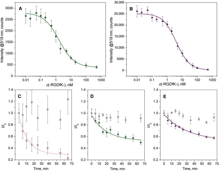
2 Pallarola et al.: Focal adhesion stabilization by enhanced integrin-cRGD binding affinity
In recent years, novel strategies were developed, for a specific integrin subtype [32]. Apart from strategies
based on the engineering of biomimetic matrices for con- for restricting the conformational space of a ligand, multi-
trolled stimulation of cells in vitro, particularly in key bio- valency can further enhance its affinity for target cells by
medical applications such as stimulation of immune cells, displaying additional epitopes able to promote rebinding
controlling pluripotency and regulating cell migration [9, [33–35]. Notably, a minimum distance between the RGD
10]. The use of such surfaces enabled testing the effects peptides and the anchoring substrate is required, such
of ECM ligands diversity, as well as the effects of integ- that the binding motifs are adequately oriented to facili-
rin receptor occupancy and clustering [11]. Early studies tate integrin binding [36, 37]. Aliphatic [38, 39] and poly-
focused on the process of integrin aggregation and FA for- ethylene glycol [40, 41] (PEG)-based short polymers, as
mation in different ECM environments in vitro [12, 13]. It well as amino acid chains [42, 43] (e.g. polyglycine, poly-
was shown that by controlling cluster size, affinity, and proline) have been used as spacers for peptide immobiliza-
average ligand density and spacing, cell migration and tion. Nonetheless, despite their wide use, ligands bearing
spreading behaviors, as well as FA assembly and matura- aliphatic and PEG spacers may experience a decrease
tion could be affected. Supported by theoretical modeling in binding affinity [10, 44]. Densely packed monolay-
[14], these studies suggested that by increasing receptor- ers and spacer flexibility may prevent optimal exposure
ligand interactions, and thus the amount of occupied of the integrin ligands. Recently, we compared the influ-
receptors, clustered ligands enhanced binding affinity ence of different spacer systems, namely alkane-, PEG-,
and, concomitantly, adhesion strength. Nevertheless, and polyproline-based sequences, on the affinity of a c(-
these approaches suffered from variations in ligand distri- RGDfX-)-containing ligand to αvβ3 integrin [43]. Our find-
bution due to random ligand grafting. Recent progress in ings demonstrated that the more extended nature of the
surface patterning techniques made it possible to control polyproline spacers and the low-density assemblies they
the precise placement of individual anchoring ligands at yield, the more precisely and constructively ligand display
nanoscale resolution [15–17]. These studies indicated that could be controlled, thus enhancing cRGD-integrin inter-
integrin binding and clustering, the consequent assem- actions at the adhesion sites.
bly of FAs and cell spreading are strongly influenced by In the present work, we aim to provide insights into
fine changes in adhesive cues. It has been proposed that the role of ligand binding and integrin adhesion ligand
inter-ligand spacing ranging from 58 to 73 nm is required clustering in regulating cell adhesive responses, by com-
for successful integrin-mediated signaling activation [15, paring cell behavior on substrates that present c(-RGDfX-)-
16], and that local, more than global ligand distribution containing ligands of different binding affinities to its
seems to be a key surface parameter for the assembly and primary target, αvβ3 integrin. Ligands were precisely
stability of FA complexes [18–20]. At the nanoscale, the positioned at different lateral distances by means of a
number and geometric distribution of receptors, as well gold nanoparticle (AuNP) pattern, enabling for variations
as their binding strengths, exert a profound effect on cell in the density of anchor binding points at the nanoscale.
adhesion strength, by means of a cooperative integrin Interactions between the integrin extracellular domains
clustering mechanism [20–22]. Although the physiologi- and the c(-RGDfX-)-binding domains were evaluated in
cal relevance of ligand spacing regulation is still unclear, response to variations in ligand presentation, by means of
these findings clearly expose the exquisite cellular sensing different spacer systems.
machinery, thus providing a sound approach for further
eliciting and unraveling specific cellular responses to
their environment.
Among the peptides known to support cell adhe- Results and discussion
sion, the tri-amino acid sequence RGD is the most widely
studied [23–25]. This sequence is ubiquitously expressed Our approach to engineer cellular environments with the
in many ECM components [3, 26], and serves as a minimal ability to enable specific cell-cRGD interactions at precisely
essential binding motif for several different integrins [25], localized positions on a non-adhesive PEG-background
although it binds primarily to the integrin subtype αvβ3 was based on a previously established technique, namely
[27]. Cyclization [28], which confers structural rigidity and diblock-copolymer micelle nanolithography (BCML) [45].
thus chemical stability, and other non-natural peptide In detail, glass coverslips were patterned with AuNPs of 6
modifications such as D-amino acid incorporation [29] and 9 nm diameter arranged in a quasi-hexagonal struc-
and N-methylation [30, 31], are commonly employed to ture, with an average interparticle distance varying from
improve the selectivity and affinity of the RGD sequence 62 to 162 nm. The glass surface between the AuNPs was
Brought to you by | Weizmann Institute of Science
Authenticated
Download Date | 2/28/17 5:08 PM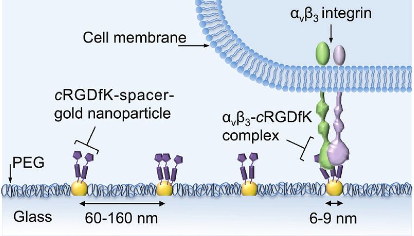
Pallarola et al.: Focal adhesion stabilization by enhanced integrin-cRGD binding affinity 3
then passivated with PEG-terminated siloxane [46], to AuNPs were functionalized with different cRGD-based
render a biologically inert background that does not ini- thiol ligands. The cRGD-based pentapeptides selected
tiate any cell activation. This approach is very powerful, for this study exhibit different integrin αvβ3 binding
since it allows for variations in both the average surface affinities, as tested in a soluble adhesion-inhibition
density, and the local spatial distribution of ligands assay (Table 1). As shown in Figure 1, each ligand con-
in well-defined nanoscopic geometries. Subsequently, sists of three main elements: (i) a monomeric or dimeric
integrin-binding group; (ii) a spacer molecule; and (iii) a
thiol-based anchoring group. Compounds 1 and 2 bear a
Table 1: Inhibition of αvβ3 integrin binding to vitronectin by cRGD flexible spacer such as an aminohexanoic acid (Ahx) or
pentapeptides.
a PEG-based spacer, respectively, while compound 3 con-
sists of a c(-RGDfE-) dimer ligated to a more rigid polypro-
Peptide description IC50a αvβ3, nm
line sequence. The influence of spacer type and length has
1 c(-RGDfK[Ahx-MPA]-) 15.7 ± 4.5 a remarkable effect on the overall binding affinity towards
2 c(-RGDfK[Hegas-(cta)3]-) 19.5 ± 2.9
αvβ3 integrin [43], making these compounds excellent
3 [c(-RGDfE[HexPPPPPP]-)]2K-cta 0.17 ± 2.4
c(-RGDfK-)b 2.6 ± 0.6
candidates for studying the cellular response to chemical
Cilengitidec 0.54 ± 0.02 signaling of varying affinities within its environment.
In the following, the impact of (i) three chemically dif-
a
IC50 values were obtained from a competitive ELISA using the
ferent ligands, (ii) ligand density (i.e. the distance between
natural ligand, vitronectin (Vn), and the soluble integrin αvβ3 [43].
b
This cyclic pentapeptide was used as precursor for the design of gold dots) and (iii) particle size, on the integrin-mediated
1–3, and as a competitive agent in αvβ3 integrin binding assays. cell adhesion were examined. Rat embryonic fibroblasts
c
Cilengitide, c(-RGDfMeV-) [30], was used as an internal reference [REF52 wild-type (WT) cells] were seeded on the individ-
compound for the integrin αvβ3 ELISA assay. ual cRGD-functionalized adhesive surfaces for 4 h, and
Ahx, 6-amino-hexanoic acid; cta, cystamine; Hegas, heptaethylene
then visualized by phase contrast microscopy (Figure 2).
glycol amino acid (PEG thiol acid); Hex, 4-(1-(2-aminoethyl)-1H-1,2,3-
triazol-4-yl)butanoic acid; MPA, mercaptopropionic acid.
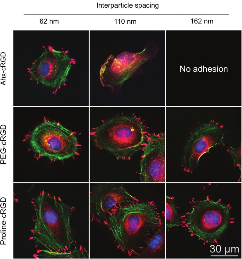
It is evident that fibroblasts spread on the 62 nm patterns,
Figure 1: Molecular structure of the peptides examined in this study. 1 and 2) Ligand peptide composed of a cRGD headgroup with an
aminohexanoic acid (Ahx) (1), or a polyethylene glycol (PEG)-type spacer (2). 3) Ligand peptide consisting of a cRGD headgroup dimer ligated
to a polyproline spacer.
Spacer length in brackets was calculated for the all-trans configuration of aliphatic and PEG spacers between Cα Lys(K) and thiol, and for
proline spacers between Cα Glu(E) and thiol. All compounds have a thiol system for surface attachment (gold-SH bond).
Brought to you by | Weizmann Institute of Science
Authenticated
Download Date | 2/28/17 5:08 PM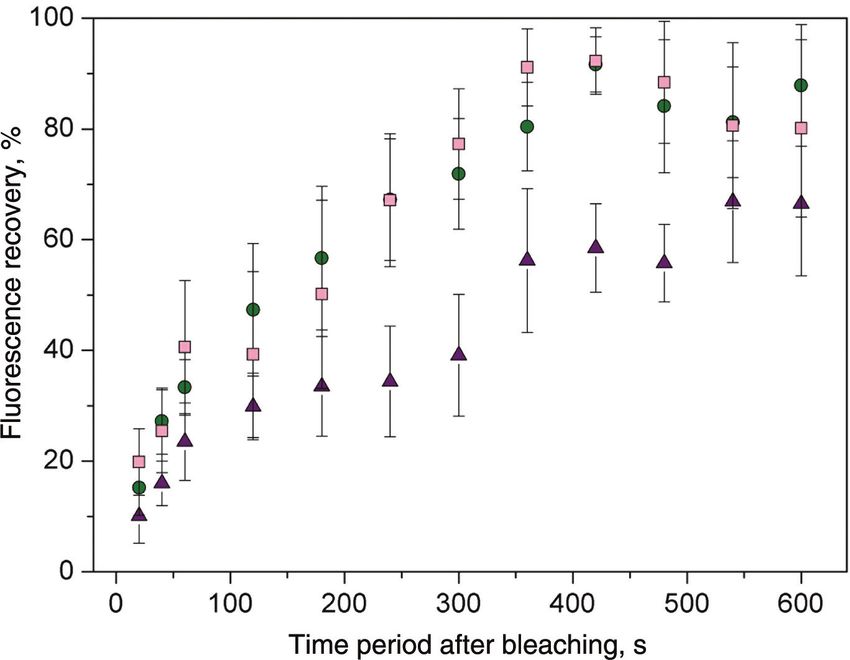
4 Pallarola et al.: Focal adhesion stabilization by enhanced integrin-cRGD binding affinity
Figure 2: Phase contrast images of REF52 WT cells 4 h after seeding on nanostructured glass surfaces functionalized with compound 1
(c(-RGDfK[Ahx-MPA]-), 2 (c(-RGDfK[Hegas-(cta)3]-), and 3 ([c(-RGDfE[HexPPPPPP]-)]2K-cta) with varying particle sizes (6–9 nm) and interparti-
cle distances (62–162 nm).
Scale bar: 100 μm. Insets: Magnification of the selected areas. Scale bar: 30 μm.
as indicated by their typical morphology. However, dis-
tinct differences in adhesion-based cell responses are
noticeable between the ligands on substrates with 110
and 162 nm interparticle distances. Ahx-based ligand led
to limited cell spreading, characterized by elongated cel-
lular morphology. This effect was even more pronounced
on substrates with 162 nm-spaced AuNPs, where more
elongated cell spreading and an increased number of qui-
escent cells could be observed. Conversely, 62 and 110 nm
patterns functionalized with the PEG-based ligand were
sufficient to support spreading via αvβ3 integrin-cRGD
interactions, while cells plated on 162 nm patterns exhibit
a behavior similar to that observed on 110 nm patterns
functionalized with the Ahx-based ligand.
Cell behavior on AuNPs-structured surfaces func- Figure 3: Projected cell area analysis of REF52 WT cells 4 h after
tionalized with the polyproline-based ligand is remark- seeding on 9 nm AuNP-patterned glass surfaces functionalized with
able. Well-spread cells with a radial morphology can be the different cRGD pentapeptides.
Data are presented as mean ± s.e.m., n > 50, p < 0.01.
observed, regardless of the distance between AuNPs.
1: c(-RGDfK[Ahx-MPA]-) (black); 2: c(-RGDfK[Hegas-(cta)3]-) (gray);
Similar results were obtained with mouse calvaria osteo- 3: [c(-RGDfE[HexPPPPPP]-)]2K-cta (white).
blasts (MC3T3 cells), indicating a more generalized cell
adhesion behavior (Figure S3). These findings suggest
that the divalent polyproline-based ligand provides place when the particle spacing is increased above 220 nm
superior binding affinity to αvβ3 integrin, thus support- (Figure 3). This critical interparticle distance is more than
ing cell adhesion and spreading even at very low ligand three times the value previously reported [15], and veri-
densities. These observations are quantitatively summa- fied in this work, for the Ahx-based ligand, highlighting
rized through the projected cell area analysis as depicted the critical influence of the ligand-binding affinity at the
in Figure 3. Furthermore, we characterized the cellular integrin-adhesion site.
response on gold nanopatterns functionalized with the To determine whether the observed cell behav-
polyproline-based ligand at larger interparticle distances ior is purely controlled by the chemical features of the
(162–280 nm), showing that limited cell adhesion takes ligand-presenting molecules or is also influenced by the
Brought to you by | Weizmann Institute of Science
Authenticated
Download Date | 2/28/17 5:08 PMPallarola et al.: Focal adhesion stabilization by enhanced integrin-cRGD binding affinity 5
dimensions of the binding domains, we varied the AuNP different ligands on the assembly of FAs and actin fibers.
size from 6 to 9 nm, to measure the sensitivity of the REF52 WT cells were plated for 4 h on nanopatterned
adherent tissue cells to variations in the presentation of surfaces with different inter-ligand spacings, then fixed
adhesive ligands at the anchoring points. In the case of and stained for paxillin, zyxin and actin. Paxillin resides
the Ahx- and PEG-based ligands, changes in cell adhe- in both initial adhesion contacts [58]; namely, focal
sive response are barely noticeable if the size of AuNPs is complexes (FXs), and mature adhesions, while zyxin is
increased: a slight reduction in the number of quiescent recruited during the maturation of these FXs into FAs, and
cells and a discrete widening of the lamellipodia is seen constitutes a distinctive protein of mature FAs [59]. Exami-
in 110 nm patterns functionalized with the alkane-based nation of the labeled cells revealed extensive cell adhe-
compound, and 162 nm patterns functionalized with the sion and spreading on the 62 nm cRGD-nanopatterns,
PEG-based compound, compared with the analogous indicating successful integrin-ligand interactions with all
6 nm AuNP patterns. Cells seeded on gold patterned sub- the different compounds (Figure 4, first column). Similar
strates bearing the polyproline-based ligand showed no paxillin- and zyxin-rich FA distribution was observed for
visible dependence on particle size within the evaluated the different compounds, as well as actin fibers organ-
regime. We can thus infer that variation in cell-substratum ized as a dense meshwork of peripheral actin filaments,
adhesion interaction is mainly governed by the spatial although with a higher density of stress fibers in the case
organization and the chemical features of the cRGD-based of the polyproline-based compound (Figure S4). In agree-
ligands. These observations also suggest that more than ment with Figure 2, cells growing on 110 nm patterns
one integrin dimer interacting with one single domain is exhibited distinctive features (Figure 4, second column).
an unlikely scenario. Considering that the diameter of an On substrates functionalized with the Ahx-based ligand,
integrin dimer varies between 8 and 12 nm [47], it is rea- cells were considerably less spread than those plated on
sonable to assume that each cRGD-coated AuNP provides the 62 nm nanopatterns, and failed to develop FAs and
an individual binding site, due to steric hindrance. Thus, to induce stress fiber assembly. In contrast, cells plated
potential multiple integrin binding does not account on 110 nm patterns functionalized with the PEG- or poly-
for cell adhesion-associated responses on 9 nm AuNP proline-based ligand showed stable integrin-mediated
nanoarrays. Moreover, the height of the nanoparticles was adhesion characterized by radial spreading, and co-
adjusted to at least the height of the PEG layer ( ~ 6 nm), localized paxillin and zyxin patches mainly distributed
resulting in adequate exposure of the integrin-binding at the periphery of the cells (Figure S4). The effect of
group. Nevertheless, topographical effects [48–52] and the cRGD peptide spacing on the cellular response was even
promotion of integrin rebinding [53–56] due to increase in greater when AuNPs were separated by 162 nm (Figure
particle size and ligand population cannot be ruled out. 4, third column). Cells plated on substrates coated with
However, the observed phenomena could be explained the alkane-based compound showed very poor adhesion
in terms of the availability of the cRGD head group for and spreading, resulting in complete removal of cells
binding, as well as its ability to promote ligand rebinding. after gentle rinsing. Cell behavior observed on nanopat-
These two features are mainly attributed to the chemi- terns functionalized with the PEG-based ligand is consist-
cal nature of the ligand-presenting molecule, and will ent with a spreading and motility regime characterized
be discussed in further detail later [43]. Notably, cells in by repeated extension-retraction cycles [16]. Such cells
control experiments performed on PEG-passivated sur- became highly polarized, displaying small paxillin and
faces lacking AuNPs and/or cRGD functionalization did zyxin clusters at low density, restricted to the cell edges
not adhere to the substrates. Furthermore, X-ray photo- (Figure S4). Conversely, the polyproline-based compound
electron spectroscopy of non-patterned substrates incu- is able to circumvent these limitations, providing a suit-
bated with the different cRGD pentapeptides showed no able environment for the cells to attach and spread. These
evidence of ligand intercalation within the PEG brush substrates supported good cell adhesion with well-consti-
layer (data not shown). This confirms that cell adhesion tuted paxillin- and zyxin-rich adhesions, as well as organ-
responses as reported herein are entirely due to the activa- ized actin fibers (Figure S4), similar to what was observed
tion of αvβ3 integrin, through its interaction with cRGD- on dense nanopatterns.
functionalized AuNPs. Ligand binding to integrins induces conformational
A hallmark of integrin-mediated cell adhesion is the reorganization of the α and β-integrin dimer, leading to
formation of FAs and the assembly actin stress fibers [1, integrin activation and clustering, and the subsequent
57]. To address whether those tailored surfaces activate signal propagation that leads to events such as cell adhe-
integrin signaling, we compared the influence of the three sion and proliferation [60]. This highly regulated process
Brought to you by | Weizmann Institute of Science
Authenticated
Download Date | 2/28/17 5:08 PM6 Pallarola et al.: Focal adhesion stabilization by enhanced integrin-cRGD binding affinity
Figure 4: Fluorescent micrographs of representative REF52 WT cells on nanopatterns with different interparticle distances, and functional-
ized with c(-RGDfK[Ahx-MPA]-, c(-RGDfK[Hegas-(cta)3]-, and [c(-RGDfE[HexPPPPPP]-)]2K-cta.
Following 4 h of incubation, cells were fixed and stained for paxillin (red), zyxin (pink), actin (green) and nuclei (blue).
is essential for initiation of FAs, which are tightly asso- compared with similar studies performed with human
ciated with the cytoskeletal network, enabling cells to foreskin fibroblasts on the randomly immobilized ligand
respond to diverse physical and chemical environmental GRGDY, in which case a minimal peptide-to-peptide
signals [1]. As integrin epitope activity promotes the for- spacing of 140 nm was required for FA and stress fiber for-
mation of stable integrin-cRGD complexes, it is also likely mation [12]. This difference was mainly attributed to the
that high-affinity interactions between the receptors and much lower affinity of the YGRGD ligand compared to the
binding sites may promote enhancement of specific cel- GRGDY ligand, which implies that a much higher ligand
lular responses. An example of this finely-tuned crosstalk density would be required for YGRGD to obtain equivalent
between FAs and actin filaments is the appearance of values of receptor occupancy. Although both approaches
high-frequency FA nucleation in cells adhering to high- lack a spatial distribution of ligands that is precisely
affinity substrates [61, 62]. Minimal requirements for cell localized and predefined, these studies provide compel-
adhesion were also found to be sensitively dependent of ling evidence for the importance of integrin-ECM binding
ligand affinity. WT NR6 fibroblasts seeded on substrates affinity in stimulating adhesion-mediated signaling.
grafted with star-shaped PEG macromolecules functional- In order to shed light on the role of biomimetic surface
ized with YGRGD peptide exhibited a substantially lower properties in regulating adhesive interactions, we inves-
threshold spacing for FA and stress fiber development [13], tigated the effects of the different cRGD ligands on the
Brought to you by | Weizmann Institute of Science
Authenticated
Download Date | 2/28/17 5:08 PMPallarola et al.: Focal adhesion stabilization by enhanced integrin-cRGD binding affinity 7
binding of αvβ3 integrin. Two different in vitro binding with either the PEG- or the polyproline-based ligand are
assays were conducted, to determine: a) the binding shown in Figure 5A and B, respectively. Data from these
affinity of αvβ3 integrin, and b) the dissociation rate of assays were fit to a 4-parameter logistic model; the derived
αvβ3 integrin-cRGD complexes. The binding affinity of binding affinities are summarized in Table 2. Consistent
αvβ3 integrin towards cRGD nanopatterns was evalu- with the results of the previously described cell adhesion
ated as the median inhibitory concentration (IC50) in a experiments, the polyproline-based ligand displayed a
competitive ELISA. cRGD-functionalized nanopatterns higher affinity for αvβ3 integrin binding than the ana-
with a 62 nm interparticle distance and 9 nm particle size, logue bearing a PEG-based spacer (i.e. IC503 > IC502). Data
soluble αvβ3 integrin, and soluble c(-RGDfK-) as a com- for the alkane-based ligand could not be evaluated, due to
petitive binding reagent were used. Dose-response curves the low fluorescence intensity measured under the experi-
for the binding of αvβ3 integrin in the presence of differ- mental conditions (Figure S5). The efficacy of the poly-
ent concentrations of c(-RGDfK-) to nanopatterns coated proline-based spacer as a ligand-presenting molecule is
Figure 5: (A and B) Inhibition of αvβ3 integrin binding to nanopatterns functionalized with compound 2 (c(-RGDfK[Hegas-(cta)3]-), and
3 ([c(-RGDfE[HexPPPPPP]-)]2K-cta) by the soluble pentapeptide c(-RGDfK-).
Inhibition data at each concentration of c(-RGDfK-) are an average of three independent experiments ± standard deviation. The dose-
response data are fit to a 4-parameter logistics model. (C–E) Effect of the soluble pentapeptide c(-RGDfK-) (100 μM) on αvβ3-cRGD
complex dissociation on nanopatterns functionalized with compound 1 (c(-RGDfK[Ahx-MPA]-) (C), 2 (c(-RGDfK[Hegas-(cta)3]-) (D), and 3
([c(-RGDfE[HexPPPPPP]-)]2K-cta) (E). The data are fit to a first-order kinetic equation to obtain the dissociation rate constants (kdiss) and half-
lives (t1/2) of the αvβ3-ligand complex. Data at each time point are presented as an average of three independent experiments ± standard
deviation. Each plot (C–E) includes the data (gray symbol) corresponding to the αvβ3-cRGD complex dissociation kinetics, in the absence of
c(-RGDfK-).
Brought to you by | Weizmann Institute of Science
Authenticated
Download Date | 2/28/17 5:08 PM8 Pallarola et al.: Focal adhesion stabilization by enhanced integrin-cRGD binding affinity
Table 2: Characterization of αvβ3 integrin binding to cRGD-func- obtained for the different substrates can be ascribed to the
tionalized nanopatterns. accessibility of the binding complex, and to the ability of
the ligand to promote rebinding upon complex dissocia-
Peptide IC50a αvβ3, nm kdissb, 103 min − 1 t1/2b, min
tion. These opposite effects are intimately linked to ligand
1 n.d.c 17.92 ± 7.90 38.7 ± 17.0 density and orientation at the binding nanodomains (i.e.
2 1.5 ± 0.7 9.03 ± 2.41 76.7 ± 20.4 AuNPs). High accessibility to the ligand-binding pocket
3 3.3 ± 0.6 7.53 ± 1.64 92.0 ± 20.1
increases both the association and dissociation rates of
a
IC50 values were obtained by fitting the competition binding curves the integrin-ligand complex and, at the same time, pro-
(Figure 5A and B) according to a 4-parameter logistics model. vides a favorable scenario for ligand rebinding, thereby
b
Kinetics parameters kdiss and t1/2, were extracted by fitting the dis-
contributing to the stability of the complex [55]. Several
sociation curves (Figure 5C–E) to a first-order kinetic equation.
c
Experimental conditions were such that the intensity of the fluores-
studies conducted on the recognition-driven assembly of
cence signal was very low, making the analysis unviable. proteins showed that the binding accessibility of a protein
is decreased in densely packed assemblies of the target
molecule, compared to those with lower molecule density
reflected by the amplitude of the response plotted on the [13, 37, 64, 65], a finding mainly attributed to steric inhibi-
Y-axis. The fluorescence intensity value obtained for nan- tion of the binding. Orientation and presentation of the
opatterns coated with this ligand in the absence of com- binding domain very much depends on the nature of the
petition is almost 10-fold higher than that seen with the spacer.
PEG-based ligand (Figure S5). It is also worth mentioning Conformational changes caused by a flexible and/or
that at higher concentrations of c(-RGDfK-), both curves too long spacer can result in the shielding of the active site
reach similar low values, indicating negligible, unspecific [10, 36, 44, 66]. In a recent study, we showed that the bulky
binding of αvβ3 integrin to the substrates. Moreover, no and more rigid polyproline sequence used in compound 3
binding inhibition was observed when a highly selective leads to a reduction in packing density when self-assem-
α5β1-antagonist, c(-phg-isoDGRk-) [63], was used, even at bled on gold, compared with the alkane-based ligand [43].
a concentration of 500 nM, demonstrating the specificity In the context of our results, this means that ligand rebind-
of the integrin binding process. ing plays a leading role in the observed behavior. These
Dynamic monitoring of fluorescently labeled αvβ3 observations certainly emphasize the dynamic nature of
integrin was carried out to assess and compare the disso- the integrin-cRGD interactions, providing insights into
ciation rate of the αvβ3 integrin-cRGD complexes prepared cellular responses to environmental chemical signaling.
in vitro, as described in the Methods section. The integrin- The turnover rates of β3-integrins within FAs estab-
specific association with the cRGD-particle adducts was lished on different cRGD-coated surfaces were analyzed
conducted in the absence of competitive ligands over a using fluorescence recovery after photobleaching (FRAP),
24 h period. Surfaces were then rinsed to remove loosely in order to understand whether the motile behavior of
bound integrins, and incubated with 100 μM c(-RGDfK-). integrins could be correlated to the affinity of the cRGD
The corresponding dissociation curves for the different ligand, and the corresponding stability of the integrin-
cRGD-coated nanopatterns are shown in Figure 5C–E. Dis- cRGD complex. Single FAs localized at the periphery of
sociation data were fit to a simple exponential decay curve the cell were bleached by application of high-intensity
to extract the dissociation constants (kdiss) and half-life laser light, and the recovery of GFP fluorescence meas-
(t1/2) of the αvβ3 integrin-cRGD complexes, as presented ured at different time periods (Figure 6). Cells plated on
in Table 2. The results clearly show the reversible nature nanopatterns functionalized with the polyproline-based
of the interaction between αvβ3 integrin and the cRGD ligand exhibited the lowest exchange of β3-integrins.
head group; furthermore, disruption of this interaction Within 350 s, 50% of the integrin fluorescence recovered
is highly dependent on the ligand-presenting molecule. in the bleached contacts, whereas the exchange on sur-
The αvβ3 integrin-cRGD complex dissociates rapidly from faces coated with the Ahx-based ligand was twice as fast.
nanopatterns functionalized with the Ahx-based ligand, These results compare well with the in vitro αvβ3 integrin
at a rate that is 2- to 3-fold greater than the analogues with binding assays. No significant differences were observed
the PEG- and polyproline-based ligands, respectively. between the Ahx- and the PEG-based ligands, although
Once established, the inherent stability of the αvβ3 this discrepancy could be interpreted by considering each
integrin-cRGD complex is equivalent for the different pool of integrin measured in the experiment. In FRAP, all
ligands, since the pharmacophoric molecule remains integrins, whether bound to the ligand or not, are fluores-
the same. Therefore, the variations in dissociation rates cently labeled, and within the membrane region probed,
Brought to you by | Weizmann Institute of Science
Authenticated
Download Date | 2/28/17 5:08 PMPallarola et al.: Focal adhesion stabilization by enhanced integrin-cRGD binding affinity 9
[68]. These experiments, together with the previous cell
adhesion and biochemical experiments, clearly prove that
small changes in adhesive chemical cues have a strong
influence on the cell adhesion process.
Our most striking finding is that cell adhesive response
on cRGD-coated substrates strongly depends not only on
ligand spatial organization but also on how the ligands
are exposed to the integrin extracellular domains. The
polyproline sequence, rather than Ahx or PEG-containing
spacer, exhibited improved ligand availability at the nano-
metric scale and as such it stabilized FAs. The extended
nature of the polyproline-based dimeric construct is able
to display in a more efficient manner the binding moieties
leading to higher integrin-binding affinity. This results in
higher receptor occupancy and, consequently, provides
Figure 6: Representative FRAP curves for β3-GFP integrins. FRAP was
performed on FAs localized at the edge of REF52 WT cells transiently more suitable nanometric sites for integrin clustering.
expressing β3-GFP integrins plated on nanopatterns functionalized Consistent with this is the observation that the poly
with 1 (pink squares), 2 (green circles) and 3 (violet triangles). proline sequence yields more stable αvβ3 integrin-cRGD
Values are normalized to the pre-bleaching intensity. Each curve complexes characterized by a reduced lateral mobility of
represents fluorescence intensity measurements from several (4–7)
integrins, which can be attributed to an effective receptor
cells and three to five individual FAs.
rebinding. Since integrin clustering is a highly regulated
and dynamic process, which involves the recruitment of
additional components, it is very likely that the longer
contribute to the signal [67–69]. Furthermore, it could be
residence time of integrin at the binding site favors this
that the great heterogeneity in lateral mobility could be
process.
associated with the experimental conditions. Cells on
Further investigations aiming to identify the number
cRGD-coated nanopatterns were evaluated after a 4 h
of bound integrins at the binding sites (i.e. AuNPs) with
incubation period to match the conditions of the other
nanometric resolution, as well as essential proteins such
cell assays, and to minimize the influence of cell-surface
as talin or vinculin for integrin activation will be required
interactions provided by the ECM components secreted by
to achieve a detailed knowledge of the AuNP-binding
the cells, which are thought to be dominant over longer
domain. This challenging endeavor will provide deeper
incubation times. Within the 4 h time frame, inherent lim-
insights into the interplay of physical and biochemi-
itations such as adhesion sliding arise in analyzing FAs
cal signals governing cell adhesion and influencing cell
within an advancing lamellipodia.
behavior.
The results obtained for fibroblasts on nanopat-
terns functionalized with the polyproline-based ligand
enable us to infer that β3 integrins display a slower rate
of integrin-ligand dissociation, implying longer residence Conclusions
at FAs. A slower diffusion rate can also be associated
with higher integrin-ligand affinity [68, 70, 71]. Several This article describes the important role of ligand-binding
studies have shown that increasing the affinity between affinity in regulating integrin-mediated cell adhesive
integrins and the binding domain restricts lateral diffu- responses, by comparing REF52 and MC3T3 cell adhe-
sion. It was recently reported that activation of the high- sion behavior on substrates that present c(-RGDfX-)-
affinity integrin receptor lymphocyte function-associated pentapeptides with different ligand-presenting features.
antigen 1 (LFA-1) by extracellular Ca2 + depletion resulted For this purpose, we used nanopatterned surfaces con-
in reduction of the TS2/4-ATTO520-labeled LFA-1 mobile taining cRGD-biofunctionalized AuNPs surrounded by
fraction [71]. In another study, a comparison of lateral passivated regions. By varying the chemical nature of
diffusion rates between the WT αPS2CβPS and the high- the spacer, required for peptide immobilization on the
affinity mutant αPS2CβPS409D was made by single-par- AuNPs, we were able to achieve varying rates of expo-
ticle tracking, resulting in a decreased mobile fraction sure of the ligand to the integrin extracellular domains.
and a slower diffusion coefficient for the mutant integrin We showed that cells plated on nanopatterned surfaces
Brought to you by | Weizmann Institute of Science
Authenticated
Download Date | 2/28/17 5:08 PM10 Pallarola et al.: Focal adhesion stabilization by enhanced integrin-cRGD binding affinity
having polyproline spacers for peptide immobiliza- MilliQ water for 2 h at room temperature. The physisorbed material
tion could tolerate higher ligand spacings (162 nm) (that was removed by thorough rinsing with MilliQ water and PBS. In vitro
αvβ3 integrin binding assays and cell adhesion experiments were
means lower ligand density) for FA formation in compari-
carried out immediately after this step.
son to cells on the surfaces with cRGD immobilized via
PEG (110 nm) or Ahx (62 nm) spacers. These results are
in partial agreement with our previous studies where we Cell adhesion experiments
showed that surfaces coated with a cRGD peptide bearing
Ahx as spacer failed to induce the formation of FAs and REF52 WT cells were cultured in DMEM medium supplemented with
stress fibers at an interparticle spacing longer than 70 nm 10% FBS (Invitrogen, Germany) at 37 °C and 5% CO2. For adhesion
[15, 16]. The current findings indicate that cell adhesive experiments, cells in culture were rinsed with PBS at 37 °C and adher-
response on cRGD-coated substrates strongly depends ent cells were removed from the culture surface by treatment with
trypsin-EDTA 0.25% (Invitrogen, Germany) for 5 min at 37 °C. Cells
not only on ligand spatial organization but also on the
were seeded at a density of 150 cells/mm2 on the respective function-
way the ligands are exposed to the integrin extracellular
alized surfaces in DMEM containing 1% FBS, followed by incubation
domains. The hypothesis that the extended nature of the for 4 h at 37 °C and 5% CO2. Live cell phase contrast microscopy inves-
polyproline-based dimeric construct is able to display the tigation was performed with a 10x/0.25 Ph1 A-Plan objective (Carl
binding moieties leading to higher integrin-binding affin- Zeiss, Jena, Germany) using an Axiovert 40 CFL microscope (Carl
ity in a more efficient manner, was verified on the cellular Zeiss, Jena, Germany). Projected cell area was determined manually
using ImageJ 1.48 (NIH, http://rsb.info.nih.gov/ij/).
level by FRAP measurements. Moreover, the binding affin-
ity assay of αvβ3 integrin and the dissociation rate assay
of αvβ3 integrin-cRGD complexes were conducted, to
Immunofluorescence staining
confirm the advantageous binding of cRGD immobilized
ligands via PEG or polyproline spacers. These findings rep-
After 4 h on the nanopatterned surfaces, REF52 WT cells were washed
resent an important step towards a deeper understanding with PBS at 37 °C and fixed with 2.5% paraformaldehyde in PBS for
of the interactions between cells and their environment, 10 min. Cells were then permeabilized with 0.1% Triton X-100 in PBS,
and provide further means to engineer adhesive surfaces blocked with 5% goat serum (Invitrogen, Germany) in PBS for 1 h at
to study the mechanisms cells use to sense and respond to room temperature, and incubated with a 1 : 100 dilution of mouse
anti-paxillin (Abcam, USA) and with a 1 : 100 dilution of rabbit anti-
different chemical cues.
zyxin (Sigma-Aldrich, Germany) for 1 h at room temperature. Cells
were then labeled with a 1 : 100 dilution of goat anti-rabbit Alexa
594-conjugated secondary antibody and with a 1 : 100 dilution of goat
Materials and methods anti-mouse Alexa 647-conjugated secondary antibody (Invitrogen,
Germany), in 5% goat serum in PBS for 1 h at room temperature. Fila-
mentous actin and nuclei were labeled with Alexa 488-conjugated
Chemical synthesis phalloidin and DAPI (Invitrogen, Germany), respectively. Cells were
examined with a 63x/1.25 Oil Ph3 Antiflex Plan-Neofluar objective
Peptide synthesis was carried out using TCP resin, following standard (Carl Zeiss, Jena, Germany) using an Axiovert 200 epi-fluorescence
Fmoc-strategy [72]. All tested compounds exhibited ≥ 95% purity, as microscope (Carl Zeiss, Jena, Germany) equipped with a Hamamatsu
determined by RP-HPLC-(MS). A detailed description of the synthetic (model C10600-10B-H) digital CCD camera (Hamamatsu Photon-
procedures was published elsewhere [43]. All synthesized peptides ics, Germany). Image processing was achieved with the AxioVision
were tested in vitro in a competitive ELISA assay using the natural image viewer (Carl Zeiss, Jena, Germany).
ligand, vitronectin (Vn), and the soluble human αvβ3 integrin puri-
fied receptor [43, 72] (Millipore, Schwalbach/Ts., Germany) (Table 1).
In vitro αvβ3 integrin binding assays
Biofunctionalized nanopatterns Nanopatterned glass surfaces with a 62 nm interparticle distance and
9 nm particle size, and functionalized with the different cRGD penta-
AuNP quasi-hexagonal patterns were prepared on glass coverslips peptides, were employed to conduct the integrin binding assays. Two
(Carl Roth, Germany) by means of diblock-copolymer micelle nano- different types of experiments were performed: a) binding affinity of
lithography (BCML) as previously described [45]. Details concern- αvβ3 integrin, and b) dissociation rate of αvβ3 integrin-cRGD com-
ing the applied diblock copolymers and the casting process are plexes. Binding affinity of αvβ3 integrin towards cRGD-nanopatterns
presented in Supporting Information (Table S1, Figures S1 and S2). was determined in a competitive ELISA-type assay using the solu-
The area between AuNPs was passivated with mPEG-triethoxysilane ble pentapeptide c(-RGDfK-) and human αvβ3 integrin (Millipore,
(2000) to prevent non-specific adhesion according to a procedure Schwalbach/Ts., Germany). The amount of αvβ3 integrin adsorbed
described elsewhere [46]. Each surface was functionalized with the on the nanopatterns was assessed by immunohistochemistry using
corresponding cRGD pentapeptide at a concentration of 25 μM in primary antibody mouse anti-human CD51/61 (BD Biosciences,
Brought to you by | Weizmann Institute of Science
Authenticated
Download Date | 2/28/17 5:08 PMPallarola et al.: Focal adhesion stabilization by enhanced integrin-cRGD binding affinity 11
Heidelberg, Germany) and anti-mouse Alexa 488-conjugated sec- the support of the Alexander von Humboldt Foundation.
ondary antibody (Invitrogen, Germany). A similar procedure was car- D.P. is staff researcher of CONICET. J.P.S. is the Weston
ried out to prepare the samples for determination of the dissociation
Visiting Professor at the Weizmann Institute of Science.
rate of αvβ3 integrin-cRGD complexes. Human integrin αvβ3 was
incubated for 24 h in the absence of competition, while fluorescence B.G. is the incumbent of the Erwin Neter Professorial
measurements were conducted in the presence of c(-RGDfK-) 100 μM. Chair in Cell and Cancer Biology. The Max Planck Society
All experiments were performed at 37 °C, using a microscopy system is appreciated for its general support of all aspects of our
previously described [73]. Details concerning incubation and wash- research. Author’s Statement
ing conditions, as well as measurement conditions and settings, are
presented in Supporting Information.
Author’s statement
Conflict of interest: Authors state no conflict of interest.
FRAP measurements Material and methods:
Informed consent: Informed consent has been obtained
REF52 WT cells were transfected with β3-EGFP-integrin plasmid [74] from all individuals included in this study.
Lipofectamine® 2000 reagent (Invitrogen, Germany), according to Ethical approval: The research related to human use
the standard manufacturer’s protocol. REF52 WT cells expressing β3-
has been complied with all the relevant national regula-
GFP integrins were harvested from culture by treatment with trypsin-
EDTA 0.25% solution (Gibco Laboratories, Germany). Cells were tions, institutional policies and in accordance the tenets
seeded at a density of 150 cells/mm2 on the respective functionalized of the Helsinki Declaration, and has been approved by
surfaces in DMEM containing 1% FBS. Following 4 h of incubation at the authors’ inistitutional review board or equivalent
37 °C and 5% CO2, nanopatterned glass substrates (62 nm interpar- committee.
ticle distance and 9 nm particle size) were mounted on an inverted
confocal laser-scanning microscope equipped with an incubation
chamber (Leica TCS SP5 X, Leica Mikrosysteme GmbH, Wetzlar, Ger-
many). Confocal images of FA sites were recorded with 5%–6% of the References
intensity of Ar-Ion gas laser 488 nm line excited via a 63x oil objec-
tive (HCX PL APO 63x/1.40-0.60; Leica Mikrosysteme GmbH, Wetzlar, 1. Geiger B, Spatz JP, Bershadsky AD. Environmental sensing
Germany). Five bleach cycles at 50% intensity were used to eliminate through focal adhesions. Nat Rev Mol Cell Biol. 2009;10:21–33.
the GFP fluorescence at FAs localized at the cell edges. FRAP curves 2. Hynes RO. Integrins – Versatility, modulation, and signaling in
per cell were obtained by considering the fluorescence intensity of cell-adhesion. Cell 1992;69:11–25.
three to five individual FA spots. Similar to a previous investigation 3. Luo BH, Carman CV, and Springer TA. Structural basis of integrin
[74], no newly synthesized β3-GFP integrins were detected during the regulation and signaling. Annu Rev Immunol. 2007;25:619–47.
recovery period (up to 10 min). 4. Takagi J, Springer TA. Integrin activation and structural rear-
rangement. Immunol Rev. 2002;186:141–63.
Supplemental material: Experimental details, SEM and 5. Calderwood DA. Integrin activation. J Cell Sci. 2004;117:657–66.
6. Geiger B, Bershadsky A, Pankov R, Yamada KM. Transmembrane
TEM characterization of gold nanopatterns, fluorescence
extracellular matrix-cytoskeleton crosstalk. Nat Rev Mol Cell
micrographs of αvβ3 integrin binding, and supplemen- Biol. 2001;2:793–805.
tary MC3T3 osteoblast adhesion experiments. 7. Zaidel-Bar R, Itzkovitz S, Ma’ayan A, Iyengar R, Geiger B.
Functional atlas of the integrin adhesome. Nat Cell Biol.
Acknowledgements: D.P. acknowledges financial sup- 2007;9:858–68.
8. Kanchanawong P, Shtengel G, Pasapera AM, Ramko EB,
port from Max-Planck-Gesellschaft (Max Planck Partner
Davidson MW, Hess HF, et al. Nanoscale architecture of integrin-
Group Nanoelectronics for Cellular Interfaces). Parts of based cell adhesions. Nature 2010;468:58–U262.
the research leading to these results have received fund- 9. Yao X, Peng R, Ding J. Cell-material interactions revealed
ing from the European Union Seventh Framework Pro- via material techniques of surface patterning. Adv Mater.
gramme (FP7/2007–2013) under grant agreement Nos. 2013;25:5257–86.
NMP4-LA-2009-229289 NanoII (J.P.S. and B.G.) and NMP3- 10. Rodda AE, Meagher L, Nisbet DR, Forsythe JS. Specific control
of cell-material interactions: Targeting cell receptors using
SL-2009-229294 NanoCARD (J.P.S. and B.G.), as well as
ligand-functionalized polymer substrates. Prog Polym Sci.
from an ERC Advanced Grant under grant agreement No. 2014;39:1312–47.
294852-SynAd (J.P.S. and B.G.). This work is also part of 11. Balaban NQ, Schwarz US, Riveline D, Goichberg P, Tzur G, Saba-
the MaxSynBio consortium, which is jointly funded by the nay I, et al. Force and focal adhesion assembly: a close relation-
Federal Ministry of Education and Research of Germany ship studied using elastic micropatterned substrates. Nat.Cell
Biol. 2001;3:466–72.
and the Max Planck Society. The authors acknowledge the
12. Massia SP, Hubbell JA. An RGD spacing of 440 nm is sufficient
support of the excellence cluster CellNetworks at the Uni- for integrin alpha-V-beta-3-mediated fibroblast spreading and
versity of Heidelberg. J.P.S. and E.A.C. acknowledge the 140nm for focal contact and stress fiber formation. J Cell Biol.
support from DFG SFB 1129 (P1 and P15). I.P. acknowledges 1991;114:1089–100.
Brought to you by | Weizmann Institute of Science
Authenticated
Download Date | 2/28/17 5:08 PM12 Pallarola et al.: Focal adhesion stabilization by enhanced integrin-cRGD binding affinity
13. Maheshwari G, Brown G, Lauffenburger DA, Wells A, Griffith LG. 33. Kiessling LL, Gestwicki JE, Strong LE. Synthetic multivalent
Cell adhesion and motility depend on nanoscale RGD clustering. ligands as probes of signal transduction. Angew Chem Int Edit.
J Cell Sci. 2000;113:1677–86. 2006;45:2348–68.
14. Irvine DJ, Hue KA, Mayes AM, Griffith LG. Simulations of cell- 34. Lössner D, Kessler H, Thumshirn G, Dahmen C, Wiltschi B,
surface integrin binding to nanoscale-clustered adhesion Tanaka M, et al. Binding of small mono- and oligomeric integrin
ligands. Biophys. J. 2002;82:120–32. ligands to membrane-embedded integrins monitored by surface
15. Arnold M, Cavalcanti-Adam EA, Glass R, Blümmel J, Eck W, plasmon-enhanced fluorescence spectroscopy. Anal Chem.
Kantlehner M, et al. Activation of integrin function by nanopat- 2006;78:4524–33.
terned adhesive interfaces. ChemPhysChem 2004;5:383–8. 35. Wängler C, Maschauer S, Prante O, Schäfer M, Schirrmacher R,
16. Cavalcanti-Adam EA, Volberg T, Micoulet A, Kessler H, Geiger Bartenstein P, et al. Multimerization of cRGD peptides by click
B, Spatz JP. Cell spreading and focal adhesion dynamics chemistry: synthetic strategies, chemical limitations, and influ-
are regulated by spacing of integrin ligands. Biophys J. ence on biological properties. Chembiochem 2010;11:2168–81.
2007;92:2964–74. 36. Hersel U, Dahmen C, Kessler H. RGD modified polymers: bioma-
17. Schvartzman M, Palma M, Sable J, Abramson J, Hu X, Sheetz MP, terials for stimulated cell adhesion and beyond. Biomaterials
et al. Nanolithographic control of the spatial organization of cel- 2003;24:4385–415.
lular adhesion receptors at the single-molecule level. Nano Let. 37. Salinas CN, Anseth KS. The influence of the RGD peptide
2011;11:1306–12. motif and its contextual presentation in PEG gels on human
18. Arnold M, Schwieder M, Blümmel J, Cavalcanti-Adam EA, López- mesenchymal stem cell viability. J Tissue Eng Regen Med.
Garcia M, Kessler H, et al. Cell interactions with hierarchically 2008;2:296–304.
structured nano-patterned adhesive surfaces. Soft Matter 38. Kantlehner M, Finsinger D, Meyer J, Schaffner P, Jonczyk A,
2009;5:72–7. Diefenbach B, et al. Selective RGD-mediated adhesion of
19. Huang J, Grater SV, Corbellini F, Rinck S, Bock E, Kemkemer R, osteoblasts at surfaces of implants. Angew Chem Int Edit.
et al. Impact of order and disorder in RGD nanopatterns on cell 1999;38:560–2.
adhesion. Nano Lett. 2009;9:1111–6. 39. Mas-Moruno C, Fraioli R, Albericio F, Manero JM, Gil FJ. Novel
20. Deeg JA, Louban I, Aydin D, Selhuber-Unkel C, Kessler H, Spatz peptide-based platform for the dual presentation of biologically
JP. Impact of local versus global ligand density on cellular adhe- active peptide motifs on biomaterials. ACS Appl Mater Inter.
sion. Nano Lett. 2011;11:1469–76. 2014;6:6525–36.
21. Helenius J, Heisenberg CP, Gaub HE, Muller DJ. Single-cell force 40. Kalinina S, Gliemann H, López-García M, Petershans A,
spectroscopy. J Cell Sci. 2008;121:1785–91. Auernheimer J, Schimmel T, et al. Isothiocyanate-functionalized
22. Liu Y, Medda R, Liu Z, Galior K, Yehl K, Spatz JP, et al. Nanoparti- RGD peptides for tailoring cell-adhesive surface patterns.
cle tension probes patterned at the nanoscale: impact of integrin Biomaterials 2008;29:3004–13.
clustering on force transmission. Nano Lett. 2014;14:5539–46. 41. Kilian KA, Mrksich M. Directing stem cell fate by controlling the
23. Ruoslahti E. RGD and other recognition sequences for integrins. affinity and density of ligand-receptor interactions at the bioma-
Annu Rev Cell Dev Biol. 1996;12:697–715. terials interface. Angew Chem Int Edit. 2012;51:4891–5.
24. Plow EF, Haas TA, Zhang L, Loftus J, Smith JW. Ligand binding to 42. Petrie TA, Capadona JR, Reyes CD, García AJ. Integrin specificity
integrins. J Biol Chem. 2000;275:21785–8. and enhanced cellular activities associated with surfaces pre-
25. Humphries JD, Byron A, Humphries MJ. Integrin ligands at a senting a recombinant fibronectin fragment compared to RGD
glance. J Cell Sci. 2006;119:3901–3. supports. Biomaterials 2006;27:5459–70.
26. Hynes RO. The extracellular matrix: not just pretty fibrils. 43. Pallarola D, Bochen A, Boehm H, Rechenmacher F, Sobahi TR,
Science 2009;326:1216–9. Spatz JP, et al. Interface immobilization chemistry of cRGD-
27. Zamir E, Geiger B. Components of cell-matrix adhesions. J Cell based peptides regulates integrin mediated cell adhesion. Adv
Sci. 2001;114:3577–9. Funct Mater. 2014;24:943–56.
28. Aumailley M, Gurrath M, Müller G, Calvete J, Timpl R, Kessler H. 44. Kingshott P, Thissen H, Griesser HJ. Effects of cloud-point graft-
Arg-Gly-Asp constrained within cyclic pentapeptides – strong ing, chain length, and density of PEG layers on competitive
and selective inhibitors of cell-adhesion to vitronectin and adsorption of ocular proteins. Biomaterials 2002;23:2043–56.
laminin fragment-P1. FEBS Lett. 1991;291:50–4. 45. Glass R, Möller M, Spatz JP. Block copolymer micelle nanolithog-
29. Haubner R, Finsinger D, Kessler H. Stereoisomeric peptide raphy. Nanotechnology 2003;14:1153–60.
libraries and peptidomimetics for designing selective inhibitors 46. Blümmel J, Perschmann N, Aydin D, Drinjakovic J, Surrey T,
of the alpha(V)beta(3) integrin for a new cancer therapy. Angew Lopez-Garcia M, et al. Protein repellent properties of covalently
Chem Int Edit. 1997;36:1375–89. attached PEG coatings on nanostructured SiO2-based inter-
30. Mas-Moruno C, Rechenmacher F, Kessler H. Cilengitide: the faces. Biomaterials 2007;28:4739–47.
first anti-angiogenic small molecule drug candidate design, 47. Xiong JP, Stehle T, Zhang R, Joachimiak A, Frech M, Goodman SL,
synthesis and clinical evaluation. Anticancer Agents Med Chem. et al. Crystal structure of the extracellular segment of integrin
2010;10:753–68. alpha V beta 3 in complex with an Arg-Gly-Asp ligand. Science
31. Chatterjee J, Rechenmacher F, Kessler H. N-methylation of 2002;296:151–5.
peptides and proteins: an important element for modulating 48. Curtis A, Riehle M. Tissue engineering: the biophysical back-
biological functions. Angew Chem Int Edit. 2013;52:254–69. ground. Phys Med Biol. 2001;46, R47–65.
32. Temming K, Schiffelers RM, Molema G, Kok RJ. RGD-based strate- 49. Dalby MJ, Riehle MO, Johnstone H, Affrossman S, Curtis AS.
gies for selective delivery of therapeutics and imaging agents to In vitro reaction of endothelial cells to polymer demixed nano-
the tumour vasculature. Drug Resist Updates 2005;8:381–402. topography. Biomaterials 2002;23:2945–2954.
Brought to you by | Weizmann Institute of Science
Authenticated
Download Date | 2/28/17 5:08 PMYou can also read