Hepatitis C Virus and Hepatocellular Carcinoma: When the Host Loses Its Grip - MDPI
←
→
Page content transcription
If your browser does not render page correctly, please read the page content below
International Journal of
            Molecular Sciences

Review
Hepatitis C Virus and Hepatocellular Carcinoma:
When the Host Loses Its Grip
Kaku Goto 1,2 , Armando Andres Roca Suarez 1,2 , Florian Wrensch 1,2 ,
Thomas F. Baumert 1,2,3,4, * and Joachim Lupberger 1,2, *
 1    Université de Strasbourg, F-67000 Strasbourg, France; kgoto@unistra.fr (K.G.);
      andres.roca-suarez@etu.unistra.fr (A.A.R.S.); wrensch@unistra.fr (F.W.)
 2    Institut National de la Santé et de la Recherche Médicale, U1110, Institut de Recherche sur les Maladies
      Virales et Hépatiques, Université de Strasbourg (IVH), F-67000 Strasbourg, France
 3    Pôle Hépato-digestif, Institut Hopitalo-Universitaire, F-67000 Strasbourg, France
 4    Institut Universitaire de France, F-75231 Paris, France
 *    Correspondence: thomas.baumert@unistra.fr (T.F.B.); Joachim.Lupberger@unistra.fr (J.L.);
      Tel.: +33-3-68-85-37-03 (T.F.B. & J.L.); Fax: +33-3-68-85-37-24 (T.F.B. & J.L.)

 Received: 27 March 2020; Accepted: 24 April 2020; Published: 26 April 2020

 Abstract: Chronic infection with hepatitis C virus (HCV) is a major cause of hepatocellular carcinoma
 (HCC). Novel treatments with direct-acting antivirals achieve high rates of sustained virologic
 response; however, the HCC risk remains elevated in cured patients, especially those with advanced
 liver disease. Long-term HCV infection causes a persistent and accumulating damage of the liver
 due to a combination of direct and indirect pro-oncogenic mechanisms. This review describes
 the processes involved in virus-induced disease progression by viral proteins, derailed signaling,
 immunity, and persistent epigenetic deregulation, which may be instrumental to develop urgently
 needed prognostic biomarkers and as targets for novel chemopreventive therapies.

 Keywords: HCV; HCC; epigenetics; signaling; tumor immunity; clinical impact

1. Introduction
     Globally, liver cancer is the sixth most commonly diagnosed cancer type and the fourth leading
cause of cancer mortality [1,2]. With 70–80% of cases, hepatocellular carcinoma (HCC) is the most
frequent liver cancer [1] and chronic infection with hepatitis C virus (HCV) has been recognized as
a major cause of HCC [3]. In recent years, the direct-acting antiviral agents (DAAs) revolutionized
the standard therapy, achieving high rates of sustained virologic response (SVR), which is associated
with a largely reduced risk of mortality and HCC [4,5]. However, despite their efficacy, the novel
therapies cannot fully eradicate liver cancer risk, especially in HCV-cured patients with advanced liver
disease [6], suggesting an accumulation of irreversible damages to the liver during long-term HCV
infection. Cirrhosis is an important factor in HCC development since the majority of HCV-associated
HCCs occur in cirrhotic livers. Moreover, patients with established cirrhosis have a persistently
elevated risk of HCC, even many years after SVR [4,7]. An association of DAA regimens with HCC
development and recurrence was initially discussed but has not been confirmed by additional studies
and meta-analyses [2,4,8,9]. Many studies suggest that the accumulation of liver damage during
chronic HCV infection is caused by a complex interaction of direct and indirect mechanisms, which
forces the liver towards a tilting point of no return in terms of HCC development. This review aims to
summarize pro-oncogenic events induced by viral proteins, deranged host signaling, inflammation
and immunity. Moreover, it highlights the role of epigenetic dysregulation by HCV and fibrosis in
the pathogenic memory post infection, pointing towards novel and urgently needed biomarkers and
chemopreventive concepts to identify and help patients at considerable HCC risk after cure.

Int. J. Mol. Sci. 2020, 21, 3057; doi:10.3390/ijms21093057                             www.mdpi.com/journal/ijms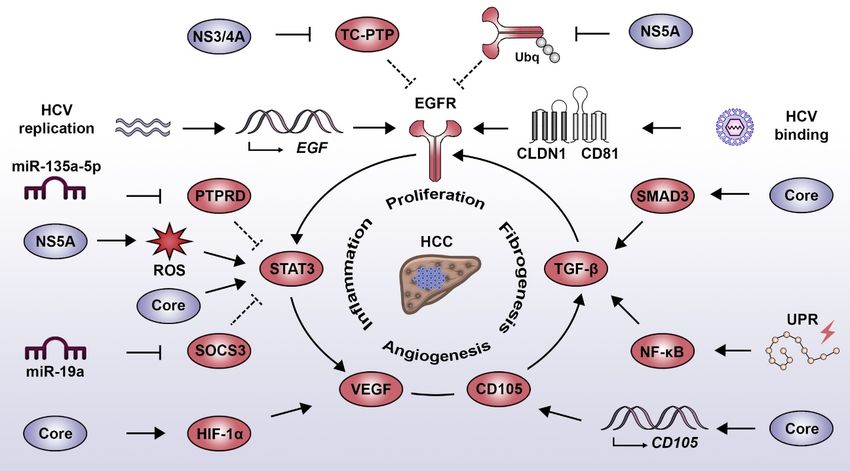
Int. J. Mol. Sci. 2020, 21, 3057                                                                    2 of 17

2. HCV Life Cycle
      HCV is an enveloped positive single-stranded RNA virus of the Flaviviridae family, which was
discovered in 1989 as a cause of non-A, non-B hepatitis [10]. Viral particles expose heterodimers
of the two viral glycoproteins E1 and E2, which are the main targets for neutralizing antibodies.
Moreover, viral particles are associated with lipids and lipoproteins forming lipoviral particles of low
and very-low buoyant density, which contributes to viral entry and a shielding from neutralizing
antibodies [11]. HCV infects predominantly hepatocytes, but additional reservoirs in peripheral blood
mononuclear cells (PBMC), including dendritic cells, B cells, and T cells have been suggested [12–15].
HCV entry in hepatocytes requires an unusually large number of host factors for binding, post-binding,
internalization and fusion with endosomal membranes (reviewed in [16]), also engaging host signaling
pathways involved in cell proliferation and survival. Translation of the released viral genome at the
endoplasmic reticulum is initiated by a viral internal ribosomal entry site (IRES), which requires the
microRNA (miRNA) miR-122 for its stabilization [17,18]. The viral polyprotein is then processed by
host proteases and the viral proteases NS2/NS3 and NS3/4A to mainly ten viral proteins comprising
structural proteins (core, E1, E2) and nonstructural (NS) proteins (p7, NS2, NS3, NS4A, NS4B, NS5A,
NS5B) [19]. Viral proteins and the HCV-recruited host lipid kinase phosphatidylinositol 4-kinase III
induce a deformation of the endoplasmic reticulum membranes to form the “membranous web”-termed
replication complex [20]. The replication complex accumulates lipid droplets and lipoproteins, which
are essential for virus assembly [19]. HCV lipoviral particles are released via the Golgi compartment in
a non-lytic manner to the extracellular space [21] or are transmitted to the neighboring hepatocytes in
a cell-free manner [16].

3. Pro-Oncogenic Impact of Viral Proteins
     HCV does not code for classical viral oncogenes like v-src from Rous sarcoma virus [22] or E6/E7
from human papilloma virus [23]. However, some HCV proteins manipulate host pathways to favor
tumor development by promoting cell proliferation and survival. Thus, HCV proteins contribute to a
pro-oncogenic environment during chronic HCV infection [24]. Most of the evidence from cell culture
and transgenic animal models supports the notion that mainly the HCV proteins core and the NS
proteins 3 and 5A may have an active role in the development and progression of HCV-associated liver
disease and HCC.

3.1. Core Protein
      HCV core is an RNA-binding protein that, in combination with the viral genome, constitutes
the nucleocapsid [25]. Core has been implicated in the development of several hepatic complications.
In transgenic animal models, core expression alone is sufficient to induce hepatic steatosis [26], insulin
resistance [27] and HCC [28]. It was suggested that core expression increases the production of
reactive oxygen species (ROS), which results in an impaired mitochondrial β-oxidation [29]. These data
indicate that HCV core promotes hepatocyte proliferation, which is emphasized by an accelerated liver
regeneration following partial hepatectomy in core-transgenic mice [30]. This is further supported by
the association of distinct core mutations in HCV genotype 1 with an elevated HCC risk in patients [31].
Interestingly, the very same mutations were still associated with an increased HCC risk, even after
HCV elimination [32].

3.2. NS3
     The NS3 protein of HCV is a multifunctional protein that acts as viral protease, RNA helicase and
nucleoside triphosphatase (NTPase) during the viral life cycle [33]. Its serine protease activity cleaves
the HCV polyprotein at four distinct sites to generate NS4A, NS4B, NS5A and NS5B [34]. The RNA
helicase and NTPase function of NS3 are essential components of the HCV replication complex [35].
The direct role of NS3 as an inducer of cell transformation was initially demonstrated in vitro by the
Int. J.
Int. J. Mol.
Mol. Sci. 2020, 21,
Sci. 2020, 21, x3057
FOR PEER REVIEW 33 of
of 17
17
[35]. The direct role of NS3 as an inducer of cell transformation was initially demonstrated in vitro
by the overexpression
overexpression of NS3of inNS3
NIHin3T3NIH 3T3 fibroblasts.
fibroblasts. The subsequent
The subsequent injection
injection of these
of these cellsnude
cells into into nude
mice
mice led to the formation of ectopic tumors [36]. Moreover, NS3 promotes liver disease progression
led to the formation of ectopic tumors [36]. Moreover, NS3 promotes liver disease progression by
by stimulating
stimulating hepatic
hepatic fibrosis
fibrosis in HCV-infected
in HCV-infected chimeric
chimeric mice,mice,
whichwhich
can becan be attenuated
attenuated by treatment
by treatment with
with
an an anti-NS3
anti-NS3 antibody
antibody [37]. [37].
3.3. NS5A
3.3. NS5A
The viral
The viral phosphoprotein
phosphoprotein NS5A NS5A has
has aa key
key role
role during
during RNA
RNA replication
replication andand virion
virion assembly
assembly [38][38]
and confers viral resistance to interferon (IFN) [39,40]. It is a driver of liver disease, inducing hepatic
and confers viral resistance to interferon (IFN) [39,40]. It is a driver of liver disease, inducing hepatic
steatosis, as
steatosis, as demonstrated
demonstrated in in NS5A
NS5A transgenic
transgenic micemice [41].
[41]. Transgenic
Transgenic mice mice expressing NS5A are
expressing NS5A are
protected against hepatic apoptosis following tumor necrosis factor alpha (TNF-α) injection [42].
protected against hepatic apoptosis following tumor necrosis factor alpha (TNF-α) injection [42].
However, NS5A
However, NS5A expression
expression alone
aloneisisnot
notsufficient
sufficienttotoinduce
induceliver
livertumors
tumors [43,44], suggesting
[43,44], suggesting thatthat
its
oncogenic
its oncogenicroleroleresults from
results its interaction
from its interactionwithwith
additional cancer-related
additional cancer-related pathways
pathways[45] [45]
or inorthe
in
context of co-morbidities. For example, NS5A transgenic mice develop liver cancer if fed with high-
the context of co-morbidities. For example, NS5A transgenic mice develop liver cancer if fed with
fat diet by
high-fat dietexhibiting
by exhibiting a pronounced
a pronounced mesenchymal
mesenchymal phenotype
phenotype [46]. NS5A
[46]. NS5Ahyperphosphorylation
hyperphosphorylation is
essential
is forfor
essential itsitsfunction
functionduring
duringthetheviral
virallife
lifecycle,
cycle,indicating
indicatingaatight
tight interaction
interaction with
with host
host signaling
signaling
pathways [18,33,47,48].
pathways [18,33,47,48].
4. HCV-Induced Proliferative Signaling Associated with Liver Disease
4. HCV-Induced Proliferative Signaling Associated with Liver Disease
HCV infection has been reported to induce a wide range of cell signaling alterations, which
HCV infection has been reported to induce a wide range of cell signaling alterations, which
directly or indirectly contribute to the development and progression of liver diseases [49,50]. In the
directly or indirectly contribute to the development and progression of liver diseases [49,50]. In the
following paragraphs, we illustrate this aspect by describing the relationship between HCV and four
following paragraphs, we illustrate this aspect by describing the relationship between HCV and four
major signaling drivers that play an important role in liver disease progression towards HCC: the
major signaling drivers that play an important role in liver disease progression towards HCC: the
epidermal growth factor (EGF), signal transducer and activator of transcription 3 (STAT3), transforming
epidermal growth factor (EGF), signal transducer and activator of transcription 3 (STAT3),
growth factor beta (TGF-β) and vascular endothelial growth factor (VEGF). In its evolution, HCV
transforming growth factor beta (TGF-β) and vascular endothelial growth factor (VEGF). In its
diverted these pathways to promote its replication and persistence with important consequences to
evolution, HCV diverted these pathways to promote its replication and persistence with important
viral pathogenesis
consequences andpathogenesis
to viral liver disease and
(Figure
liver1).disease (Figure 1).
Figure 1.1.Interplay
Figure Interplaybetween
between HCV-induced
HCV-induced oncogenic
oncogenic signaling
signaling pathways.
pathways. EGF pathway:
EGF pathway: HCV
HCV binding
binding
to toreceptor
its entry its entrycomplex
receptor complex
(i.e., (i.e., CLDN1/CD81)
CLDN1/CD81) induces EGFRinduces EGFR phosphorylation.
phosphorylation. This is sustainedThis is
by the
sustained
action by the and
of NS3/4A action of NS3/4A
NS5A and NS5Aregulate
which negatively which negatively regulate
the phosphatase the phosphatase
TC-PTP TC-PTP
and the process and
of EGFR
the process ofrespectively.
degradation, EGFR degradation, respectively.
Additionally, Additionally,
HCV replication HCV
has been replication
linked has been expression
to the increased linked to the
of
increased expression of EGF and activation of the TGF-β pathway, both contributing to EGFR
signaling. STAT3 pathway: STAT3 activation results from the direct action of the core protein andInt. J. Mol. Sci. 2020, 21, 3057 4 of 17
EGF and activation of the TGF-β pathway, both contributing to EGFR signaling. STAT3 pathway:
STAT3 activation results from the direct action of the core protein and indirectly via EGFR activation and
the NS5A protein, which favors the production of ROS. Moreover, HCV employs miR-135a-5p
and miR-19a to decrease the expression of the negative STAT3 regulators PTPRD and SOCS3,
respectively. TGF-β pathway: HCV induces the activation of the TGF-β pathway by intermediary of
the UPR, which favors NF-κB activity and via the core protein, which directly interacts with SMAD3.
The HCV-mediated expression of endoglin (CD105) favors activation of the TGF-β pathway and the
induction of angiogenesis signaling. VEGF pathway: HCV core induces the activation of HIF-1α, which
leads to an increased expression of VEGF. Similarly, an increased VEGF expression is promoted by
HCV via the STAT3-dependent activation of androgen receptor. Abbreviations: CLDN1, claudin 1; EGF,
epidermal growth factor; EGFR, epidermal growth factor receptor; HCC, hepatocellular carcinoma;
HCV, hepatitis C virus; HIF-1α, hypoxia-inducible factor 1 alpha; NF-κB, nuclear factor kappa B;
PTPRD, protein tyrosine phosphatase receptor type delta; ROS, reactive oxygen species; SMAD3,
SMAD family member 3; SOCS3, suppressor of cytokine signaling 3; STAT3, signal transducer and
activator of transcription 3; TC-PTP, T cell protein tyrosine phosphatase; TGF-β, transforming growth
factor beta; UPR, unfolded protein response; VEGF, vascular endothelial growth factor.
4.1. EGF Signaling Pathway
In the context of HCV infection, one of the best characterized signaling components associated
with the development and progression of liver disease is the epidermal growth factor receptor (EGFR)
pathway [51]. EGFR is an essential host factor regulating entry of the virus into hepatocytes [52]
and is chronically deregulated in the liver of HCV-infected patients [52,53]. HCV has developed
multiple mechanisms to induce and maintain EGFR signaling. In the early stages of the viral life
cycle, HCV binding to its entry receptor complex, i.e., CD81 and claudin-1 (CLDN1), induces EGFR
phosphorylation [54] and downstream signaling [55], thereby facilitating viral particle internalization.
EGFR activity is prolonged by the NS5A-mediated alteration of EGFR trafficking [56] and by stimulated
Netrin-1 expression, which impedes EGFR recycling [57]. Moreover, HCV replication itself promotes
the expression of the receptor ligand EGF [58]. Additionally, NS3/4A activity induces the proteolytic
cleavage of the EGFR phosphatase T cell protein tyrosine phosphatase (TC-PTP), thus sustaining
EGFR activation [59]. Beyond viral entry and replication, persistent EGFR signaling contributes to
the viral evasion from the antiviral activity of type I IFNs. EGFR activity suppresses the expression
of suppressor of cytokine signaling 3 (SOCS3), a negative feedback regulator of STAT3 resulting in a
blunting of the antiviral IFN response [60]. The persistent virus-induced signal transduction by the
EGFR pathway leads to drastic changes not only in the infected hepatocytes but also in the immediate
liver microenvironment with important consequences to liver pathogenesis. EGF expression is a main
driver of liver fibrosis and HCC [61] and is part of a prognostic transcriptional signature associating
with HCC development and patient survival [62–64]. Moreover, a recent study has shown that HCV
infection induces the EGFR-dependent expression of invadopodia-related genes, therefore enhancing
intra- and extrahepatic HCC dissemination in vivo [65].
4.2. STAT3 Signaling Pathways
Following liver injury, the release of inflammatory cytokines induces the activation of signaling
pathways which prime hepatocytes for proliferation and allows liver regeneration via compensatory
hyperplasia. Similarly to EGFR, STAT3 plays a central role in the tight regulation of this process,
as observed in animal models of partial hepatectomy [66]. However, in the context of a persistent
inflammatory response, as observed during HCV infection, this sustained STAT3 activation favors
liver disease development [67]. STAT3 is also a host factor promoting HCV replication [55,68]. HCV
induces STAT3 activation in a direct manner via its interaction with the core protein [69] and indirectly
through NS5A and the production of ROS [70]. Moreover, miR-135a-5p, which is a host factor for
HCV by itself [71], is upregulated following HCV infection targeting the STAT3 phosphatase protein
tyrosine phosphatase receptor type delta (PTPRD) for degradation [72]. Consequently, impairedInt. J. Mol. Sci. 2020, 21, 3057 5 of 17
PTPRD expression leads to an enhanced STAT3 transcriptional activity [72]. Overactivated STAT3
has been shown to control microtubule dynamics through contact inhibition with stathmin, thus
enhancing intracellular trafficking of the virus and increasing replication [68]. The activation of STAT3
is not limited to hepatocytes, as it has been demonstrated that HCV-infected cells secrete miR-19a in
exosomes, which promotes STAT3 phosphorylation in hepatic stellate cells (HSCs) via downregulation
of SOCS3. This stimulates HSC activation and virus-induced pro-fibrotic TGF-β1 signaling [73] and
thus, contributes to liver disease progression and cirrhosis [74]. Consistently, STAT3 activation is
enhanced in more aggressive HCC tumors [75].
4.3. TGF-β Signaling Pathway
The activation of the TGF-β pathway has been established as one of the main cellular signaling
events associated with the development of liver fibrosis [76]. In HCCs, this cytokine has a dual
role as tumor suppressor in the early stages of HCC development and as tumor promoter at later
stages by stimulating the expression of antiapoptotic genes [77,78]. Interestingly, the dissection of the
molecular mechanism attributed the oncogenic effect of TGF-β to its capacity to activate the EGFR
pathway [79,80]. HCV infection stimulates TGF-β and EGFR signaling, which in turn promotes HCV
entry and replication [52,81]. Consistently, the expression level of TGF-β1 decreases following viral
clearance [82]. On the one hand, HCV indirectly favors TGF-β signaling via nuclear factor kappa
B (NF-κB) by inducing an unfolded protein response (UPR), which is triggered by chronic infection
and membrane remodeling [83]. On the other hand, HCV directly induces TGF-β signaling via the
interaction of HCV core with SMAD family member 3 (SMAD3) [84] and via the core-stimulated
expression of endoglin (CD105) on the surface of hepatocytes [85]. Endoglin is a component of the
TGF-β receptor complex favoring signaling pathways related to liver fibrosis and tumor growth [85].
Moreover, endoglin also plays a role in signal transduction relevant to angiogenesis and is highly
expressed in the vasculature of HCC tumors and endoglin expression is correlated with a poor
prognosis [86].
4.4. VEGF Signaling Pathway
A key regulator of angiogenesis is VEGF signaling, which is deregulated in the majority of solid
malignancies, since the growth of liver tumors requires the formation of new blood vessels to cope with
the increased metabolic demands and with tissue hypoxia [87]. The involvement of this process during
HCV infection is highlighted by the higher micro-vessel density in livers of HCV-infected patients
as compared to chronic hepatitis B patients [88]. HCV induces the formation of new vasculature by
multiple mechanisms such as the core-mediated activation of hypoxia-inducible factor 1 alpha (HIF-1α),
which leads to the increased expression of VEGF [89]. Moreover, the HCV-induced activation of STAT3
enhances androgen receptor transcriptional activity, which also results in an increased expression of
VEGF [90]. VEGF signaling has a proviral effect, facilitating HCV entry by altering occludin localization
and by perturbing tight junction integrity [91]. This is potentially relevant to HCV-associated HCC,
since the tissue and serum levels of VEGF correlate with patient survival after tumor resection [92].
5. Immune-Mediated Contribution to Liver Disease Progression during Chronic HCV Infection
HCV does not possess a latent phase in its life cycle and is considered to be largely noncytopathic,
although also induction of apoptosis has been described [93]. It therefore poses a constant challenge
to liver homeostasis, causing stress and inflammation. Triggered by innate immune responses,
cytokine-stimulated non-parenchymal cells form a proinflammatory microenvironment as a major
determinant of liver disease progression from fibrosis to cirrhosis and HCC (Figure 2). Indeed, 70–90%
of all HCCs develop after a long history of liver disease due to chronic inflammation [94].interleukins (IL) and an activation of the inflammasome [97] without IFN induction. In vitro data
suggest that sensing of HCV-infected hepatocytes by macrophages triggers NLR3P inflammasomes
and induces IL-18 secretion, which activates natural killer (NK) cells [98]. Since the host fails to
overcome HCV infection, the persistent deregulation of immune factors such as IFN signaling,
activation of NF-κB, TNF-α and IL-6-mediated signaling were found to be significantly associated
Int. J. Mol. Sci. 2020, 21, 3057 6 of 17
with a poor prognosis for HCC development [62] and thus, presumably contribute to liver disease
progression.
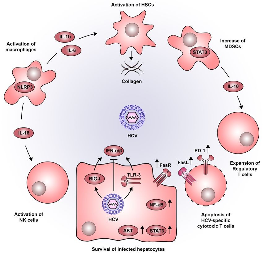
Figure
Figure 2. 2. Pro-oncogenic
Pro-oncogenic inflammatory
inflammatory microenvironment
microenvironment inducedinduced by by HCV.
HCV. HCVHCV infection
infection inin
hepatocytes isisdetected
hepatocytes detectedbybyviral
viral sensors
sensors such
such as RIG-I
as RIG-I andand
TLR3,TLR3, leading
leading to thetoproduction
the production
of typeofI
type I IFNs. As for most viruses, HCV has developed a wide variety of strategies
IFNs. As for most viruses, HCV has developed a wide variety of strategies to dampen this antiviral to dampen this
antiviral response. The persistent inflammatory environment in the liver combined
response. The persistent inflammatory environment in the liver combined with the action of viral with the action of
viral proteins, establishes a sustained activation of signaling pathways associated to
proteins, establishes a sustained activation of signaling pathways associated to cell survival (e.g.,cell survival (e.g.,
STAT3, AKT,
STAT3, AKT, NF-κB
NF-κB andand FasR).
FasR). The
The sensing
sensing of
of HCV-infected
HCV-infected hepatocytes
hepatocytes by by macrophages
macrophages triggers
triggers
NLRP3 inflammasomes, inducing the secretion of IL-18 which activates NK
NLRP3 inflammasomes, inducing the secretion of IL-18 which activates NK cells. Moreover, IL-1bcells. Moreover, IL-1b
and IL-6 produced by macrophages favor the activation of HSCs which are central
and IL-6 produced by macrophages favor the activation of HSCs which are central components in the components in
progressive deposition of collagen associated to liver cirrhosis. Additionally, STAT3 also plays a rolea
the progressive deposition of collagen associated to liver cirrhosis. Additionally, STAT3 also plays
role in the development of MDSCs which produce IL-10 and favor the expansion of regulatory T cells.
in the development of MDSCs which produce IL-10 and favor the expansion of regulatory T cells.
This altered immune response, is further accentuated by the increased expression of PD-1 and FasL,
This altered immune response, is further accentuated by the increased expression of PD-1 and FasL,
impairing cytotoxic T lymphocyte function and inducing their apoptosis. Abbreviations: AKT, AKT
impairing cytotoxic T lymphocyte function and inducing their apoptosis. Abbreviations: AKT, AKT
serine/threonine kinase; FasL, Fas ligand; FasR, Fas receptor; HCV, hepatitis C virus; HSCs, hepatic
stellate cells; IFN, interferon; IL, interleukin; MDSCs, myeloid-derived suppressor cells; NF-κB, nuclear
factor kappa B; NK, natural killer; NLRP3, NOD- LRR- and pyrin domain-containing protein 3; PD-1,
programmed cell death 1; RIG-I, retinoic acid-inducible gene I; STAT3, signal transducer and activator
of transcription 3; TLR3, toll like receptor 3.
During viral infections, pathogen-associated molecular patterns (PAMPS) are recognized by
innate immune sensors, i.e., toll-like receptors (TLRs), retinoic acid-inducible gene I (RIG-I), and
cyclic GMP-AMP synthase (cGAS), triggering a rapid IFN type I response against infection [95]. As aInt. J. Mol. Sci. 2020, 21, 3057 7 of 17
prototype positive-stranded RNA virus, HCV is mainly recognized by the TLR3 and RIG-I, although it
has developed several mechanisms to evade innate immune sensing and to blunt the resulting IFN
response (reviewed in more detail in [96]). During chronic infection, HCV triggers TLR3 signaling
also in monocytes and macrophages leading to the secretion of proinflammatory cytokines including
interleukins (IL) and an activation of the inflammasome [97] without IFN induction. In vitro data
suggest that sensing of HCV-infected hepatocytes by macrophages triggers NLR3P inflammasomes and
induces IL-18 secretion, which activates natural killer (NK) cells [98]. Since the host fails to overcome
HCV infection, the persistent deregulation of immune factors such as IFN signaling, activation of
NF-κB, TNF-α and IL-6-mediated signaling were found to be significantly associated with a poor
prognosis for HCC development [62] and thus, presumably contribute to liver disease progression.
In addition to the above-mentioned pro-viral and proliferative role of STAT3 signaling, this
pathway also exhibits an important role during inflammation. The proinflammatory cytokines IL-6
and TNF-α activate the transcription factors STAT3 and NF-κB, which if persistently stimulated can
aggravate liver disease progression and HCC development [99]. NF-κB signaling induces IFN-β
production, which triggers JAK/STAT signaling in neighboring cells and consequently leads to the
expression of antiviral IFN stimulated genes. However, these events are directly targeted by HCV
proteins blunting the innate antiviral response. In contrast, HCV proteins core and NS5A also block
apoptosis of infected cells by activation of AKT serine/threonine kinase (AKT) and NF-kB [100].
Chronic HCV infection stimulates the expression of Fas cell surface death receptor (FAS) in hepatocytes
promoting cell survival of infected cells and adding to the pro-oncogenic environment caused by the
virus. The same mechanism triggers the apoptosis of inflammatory T cells [101], and thus, promotes
the survival of infected pro-oncogenic cells. Chronic HCV infection results in the dysregulation of the
balance between Fas receptor (FasR, CD95) and ligand (FasL, CD95L). FasL positive T cells have been
shown to interact with FasR-exposing hepatocytes, which results in liver cell apoptosis. During chronic
HCV infection, upregulation of FasR expression on hepatocytes as well as FasL upregulation in T cells
significantly correlate with the severity of liver inflammation [102,103]. In addition, FasR expression
is almost undetectable in HCC [104], suggesting a reduced susceptibility towards T cell-mediated
cytotoxicity, which potentially results in an increased survival of tumorigenic cells.
Contributing to this effect is also the lack of an effective T cell response during chronic infection.
The constant and prolonged exposure of T cells to HCV antigens causes exhaustion, especially of CD8+
T cells [105,106]. Additionally, HCV-specific T cells undergo massive apoptosis during the chronic
phase of infection, which in turn may contribute to the chronic inflammatory environment during
HCV infection [107]. A key inhibitory marker on exhausted T cells is programmed cell death 1 (PD-1).
Its expression on exhausted T cells is at least partially maintained even following DAA cure [108,109],
potentially due to epigenetic modifications induced by long term exposure to HCV. Additionally,
multiple reports suggest that immune cells, including T cells can also be productively infected by
HCV [12–15], although it remains unclear to which degree this might affect the HCV-specific immune
response [13,15,16]. HCV infection has been linked to the presence of myeloid-derived suppressor
cells (MDSCs), a population of myeloid cells that negatively regulates the function of NK, CD4+ and
CD8+ T cells [110]. Again, STAT3 activation is a central driver in the development of MDSCs, as it
induces the expression of suppressive genes, i.e., IL-10, which in turn favor the expansion of regulatory
T (Treg) cells [111,112]. Evidence suggests that MDSCs increase tumor burden and metastasis rate in
liver cancer animal models and patients with HCC [113].
HSCs are important regulators for the liver extracellular matrix and wound healing. During chronic
injury, activated stellate cells are the main drivers of liver fibrosis, which is characterized by an excessive
deposition of collagen scar tissue. If unstopped, this process is a main cause of impaired liver function
and cirrhosis [114]. Importantly, NF-κB signaling and the secretion of proinflammatory cytokines, i.e.,
IL-6 and IL-1b into the microenvironment of infected hepatocytes also activate HSCs [115–118] and
thus cause an acceleration of collagen deposition into the extracellular space and liver fibrosis.Int. J. Mol. Sci. 2020, 21, 3057 8 of 17
6. Virus-Induced Epigenetic Dysregulation
It became evident that HCV not only promotes pro-oncogenic events and liver fibrosis but also
perturbs epigenetic regulatory circuits in hepatocytes with important long-term consequences to
the host. Epigenetic homeostasis via DNA methylation of regulatory elements, post-translational
modifications of histones, and non-coding RNA (ncRNA)-mediated gene silencing is essential for
the memory of genetic regulation in the context of environmental conditions [119]. It is therefore
widely accepted that epigenetic dysregulation is an important factor in the development of pathologies
including cancer [120,121].
6.1. CpG Methylation of Host DNA
During chronic HCV infection, a combination of direct and indirect factors can influence the
epigenome. Viral proteins, such as core and NS5A possess a nuclear localization signal and can be
detected in the nucleus and thus being close to host DNA [122–125]. HCV core protein markedly
increases the expression of DNA methyltransferase (DNMT)-1 and histone deacetylase (HDAC)-1 [126]
and has been suggested to cause epigenetic silencing of tumor suppressor gene expression by
DNA-methylation of cytosine-phospho-guanine (CpG) dinucleotides in regulatory gene elements [127].
Tumor suppressor genes silenced by core include secreted frizzled-related protein (SFRP) [126], which
promotes epithelial-mesenchymal transition (EMT) and deregulates Wnt/ß-catenin signaling as major
pathways involved in HCC development [128]. HCV-induced hypermethylation of cancer-related
genes such as APC, p73, p14, and O6 MGMT (summarized in [129]) and perturbed methylation in
repetitive DNA elements have been observed [130], suggesting a relevance for HCV-related HCC.
6.2. Non-Coding RNA
Gene expression is also regulated by ncRNAs [119], known to affect biological processes
encompassing differentiation, proliferation, cell death, and cancer [131]. ncRNAs comprise small
ncRNA (sncRNA) and long ncRNA (lncRNA), which are both involved in HCC, as reviewed in [131].
Among tens of thousands of lncRNAs many are reportedly related to HCC, including HOTAIR [132,133]
and PVT1 [134] functioning via epigenetic repression and cell cycle progression. sncRNAs, in particular
miRNAs are relevant in HCCs, which besides small nucleolar RNAs (snoRNAs) and piwi-interacting
RNAs (piRNAs) have been studied most extensively [119]. miRNAs are of 18-25 nucleotides length and
regulate gene expression on the post-transcriptional level. They bind to complementary sequences in
the 30 -untranslated region of mRNAs, which results in a translational suppression or degradation [135].
HCV affects miRNA expression with important impact on liver pathogenesis [71,72,136]. In addition
to the above-mentioned regulatory roles of miR-19a and miR-135-5p in virus-induced signaling,
miR-146a-5p [136] and the liver specific miR-122 [17] are also deregulated by HCV infection and are
linked to liver disease and HCC development [131].
6.3. Histone Modifications
Posttranslational modifications of histones regulate the binding affinity of DNA to histone proteins
and therefore influence accessibility of genes within chromatin to the transcriptional machinery. These
modifications comprise acetylation, methylation, phosphorylation, and ubiquitination [127]. A recent
combined genome-wide analysis of the HCV-related epigenome using Chip-Seq and transcriptomic data
(RNA-Seq) from HCV-infected liver tissues highlighted a potential role of virus-induced acetylation of
histone H3 at position lysine 27 (H3K27Ac) in liver pathogenesis [137,138]. H3K27Ac is considered to be
an activation mark promoting the transcription of associated genes by distinguishing active enhancers
from inactive/poised enhancer elements [139]. Strikingly, these studies showed that chronic HCV
infection induces specific genome-wide changes in H3K27Ac, which correlated with the expression of
known cancer risk genes [137]. Similar observations have been made in HCV-infected cell culture for
histone H3 lysine 9 acetylation (H3K9Ac) activation mark [140]. Importantly, since epigenetic regulationInt. J. Mol. Sci. 2020, 21, 3057 9 of 17
of gene expression can be considered as a lasting “epigenetic memory” [141], it may suggest that
HCV-induced changes persist even after viral cure. Indeed, an epigenetic viral footprint can be detected
in HCV-cured cell culture, chimeric mice and patients, which are correlated with elevated cancer-risk
gene expression and lower patient survival after surgical resection of HCV-associated HCC [137,140].
This footprint is certainly a combination of direct, virus-induced effects and indirect fibrosis-related
effects, which in patients with chronic HCV infection are closely linked. The comparison of H3K27Ac
Chip-Seq and RNA-Seq profiles in HCV-cured fibrotic patient livers with livers of non-fibrotic
HCV-cured chimeric mice yielded an HCV-specific persistent epigenetic and transcriptomic “footprint”
of 65 cancer-risk genes [137]. Moreover, a fibrosis-associated “footprint” of 1693 cancer-risk genes
was identified in fibrotic livers of HCV patients and in patients with non-alcoholic fatty liver disease
(NAFLD). Dysregulation of a subset of 25 genes of this fibrotic epigenetic footprint is termed the
prognostic epigenetic signature (PES) and is predictive for cancer risk in patients [138]. Importantly,
epidrugs can remove these epigenetic persistent footprints and reduce cancer risk in cell culture and
animal models [138], highlighting a potential for future chemopreventive strategies [49].
7. Clinical Implications for Biomarker Discovery to Predict HCV-Induced HCC Risk
Only 30 years after its discovery, HCV is now a curable disease thanks to an unprecedented effort
of the combined work of scientists, physicians and pharma. However, the treatment-induced viral
cure cannot fully eradicate HCV-associated complications and HCC risk especially in patients with
advanced liver disease [142]. Due to the relatively long delay between virus-induced liver injury and
the development of HCC, it is assumed that the epidemiologic peak of HCV-associated liver disease
and HCC is yet to come. This highlights two urgent unmet medical needs for the clinical management
of patients with SVR: reliable biomarkers to identify the fraction of SVR patients with elevated HCC
risk and efficient and safe chemopreventive strategies targeting virus-specific pro-oncogenic pathways,
epigenetic footprints and liver fibrosis to help these patients.
Long-term chronic infection with HCV causes liver disease progression from fibrosis to liver
cirrhosis and the occurrence of pre-neoplastic lesions with a certain accumulation of genetic mutations
including telomerase promoter, p53 and beta catenin pathway [143,144]. This relative diversity of
individual defects makes it difficult to identify a common and reliable biomarker predictive for
HCC risk. A 186-gene transcriptional signature has been identified in non-tumor tissue adjacent to
HCC lesions and in HCV-related early-stage cirrhosis, which is predictive for HCC risk [62,63]. This
so-called prognostic liver signature (PLS) can recapitulate HCC risk in patients independent from
the underlying liver disease etiology [64]. Recently discovered signatures based on virus-induced
epigenetic modifications [137], such as the PES [138], provide a novel perspective to assess residual
HCC risk in HCV patients after SVR and allow to select these patients for clinical trials to evaluate
chemoprevention of HCC.
Many of the above-mentioned virus-induced pro-oncogenic pathways are also drivers in
non-HCV-associated liver disease. Namely the pro-inflammatory, proliferative and pro-fibrinogenic
signaling pathways EGF, IL-6, and TGF-ß are also part of the etiology independent biomarker signature
PLS [64]. Chronic HCV infection has been associated with a range of extrahepatic complications, such
as mixed cryoglobulinemia and B cell lymphomas [145]. Although the mechanisms involved are not
fully understood, it has been suggested that TGF-β and IL-6 play a potential role in the development of
these complications [146,147]. It is thus conceivable that persistent HCV-induced signaling alterations
and deranged cytokine production may promote the development of extrahepatic manifestations in
immune cells [148]. Several compounds targeting these signaling pathways have been suggested for
chemoprevention of HCC (reviewed in detail in [149,150]) and targeting HCV-relevant pathways to
treat established HCC have been suggested [151–153]. Therefore, learning from HCV-induced liver
disease will help to develop novel diagnostics and personalized therapeutic concepts that may be useful
to prevent and potentially also to treat HCC from other related liver disease etiologies like NAFLD.Int. J. Mol. Sci. 2020, 21, 3057 10 of 17
Funding: This work was funded by the European Union (ERC-AdG-2014 HEPCIR ERC POC PRELICAN 755460,
ERC POC HEPCAN 862551 to T.F.B., EU H2020 HEPCAR 667273 to T.F.B. and J.L.), the French Cancer Agency
(TheraHCC2.0 IHU201901299 to T.F.B.), the Agence Nationale de Recherche sur le Sida et les hépatites virale (ANRS
ECTZ103701 to J.L. and T.F.B), The French Fondation pour la Recherche Médicale (FDT201805005763 to A.A.R.S)
the National Institutes (NIAID U19AI12386, NIAID R03AI13106, NCI 1R21CA209940, NIDDK R01CA233794 to
T.F.B), the Fondation de l’Université de Strasbourg (HEPKIN) (TBA-DON-0002), the Inserm Plan Cancer 2019-2023
to T.F.B. and German Research Foundation (DFG-395783133 to F.W.). This work has benefitted from support by the
Initiative of excellence IDEX-Unistra (ANR-10-IDEX-0002-02 to J.L.) and has been published under the framework
of the LABEX ANR-10-LAB-28 (HEPSYS). Inserm Plan Cancer, IDEX and LABEX are initiatives from the French
program “Investments for the future”.
Conflicts of Interest: The authors declare no conflicts of interest.
References
1. Bray, F.; Ferlay, J.; Soerjomataram, I.; Siegel, R.L.; Torre, L.A.; Jemal, A. Global cancer statistics 2018:
GLOBOCAN estimates of incidence and mortality worldwide for 36 cancers in 185 countries. CA Cancer J.
Clin. 2018, 68, 394–424. [CrossRef] [PubMed]
2. Yang, J.D.; Hainaut, P.; Gores, G.J.; Amadou, A.; Plymoth, A.; Roberts, L.R. A global view of hepatocellular
carcinoma: Trends, risk, prevention and management. Nat. Rev. Gastroenterol. Hepatol. 2019, 16, 589–604.
[CrossRef]
3. Axley, P.; Ahmed, Z.; Ravi, S.; Singal, A.K. Hepatitis C Virus and Hepatocellular Carcinoma: A Narrative
Review. J. Clin. Transl. Hepatol. 2018, 6, 79–84. [CrossRef] [PubMed]
4. Carrat, F.; Fontaine, H.; Dorival, C.; Simony, M.; Diallo, A.; Hezode, C.; De Ledinghen, V.; Larrey, D.;
Haour, G.; Bronowicki, J.P.; et al. Clinical outcomes in patients with chronic hepatitis C after direct-acting
antiviral treatment: A prospective cohort study. Lancet 2019, 393, 1453–1464. [CrossRef]
5. Hsu, S.J.; Yang, S.S.; Kao, J.H. Risk of hepatocellular carcinoma development after hepatitis C virus eradicated
by direct-acting antivirals: Fact or fiction? J. Formos. Med. Assoc. 2020, 119 Pt 1, 3–11. [CrossRef]
6. Ioannou, G.N.; Green, P.K.; Beste, L.A.; Mun, E.J.; Kerr, K.F.; Berry, K. Development of models estimating
the risk of hepatocellular carcinoma after antiviral treatment for hepatitis C. J. Hepatol. 2018, 69, 1088–1098.
[CrossRef]
7. Ioannou, G.N.; Beste, L.A.; Green, P.K.; Singal, A.G.; Tapper, E.B.; Waljee, A.K.; Sterling, R.K.; Feld, J.J.;
Kaplan, D.E.; Taddei, T.H.; et al. Increased Risk for Hepatocellular Carcinoma Persists Up to 10 Years
After HCV Eradication in Patients with Baseline Cirrhosis or High FIB-4 Scores. Gastroenterology 2019, 157,
1264–1278.e4. [CrossRef]
8. Baumert, T.F.; Juhling, F.; Ono, A.; Hoshida, Y. Hepatitis C-related hepatocellular carcinoma in the era of new
generation antivirals. BMC Med. 2017, 15, 52. [CrossRef]
9. Waziry, R.; Hajarizadeh, B.; Grebely, J.; Amin, J.; Law, M.; Danta, M.; George, J.; Dore, G.J. Hepatocellular
carcinoma risk following direct-acting antiviral HCV therapy: A systematic review, meta-analyses, and
meta-regression. J. Hepatol. 2017, 67, 1204–1212. [CrossRef]
10. Alter, H.J.; Purcell, R.H.; Shih, J.W.; Melpolder, J.C.; Houghton, M.; Choo, Q.L.; Kuo, G. Detection of antibody
to hepatitis C virus in prospectively followed transfusion recipients with acute and chronic non-A, non-B
hepatitis. N. Engl. J. Med. 1989, 321, 1494–1500. [CrossRef]
11. Fauvelle, C.; Felmlee, D.J.; Crouchet, E.; Lee, J.; Heydmann, L.; Lefevre, M.; Magri, A.; Hiet, M.S.; Fofana, I.;
Habersetzer, F.; et al. Apolipoprotein E Mediates Evasion from Hepatitis C Virus Neutralizing Antibodies.
Gastroenterology 2016, 150, 206–217.e4. [CrossRef] [PubMed]
12. Garcia, F., Jr.; Garcia, F.; Roldan, C.; Lopez, I.; Martinez, N.M.; Alvarez, M.; Bernal, M.C.; Hernandez, J.;
Maroto, M.C. Detection of HCV and GBV-CHGV RNA in peripheral blood mononuclear cells of patients
with chronic type C hepatitis. Microbios 2000, 103, 7–15. [PubMed]
13. Lambotin, M.; Baumert, T.F.; Barth, H. Distinct intracellular trafficking of hepatitis C virus in myeloid and
plasmacytoid dendritic cells. J. Virol. 2010, 84, 8964–8969. [CrossRef] [PubMed]
14. Lerat, H.; Rumin, S.; Habersetzer, F.; Berby, F.; Trabaud, M.A.; Trepo, C.; Inchauspe, G. In vivo tropism of
hepatitis C virus genomic sequences in hematopoietic cells: Influence of viral load, viral genotype, and cell
phenotype. Blood 1998, 91, 3841–3849. [CrossRef] [PubMed]
15. Skardasi, G.; Chen, A.Y.; Michalak, T.I. Authentic Patient-Derived Hepatitis C Virus Infects and Productively
Replicates in Primary CD4(+) and CD8(+) T Lymphocytes In Vitro. J. Virol. 2018, 92, e01790-17. [CrossRef]Int. J. Mol. Sci. 2020, 21, 3057 11 of 17
16. Colpitts, C.C.; Tsai, P.L.; Zeisel, M.B. Hepatitis C Virus Entry: An Intriguingly Complex and Highly Regulated
Process. Int. J. Mol. Sci. 2020, 21, 2091. [CrossRef]
17. Luna, J.M.; Scheel, T.K.; Danino, T.; Shaw, K.S.; Mele, A.; Fak, J.J.; Nishiuchi, E.; Takacs, C.N.; Catanese, M.T.;
de Jong, Y.P.; et al. Hepatitis C virus RNA functionally sequesters miR-122. Cell 2015, 160, 1099–1110.
[CrossRef]
18. Lupberger, J.; Casanova, C.; Fischer, B.; Weiss, A.; Fofana, I.; Fontaine, N.; Fujiwara, T.; Renaud, M.; Kopp, A.;
Schuster, C.; et al. PI4K-beta and MKNK1 are regulators of hepatitis C virus IRES-dependent translation. Sci.
Rep. 2015, 5, 13344. [CrossRef]
19. Morozov, V.A.; Lagaye, S. Hepatitis C virus: Morphogenesis, infection and therapy. World J. Hepatol. 2018,
10, 186–212. [CrossRef]
20. Reiss, S.; Rebhan, I.; Backes, P.; Romero-Brey, I.; Erfle, H.; Matula, P.; Kaderali, L.; Poenisch, M.; Blankenburg, H.;
Hiet, M.S.; et al. Recruitment and activation of a lipid kinase by hepatitis C virus NS5A is essential for integrity
of the membranous replication compartment. Cell Host Microbe 2011, 9, 32–45. [CrossRef]
21. Shimotohno, K. HCV Assembly and Egress via Modifications in Host Lipid Metabolic Systems. Cold Spring
Harb. Perspect. Med. 2020. Epub ahead of print. [CrossRef] [PubMed]
22. Vogt, P.K. Retroviral oncogenes: A historical primer. Nat. Rev. Cancer 2012, 12, 639–648. [CrossRef] [PubMed]
23. Hoppe-Seyler, K.; Bossler, F.; Braun, J.A.; Herrmann, A.L.; Hoppe-Seyler, F. The HPV E6/E7 Oncogenes: Key
Factors for Viral Carcinogenesis and Therapeutic Targets. Trends Microbiol. 2018, 26, 158–168. [CrossRef]
[PubMed]
24. Banerjee, A.; Ray, R.B.; Ray, R. Oncogenic potential of hepatitis C virus proteins. Viruses 2011, 2, 2108–2133.
[CrossRef] [PubMed]
25. Polyak, S.J.; Klein, K.C.; Shoji, I.; Miyamura, T.; Lingappa, J.R. Assemble and Interact: Pleiotropic Functions
of the HCV Core Protein. In Hepatitis C Viruses: Genomes and Molecular Biology; Tan, S.L., Ed.; Horizon
Bioscience: Norfolk, UK, 2006.
26. Moriya, K.; Yotsuyanagi, H.; Shintani, Y.; Fujie, H.; Ishibashi, K.; Matsuura, Y.; Miyamura, T.; Koike, K. Hepatitis
C virus core protein induces hepatic steatosis in transgenic mice. J. Gen. Virol. 1997, 78 Pt 7, 1527–1531. [CrossRef]
27. Shintani, Y.; Fujie, H.; Miyoshi, H.; Tsutsumi, T.; Tsukamoto, K.; Kimura, S.; Moriya, K.; Koike, K. Hepatitis
C virus infection and diabetes: Direct involvement of the virus in the development of insulin resistance.
Gastroenterology 2004, 126, 840–848. [CrossRef]
28. Moriya, K.; Fujie, H.; Shintani, Y.; Yotsuyanagi, H.; Tsutsumi, T.; Ishibashi, K.; Matsuura, Y.; Kimura, S.;
Miyamura, T.; Koike, K. The core protein of hepatitis C virus induces hepatocellular carcinoma in transgenic
mice. Nat. Med. 1998, 4, 1065–1067. [CrossRef]
29. Okuda, M.; Li, K.; Beard, M.R.; Showalter, L.A.; Scholle, F.; Lemon, S.M.; Weinman, S.A. Mitochondrial
injury, oxidative stress, and antioxidant gene expression are induced by hepatitis C virus core protein.
Gastroenterology 2002, 122, 366–375. [CrossRef]
30. Kawamura, H.; Govindarajan, S.; Aswad, F.; Machida, K.; Lai, M.M.; Sung, V.M.; Dennert, G. HCV core
expression in hepatocytes protects against autoimmune liver injury and promotes liver regeneration in mice.
Hepatology 2006, 44, 936–944. [CrossRef]
31. Nakamoto, S.; Imazeki, F.; Fukai, K.; Fujiwara, K.; Arai, M.; Kanda, T.; Yonemitsu, Y.; Yokosuka, O.
Association between mutations in the core region of hepatitis C virus genotype 1 and hepatocellular
carcinoma development. J. Hepatol. 2009, 52, 72–78. [CrossRef]
32. Akuta, N.; Suzuki, F.; Hirakawa, M.; Kawamura, Y.; Sezaki, H.; Suzuki, Y.; Hosaka, T.; Kobayashi, M.; Saitoh, S.;
Arase, Y.; et al. Amino acid substitutions in hepatitis C virus core region predict hepatocarcinogenesis following
eradication of HCV RNA by antiviral therapy. J. Med. Virol. 2011, 83, 1016–1022. [CrossRef] [PubMed]
33. Neufeldt, C.J.; Cortese, M.; Acosta, E.G.; Bartenschlager, R. Rewiring cellular networks by members of the
Flaviviridae family. Nat. Rev. Microbiol. 2018, 16, 125–142. [CrossRef] [PubMed]
34. Tomei, L.; Failla, C.; Santolini, E.; De Francesco, R.; La Monica, N. NS3 is a serine protease required for
processing of hepatitis C virus polyprotein. J. Virol. 1993, 67, 4017–4026. [CrossRef] [PubMed]
35. Gosert, R.; Egger, D.; Lohmann, V.; Bartenschlager, R.; Blum, H.E.; Bienz, K.; Moradpour, D. Identification of
the hepatitis C virus RNA replication complex in Huh-7 cells harboring subgenomic replicons. J. Virol. 2003,
77, 5487–5492. [CrossRef] [PubMed]
36. Sakamuro, D.; Furukawa, T.; Takegami, T. Hepatitis C virus nonstructural protein NS3 transforms NIH 3T3
cells. J. Virol. 1995, 69, 3893–3896. [CrossRef] [PubMed]Int. J. Mol. Sci. 2020, 21, 3057 12 of 17
37. Sakata, K.; Hara, M.; Terada, T.; Watanabe, N.; Takaya, D.; Yaguchi, S.; Matsumoto, T.; Matsuura, T.;
Shirouzu, M.; Yokoyama, S.; et al. HCV NS3 protease enhances liver fibrosis via binding to and activating
TGF-beta type I receptor. Sci. Rep. 2013, 3, 3243. [CrossRef]
38. Appel, N.; Zayas, M.; Miller, S.; Krijnse-Locker, J.; Schaller, T.; Friebe, P.; Kallis, S.; Engel, U.; Bartenschlager, R.
Essential role of domain III of nonstructural protein 5A for hepatitis C virus infectious particle assembly.
PLoS Pathog. 2008, 4, e1000035. [CrossRef]
39. Enomoto, N.; Sakuma, I.; Asahina, Y.; Kurosaki, M.; Murakami, T.; Yamamoto, C.; Ogura, Y.; Izumi, N.;
Marumo, F.; Sato, C. Mutations in the nonstructural protein 5A gene and response to interferon in patients
with chronic hepatitis C virus 1b infection. N. Engl. J. Med. 1996, 334, 77–81. [CrossRef]
40. Gale, M.J., Jr.; Korth, M.J.; Tang, N.M.; Tan, S.L.; Hopkins, D.A.; Dever, T.E.; Polyak, S.J.; Gretch, D.R.;
Katze, M.G. Evidence that hepatitis C virus resistance to interferon is mediated through repression of the
PKR protein kinase by the nonstructural 5A protein. Virology 1997, 230, 217–227. [CrossRef]
41. Wang, A.G.; Lee, D.S.; Moon, H.B.; Kim, J.M.; Cho, K.H.; Choi, S.H.; Ha, H.L.; Han, Y.H.; Kim, D.G.;
Hwang, S.B.; et al. Non-structural 5A protein of hepatitis C virus induces a range of liver pathology in
transgenic mice. J. Pathol. 2009, 219, 253–262. [CrossRef]
42. Majumder, M.; Ghosh, A.K.; Steele, R.; Zhou, X.Y.; Phillips, N.J.; Ray, R.; Ray, R.B. Hepatitis C virus NS5A
protein impairs TNF-mediated hepatic apoptosis, but not by an anti-FAS antibody, in transgenic mice.
Virology 2002, 294, 94–105. [CrossRef] [PubMed]
43. Kriegs, M.; Burckstummer, T.; Himmelsbach, K.; Bruns, M.; Frelin, L.; Ahlen, G.; Sallberg, M.; Hildt, E.
The hepatitis C virus non-structural NS5A protein impairs both the innate and adaptive hepatic immune
response in vivo. J. Biol. Chem. 2009, 284, 28343–28351. [CrossRef] [PubMed]
44. Majumder, M.; Steele, R.; Ghosh, A.K.; Zhou, X.Y.; Thornburg, L.; Ray, R.; Phillips, N.J.; Ray, R.B. Expression
of hepatitis C virus non-structural 5A protein in the liver of transgenic mice. FEBS Lett. 2003, 555, 528–532.
[CrossRef]
45. Akkari, L.; Gregoire, D.; Floc’h, N.; Moreau, M.; Hernandez, C.; Simonin, Y.; Rosenberg, A.R.; Lassus, P.;
Hibner, U. Hepatitis C viral protein NS5A induces EMT and participates in oncogenic transformation of
primary hepatocyte precursors. J. Hepatol. 2012, 57, 1021–1028. [CrossRef] [PubMed]
46. Uthaya Kumar, D.B.; Chen, C.L.; Liu, J.C.; Feldman, D.E.; Sher, L.S.; French, S.; DiNorcia, J.; French, S.W.;
Naini, B.V.; Junrungsee, S.; et al. TLR4 Signaling via NANOG Cooperates with STAT3 to Activate Twist1
and Promote Formation of Tumor-Initiating Stem-Like Cells in Livers of Mice. Gastroenterology 2015, 150,
707–719. [CrossRef]
47. Berger, K.L.; Kelly, S.M.; Jordan, T.X.; Tartell, M.A.; Randall, G. Hepatitis C virus stimulates the
phosphatidylinositol 4-kinase III alpha-dependent phosphatidylinositol 4-phosphate production that is
essential for its replication. J. Virol. 2011, 85, 8870–8883. [CrossRef]
48. Burckstummer, T.; Kriegs, M.; Lupberger, J.; Pauli, E.K.; Schmittel, S.; Hildt, E. Raf-1 kinase associates with
Hepatitis C virus NS5A and regulates viral replication. FEBS Lett. 2006, 580, 575–580. [CrossRef]
49. Hoshida, Y.; Fuchs, B.C.; Bardeesy, N.; Baumert, T.F.; Chung, R.T. Pathogenesis and prevention of hepatitis C
virus-induced hepatocellular carcinoma. J. Hepatol. 2014, 61, S79–S90. [CrossRef]
50. Lupberger, J.; Croonenborghs, T.; Roca Suarez, A.A.; Van Renne, N.; Juhling, F.; Oudot, M.A.; Virzi, A.;
Bandiera, S.; Jamey, C.; Meszaros, G.; et al. Combined Analysis of Metabolomes, Proteomes, and
Transcriptomes of Hepatitis C Virus-Infected Cells and Liver to Identify Pathways Associated with Disease
Development. Gastroenterology 2019, 157, 537–551.e9. [CrossRef]
51. Roca Suarez, A.A.; Baumert, T.F.; Lupberger, J. Beyond viral dependence: The pathological consequences of
HCV-induced EGF signaling. J. Hepatol. 2018, 69, 564–566. [CrossRef]
52. Lupberger, J.; Zeisel, M.B.; Xiao, F.; Thumann, C.; Fofana, I.; Zona, L.; Davis, C.; Mee, C.J.; Turek, M.; Gorke, S.;
et al. EGFR and EphA2 are host factors for hepatitis C virus entry and possible targets for antiviral therapy.
Nat. Med. 2011, 17, 589–595. [CrossRef]
53. Mailly, L.; Xiao, F.; Lupberger, J.; Wilson, G.K.; Aubert, P.; Duong, F.H.T.; Calabrese, D.; Leboeuf, C.;
Fofana, I.; Thumann, C.; et al. Clearance of persistent hepatitis C virus infection in humanized mice using a
claudin-1-targeting monoclonal antibody. Nat. Biotechnol. 2015, 33, 549–554. [CrossRef] [PubMed]
54. Diao, J.; Pantua, H.; Ngu, H.; Komuves, L.; Diehl, L.; Schaefer, G.; Kapadia, S.B. Hepatitis C virus induces
epidermal growth factor receptor activation via CD81 binding for viral internalization and entry. J. Virol.
2012, 86, 10935–10949. [CrossRef] [PubMed]Int. J. Mol. Sci. 2020, 21, 3057 13 of 17
55. Zona, L.; Lupberger, J.; Sidahmed-Adrar, N.; Thumann, C.; Harris, H.J.; Barnes, A.; Florentin, J.; Tawar, R.G.;
Xiao, F.; Turek, M.; et al. HRas signal transduction promotes hepatitis C virus cell entry by triggering
assembly of the host tetraspanin receptor complex. Cell Host Microbe 2013, 13, 302–313. [CrossRef] [PubMed]
56. Mankouri, J.; Griffin, S.; Harris, M. The hepatitis C virus non-structural protein NS5A alters the trafficking
profile of the epidermal growth factor receptor. Traffic 2008, 9, 1497–1509. [CrossRef] [PubMed]
57. Plissonnier, M.L.; Lahlali, T.; Michelet, M.; Lebosse, F.; Cottarel, J.; Beer, M.; Neveu, G.; Durantel, D.;
Bartosch, B.; Accardi, R.; et al. Epidermal Growth Factor Receptor-Dependent Mutual Amplification between
Netrin-1 and the Hepatitis C Virus. PLoS Biol. 2016, 14, e1002421. [CrossRef]
58. Groepper, C.; Rufinatscha, K.; Schroder, N.; Stindt, S.; Ehlting, C.; Albrecht, U.; Bock, H.H.; Bartenschlager, R.;
Haussinger, D.; Bode, J.G. HCV modifies EGF signalling and upregulates production of CXCR2 ligands:
Role in inflammation and antiviral immune response. J. Hepatol. 2018, 69, 594–602. [CrossRef]
59. Brenndorfer, E.D.; Karthe, J.; Frelin, L.; Cebula, P.; Erhardt, A.; Schulte am Esch, J.; Hengel, H.;
Bartenschlager, R.; Sallberg, M.; Haussinger, D.; et al. Nonstructural 3/4A protease of hepatitis C virus
activates epithelial growth factor-induced signal transduction by cleavage of the T-cell protein tyrosine
phosphatase. Hepatology 2009, 49, 1810–1820. [CrossRef]
60. Lupberger, J.; Duong, F.H.; Fofana, I.; Zona, L.; Xiao, F.; Thumann, C.; Durand, S.C.; Pessaux, P.; Zeisel, M.B.;
Heim, M.H.; et al. Epidermal growth factor receptor signaling impairs the antiviral activity of interferon-alpha.
Hepatology 2013, 58, 1225–1235. [CrossRef]
61. Fuchs, B.C.; Hoshida, Y.; Fujii, T.; Wei, L.; Yamada, S.; Lauwers, G.Y.; McGinn, C.M.; DePeralta, D.K.; Chen, X.;
Kuroda, T.; et al. Epidermal growth factor receptor inhibition attenuates liver fibrosis and development of
hepatocellular carcinoma. Hepatology 2014, 59, 1577–1590. [CrossRef]
62. Hoshida, Y.; Villanueva, A.; Kobayashi, M.; Peix, J.; Chiang, D.Y.; Camargo, A.; Gupta, S.; Moore, J.;
Wrobel, M.J.; Lerner, J.; et al. Gene expression in fixed tissues and outcome in hepatocellular carcinoma. N.
Engl. J. Med. 2008, 359, 1995–2004. [CrossRef]
63. Hoshida, Y.; Villanueva, A.; Sangiovanni, A.; Sole, M.; Hur, C.; Andersson, K.L.; Chung, R.T.; Gould, J.;
Kojima, K.; Gupta, S.; et al. Prognostic gene expression signature for patients with hepatitis C-related
early-stage cirrhosis. Gastroenterology 2013, 144, 1024–1030. [CrossRef] [PubMed]
64. Nakagawa, S.; Wei, L.; Song, W.M.; Higashi, T.; Ghoshal, S.; Kim, R.S.; Bian, C.B.; Yamada, S.; Sun, X.;
Venkatesh, A.; et al. Molecular Liver Cancer Prevention in Cirrhosis by Organ Transcriptome Analysis and
Lysophosphatidic Acid Pathway Inhibition. Cancer Cell 2016, 30, 879–890. [CrossRef] [PubMed]
65. Ninio, L.; Nissani, A.; Meirson, T.; Domovitz, T.; Genna, A.; Twafra, S.; Srikanth, K.D.; Dabour, R.;
Avraham, E.; Davidovich, A.; et al. Hepatitis C Virus Enhances the Invasiveness of Hepatocellular Carcinoma
via EGFR-Mediated Invadopodia Formation and Activation. Cells 2019, 8, 1395. [CrossRef] [PubMed]
66. Li, W.; Liang, X.; Kellendonk, C.; Poli, V.; Taub, R. STAT3 contributes to the mitogenic response of hepatocytes
during liver regeneration. J. Biol. Chem. 2002, 277, 28411–28417. [CrossRef] [PubMed]
67. Roca Suarez, A.A.; Van Renne, N.; Baumert, T.F.; Lupberger, J. Viral manipulation of STAT3: Evade, exploit,
and injure. PLoS Pathog. 2018, 14, e1006839. [CrossRef] [PubMed]
68. McCartney, E.M.; Helbig, K.J.; Narayana, S.K.; Eyre, N.S.; Aloia, A.L.; Beard, M.R. Signal transducer and activator
of transcription 3 is a proviral host factor for hepatitis C virus. Hepatology 2013, 58, 1558–1568. [CrossRef]
69. Yoshida, T.; Hanada, T.; Tokuhisa, T.; Kosai, K.; Sata, M.; Kohara, M.; Yoshimura, A. Activation of STAT3 by
the hepatitis C virus core protein leads to cellular transformation. J. Exp. Med. 2002, 196, 641–653. [CrossRef]
70. Gong, G.; Waris, G.; Tanveer, R.; Siddiqui, A. Human hepatitis C virus NS5A protein alters intracellular
calcium levels, induces oxidative stress, and activates STAT-3 and NF-kappa B. Proc. Natl. Acad. Sci. USA
2001, 98, 9599–9604. [CrossRef]
71. Sodroski, C.; Lowey, B.; Hertz, L.; Jake Liang, T.; Li, Q. MicroRNA-135a Modulates Hepatitis C Virus Genome
Replication through Downregulation of Host Antiviral Factors. Virol. Sin. 2018, 34, 197–210. [CrossRef]
72. Van Renne, N.; Roca Suarez, A.A.; Duong, F.H.T.; Gondeau, C.; Calabrese, D.; Fontaine, N.; Ababsa, A.;
Bandiera, S.; Croonenborghs, T.; Pochet, N.; et al. miR-135a-5p-mediated downregulation of protein tyrosine
phosphatase receptor delta is a candidate driver of HCV-associated hepatocarcinogenesis. Gut 2018, 67,
953–962. [CrossRef] [PubMed]
73. Devhare, P.B.; Sasaki, R.; Shrivastava, S.; Di Bisceglie, A.M.; Ray, R.; Ray, R.B. Exosome-Mediated Intercellular
Communication between Hepatitis C Virus-Infected Hepatocytes and Hepatic Stellate Cells. J. Virol. 2017,
91, e02225-16. [CrossRef] [PubMed]You can also read