Maintenance of Cell Wall Integrity under High Salinity
←
→
Page content transcription
If your browser does not render page correctly, please read the page content below
International Journal of
               Molecular Sciences

Review
Maintenance of Cell Wall Integrity under High Salinity
Jianwei Liu 1 , Wei Zhang 1,2 , Shujie Long 1,2 and Chunzhao Zhao 1, *

                                          1   Shanghai Center for Plant Stress Biology, CAS Center for Excellence in Molecular Plant Sciences,
                                              Chinese Academy of Sciences, Shanghai 200032, China; jwliu@psc.ac.cn (J.L.); weizhang@psc.ac.cn (W.Z.);
                                              shjlong@psc.ac.cn (S.L.)
                                          2   University of the Chinese Academy of Sciences, Beijing 100049, China
                                          *   Correspondence: czzhao@psc.ac.cn; Tel.: +86-021-5707-8274

                                          Abstract: Cell wall biosynthesis is a complex biological process in plants. In the rapidly growing
                                          cells or in the plants that encounter a variety of environmental stresses, the compositions and the
                                          structure of cell wall can be dynamically changed. To constantly monitor cell wall status, plants
                                          have evolved cell wall integrity (CWI) maintenance system, which allows rapid cell growth and
                                          improved adaptation of plants to adverse environmental conditions without the perturbation of cell
                                          wall organization. Salt stress is one of the abiotic stresses that can severely disrupt CWI, and studies
                                          have shown that the ability of plants to sense and maintain CWI is important for salt tolerance.
                                          In this review, we highlight the roles of CWI in salt tolerance and the mechanisms underlying the
                                          maintenance of CWI under salt stress. The unsolved questions regarding the association between the
                                          CWI and salt tolerance are discussed.

                                          Keywords: cell wall integrity; cell wall sensor; salt stress; salt tolerance; LRXs; CrRLK1Ls


Citation: Liu, J.; Zhang, W.; Long, S.;
                                          1. Introduction
Zhao, C. Maintenance of Cell Wall
Integrity under High Salinity. Int. J.           High salinity is an adverse environmental stress that severely affects the growth and
Mol. Sci. 2021, 22, 3260. https://        yield of crops. Excessive accumulation of sodium in plants confers both ion toxicity and
doi.org/10.3390/ijms22063260              osmotic stress, which in turn dramatically affect the morphological, physiological, biochem-
                                          ical, and metabolic status of plants [1]. Currently, more than 20% of the irrigated lands in
Academic Editor: Raffaella                the world are threatened by high salinity, and the area of saline soils is increasing gradually
Maria Balestrini                          every year accompanied by the global climate change and poor irrigation practices [2–4].
                                          It is expected the global population will reach to nearly 10 billion in 2050, and to meet the
Received: 27 February 2021                increasing food demand in future, the utilization of saline soils to grow major crops tends
Accepted: 19 March 2021                   to be inevitable. Therefore, the cultivation of crops with increased salt tolerance is a major
Published: 23 March 2021                  objective in salt stress community.
                                                 To avoid the damage caused by excessive salts in soil, plants have evolved various
Publisher’s Note: MDPI stays neutral      strategies to overcome the problems caused by high salinity. Ion homeostasis, osmotic
with regard to jurisdictional claims in   adjustment, ROS balance, and metabolic adjustment are the major factors that are associated
published maps and institutional affil-   with the tolerance of plants to salt stress. Based on the capacity of plants to adapt to salt
iations.
                                          stress, plants can be classified into glycophytes and halophytes. Our major crops, such as
                                          rice, maize, and wheat, are glycophytes that are unable to complete their life cycle when
                                          they are being exposed to high salinity. Halophytes, however, have developed various
                                          strategies to adapt to the environments with a high concentration of sodium. For example,
Copyright: © 2021 by the authors.         halophytes are able to extrude salts via glands or store excessive Na+ in the vacuoles of
Licensee MDPI, Basel, Switzerland.        epidermal bladder cells [5,6].
This article is an open access article           More and more studies point out that maintenance CWI is also critical for the adapta-
distributed under the terms and           tion of plants to high salinity. Plant cell walls, which mainly consist of polysaccharides and
conditions of the Creative Commons
                                          structural proteins, are essential for the establishment of plant morphology and protection
Attribution (CC BY) license (https://
                                          of plants against adverse environmental changes [7]. During plant growth and develop-
creativecommons.org/licenses/by/
                                          ment or in response to environmental stresses, the cell wall compositions and structures are
4.0/).

Int. J. Mol. Sci. 2021, 22, 3260. https://doi.org/10.3390/ijms22063260                                          https://www.mdpi.com/journal/ijms
Int. J. Mol. Sci. 2021, 22, 3260 2 of 19
dynamically modulated, allowing rapid cell elongation and increased stress tolerance [8].
To maintain CWI during the reorganization of cell wall, plants need to constantly moni-
tor the chemical and mechanical properties of the cell walls and also need to process an
ability to repair cell wall once they are seriously disrupted. It has been shown that CWI
maintenance mechanism exists in plants and is essential for the regulation of growth and
development and in response to stress conditions [9,10]. The progresses about CWI sensing
and maintenance system in plants have been summarized in several outstanding review
papers [8,11,12]. In this review, we focus on the elucidation of the associations between
CWI and salt tolerance in plants.
2. Importance of Cell Wall Biosynthesis in Salt Tolerance
The plant cell wall is a dynamic network composed of cellulose, hemicellulose, pectin,
lignin, and multiple types of structural proteins [13,14]. Moreover, cell wall-remodeling
enzymes, various ions, and reactive oxygen species (ROS) also exist in the apoplast and
are involved in the regulation of CWI. Upon exposure to high salinity, several changes
in the cell wall have been identified, including the reduction of cellulose content [15,16],
disruption of the cross-linking of pectins [9], and accumulation of lignin [17]. Studies have
shown that the plants that are defective in cell wall biosynthesis are hypersensitive to salt
stress, suggesting that maintenance of CWI is important for the adaptation of plants to
high salinity.
2.1. Cellulose
Cellulose is the most abundant organic component in the cell wall of terrestrial
vascular plants. Cellulose micro-fibrils are composed of β-1,4-linked glucan chains, which
are synthesized at the cell surface by cellulose synthase (CesA) complexes (CSCs) [18,19].
Each CSC is assembled into a hexameric rosette structure, harboring CesA catalytic subunits
and several accessory proteins. In Arabidopsis, there are ten CesA proteins [18]. It is well
known that CesA1, CesA3, and CesA6 are assembled in a CSC to synthesize cellulose
in the primary cell wall, while CesA4, CesA7, and CesA8 are mainly involved in the
synthesis of cellulose in the secondary cell wall [20]. Experimental data have shown that
the cellulose contents are significantly reduced after salt treatment and the plants with a
loss of function of CESA1 and CESA6 gene display reduced root elongation and severe root
tip swelling under salt stress, indicating that cellulose biosynthesis is important for salt
tolerance in plants [21,22]. Clear evidences have indicated that the CSCs are dissociated
from plasma membrane within 30 min after exposure to high salinity. However, during
the growth recovery phase after salt treatment, the CSCs can be reassembled at the plasma
membrane to synthesize new cellulose, and the capacity to reassemble CSCs during the
growth recovery stage is critical for plants to maintain root and hypocotyl growth under
salt stress [16].
Apart from the CesAs, several cellulose biosynthesis-related proteins have also been
reported involved in salt tolerance. For example, KORRIGAN1 (KOR1), a putative endo-
1,4-β-D-glucanase, is an integral part of the primary cell wall CSC and is required for root
elongation under salt stress [22,23]. Cellulose synthase interacting protein 1 (CSI1) and
companion of cellulose synthase 1 (CC1 and CC2) proteins, acting as companions of CesAs,
are both required for cellulose biosynthesis [16,21]. Mutations in CSI1 or CC1 and CC2 lead
to reduced root or hypocotyl elongation under salt stress. CTL1 encodes a chitinase-like
protein that participates in the deposition of the ordered cellulose, and mutation of this
gene results in increased sensitivity to high salinity [24] (Table 1).Int. J. Mol. Sci. 2021, 22, 3260 3 of 19
Table 1. List of the cell wall biosynthesis-related genes that are involved in salt stress response.
Name Gene ID Annotation Function Reference(s)
AtCesA1/RSW1 At4g32410 Cellulose synthesis in the primary cell wall [22]
Cellulose synthase catalytic subunit
AtCesA8/IRX1 At4g18780 Cellulose synthesis in the secondary cell wall [25]
AtCC1 At1g45688
Cellulose synthase companion protein Cortical microtubules assembly and cellulose biosynthesis under salt stress [16]
AtCC2 At5g42860
AtCTL/POM1 At1g05850 Chitinase-like protein 1 Involved in the assembly of glucan chains [24,26]
AtCSI1/POM2 At2g22125 Cellulose synthase-interactive protein 1 Companion of CesAs; required for cell elongation in root [21]
AtCCoAOMT1 At4g34050 Caffeoyl-CoA 3-O-methyltransferase Involved in lignin synthesis [17]
AtKOR/RSW2 At5g49720 Endo-β-1,4-glucanase Integral component of CSC; required for cell elongation in root [23]
AtHSR8/MUR4 At1g30620 Golgi-localized UDP-D-xylose 4-epimerase Arabinose biosynthesis; related to the modification of polysaccharides and glycoproteins [27]
Transfer of galactose from UDP-α-d-Gal or arabinopyranose from UDP-β-l-Arap to
AtGALS1 At2g33570 β-1,4-galactan synthase [28,29]
growing β-1,4-galactan chains
Xyloglucan endotrans Cleave or rejoin the xyloglucan; xth30 mutation decreases crystalline cellulose content
AtXTH30 At1g32170 [30]
glucosylase-hydrolase and affects the depolymerization of microtubules under salt stress
AtPMEI13 At5g62360 Pectin methyl-esterase inhibitor 13 Inhibits the activity of PMEs [31]
AtBPC1 At2g01930
BPC-type transcription factor Regulation of the expression of AtGALS1 [28]
AtBPC2 At1g14685
AtGCN5 At3g54610 Histone acetyltransferase Epigenetic regulation of cell wall-related genes [32]
OsTSD2 Os02g51860 Pectin methyltransferase Regulation of pectin metabolism [33]
OsBURP16 Os10g26940 β subunit precursor of polygalacturonase 1 Involved in cell wall pectin degradation [34]Int. J. Mol. Sci. 2021, 22, 3260 4 of 19
2.2. Hemicellulose
Hemicelluloses are grouped into xyloglucans (XyG), xylans, mannans, and β-(1,3;1,4)-
glucans, and the abundance and structure of these polysaccharides vary greatly in different
plants species [35]. Xylan is considered as a cross-linking polysaccharide in the establish-
ment of cell wall architecture [35,36]. XyG contributes to the strengthening of cell wall
during cell elongation by binding to cellulose micro-fibrils with hydrogen bonds [37,38].
XyG can be cleaved by the cell wall remodeling enzymes xyloglucan endotransglucosy-
lase/hydrolases (XTHs) [39]. After cleavage, the reducing end of the XyG is attached
to the non-reducing end of another XyG oligomer or polymer to produce chimeric XyG
molecules [39]. The XTHs-mediated modification of XyG is considered to be important
for controlling cell wall extensibility. Studies have reported that XTHs are involved in
salt stress response in plants. Arabidopsis XTH30, encoding a xyloglucan endotransglu-
cosylase/hydrolase 30, is strongly upregulated under salt stress [30]. Loss of function of
the XTH30 gene leads to increased salt tolerance, which is mainly caused by the slower
reduction of crystalline cellulose content and alleviated depolymerization of microtubules
in response to salt stress [30]. This result suggests that XTH30 plays a negative role in salt
tolerance. However, the positive roles of XTHs in salt tolerance have also been reported.
Constitutive expression of CaXTH3 in hot pepper [40,41] and PeXTH in Populus euphrat-
ica [42] enhance tolerance to salt stress, and disruption of XTH19 and XHT23 genes in
Arabidopsis results in decreased salt tolerance [43].
2.3. Pectin
Pectin is a group of acidic polysaccharides that are enriched with α-(1, 4)-linked galac-
turonic acids in the backbone [44]. Pectin accounts for up to 40% of the dry weight of higher
plant cell walls [44] and plays critical roles in plant growth and development [45], leaf senes-
cence [46], biotic [47] and abiotic stress responses [48]. Pectin is composed of three major
types: homogalacturonan (HG), rhamnogalacturonan-I (RG-I), and rhamnogalacturonan-II
(RG-II) [7,44]. HG is synthesized in the Golgi apparatus and secreted to the apoplast in
a highly methy-esterified form and later it is selectively de-esterified by pectin methyl
esterases (PMEs) during cell growth and in response to environmental stimuli [7,44]. The
degree and pattern of the methyl-esterification of pectin in some extent determines the
stiffness of cell walls [49]. In Arabidopsis, there are around 66 members of PME family pro-
tein, and for most of PMEs, their activities can be inhibited by endogenous PME inhibitors
(PMEIs) or a natural inhibitor epigallocatechin gallate (EGCG) [50,51]. High salinity trig-
gers the demethyl-esterification of loosely bound pectins to inhibit cell swelling [52] and
previous studies showed that the activity of PMEs is either positively or negatively associ-
ated with salt tolerance in plants [53]. For instance, null Arabidopsis function mutant pme13
is hypersensitive to Na+ toxicity in seed germination and seedling growth [53]. In contrast,
overexpression of Chorispora bungeana PMEI1 or AtPMEI13 in Arabidopsis causes decreased
PMEs activity and enhanced methyl-esterification level of pectins, which subsequently
improves seeds germination and survival rate under salt stress [31]. The de-esterified
HG molecules can be cross-linked to form the so called egg-box structure, the process of
which is mediated by divalent cations, such as Ca2+ , and the formation of egg-box structure
promotes cell wall stiffening [54]. In the presence of high concentration of Na+ , the ratio of
Na+ /Ca2+ in the apoplast is increased, and Na+ is supposed to replace Ca2+ to bind pectins
and thus disturbs the cross-linking of pectins, leading to reduced cell elongation [55].
Besides, the borate-mediated cross-linking of RG-II contributes to the strength of cell wall
and is required for the regulation of growth recovery after exposure to high salinity [56,57].
The roles of pectin in salt tolerance have also been reported in rice. Polygalacturonase
1 (PG1) is a cell wall hydrolase that is responsible for the degradation of cell wall pectin.
Overexpression of OsBURP16, which encodes a non-catalytic β subunit of PG1, results in
an increased pectin degradation and increased salt-hypersensitivity in rice [34]. OsTSD2
encodes a pectin methyltransferase in rice, and mutation in OsTSD2 leads to a higher
content of Na+ and a lower level of K+ in rice shoot under high salinity, which is mainlyInt. J. Mol. Sci. 2021, 22, 3260 5 of 19
caused by the reduced expression of genes that are responsible for the maintenance of ion
homeostasis, such as OsHKT1;5, OsSOS1, and OsKAT1 [33] (Table 1).
2.4. Lignin
As one of the most abundant organic compound in plants, lignin is composed of
phenylalanine-derived [58] or tyrosine-derived [59] aromatic monomer substances and
is important for the secondary cell wall formation and the responses to a variety of en-
vironmental stresses [60]. High salinity induces the accumulation of lignin content and
cell wall thickening via the activation of lignin biosynthesis pathway [60]. The accumu-
lation of lignin contributes to the mechanical strengthening of cell wall and protection
of membrane integrity under salt stress [61]. The effects of lignin accumulation on salt
tolerance have been reported in different crops, including soybean [62], wheat [63], and
tomato [64]. CCoAOMT encodes a caffeoyl CoA O-methyltransferase (CCoAOMT), which
catalyzes caffeoyl CoA to feruloyl CoA in lignin biosynthesis pathway. The expression
of CCoAOMT is induced in salt-adapted cell, and the plants with a loss-of-function of
CCoAOMT are hypersensitive to salt stress [17]. BpMYB46 and BpNAC012, encoding two
transcription factors in white birch (Betula platyphylla), are required for the up-regulation
of lignin biosynthetic genes and salt stress-responsive genes, and overexpression of these
two genes enhances salt tolerance in B. platyphylla [65,66]. AgNAC1, a nuclear-localized
protein in celery, acts as a positive regulator in inducing the expression of lignin-related
and salt stress-responsive genes, and overexpression of AgNAC1 enhances the formation of
secondary walls and plant salt tolerance [67].
3. The Roles of the Cell Wall-Localized Glycoproteins in Salt Stress Response
In addition to dynamic and complex polysaccharide networks, several types of cell
wall proteins (CWPs) have been identified in the apoplast. CWPs play critical roles in cell
wall modifications and cell wall stress signals transduction. Hydroxyproline (Hyp)-rich
glycoproteins (HRGPs), proline-rich proteins (PRPs), glycine-rich proteins (GRPs), and
arabinogalactan proteins AGPs are the major types of CWPs [68]. For most of CWPs, they
are secreted into the apoplast in a glycosylation-modified form [69–71].
Extensins (EXTs) are a group of cell wall glycoproteins that belong to the HRGPs
family. EXTs are typically characterized for the enrichment of Ser-(Hyp)3–5 repeats in their
protein sequences [72], and each Hyp residue is decorated with up to five arabinose units
by several different arabinosyltransferases, including HPAT1-HPAT3 [73], RRA1-RRA3 [74],
XEG113 [75], and ExAD [76]. The arabinosylation of EXTs is suggested to be important for
the fulfillment of their biological functions. Our recent study showed that the mutation of
MUR4, which encodes an UDP-Xyl 4-epimerase that is essential for the conversion of UDP-
Xyl to UDP-Arap in Golgi, results in reduced root elongation under salt stress, suggesting
that arabinose biosynthesis and subsequently the modification of polysaccharides and
glycoproteins by arabinose are important for salt tolerance in plants [27].
Leucine-rich repeat extensins (LRXs) are chimeric proteins that contain an N-terminal
leucine-rich repeat (LRR) domain that binds with interacting partners and a C-terminal
extensin domain that is likely linked with the EXT network or polysaccharides in the
apoplast [77]. LRXs gene family consists of 11 members in Arabidopsis, among of which
LRX3, LRX4, and LRX5 are dominantly expressed in vegetative tissues [77]. The biological
functions of these three LRX proteins are redundant, as mutation of each single gene
does not cause any obvious phenotypes, but lrx34 double and lrx345 triple mutants both
exhibit dwarfism, increased accumulation of anthocyanin, and increased sensitivity to high
salinity [10]. It is worth noting that all these phenotypes are more severe in the lrx345 triple
mutant than that in the lrx34 double mutant. Our study indicated that fer-4 mutant as well
as the transgenic plants overexpressing RALF22 and RALF23 exhibit similar phenotypes
as lrx345 in terms of plant growth and salt sensitivity, and biochemical data show that
RALF22 and RALF23 are physically associated with LRX3/4/5 [10]. Combining the data
showing that FER is the receptor of RALFs [9], we can conclude that the LRX3/4/5, theInt. J. Mol. Sci. 2021, 22, x FOR PEER REVIEW 6 of 19
Int. J. Mol. Sci. 2021, 22, 3260 as the transgenic plants overexpressing RALF22 and RALF23 exhibit similar phenotypes 6 of 19
as lrx345 in terms of plant growth and salt sensitivity, and biochemical data show that
RALF22 and RALF23 are physically associated with LRX3/4/5 [10]. Combining the data
showing that FER is the receptor of RALFs [9], we can conclude that the LRX3/4/5, the
secreted peptide
secreted peptide RALFs,
RALFs, and
and the receptor-like kinase
the receptor-like kinase FER
FER function
function as
as aa module
module to mediate
to mediate
salt stress response in the apoplast. It is supposed that the extensin domain
salt stress response in the apoplast. It is supposed that the extensin domain of LRXs of LRXs is
is able
able to anchor polysaccharides in the cell wall [77,78], but it is still unknown
to anchor polysaccharides in the cell wall [77,78], but it is still unknown whether LRXs whether
LRXs directly
directly participate
participate in the sensing
in the sensing of CWIoforCWI or coordinate
coordinate with FER with FER to perceive
to perceive CWI
CWI (Figure
(Figure
1). 1).
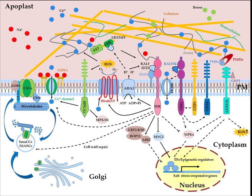
Figure
Figure 1. 1. Sensing
Sensingandandmaintenance
maintenanceofofcell cellwall integrity
wall integrityunder
undersaltsalt
stress. SaltSalt
stress. stress-induced
stress-inducedcell wall changes
cell wall are proposed
changes are pro-
to be sensed
posed by multiple
to be sensed receptor-like
by multiple kinases,kinases,
receptor-like including FER, THE1,
including MIK2, FEI1/2,
FER, THE1, and WAK1/2.
MIK2, FEI1/2, and WAK1/2.As one Asofone
theofmost
the
most important
important cellintegrity
cell wall wall integrity
(CWI)(CWI) sensors,
sensors, FER may FER function
may function
alonealone or together
or together with with LRX3/4/5-RALF22/23
LRX3/4/5-RALF22/23 module
module to
to perceive
perceive thethe perturbation
perturbation of CWI
of CWI caused
caused by high
by high salinity.
salinity. The AHA2-mediated
The AHA2-mediated acidification
acidification of theof the apoplastic
apoplastic pH in-
pH increases
creases
the the of
affinity affinity
LRXs of LRXs
with with while
RALFs, RALFs, thewhile the alkaline
alkaline state
state in the in the apoplast
apoplast promotespromotes
the bindingtheofbinding
RALFsofwith RALFsFER.with
FERFER.
and
probably also other cell wall sensors convert salt-triggered cell wall signals to multiple intracellular signals, including Cain-
FER and probably also other cell wall sensors convert salt-triggered cell wall signals to multiple intracellular signals, 2+ ,
cluding Ca2+, ROS, abscisic acid (ABA), jasmonic acid (JA), and MPKs, which in turn regulate the expression of salt stress-
ROS, abscisic acid (ABA), jasmonic acid (JA), and MPKs, which in turn regulate the expression of salt stress-responsive
responsive genes in the nucleus. Salt stress can alter the redox status in the apoplast, and RbohD/F-mediated production
genes in the nucleus. Salt stress can alter the redox status in the apoplast, and RbohD/F-mediated production of the
of the apoplastic H2O2 may affect the cross-linking of cell wall polymers and activate H2O2 sensor HPCA1. Glycosyl inositol
apoplastic H2 O2 may(GIPC)
phosphorylceramide affect the cross-linking
sphingolipids of cell wall
participate in polymers
the sensing andof activate H2 O2salt
extracellular sensor HPCA1.binding
by directly Glycosyl to inositol
sodium
phosphorylceramide (GIPC) sphingolipids
ions. Cell wall biosynthesis- participate in
and modification-related the sensing including
components, of extracellular
pectinsalt by directly
methyl binding
esterases (PMEs),to sodium ions.
PME inhib-
Cell
itorswall biosynthesis-
(PMEIs), and modification-related
and cellulose synthase (CesA), are components,
involved in the including pectin
regulation methyl
of salt esterases
tolerance (PMEs),
in plants. Upon PME inhibitors
initial expo-
sure to salt
(PMEIs), andstress, cortical
cellulose microtubules
synthase depolymerized
(CesA), are involved and cellulose
in the regulation synthase
of salt complex
tolerance (CSC)
in plants. together
Upon initialwith its com-
exposure to
panions
salt stress,CSI1 andmicrotubules
cortical CC1/2 are internalized into small
are depolymerized CesA compartments/microtubule-associated
and cellulose synthase complex (CSC) together withCesA compartments
its companions CSI1
(smaCCs/MASCs).
and At the growth
CC1/2 are internalized recovery
into small stage after salt application, FER is probably
CesA compartments/microtubule-associated required for the
CesA compartments regulation of the
(smaCCs/MASCs).
reassembly of cortical microtubules and the relocation of CSCs to the plasma membrane
At the growth recovery stage after salt application, FER is probably required for the regulation of the reassembly to synthesize cellulose, which
of cortical
subsequently enhances the adaptation of plants to salt stress. Solid lines represent direct regulations, and dashed lines
microtubules and the relocation of CSCs to the plasma membrane to synthesize cellulose, which subsequently enhances
represent in-direct or potential regulations.
the adaptation of plants to salt stress. Solid lines represent direct regulations, and dashed lines represent in-direct or
potential regulations.
AGPs are highly glycosylated with arabinogalactan chains and are proposed to play
important roles in salt stress response [69]. Our study showed that the reduced root elon-
gation of the mur4 mutant under high salinity is partially caused by the decreased AGPs,Int. J. Mol. Sci. 2021, 22, 3260 7 of 19
as application of gum arabic, a commercial source of Acacia senegal AGPs, restores the
root elongation of the mur4 mutant under salt stress [27]. As a glycosylphosphatidyli-
nositol (GPI)-anchored fasciclin-like AGP, salt overly sensitive 5 (SOS5)/fasciclin-like
arabinogalactan-protein 4 (FLA4) was identified based on a screening of mutants with
increased sensitivity to salt stress. The sos5/fla4 mutant exhibits reduced root elongation and
severe root tip swelling under salt stress [79,80]. SOS5 is glycosylated by galactosyltrans-
ferase 2 (GALT2) and GALT5, both of which belong to AGP-specific galactosyltransferases.
The galt2 galt5 double mutant displays a similar phenotype as the sos5/fla4 mutant in the
presence of high concentration of NaCl [80]. Recently, studies showed that AGPs are able
to cross-link with cell wall components. For instance, arabinoxylan pectin arabinogalactan
protein 1 (APAP1) is covalently linked to pectins [81], and arabinogalactan protein 31
(AGP31) physically associates with methyl-esterified polygalacturonic acid and galactans,
which are the branches of RG-I [82].
Expansins, first isolated from growing cucumber hypocotyls, consist of four subfam-
ilies: α-expansin, β-expansin, expansin-like A, and expansin-like B [83,84]. Expansins
are key regulators of cell-wall loosening and are required for cell enlargement under a
variety of environmental stresses [85]. Several studies have shown that the expression of
expansin-encoding genes is induced by high salt and the elevation of the protein levels of
expansins tends to promote salt tolerance in plants. ZmEXPB2, ZmEXPB6, and ZmEXPB8
genes in maize [86], AsEXP1 gene in turf grass [87], and OsEXPA3 gene in rice [88], are
induced upon exposure to high salinity. Down-regulation of ZmEXPB6 is correlated with
the reduced leaf growth of maize under salt stress [89]. Overexpression of rice expansin 7
(OsEXPA7) confers substantially enhanced tolerance to salt stress by lowering reactive
oxygen species (ROS) accumulation and increasing antioxidant activity in rice [90]. Ec-
topic expression of wheat expansin 2 (TaEXPA2) or TaEXPB23 improves salt tolerance in
tobacco [91,92]. Although expansins have been known to positively regulate salt stress
response in multiple species, few studies have revealed the mechanisms underlying the
expansins-mediated regulation of salt tolerance.
4. Salt Stress Alters the Redox Status in the Apoplast
Reactive oxygen species (ROS) are a class of metabolites, including hydrogen peroxide,
singlet oxygen, superoxide, and hydroxyl radicals, which are produced in chloroplasts,
mitochondria, peroxisomes, and apoplast [93]. The salt stress-triggered production of
ROS and their effects on CWI have been widely reported in plants [93–95]. ROS trig-
gers the cross-linking of cell wall compounds and enhances the mechanical strength of
cell wall under a short-term stress exposure. Under a prolonged stress treatment, the
formation of hydroxyl radicals (•OH) cleave plant polysaccharides, leading to cell wall
loosening [96]. The ROS-induced lignin biosynthesis facilitates the adaptation of plants to
high salt environment [95,97].
The production of ROS in the apoplast is mainly mediated by respiratory burst oxidase
homolog D (RbohD) and RbohF [98], two NADPH oxidases that are localized at the plasma
membrane. NADPH oxidases transfer electrons from cytosolic NADPH or NADH to
apoplastic oxygen, leading to the production of superoxide (O2 − ), which is then catalyzed
to hydrogen peroxide (H2 O2 ) by superoxide dismutases [99]. The expression of RbohD and
RbohF is induced under salt stress and rbohD rbohF double mutant is hypersensitive to salt
stress [100], suggesting that the ROS production in the apoplast is required for salt tolerance.
Salt-induced production of ROS by RbohD/F is able to activate Ca2+ channel to increase the
influx of Ca2+ into cytosol, which mediates the modulation of Na+ /K+ homeostasis [100].
The H2 O2 generated by RbohD/F during the early stage of stress treatment also acts as a
signal molecule to activate antioxidant system to attenuate salt stress-induced oxidative
damages [101]. Recent studies showed that RbohD/F form nanoclusters at the plasma
membrane in response to osmotic stress and later they are internalized into the cytoplasm
via membrane microdomains [102–104]. As high salt conditions are accompanied byInt. J. Mol. Sci. 2021, 22, 3260 8 of 19
osmotic stress, the formation of RbohD/F as nanoclusters at the plasma membrane is
perhaps also the case in the plants being exposed to high salinity (Figure 1).
Class III peroxidases are heme-containing enzymes, which are mainly localized in
the apoplast and vacuole. Class III peroxidases either positively or negatively modulate
apoplastic ROS levels [105]. Class III peroxidases explore H2 O2 and O2 − to generate •OH,
which leads to the cleavage of polysaccharides and promotes cell wall loosening [106].
Class III peroxidase 71 (PRX71), which is strongly up-regulated in response to cell wall
damage (CWD), negatively regulates growth and cell size and positively regulates ROS
accumulation [94]. GsPRX9, encoding a Class III peroxidase, is induced by salt treatment in
soybean root, and the soybean transgenic plants overexpressing GsPRX9 exhibit increased
root growth and decreased H2 O2 content under salt stress [107].
The biological significance of the salt stress-induced redox change in the apoplast
is still far from being fully understood. One of the outputs of the redox change is to
affect the formation of intra- and inter-molecular disulfide bond. A large number of cell
wall-localized glycoproteins and secreted peptides are characterized with the enrichment
of cysteines, which are potentially involved in the formation of disulfide bonds. There-
fore, we can speculate that the salt stress-induced redox change can affect the intra- and
inter-molecular disulfide bridges of cell wall glycoproteins, which in turn transduce cell
wall signals to the cellular interior. LRX8 and RALF4, which are both required for the
regulation of pollen tube growth, process cysteines that are involved in the formation of
disulfide bridges. A recent structural study showed that the formation of LRX8 homodimer
and also the physical association of RALF4 with LRX8 require oxidative environment.
Abolishment of the disulfide bonds via sites mutation or treatment of proteins with dithio-
threitol (DTT) largely prevents the formation of LRX8 homodimer and affects the affinity
of LRX8 with RALF4 [108]. These results suggest that the redox status in the cell wall
is required for the regulation of the formation of LRXs-RALFs complex. Based on this
hypothesis, we propose that the salt stress-induced change of apoplastic redox status
may affect the formation of homo- and hetero-dimers of LRX3/4/5 and also affect the
affinity of LRX3/4/5 proteins with RALFs, which finally transduce salt stress signals to the
intracellular signaling pathways.
5. The Impact of Apoplastic pH on Salt Tolerance
In the early 1970s, the acid growth theory was proposed, which states that acidification
of the apoplast promotes cell elongation, whereas alkaline state in the apoplast prevents
cell growth [109]. The reduction of apoplastic pH (apo pH) activates several cell wall
proteins, including expansins and other remodeling enzymes, resulting in the loosening of
cell wall [110]. apo pH in linear growing cells is regulated by plasma membrane-localized
H+ -ATPases (AHAs) [111]. RALFs are a class of peptides that cause the alkalinization of the
apoplast by regulating H+ -ATPases via Catharanthus roseus RLK1-like kinases (CrRLK1Ls).
FER is one of the CrRLK1L family proteins that consist of two carbohydrate-binding
malectin-like domains, a transmembrane domain, and an intracellular serine/threonine-
kinase domain [112,113]. FER inhibits the proton transport activity of AHA2 likely via
direct phosphorylation [114]. It is known that salinity triggers the transient alkalization
in the apoplast and inhibits plant growth [115], and our study showed that salt stress can
induce the formation of mature RALFs [10]. These data suggest that salt stress-induced
alkalinization of the apoplast is probably mediated by RALFs-FER-AHA2 module and the
acidification of the extracellular environment is important for salt tolerance. Two halophyte
species, Atriplex lentiformis and Chenopodium quinoa, which have a capacity to tolerate a high
concentration of sodium ion, display a high H+ -ATPase activity under salt stress, which
contributes to a low apo pH and fast Na+ efflux [116]. SOS1, encoding a plasma membrane
membrane-localized Na+ /H+ antiporter, is required for the extrusion of excessive Na+
from the cytosol [117]. The Na+ /H+ exchange activity of SOS1 is absent under normal
growth conditions. Upon salt stress, however, Na+ -induces induced formation of an
ATP-dependent pH gradient can enhance the Na+ /H+ transport activity of SOS1 [118].Int. J. Mol. Sci. 2021, 22, 3260 9 of 19
Altogether, low apo pH facilitates plant growth under salt stress, but the direct effects of low
apo pH on cell wall networks need more detailed studies.
6. Cell Wall Integrity Sensing and Signal Transduction under High Salinity
Unlike the traditional activation of plant receptor-like kinases by the corresponding
ligands, the sensing of CWI is not limited by ligand-receptor pattern, e.g., recognition
of wall fragments released from the damaged cell walls by receptor-like kinases, and is
probably also achieved via the recognition of the cell wall modifications and the alteration
of redox and apo pH status. Currently, a series of plasma membrane-localized receptor-like
kinases and cell wall glycoproteins have been identified that are involved in the sensing
and maintenance of CWI. As a universal signal molecule, Ca2+ is also involved in the
transduction of CWI signaling signals in plants.
The cell wall appears to be the largest source of Ca2+ in plant cell [119]. Under
normal conditions, Ca2+ is used to stabilize pectins via the dimerization of HG chains [120].
AGPs have been shown to bind abundant Ca2+ [121]. Under salt stress, the excessive
accumulation of Na+ in the apoplast disrupts ion homeostasis, leading to rapid sodium-
specific calcium waves occurred in roots [122]. The imported calcium ions directly bind the
EF hands of RbohD/F and improve their catalytic activity [123,124]. Ca2+ is also an initial
signal to activate the SOS signaling pathway, which promotes the extrusion of Na+ from
the cytosol [125,126].
In addition to high salinity, other abiotic stresses, such as drought, cold, and osmotic
stress, can also induce the cytosolic Ca2+ influx within a few seconds to minutes. Although
the induction of Ca2+ signaling is a common event for these different abiotic stresses,
studies have shown that the different stresses-triggered Ca2+ influx is mediated by different
components. Reduced hyperosmolality-induced [Ca2+ ]i increase 1 (OSCA1) is specifically
required for the osmotic stress-triggered uptake of Ca2+ [127], and hydrogen-peroxide-
induced Ca2+ increases 1 (HPCA1) is required for H2 O2 -, but not for salt- and osmotic stress-
, induced influx of Ca2+ [128]. Glycosyl inositol phosphorylceramide (GIPC) sphingolipids,
which are glycosylated via glucuronosyltransferase MOCA1, was discovered as a sensor
of extracellular salt by directly binding to sodium ions [129]. The moca1 mutant lacking
functional GIPCs is defective in the activation of Ca2+ waves when being exposed to high
concentration of Na+ , K+ , or Li+ ion. GIPCs can bind Na+ to gate Ca2+ influx channels
and trigger the activation of SOS signaling pathway. However, which Ca2+ channels
are activated by GIPCs and the mechanism underlying the activation need further study
(Figure 1).
FER is considered as a CWI sensor and required for the activation of Ca2+ influx and
maintenance of CWI under salt stress [9]. Mutation of FER reduces salt-induced Ca2+ influx
in the root epidermis and increases sensitivity to high salinity. FER contains two malectin
domains that have been experimentally demonstrated to directly bind with de-methyl-
esterified HG in vitro and in vivo [9,130], suggesting that FER probably senses the cell wall
changes directly via its extracellular domain and then transduces the cell wall signals to
cellular interior via its cytoplasmic kinase domain. However, how the modification of
pectin affects the activity of FER is still elusive. Our recent study showed that LRX3/4/5,
RALFs, and FER function as a module to regulate salt stress response, which implies that
FER-mediated perception of CWI probably needs the aid of LRX3/4/5-RALFs regulatory
module [10]. Salt stress may dissociate the LRX3/4/5-RALFs complex via the salt stress-
induced redox and pH changes in the apoplast, and the released RALFs bind to LLG1-FER
complex and thereby allow the transduction of cell wall signals. The mechanism behind
the dissociation of LRX3/4/5 and RALFs under salt stress needs to be further investigated.
THESEUS1 (THE1) is a CrRLK1L family protein that was first identified in a screening
for the suppressors of prc1-1 [131]. The null mutation of the1 partially suppresses the
stunted growth and lignin deposition of the prc1-1 mutant, despite the reduced cellulose
content in the prc1-1 is not restored [131]. HERKULES1 (HERK1) is another CrRLK1L
protein that is phylogenetically closely related to FER and THE1. Double mutant herk1Int. J. Mol. Sci. 2021, 22, 3260 10 of 19
the1-4 displays similar phenotypes as fer-4 in terms of growth and salt stress response [52].
A recent study indicates that THE1 acts as the receptor of RALF34 to fine-tune lateral root
initiation [132]. These results suggest that FER, THE, and HERK1 may work together to
replay RALFs-mediated cell wall signals, but the biochemical associations among these
three CrRLK1L proteins are still largely unknown.
Male discoverer 1-interacting receptor like kinase 2 (MIK2) is a leucine-rich repeat
receptor-like kinase (LRR-RLK) that was identified by a genome-wide association study
(GWAS) based on the natural variations in response to salinity stress [133]. MIK2 controls
root growth direction under salt stress in a THE1-dependent manner [134]. The salt-
hypersensitive phenotype of mik2 mutant can be suppressed by the1-1, a null mutation
of THE1 [134]. Recently, the serine rich endogenous peptide (SCOOP) phytocytokines
were identified as the ligands of MIK2 to trigger immune responses [135], but whether
the SCOOP peptides participate in MIK2-mediated regulation of salt tolerance is still
unknown. FEI1 and FEI2 are two LRR-RLKs that are associated with cellulose synthesis
and anisotropic cell expansion and are involved in CWI sensing [136]. Double mutant fei1
fei2 displays root swelling and reduced cellulose biosynthesis under high sucrose or high
salt conditions [137]. Genetic analysis indicated that FEI2 functions downstream of THE1
in mediating CWI perception [138]. Mid1-complementing activity 1 (MCA1) is a plasma
membrane–localized stretch-activated Ca2+ channel and functions downstream of THE1 in
Arabidopsis [95,139]. Like the1-1 mutant, mca1 seedlings exhibit reduced deposition of lignin
and decreased jasmonic acid and salicylic acid biosynthesis in response to isoxaben-induced
CWD [138].
Wall-associated kinases (WAKs) are a family of receptor-like Ser/Thr kinases whose
extracellular domains are cross-linked with pectin fraction in a high affinity [140,141]. The
EGF-like domain of WAK1/2 preferentially binds to de-methyl-esterified HG over methyl-
esterified HG, and WAK1 also exhibits a high affinity with oligogalacturonides (OGs)
in vitro [140,142]. The binding of WAKs to pectin and OGs occurs only in the presence
of Ca2+ [140]. GRP-3, a glycine-rich cell wall protein, also acts as a potential switch for
the kinase activity of WAK1 and negatively regulates the defense responses elicited by
OGs [143]. A dominant allele of wak2 mutant exhibits constitutive activation of stress
responses, including increased ROS accumulation and cell wall biogenesis [142,144]. Under
long-term salt stress, tomato WAK1 mutant slwak1 exhibits disrupted osmotic homeostasis
and elevated sucrose content in roots, which in turn negatively affects fruit yield [145].
Similarly, Ds transposon insertion mutant of HvWAK1 in barley displays decreased salt
tolerance [146]. Although the WAKs have been shown to participate in the salt stress
response, the existing experimental evidences to elaborate the roles of WAKs in sensing the
CWI under salinity are still lacking. Recently, Gigli-Bisceglia et al. indicated that salinity
stress-induced de-methyl-esterification of pectin activates stress signaling pathways, which
may provide a direction to study the roles of WAKs in salt stress response [52] (Figure 1).
The CWD caused by salinity stress, isoxaben, an inhibitor of cellulose biosynthesis, or
driselase, a cell wall-degrading enzyme, can increase the protein levels of hormone-like
peptides PROPEP1/3, the precursors of plant elicitor peptide 1/3 (Pep1/3) [138,147]. The
Pep3 knockdown plants and the null mutant of Pep1 receptor 1 (PEPR1) both exhibit salt-
hypersensitivity [148]. These results suggest that the activation of PEPR1 by PROPEP3
positively regulates salt tolerance in Arabidopsis. Currently, the majority of studies on
Peps-PEPRs complexes focus on their roles in plant immunity, and in future the roles
of the Peps-PEPRs complexes-mediated signaling in abiotic stress responses need more
investigations.
HPCA1 is a LRR-RLK required for the sense of extracellular H2 O2 [128]. The two
pairs of cysteine residues in the extracellular domain of HPCA1 are covalently modified by
extracellular H2 O2 , which leads to the activation of HPCA1 and elevation of Ca2+ influx.
In hpca1 mutant seedlings, the extracellular H2 O2 -induced Ca2+ influx, the activation of
ABA signaling, and the phosphorylation of MPK3/6 are all inhibited [128]. It was shown
that HPCA1 is not required for the salt stress-induced influx of Ca2+ , but considering thatInt. J. Mol. Sci. 2021, 22, 3260 11 of 19
high salinity can affect the redox status in the apoplast, so whether HPCA1 is also required
for the sense of salt stress-induced redox changes worth further investigations.
7. Salt Stress-Triggered Intracellular Signaling Pathway Regulated by Cell Wall Sensors
Although several plasma membrane-localized cell wall integrity sensors have been
identified that perceive cell wall changes, the intracellular signaling pathways that relay
cell wall signals are still largely unknown. The phosphorylation of MPK6 is a marker of the
environmental stimuli, and the transient activation of MPK6 under abiotic stress conditions,
including high salinity and cold, has been reported [149]. As a major signaling transducer,
the activity of MPK6 is regulated by multiple CWI sensors, such as FER, THE1, HERK1,
and HPCA1 [52,128]. In future, the regulatory mechanisms of these CWI sensors on the
activity of MPK6 need to be addressed.
After perception of CWD by cell wall sensors, plants can integrate and balance mul-
tiple hormone signals to improve salt tolerance. ABA and JA are the major hormones
involved in the response to diverse environmental stresses. In the lrx345 and fer-4 mu-
tants, the ABA and JA contents are constitutively increased and the salt-hypersensitivity of
these two mutants is largely caused by the disrupted homeostasis of phytohormones [150].
Phosphatase ABA insensitive 2 (ABI2) is a negative regulator of ABA signaling path-
way, and FER activates the guanine nucleotide exchange factor (GEF) 1/4/10/Rho of
plant 11 (ROP11) pathway to positively regulate the activity of ABI2 phosphatase, and
thereby modulating ABA signaling pathway [151,152]. MYC2, a master transcription factor
in JA signaling pathway, is also regulated by FER. FER positively regulates immunity by
inhibiting JA signaling via the phosphorylation-mediated destabilization of MYC2 [153].
It has also been shown that MYC2 negatively regulates salt tolerance via the inhibition of
proline biosynthesis [154]. In brief, these results suggest that FER controls the environmental
stress responses via the modulation of the homeostasis of phytohormones (Figure 1).
8. Cell Wall Repair under High Salinity
Upon exposure to salt stress, the cortical microtubules in the hypocotyl of seedling
are rapidly depolymerized, the process of which usually occurs within 2 h of salt appli-
cation. However, at the growth recovery stage (after salt treatment for ~8 h), the cortical
microtubules are reassembled into stable cortical arrays [16]. Evidences have shown that
the rapid depolymerization of the cortical microtubules network is important for salt
tolerance. For instance, stabilization of microtubules with paclitaxel leads to increased
salt-hypersensitivity, whereas constitutive disruption of microtubules with oryzalin or
propyzamide improves salt tolerance [155].
The depolymerization of cortical microtubules requires the alteration of the activities
of the atypical microtubule-associated protein kinase propyzamide hypersensitive 1 (PHS1)
and microtubule-associated protein SPIRAL1 (SPR1). Under normal growth conditions, the
kinase activity of PHS1 is inhibited by its own phosphatase domain, while salt or osmotic
stress blocks this inhibition and then enhances the phosphorylation and depolymeriza-
tion of α-tubulin [156]. SPR1 binds to the microtubules and antagonizes stress-induced
cortical microtubule depolymerization. Under salt stress, SPR1 is rapidly degraded by
the 26S proteasome and the inhibition of microtubule depolymerization is relieved [157].
Histone H2B monoubiquitination (H2Bub1) participates in the regulation of the expression
of protein tyrosine phosphatase 1 (PTP1) and MAP kinase phosphatase (MKP) genes, which in
turn modulate the phosphorylation and dephosphorylation of microtubule-binding pro-
teins via a PTP1/MKP-MPK3/6 signal mode, and finally promotes the rapid microtubule
depolymerization under salt stress [158].
CSCs synthesize cellulose via the binding with cortical microtubules, and the polymer-
ization status of cortical microtubules determines the movement of CSCs at the cell surface.
CSCs are assembled in the Golgi apparatus and translocated to the plasma membrane
via vesicle trafficking. Salt-induced depolymerization of microtubules is accompanied by
the internalization of CSCs into small CesA compartments/microtubule-associated CesAInt. J. Mol. Sci. 2021, 22, 3260 12 of 19
compartments (smaCCs/MASCs) [15]. At the growth recovery stage after salt treatment,
cortical microtubule is reassembled and CSCs is relocated to the plasma membrane to
synthesize cellulose. Increasing evidences have shown that the efficiency of plants to
reassembly cortical microtubule and cellulose during the growth recovery stage is critical
for salt tolerance. CC1 and its paralog CC2 were identified as companions of CSCs and are
required for the reassembly of cortical microtubule and subsequently cellulose biosynthesis
during the growth recovery stage [16]. In cc1 cc2 double mutants, CSCs dissociate from the
microtubules after salt treatment, but a stress-tolerant microtubule complex cannot be re-
produced, resulting in the abolishment of the localization of CSCs at the plasma membrane
and decreased cellulose synthesis. Microtubules-associated proteins 65-1 (MAP65-1) is a
plant microtubule-bundling protein, which participates in the polymerization and bundling
of cortical microtubules [159]. Phosphatidic acid (PA), a product of phospholipase D (PLD),
binds to MAP65-1 and increases its activity to enhance microtubule polymerization and
bundling [160]. The pldα1 mutant exhibits a defect in microtubule organization under salt
stress and increased salt-hypersensitivity. Moreover, 16:0–18:2 PA can activate MPK6 via
directly binding to MPK6 and the salt-induced transient activation of MPK6 is abolished in
the pldα1 mutant [149].
Brassinosteroid insensitive 2 (BIN2), a master negative factor in brassinosteroid signal
pathway, regulates the balance between salt stress response and growth recovery [161].
BIN2 is required for the negative regulation of cellulose biosynthesis. BIN2 phosphorylates
CESA1 to inhibit the activity of CSCs [162]. By exploring turboID-mediated proximity
labeling technology, Kim et al. found that BIN2 interacts with FER, but the biological
significance of this interaction has not yet been resolved [163]. It is possible that FER
regulates the activity of BIN2 via phosphorylation, and then modulates CesAs activity and
cellulose biosynthesis under salt stress.
9. Transcriptional Regulation of Cell Wall-Associated Genes under Salt Stress
Under salinity stress, plant cells sense salt signals via receptors or sensors and then
transmit the signals to the downstream regulatory networks to trigger the transcription of
salt stress-responsive genes, which in turn promote the adjustment of the physiological,
biochemical, and metabolic properties of plant cells to adapt to high salinity.
The transcriptional regulation of genes largely depends on the activity of the corre-
sponding transcription factors. Some transcription factors have been identified that are
required for the regulation of cell wall-associated genes in response to salt stress. For ex-
ample, salt stress induces the accumulation of β-1,4-galactan in root cell walls through the
up-regulation the of galactan synthase 1 (GALS1) gene. Based on a genetic screening, two
transcription factors basic pentacysteine 1 (BPC1) and BPC2 were identified that directly
bind to the promoter of the GALS1 gene and repress its expression [28]. The expression
of BPC1 and BPC2 genes is significantly reduced under salt stress. The bpc1 bpc2 double
mutant, in which the accumulation of β-1,4-galactan is elevated under salt stress com-
pared with the wild type, exhibits increased salt tolerance [28]. Oryza sativa MULTIPASS
(OsMPS) encodes an R2R3-type MYB transcription factor in rice. Expression profiling
revealed that, upon ABA or salt stress treatment, the expression of expansins, such as
OsEXPA4, OsEXPA8, OsEXPB2, OsEXPB3, and OsEXPB6, and the expression of cell wall
biosynthesis genes, such as endoglucanase genes OsGLU5 and OsGLU14, are negatively
regulated by OsMPS [164]. XTH19 and XTH23, belonging to xyloglucan endotransgluco-
sylase/hydrolase group II, are up-regulated by salt stress and BR [43]. In the xth23 single
or xth19 xth23 double mutant, lateral root growth is disrupted under salt stress, whereas
overexpression of XTH19 or XTH23 enhances salt tolerance and increases lateral root initia-
tion [43]. BRI1-EMS-SUPPRESSOR 1 (BES1) is a transcription factor that is involved in BR
signaling pathway. BES1 directly binds the promoter of XTH19 and XTH23 and positively
regulates their expression under salt stress [43] (Table 1).
Gene expression is also influenced by epigenetic regulation, such as histone modi-
fication and DNA methylation. Salt stress triggers the histone H3K9/K14 acetylation ofInt. J. Mol. Sci. 2021, 22, 3260 13 of 19
some abiotic stress-responsive genes to crease their transcript levels [165]. General control
nonderepressible 5 (GCN5), encoding a histone acetyltransferase, is induced by salt stress
and acts as a maintainer of CWI. GCN5 mediates the acetylation of H3K9 and H3K14 in
the promoters of CTL1, PGX3 (polygalacturonase involved in expansion-3), and MYB54 under
salt stress, and thus fine-tunes their gene expression [32]. Constitutive expression of CTL1
partially restores the salt-hypersensitivity and CWD of the gcn5 mutant [32]. Similarly, the
H3K9 acetylation level in the genome of maize is also elevated after salt treatment, and the
increased acetylation level enhances the expression of ZmGCN5, which in turn promotes
the expression of ZmEXPB2 and ZmXET1 genes [166].
10. Conclusions and Future Perspective
Cell wall is not just a mechanical support for plant cells, but is also the frontline to
sense and transduce environmental stress signals. High salinity, as one of the globally
distributed abiotic stresses, can disrupt the CWI, and the severity of the salt-triggered
CWD largely depends on the concentration of the surrounding sodium ion combined with
other environmental conditions, such as light intensity and water availability. Study of
the mechanisms underlying the sensing and maintenance of CWI under salt stress not
only strengthens our understanding of salt stress responses in plants but also provides
new strategies for the cultivation of crops with improved salt tolerance. Regarding the
associations between CWI and salt tolerance, there are still many questions remain to be
addressed, and the most important ones could be that how the excessive accumulation of
Na+ in the apoplast affects the CWI, and how the salt-induced cell wall changes are sensed
by the cell wall sensors. Moreover, the Ca2+ channels that are required for the relay of salt-
triggered cell wall stress signals need to be identified and the cell wall repair mechanisms
under stress conditions need to be further investigated. With the development of gene
editing technologies and improved transformation efficiency, editing of CWI-related genes
in crops to generate salt-tolerant varieties can be applied in future.
Author Contributions: J.L., S.L. and W.Z. compiled the materials and wrote the first draft; J.L. and
C.Z. edited and finalized the manuscript. All authors have read and agreed to the published version
of the manuscript.
Funding: This work was supported by the National Natural Science Foundation of China (NSFC)
(Grant No. 32070295), and the Shanghai Pujiang Program (Grant No. 20PJ1414800), and the Strategic
Priority Research Program from the Chinese Academy of Sciences (Grant No. XDA27040104).
Data Availability Statement: Not applied in this study.
Conflicts of Interest: The authors declare no conflict of interest.
References
1. Munns, R.; Tester, M. Mechanisms of salinity tolerance. Annu. Rev. Plant Biol. 2008, 59, 651–681. [CrossRef] [PubMed]
2. Singh, A. Soil salinization management for sustainable development: A review. J. Environ. Manag. 2021, 277, 111383. [CrossRef]
3. Jesus, J.M.; Danko, A.S.; Fiúza, A.; Borges, M.T. Phytoremediation of salt-affected soils: A review of processes, applicability, and
the impact of climate change. Environ. Sci. Pollut. Res. Int. 2015, 22, 6511–6525. [CrossRef] [PubMed]
4. Rengasamy, P. World salinization with emphasis on Australia. J. Exp. Bot. 2006, 57, 1017–1023. [CrossRef] [PubMed]
5. Van Zelm, E.; Zhang, Y.; Testerink, C. Salt tolerance mechanisms of plants. Annu. Rev. Plant Biol. 2020, 71, 403–433. [CrossRef]
[PubMed]
6. Zhao, C.; Zhang, H.; Song, C.; Zhu, J.; Shabala, S. Mechanisms of plant responses and adaptation to soil salinity. Innovation 2020,
1, 100017. [CrossRef]
7. Caffall, K.H.; Mohnen, D. The structure, function, and biosynthesis of plant cell wall pectic polysaccharides. Carbohydr. Res. 2009,
344, 1879–1900. [CrossRef]
8. Voxeur, A.; Höfte, H. Cell wall integrity signaling in plants: “To grow or not to grow that’s the question”. Glycobiology 2016, 26,
950–960. [CrossRef]
9. Feng, W.; Kita, D.; Peaucelle, A.; Cartwright, H.N.; Doan, V.; Duan, Q.; Liu, M.C.; Maman, J.; Steinhorst, L.; Schmitz-Thom, I.; et al.
The FERONIA receptor kinase maintains cell-wall integrity during salt stress through Ca2+ Signaling. Curr. Biol. 2018, 28,
666–675.e5. [CrossRef]Int. J. Mol. Sci. 2021, 22, 3260 14 of 19
10. Zhao, C.; Zayed, O.; Yu, Z.; Jiang, W.; Zhu, P.; Hsu, C.C.; Zhang, L.; Tao, W.A.; Lozano-Durán, R.; Zhu, J.K. Leucine-rich repeat
extensin proteins regulate plant salt tolerance in Arabidopsis. Proc. Natl. Acad. Sci. USA 2018, 115, 13123–13128. [CrossRef]
[PubMed]
11. Rui, Y.; Dinneny, J.R. A wall with integrity: Surveillance and maintenance of the plant cell wall under stress. New Phytol. 2020,
225, 1428–1439. [CrossRef] [PubMed]
12. Bacete, L.; Hamann, T. The role of mechanoperception in plant cell wall integrity maintenance. Plants 2020, 9, 574. [CrossRef]
[PubMed]
13. Lampugnani, E.R.; Khan, G.A.; Somssich, M.; Persson, S. Building a plant cell wall at a glance. J. Cell Sci. 2018, 131. [CrossRef]
14. Somerville, C.; Bauer, S.; Brininstool, G.; Facette, M.; Hamann, T.; Milne, J.; Osborne, E.; Paredez, A.; Persson, S.; Raab, T.; et al.
Toward a systems approach to understanding plant cell walls. Science 2004, 306, 2206–2211. [CrossRef]
15. Kesten, C.; Wallmann, A.; Schneider, R.; McFarlane, H.E.; Diehl, A.; Khan, G.A.; van Rossum, B.J.; Lampugnani, E.R.;
Szymanski, W.G.; Cremer, N.; et al. The companion of cellulose synthase 1 confers salt tolerance through a Tau-like mech-
anism in plants. Nat. Commun. 2019, 10, 857. [CrossRef]
16. Endler, A.; Kesten, C.; Schneider, R.; Zhang, Y.; Ivakov, A.; Froehlich, A.; Funke, N.; Persson, S. A mechanism for sustained
cellulose synthesis during salt stress. Cell 2015, 162, 1353–1364. [CrossRef] [PubMed]
17. Chun, H.J.; Baek, D.; Cho, H.M.; Lee, S.H.; Jin, B.J.; Yun, D.J.; Hong, Y.S.; Kim, M.C. Lignin biosynthesis genes play critical roles in
the adaptation of Arabidopsis plants to high-salt stress. Plant Signal. Behav. 2019, 14, 1625697. [CrossRef] [PubMed]
18. McFarlane, H.E.; Döring, A.; Persson, S. The cell biology of cellulose synthesis. Annu. Rev. Plant Biol. 2014, 65, 69–94. [CrossRef]
19. Paredez, A.R.; Somerville, C.R.; Ehrhardt, D.W. Visualization of cellulose synthase demonstrates functional association with
microtubules. Science 2006, 312, 1491–1495. [CrossRef] [PubMed]
20. Endler, A.; Persson, S. Cellulose synthases and synthesis in Arabidopsis. Mol. Plant 2011, 4, 199–211. [CrossRef] [PubMed]
21. Zhang, S.S.; Sun, L.; Dong, X.; Lu, S.J.; Tian, W.; Liu, J.X. Cellulose synthesis genes CESA6 and CSI1 are important for salt stress
tolerance in Arabidopsis. J. Integr. Plant Biol. 2016, 58, 623–626. [CrossRef]
22. Kang, J.S.; Frank, J.; Kang, C.H.; Kajiura, H.; Vikram, M.; Ueda, A.; Kim, S.; Bahk, J.D.; Triplett, B.; Fujiyama, K.; et al. Salt
tolerance of Arabidopsis thaliana requires maturation of N-glycosylated proteins in the Golgi apparatus. Proc. Natl. Acad. Sci. USA
2008, 105, 5933–5938. [CrossRef] [PubMed]
23. Vain, T.; Crowell, E.F.; Timpano, H.; Biot, E.; Desprez, T.; Mansoori, N.; Trindade, L.M.; Pagant, S.; Robert, S.; Höfte, H.; et al. The
cellulase KORRIGAN is part of the cellulose synthase complex. Plant Physiol. 2014, 165, 1521–1532. [CrossRef] [PubMed]
24. Kwon, Y.; Kim, S.H.; Jung, M.S.; Kim, M.S.; Oh, J.E.; Ju, H.W.; Kim, K.I.; Vierling, E.; Lee, H.; Hong, S.W. Arabidopsis hot2 encodes
an endochitinase-like protein that is essential for tolerance to heat, salt and drought stresses. Plant J. 2007, 49, 184–193. [CrossRef]
[PubMed]
25. Chen, Z.; Hong, X.; Zhang, H.; Wang, Y.; Li, X.; Zhu, J.K.; Gong, Z. Disruption of the cellulose synthase gene, AtCesA8/IRX1,
enhances drought and osmotic stress tolerance in Arabidopsis. Plant J. 2005, 43, 273–283. [CrossRef]
26. Sánchez-Rodríguez, C.; Bauer, S.; Hématy, K.; Saxe, F.; Ibáñez, A.B.; Vodermaier, V.; Konlechner, C.; Sampathkumar, A.;
Rüggeberg, M.; Aichinger, E.; et al. CHITINASE-LIKE1/POM-POM1 and its homolog CTL2 are glucan-interacting proteins
important for cellulose biosynthesis in Arabidopsis. Plant Cell 2012, 24, 589–607. [CrossRef]
27. Zhao, C.; Zayed, O.; Zeng, F.; Liu, C.; Zhang, L.; Zhu, P.; Hsu, C.C.; Tuncil, Y.E.; Tao, W.A.; Carpita, N.C.; et al. Arabinose
biosynthesis is critical for salt stress tolerance in Arabidopsis. New Phytol. 2019, 224, 274–290. [CrossRef]
28. Yan, J.; Liu, Y.; Yang, L.; He, H.; Huang, Y.; Fang, L.; Scheller, H.V.; Jiang, M.; Zhang, A. Cell wall β-1,4-galactan regulated by the
BPC1/BPC2-GALS1 module aggravates salt sensitivity in Arabidopsis thaliana. Mol. Plant 2020, 14, 411–425. [CrossRef]
29. Laursen, T.; Stonebloom, S.H.; Pidatala, V.R.; Birdseye, D.S.; Clausen, M.H.; Mortimer, J.C.; Scheller, H.V. Bifunctional glycosyl-
transferases catalyze both extension and termination of pectic galactan oligosaccharides. Plant J. 2018, 94, 340–351. [CrossRef]
30. Yan, J.; Huang, Y.; He, H.; Han, T.; Di, P.; Sechet, J.; Fang, L.; Liang, Y.; Scheller, H.V.; Mortimer, J.C.; et al. Xyloglucan
endotransglucosylase-hydrolase 30 negatively affects salt tolerance in Arabidopsis. J. Exp. Bot. 2019, 70, 5495–5506. [CrossRef]
31. Chen, J.; Chen, X.; Zhang, Q.; Zhang, Y.; Ou, X.; An, L.; Feng, H.; Zhao, Z. A cold-induced pectin methyl-esterase inhibitor
gene contributes negatively to freezing tolerance but positively to salt tolerance in Arabidopsis. J. Plant Physiol. 2018, 222, 67–78.
[CrossRef]
32. Zheng, M.; Liu, X.; Lin, J.; Liu, X.; Wang, Z.; Xin, M.; Yao, Y.; Peng, H.; Zhou, D.X.; Ni, Z.; et al. Histone acetyltransferase GCN5
contributes to cell wall integrity and salt stress tolerance by altering the expression of cellulose synthesis genes. Plant J. 2019, 97,
587–602.
33. Fang, C.; Li, K.; Wu, Y.; Wang, D.; Zhou, J.; Liu, X.; Li, Y.; Jin, C.; Liu, X.; Mur, L.; et al. OsTSD2-mediated cell wall modification
affects ion homeostasis and salt tolerance. Plant Cell Environ. 2019, 42, 1503–1512. [CrossRef]
34. Liu, H.; Ma, Y.; Chen, N.; Guo, S.; Liu, H.; Guo, X.; Chong, K.; Xu, Y. Overexpression of stress-inducible OsBURP16, the β subunit
of polygalacturonase 1, decreases pectin content and cell adhesion and increases abiotic stress sensitivity in rice. Plant Cell Environ.
2014, 37, 1144–1158. [CrossRef]
35. Scheller, H.V.; Ulvskov, P. Hemicelluloses. Annu. Rev. Plant Biol. 2010, 61, 263–289. [CrossRef]
36. Zhang, B.; Gao, Y.; Zhang, L.; Zhou, Y. The plant cell wall: Biosynthesis, construction, and functions. J. Integr. Plant Biol. 2020, 63,
251–272. [CrossRef]You can also read