Targeted Nanobody-Based Molecular Tracers for Nuclear Imaging and Image-Guided Surgery - MDPI
←
→
Page content transcription
If your browser does not render page correctly, please read the page content below
antibodies
Review
Targeted Nanobody-Based Molecular Tracers for
Nuclear Imaging and Image-Guided Surgery
Pieterjan Debie , Nick Devoogdt                and Sophie Hernot *
 Laboratory for in vivo Cellular and Molecular Imaging, ICMI-BEFY/MIMA, Vrije Universiteit Brussel,
 Laarbeeklaan 103, 1090 Brussels, Belgium; pieterjan.debie@vub.be (P.D.); ndevoogd@vub.be (N.D.)
 * Correspondence: sophie.hernot@gmail.com; Tel.: +32-2-477-4991

 Received: 29 November 2018; Accepted: 7 January 2019; Published: 11 January 2019

 Abstract: Molecular imaging is paving the way towards noninvasive detection, staging, and
 treatment follow-up of diseases such as cancer and inflammation-related conditions. Monoclonal
 antibodies have long been one of the staples of molecular imaging tracer design, although their long
 blood circulation and high nonspecific background limits their applicability. Nanobodies, unique
 antibody-binding fragments derived from camelid heavy-chain antibodies, have excellent properties
 for molecular imaging as they are able to specifically find their target early after injection, with little
 to no nonspecific background. Nanobody-based tracers using either nuclear or fluorescent labels
 have been heavily investigated preclinically and are currently making their way into the clinic. In this
 review, we will discuss different important factors in nanobody-tracer design, as well as the current
 state of the art regarding their application for nuclear and fluorescent imaging purposes. Furthermore,
 we will discuss how nanobodies can also be exploited for molecular therapy applications such as
 targeted radionuclide therapy and photodynamic therapy.

 Keywords: single-domain antibody fragments; molecular imaging; molecular therapy; nuclear
 imaging; targeted fluorescence imaging; intraoperative imaging

1. Introduction
     Molecular imaging has been defined as “the visualization, characterization, and measurement of
biological processes at the molecular and cellular level in living subjects” [1]. The technique makes use
of molecular tracers in probing for biomarkers expressed in (patho)physiological processes. Molecular
tracers most often consist of a targeting moiety to direct the tracer and a signaling moiety for detection.
Following administration, the molecular tracer will specifically accumulate in areas where the biomarker
reveals itself, while unbound tracer will be eliminated. The bound molecular tracer is then visualized by
means of an appropriate imaging modality. Radioactivity and fluorescence are particularly suited for the
application because of their high detection sensitivity. Yet, the choice of detection system and associated
signaling molecule is directly linked to the intended application. In the clinic, nuclear molecular imaging
is mainly used as a noninvasive technique for diagnosis of diseases, treatment follow-up, or early
patient stratification according to the expression of predictive biomarkers [2]. Contrarily, as fluorescence
signals have limited depth penetration (several mm), the use of fluorescence imaging is restricted to
imaging of the skin surface and interventional procedures, i.e., surgery, endoscopic, and intravascular
imaging. Over the last decade, fluorescence-guided interventions have gained interest as a method to
assist surgeons in real-time by demarcating cancerous tissues for precise and complete resection [3], by
highlighting healthy tissues that should be preserved [4,5], by guiding biopsy [6,7], or by interrogating
suspicious lesions in vivo at the molecular level [8].
     The design of molecular tracers is driven by certain requirements: the tracer should remain stable
in vivo, it must accumulate specifically and in sufficient amounts into the tissue of interest, no or low

Antibodies 2019, 8, 12; doi:10.3390/antib8010012                            www.mdpi.com/journal/antibodies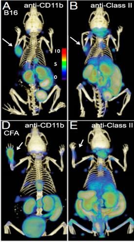
Antibodies 2019, 8, 12                                                                              2 of 21

uptake in nontargeted tissues and organs should be detected in order to reach high contrast, and an
appropriately fast imaging time point and sufficiently extended imaging window should be attained.
These parameters are predominantly defined by the pharmacokinetics of the tracer, and thus by the
choice of targeting agent. Several classes of targeting agents can be considered, including antibodies,
antibody fragments, scaffolds, peptides, or small molecules [9]. Antibodies, given their natural capacity
to recognize specific epitopes as part of the body’s humoral immune system, have been extensively
considered for in vivo targeting. However, their large size results in slow systemic clearance and hampers
deep tissue penetration [10]. In consequence, molecular imaging performed using antibody tracers often
yields low-contrast images with low specificity, and only several days after tracer administration [11].
This review will focus on nanobodies as a platform technology, since nanobody-based tracers have
been shown to possess unique characteristics in terms of versatility, specificity, and the short time
needed to attain high contrast [12]. Following the description of what nanobodies are, we will give
an overview of the application of radiolabeled nanobodies in nuclear medicine. Subsequently, we will
discuss how the attractive properties of nanobodies can also be exploited for fluorescence-guided surgery
and photodynamic therapy. The different aspects that are important to their design and utilization as
molecular tracers in these fields will be addressed in more detail.

2. Nanobodies and Their Unique Properties
      Conventional antibodies are Y-shaped and are composed of two light and two heavy polypeptide
chains. In camelids, besides these, a substantial fraction of IgG-like antibodies devoid of light chains
also occur naturally [13]. Although only half of the complementarity-determining regions (CDRs)
needed for antigen recognition are present, these heavy-chain-only antibodies retain a similar affinity
and specificity to conventional IgGs. This interesting finding has led to the exploitation of the variable
domain of heavy-chain-only antibodies for many biotechnological and medical applications. This
monomeric domain of 12–15 kDa in size and with nanometer-range dimensions is often referred to as
a single-domain antibody (sdAb) or the variable domain of a heavy-chain antibody (VH H), and has
been given the commercial name NanobodyTM [14].
      Crystallographic studies of nanobodies revealed, as for conventional VH fragments, a classical
immunoglobulin fold with nine antiparallel β-strands forming two β-sheets, connected through a
conserved disulfide bridge (Figure 1A). In camel-derived nanobodies, more so than for llama-derived
nanobodies, an additional disulfide bridge is often present between the CDR1 and CDR3 or the CDR2
and CDR3 loops. The three CDR loops are located near the amino (N)-terminal end of the domain and
opposite to the carboxy (C)-terminal end. However, adaptations are essential to compensate for the
absence of a variable light chain (VL ). On the one hand, four hydrophobic residues which normally
interact with the VL domain are changed to more hydrophilic ones [15–17]. This results in a structure
with improved water solubility and which is less prone to aggregation [18]. On the other hand, the lack
of three extra loops for antigen recognition is compensated by elongated CDR1 and CDR3 loops that
are able to adopt alternative canonical structures. As a consequence of the often more convex shape of
the paratope, nanobodies tend to bind epitopes located within cryptic clefts [19].
      An important advantage of nanobodies is their high thermal and chemical stability. They typically
exhibit melting temperatures above 60 ◦ C, and antigen-binding activity is retained even after prolonged
incubation at such high temperatures [20,21]. These properties open opportunities for novel chemical
modifications and labeling methods.
      Despite these differences, nanobodies are considered to be very weakly immunogenic due to
their high degree of homology with human variable heavy-chain (VH ) fragments [22]. This has since
been confirmed by several clinical trials where no immunogenicity or adverse effects were detected
following administration [23,24]. While preexisting anti-nanobody antibodies were found in one
clinical study using a tetravalent agonistic nanobody targeting the Death Receptor 5 as an antitumor
agent [25], no more preexisting anti-nanobody antibodies appear to be present than preexisting
autoantibodies against conventional VH fragments [26].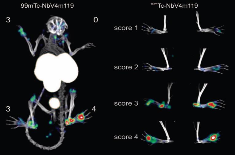
Antibodies 2019, 8, 12                                                                                3 of 21

      Nanobodies can be readily generated against many targets through the immunization of camelids
with either the antigen of interest, DNA coding for it, or cells expressing the antigen on their surface.
Subsequent amplification of the nanobody gene sequences from peripheral blood mononuclear cells
yields an immune cDNA library from which specific nanobodies can be selected through various
affinity (e.g., phage display) and/or functional screens [27]. Alternatively, synthetic libraries can be
used. These generally utilize a fixed framework, where residues in the CDR regions are randomized to
obtain specificity for different targets [28,29].
      Nanobodies with single-digit nanomolar affinities should preferentially be selected for further
in vivo applications, especially when the target is only expressed at low levels. The affinity of
nanobodies across species furthermore facilitates their preclinical characterization, as the uptake
in organs and tissues constitutively expressing the target can be better assessed.
      The selected nanobody clones can then straightforwardly be expressed in bacteria (e.g., E. coli) or
yeast strains (e.g., S. cerevisiae and P. pastoris) at yields of several milligrams of soluble nanobodies per
liter of culture. Their relatively simple and single gene form allows the engineering of nanobodies into
all kinds of formats, including the generation of multimeric and multispecific compounds, creation of
fusion proteins, and addition of small peptide sequences (tags) for later functionalization [30].
      In the last 15 years, nanobodies have been proposed as a new class of antibody-derived agents
for molecular imaging because of their unique features regarding affinity, specificity, and rapid
pharmacokinetics, ensuring good uptake in the targeted tissues and high target-to-background ratios [31].

3. Radiolabeled Nanobodies for Same-Day, High-Contrast Nuclear Imaging and Targeted
Radionuclide Therapy with Minimal Toxicity

3.1. Radiolabeling of Nanobodies
      Nuclear molecular imaging requires the targeting moiety, in this case a nanobody, to be labeled with
a diagnostic radioisotope. The latter can either be a gamma-emitting isotope for single photon emission
computed tomography (SPECT) or a positron-emitting isotope for positron emission tomography (PET).
Clinically, the higher resolution, sensitivity, and quantitative potential of PET/Computed Tomography
(CT) imaging is driving its adoption and is expected to result in a shift towards the increasing use of
this technology over SPECT/CT [32]. On the contrary, microSPECT devices specifically developed for
small-animal imaging typically achieve higher spatial resolution than microPET systems. It is thus an
attractive imaging technology for the initial in vivo screening and characterization of a set of nanobodies,
especially since nanobodies can be easily labeled with 99m Tc. Conveniently, 99m Tc–tricarbonyl reacts
site-specifically with a genetically inserted C-terminal hexahistidine tag, which can also be used for
purification purposes via immobilized metal affinity chromatography [33].
      Radiolabeling of proteins with other radiometals (e.g., 64 Cu, 68 Ga, and 89 Zr for PET or 67 Ga
and 111 In for SPECT) or radiohalogens (e.g., 18 F and 124 I for PET and 123/131 I for SPECT) usually
necessitates the use of a chelator for complexation or a prosthetic group for electrophilic substitution,
respectively. For human applications, the PET isotopes 68 Ga and 18 F are particularly suited to imaging
with nanobodies due to their short half-lives (68 and 110 min, respectively), which match up well
with the nanobodies’ biological half-life. For the attachment of chelators and prosthetic groups to
the nanobody, different conjugation strategies are possible. These can be broadly divided into two
categories: random or site-specific labeling. Random labeling typically occurs through conjugation to
primary amines (lysines) in the framework. Although this is a common and straightforward method, it
however results in a heterogenous mixture with varying amounts of labels per nanobody at different
positions. Contrarily, site-specific strategies aim to obtain homogenous and consistent tracers through
the conjugation of a single contrast agent to predetermined, specific sites. Positioning of the contrast
label opposite to the antigen-binding site furthermore avoids interference with the binding capacity of
nanobodies [34]. Different types of site-specific labeling methods with nanobodies have been explored
for in vivo applications. For example, the incorporation of a C-terminal cysteine tag enables reaction with
maleimide-functionalized agents after the prior reduction of dimeric nanobodies or nanobodies with a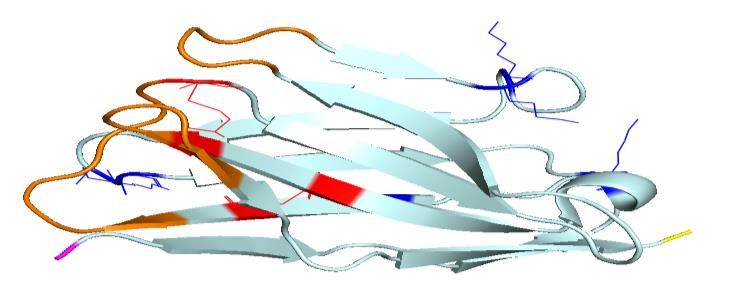
Antibodies 2019, 8, 12                                                                               4 of 21

blocked cysteine. Importantly, the reduction reaction must be carefully titrated to prevent disruption of
the nanobodies’ internal disulfide bridges [35]. Although reversal of the thioether bond is known to occur
in vivo [36,37], this is not expected to happen fast enough to pose a problem to nanobody probes due to
their fast pharmacokinetics [34]. A more elegant method for site-specific conjugation is enzyme-mediated
ligation through the transpeptidase Sortase A. Here, the enzyme catalyzes the formation of a new
peptide bond between the peptide motif LPXTG expressed C-terminally on the nanobody and the label
containing a N-terminal oligo-glycine motif [38,39]. Other methods under investigation for the design of
site-specifically labeled nanobodies for molecular imaging are alkyne–azide click reactions and those
involving the incorporation of unnatural amino acids into the nanobody structure [34,40].

3.2. In Vivo Biodistribution of Radiolabeled Nanobodies
      Upon intravenous administration, radiolabeled nanobodies are rapidly cleared from the blood
circulation. In mice, normally less than 0.5% Injected Activity (IA)/g remains present in the blood
pool at 1 h post-injection [41–44]. In humans, the early- and late-phase half-life of the anti-human
epidermal growth factor receptor 2 (HER2) 68 Ga-labeled nanobody 2Rs15d was calculated as 2.9
and 25.5 min, respectively, and at 1 h post-injection of the tracer, only 10% of the injected activity
remained in the blood pool [23]. The size of nanobodies being below 60 kDa causes them to be
filtered through the glomeruli in the kidneys. However, nanobodies are subsequently reabsorbed
by the proximal tubuli, resulting in their retention in the renal cortex. It has been previously shown
that the endocytic receptor megalin, which is abundantly expressed in the brush border, is at least
partially involved in the renal retention of nanobodies (megalin-deficient mice show 40% less renal
retention of a 99m Tc-labeled nanobody than wild-type mice) [45]. The long-term renal retention of
(radiolabeled) nanobodies and/or their (radio)catabolites can be an issue as it can possibly lead to
undesired nephrotoxicity. Furthermore, the imaging of molecular targets in the vicinity of the kidneys
is hampered due to intense renal signals. Therefore, several possible strategies to reduce the renal
reabsorption of nanobodies have been investigated. Coadministration of positively charged amino
acids or gelofusin, which competitively interact with megalin/cubulin receptors, have long been known
to reduce the renal retention of radiometal-labeled antibody fragments and peptides [46,47], and this
has also been confirmed for nanobodies [45,48]. Alternatively, the removal of charged amino-acid tags,
for example, used for purification or radiolabeling purposes, has an effect on the polarity of nanobodies
and consequently has an important impact on the degree of kidney retention. Indeed, Myc–His-
and His-tagged nanobodies show considerably higher kidney values compared to their untagged
analogues [48,49]. For clinical applications, the His tag is recommended to be removed anyway to
prevent immunogenic reactions [50,51]. Finally, the degree of kidney retention for radiohalogenated
(fluorinated and iodinated) nanobodies is significantly lower than for radiometal-labeled analogues.
Catabolites of radiohalogenated compounds formed in the kidneys are thought to be nonresidualizing
and hydrophobic and rapidly excreted via the urine [52–54].
      Other than the accumulation in kidneys and urine, the uptake of radiolabeled nanobodies in
nontargeted organs and tissues is very low (Figure 1). In combination with efficient penetration into
and diffusion through tissues and fast targeting, this consequently results in high target-to-background
ratios early after administration, allowing same-day imaging [55]. This is in stark contrast to full-length
antibodies, where due to their long circulation time, optimal tumor-to-background contrast is only
obtained several days after administration of the tracer and nonspecific uptake is generally much
higher [11]. Other non-immunoglobulin low-molecular-weight protein scaffolds (e.g., Affibodies,
DARPins, Adnectins, ADAPTs, or knottins) share many of the in vivo characteristics of nanobodies.
A recent review discusses in detail the clinical application and promising preclinical developments of
nanobodies and other small proteins for radionuclide-based imaging within the field of oncology [55].
The main advantage of nanobodies over these scaffolds remains the relatively simple process to generate
new nanobodies with high affinity through the immunization of camelids. On the other hand, further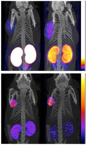
Antibodies 2019, 8, 12                                                                                   5 of 21

size-reduced compounds could eventually be produced synthetically instead of via fermentation, once a
lead compound has been identified [56].

3.3. Nuclear Medicine Applications with Nanobodies
      A major application field for nanobody-based radiotracers is cancer imaging. Radiolabeled
nanobodies targeting tumor biomarkers such as the epidermal growth factor receptor (EGFR),
carcinoembryonic antigen (CEA), mesothelin, prostate-specific membrane antigen (PSMA), CD20,
or HER2 showed high specific tumor uptake (ranging typically between 2 and 10% IA/g, depending
on the expression level of the target) in preclinical tumor models as soon as 1 h after administration,
with tumor-to-blood ratios of up to 10–30 (Figure 1B,C) [23,35,38,41,42,45,48,49,53,54,57–71]. Of particular
interest is the anti-HER2 nanobody 2Rs15d that has been selected as a lead compound for clinical
translation. In the very first clinical trial with a radiolabeled nanobody, Keyaerts et al. demonstrated
that 68 Ga–NOTA–2Rs15d PET/CT enabled the visualization of both primary lesions and/or local or
distant metastases in HER2-positive breast cancer patients, without adverse effects (Figure 1H). Good
tumor uptake was observed, with mean standard uptake values of up to 11.8 for primary tumors and
6.0 in metastases between 60 and 90 min post-injection. With the exception of the kidneys, intestines,
and liver, background uptake was low (weak uptake in glandular tissues such as the salivary glands,
pituitary, lacrimal glands, and axillary sweat glands was thought to be related to low levels of HER2
expression or chelator-mediated trapping mechanisms). Ultimately, 90 min post-injection was chosen
as the optimal imaging time point, due to decreased liver uptake compared to 60 min post-injection.
In this study, no preexisting or tracer-induced antibodies against the nanobody 2Rs15d could be detected.
These findings imply the potential application of 68 Ga–2Rs15d for the noninvasive assessment of the
HER2 status of patients [23]. A phase II study with this tracer has since been initiated, evaluating its
potential to detect brain metastasis in breast cancer patients (NCT03331601).

                             Antigen-binding domain       Framework

                         A

                                                                      C-terminus
                             N-terminus

       B                        C                     D                            E   H

       F                                              G

      Figure 1. Schematic representation of the structure of a nanobody and illustrative positron emission
      tomography (PET) and single photon emission computed tomography (SPECT) preclinical and clinical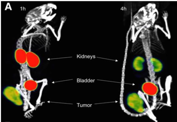
Antibodies 2019, 8, 12                                                                                         6 of 21

      images obtained using nanobodies that are labeled with distinct radionuclides in diverse medical
      applications, from oncology, immunology, atherosclerosis, and arthritis to the theranostic imaging of a
      radiotherapeutic probe. (A) Ribbon diagram of the nanobody 2Rs15d. The complementarity-determining
      regions (CDRs) are shown in orange, lysines (used for random conjugation methods) in blue, and
      cysteines and cysteine bridges in red. The C-terminus (in yellow) can be easily genetically modified for
      site-specific conjugation methods. (B) SPECT/Computed Tomography (CT) images of the biodistribution
      of 111 In-labeled JV7 nanobodies at 3 h post-injection in PSMA+ tumor-bearing mice (on the left shoulder).
      Effect on renal retention by the removal of tags (top panels: Myc–Cys-tagged nanobody, bottom:
      Cys-tagged nanobody) and coinjection of positively charged amino acids and gelofusin (left panels:
      no injection, right panels: with coinjection) is shown. Adapted from [48]. (C) SPECT/CT imaging of an
      EGFR+ tumor-bearing mouse 1 h after injection of 99m Tc-labeled 7C12 nanobody. Adapted from [64]. (D)
      PET/CT immune cell imaging 90 min after injection of 18 F-labeled nanobodies against murine CD11b
      and major histocompatibility complex (MHC) class II. Top: C57Bl/6 mice inoculated with B16 tumor cells
      on the left shoulder; bottom: animals injected with complete Freund’s adjuvant on the left paw. Adapted
      from [72]. (E) SPECT/CT coronal image taken at 2–3 h post-injection of 99m Tc-labeled cAbVCAM1-5
      nanobody, showing uptake in atherosclerotic lesions (ao) of ApoE-/- mice (bottom) and absence of
      signals in the aortic arch of C57Bl/6J mice (top) [43]. (F) SPECT/CT images of the biodistribution of
      the 131 I-labeled 2Rs15d therapeutic nanobody in a mouse model with subcutaneous HER2+ xenograft
      at 1 and 4 h post-injection. Adapted from [54]. (G) SPECT/CT imaging of arthritis in a mouse model
      with a VSIG4/CRIg-specific 99m Tc-labeled nanobody. Adapted from [73]. (H) PET/CT image of the
      biodistribution of 68 Ga-labeled anti-HER2 nanobody in a breast cancer patient 90 min post-injection
      showing uptake in breast tumor lesions. Adapted from [23].

     Next to imaging the tumor cells themselves for the prognosis and prediction of therapy response,
a different application is the characterization and quantification of specific immune cells within the
tumor environment. This approach could eventually also aid in better understanding and evaluating
drug action during drug development. In mice, radiolabeled nanobodies have been proven to be
able to track the infiltration of CD11b (macrophages, dendritic cells, and neutrophils) and major
histocompatibility complex (MHC) class II (macrophages and dendritic cells) positive cells in both
xenogeneic and syngeneic tumors, as well as after injection of complete Freund’s adjuvant [72]
(Figure 1D). Similarly, macrophage mannose receptor (MMR)-specific nanobodies were used to image
tumor-associated macrophages in mice [44,74]. The human-MMR-specific nanobody MMR3.49 labelled
with 68 Ga is currently being translated to the clinic, with a phase I safety trial to be initiated soon [75].
Of growing interest is the prediction of immune-checkpoint blockade therapy outcome. Nuclear
imaging with radiolabeled nanobodies against the T-cell marker CD8 or programmed death-ligand 1
(PD-L1) shows promising results as a tool to image antitumor immune responses [76–79].
     Macrophage-specific nanobodies have also been found to be of interest for imaging in other
inflammatory diseases, examples of which are hepatic inflammation and collagen-induced arthritis
(CIA). Indeed, radiolabeled nanobodies targeting MMR or the complement receptor of the Ig
superfamily (CRIg or VSIG4) were found to accumulate specifically in inflamed joints of mouse
CIA models (Figure 1G). Furthermore, CRIg/VSIG4 imaging appeared to be sufficiently sensitive to
detect early signs of inflammation, even before the manifestation of clinical signs [73,80–82].
     Applications that heavily rely on an elevated uptake with low nonspecific surrounding background
signals are the imaging of pancreatic islets after transplantation [83] and the imaging of vulnerable
atherosclerotic plaques. The feasibility to noninvasively detect small, inflamed atherosclerotic lesions in
the aortic arch of mice or along the aortas of atherosclerotic rabbits has been shown with radiolabeled
nanobodies targeting MMR, vascular cell adhesion molecule-1 (VCAM-1) (Figure 1E), and lectin-like
oxidized low-density lipoprotein receptor (LOX-1) [43,52,84–89]. However, their prognostic value for
the identification of high-risk patients remains to be demonstrated in clinical trials. Within this context,
the anti-VCAM-1 nanobody cAbVCAM1-5 is currently in the process of being translated into the clinic.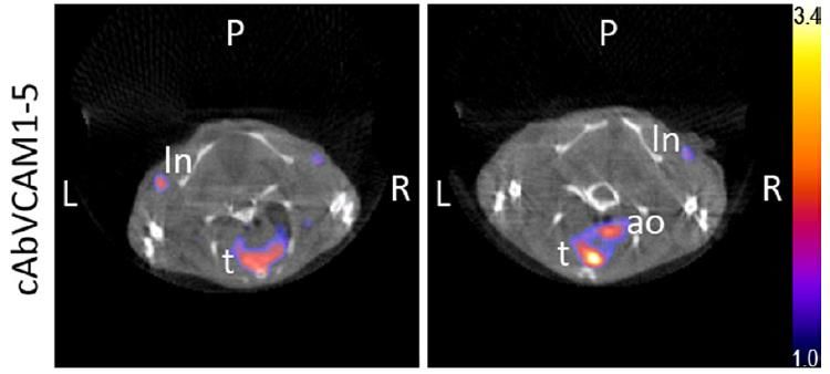
Antibodies 2019, 8, 12                                                                               7 of 21

       In analogy with molecular imaging, the incorporation of high-energy β- (177 Lu, 131 I) or α-emitters
(211 At, 213 Bi,and 225 Ac) allows the use of nanobodies for targeted radionuclide therapy (TRNT).
The nanobody is used as a vehicle to specifically bring damaging ionizing radiation to the tumor cells.
Radiation can inflict damage to tumor cells either by direct DNA damage or by the generation of
reactive oxygen species (ROS). Furthermore, the destruction of cancer cells can release antigens and
immune-triggers into the environment, activating an anticancer immune response [90,91]. β-emitters,
which have a lower linear energy transfer (LET), deposit their energy over a longer path length than
α-emitters, and can thus be advantageous in heterogenous tumors where not all malignant cells
express the molecular target. On the other hand, α-emitters deliver higher therapeutic absorbed doses,
but might require internalization as these operate at very short distances. α-TRNT is mostly suggested
for the treatment of micro-metastasis and minimal residual disease [92,93].
       In a theranostic approach, therapy is combined with molecular imaging, the latter being used
to predict susceptibility to TRNT and as a means to follow up treatment. This can be accomplished
either by using the same nanobody for both the preparation of the diagnostic and therapeutic
radio-analogue (e.g., pairing of 68 Ga/177 Lu or 123/124 I/131 I-labeled agents) or by radiolabeling the
nanobody with a radioisotope with both diagnostic and therapeutic properties (e.g., 177 Lu and
131 I) [92,93]. This strategy has successfully been applied preclinically in breast and ovarian cancer,

non-Hodgkin lymphoma, and multiple myeloma using anti-HER2 (Figure 1F), anti-CD20, and
anti-idiotypic (multiple myeloma) nanobodies, respectively [49,54,68]. Efficient tumor therapy in
a preclinical setting could be demonstrated through either significant improvement in overall survival
compared to all controls in mice inoculated subcutaneously or intraperitoneally with HER2- or
CD20-positive tumors cells, or the inhibition of disease progress in the 5T2 multiple myeloma mouse
model. Furthermore, the fast pharmacokinetics and low nonspecific uptake of nanobodies lead
to minimal toxicity. Contrarily, the prolonged blood residence time of monoclonal antibodies has
important implications for toxicity to bone marrow and other highly perfused organs such as the
spleen and liver. So far, renal toxicity due to kidney retention has not been observed in mice, even
after repeated administration. Furthermore, by using 131 I-labeled nanobodies and having nonresidual
catabolites in the kidneys, the absorbed dose to the kidneys could even be reduced below the dose
delivered to the tumor [49,54,68]. A phase I trial using a low-dose 131 I-labeled anti-HER2 nanobody
2Rs15d in breast cancer patients has recently been completed (NCT02683083) [94].
       While monovalent nanobodies are most often used for nuclear imaging and TRNT, the use
of multimeric nanobodies has also been investigated. Dimeric monospecific nanobodies generally
show similar pharmacokinetics to monovalent tracers with regard to fast blood elimination and renal
clearance. Tumor uptake of bivalent nanobodies at early time points has been found to be slightly lower
than that of monovalent ones, although the bivalent compound shows considerably longer tumor
retention [69]. However, due to the increased avidity and size of bivalent nanobodies, they likely exhibit
inferior tumor penetration properties and are more limited to the perivascular region [95,96]. Bivalent
nanobodies can consequently be used as a means to cope with on-target off-tumor uptake. This was
demonstrated by Movahedi et al., who used a bivalent unlabeled anti-MMR nanobody to modify the
biodistribution of the radiolabeled monomeric nanobody. Impact on tumor uptake was minimal, while
specific uptake in nontumor organs and tissues was almost completely blocked [44]. When extension
of the blood half-life is desirable, multimeric constructs containing one or more tumor-specific domains
and an albumin-binding domain can be engineered. The resulting radiolabeled compounds show vastly
increased blood retention, and consequently, tumor values significantly increase over time, although
so does the nonspecific uptake [69,97–99]. Half-life-extended nanobodies are thus theoretically more
relevant in a therapeutic context, and less so for diagnostic applications. The negative impact on deep
tumor penetration should also be considered.
       An overview of nanobody-based radiopharmaceuticals with potential application in clinical
nuclear medicine is provided in Table 1.
Antibodies 2019, 8, 12 8 of 21
Table 1. Overview of preclinically and clinically tested nanobody-based radiopharmaceuticals with applications in nuclear medicine.
Application Field Molecular Target Lead Compound Radiolabel Disease Development Phase References
99m Tc, 111 In, 177 Lu, 18 F, 225 Ac Preclinical [42,57–60,100]
2Rs15d Phase II ongoing
68 Ga Breast cancer [23,41]
(NCT03331601)
HER2
131 I Phase I completed
[54,94]
(NCT02683083)
5F7 125 I, 131 I, 18 F Preclinical [60–63]
7C12,7D12 99m Tc, 177 Lu, 68 Ga, 89 Zr [45,64,65,97]
EGFR Skin cancer Preclinical
D10 99m Tc [66]
Non-small cell lung
Tumor cell HER3 MSB0010853 89 Zr cancer, head and neck Preclinical [99]
imaging/therapy cancer
PSMA30 99m Tc [67]
PSMA Prostate cancer Preclinical
JVZ-007 111 In [48]
CEA CEA5 99m Tc Colon cancer Preclinical [71]
Mesothelin A1 99m Tc Breast cancer Preclinical [70]
99m Tc, 111 In, 177 Lu, 68 Ga Non-Hodgkin
CD20 9077, 9079 Preclinical [68,69]
lymphoma
HGF 1E6-Alb8, 6E10-Alb8 89 Zr Glioma Preclinical [98]
Mouse monoclonal 99m Tc, 177 Lu
R3b23 Multiple myeloma Preclinical [101]
proteinAntibodies 2019, 8, 12 9 of 21
Table 1. Cont.
Application Field Molecular Target Lead Compound Radiolabel Disease Development Phase References
Mouse CD8 VHH-X118 89 Zr Tumor immunology Preclinical [76]
B3 18 F Immune checkpoint Preclinical [78]
Mouse PD-L1
C3,E2 99m Tc [77]
Mouse dendritic cells DC1.8, DC2.1 99m Tc Tumor immunology Preclinical [102]
Mouse Cd11b VHHDC13 18 F, 64 Cu Tumor immunology Preclinical [72]
Mouse MHC class II VHH7 18 F, 64 Cu Tumor immunology Preclinical [39,72]
Human MHC class II VHH4 64 Cu Graft vs. host disease Preclinical [103]
99m Tc Tumor immunology Preclinical [44]
Tumor immunology
and inflammatory Mouse MMR 99m Tc Arthritis Preclinical [81]
diseases MMRCl1
MMR3.49 99m Tc, 18 F, 68 Ga Tumor immunology Clinical translation [74,75]
Human MMR MMR3.49 99m Tc, 64 Cu, 68 Ga Atherosclerosis Preclinical [84,86]
99m Tc, 18 F Arthritis, liver
CRIg/VSIG4 VM119 Preclinical [73,82]
inflammation
Clec4F C4m22 99m Tc Liver inflammation Preclinical [82]
VCAM-1 cAbVCAM1-5 99m Tc, 111 In, 18 F, 64 Cu, 68 Ga Atherosclerosis Clinical translation [43,52,85–87,89]
LOX-1 Lox1.14 99m Tc, 64 Cu Atherosclerosis Preclinical [86,88]
Gelsolin FAF Nb1 99m Tc Gelsolin amyloidosis Preclinical [104,105]
Amyloidosis
B-amyloid Ni3A, pa2H 99m Tc Alzheimer’s Preclinical [106]
Diabetes DPP6 4hD29 99m Tc, 111 In Diabetes Preclinical [83]
HER2: human epidermal growth factor receptor 2, EGFR: epidermal growth factor receptor, HER3: human epidermal growth factor receptor 3, PSMA: prostate-specific membrane antigen,
CEA: carcinoembryonic antigen, HGF: hepatocyte growth factor, PD-L1: programmed death-ligand 1, MMR: macrophage mannose receptor, CRIg/VSIG4: complement receptor of the
immunoglobulin family/V-set and immunoglobulin domain containing 4, Clec4F: C-type lectin domain family 4 member F, VCAM-1: vascular cell adhesion molecule 1, LOX-1: lectin-like
oxidized low-density lipoprotein receptor-1, DPP6: dipeptidyl peptidase like 6.Antibodies 2019, 8, 12 10 of 21
4. Image-Guided Surgery and Photodynamic Therapy Using Fluorescent Nanobodies
4.1. Design of Fluorescent Nanobody-Based Tracers
Alternatively to nuclear imaging, fluorescence imaging requires the presence of a fluorescent
label for sensitive detection. While a wide range of fluorescent dyes are available for biotechnological
purposes, for in vivo imaging applications, the choice of fluorophore is limited to those emitting in
the near-infrared (NIR) region, and more specifically, those with a maximal excitation and emission
wavelength between 650 and 900 nm. In this range, scattering, nonspecific tissue autofluorescence
and absorption by endogenous chromophores are minimal. These characteristics infer intrinsically
improved signal-to-background ratios and as such provide sharper contrast, better resolution, and
deeper signal detection (from several mm to 1 cm) [107]. Commonly, hydrophilic (sulfonated) variants of
cyanine dyes with a penta- or heptamethine chain are used, e.g., Cy5, Alexa Fluor (AF)680, IRDye680RD,
or IRDye800CW. The actual choice of the dye will depend on the specifications of the camera system
used for detection (the wavelength(s) the system is able to detect) and whether the tracer is intended to
be used in combination with a second tracer providing complementary information, e.g., highlighting of
the tumor tissue to be resected and the nerves to be preserved (multiplexing).
Analogous conjugation strategies to those described above for chelators and prosthetic groups are
applicable for fluorophore conjugation. However, it is increasingly being recognized that fluorophore
conjugation can have a significant impact on the pharmacokinetics of antibody tracers [108]. The impact
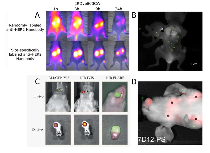
is most likely even more pronounced for smaller fragments such as nanobodies. In fact, randomly
IRDye800CW-labeled nanobodies have been demonstrated to have an atypical tissue distribution with
high background signals, high liver accumulation, and low tumor contrast [109–111]. Likewise, randomly
IRDye680RD- and AF680-labeled nanobodies do not exhibit such persistent background signals, but are
partially excreted via the hepatobiliary route [111–113]. The chosen conjugation chemistry appears to
be an additional determining factor, as the site-specific labeling of IRDye800CW and IRDye680RD via
a C-terminal cysteine tag yields nanobody tracers with normal biodistribution profiles, meaning fast
tumor targeting, renal excretion, and no nonspecific uptake (Figure 2A) [111]. Conjugation of more
than one dye per nanobody is furthermore undesirable since a higher dye/nanobody ratio may cause
quenching of the fluorescent signal due to the close proximity of the dyes.
Compared to nuclear imaging techniques, fluorescent imaging requires sufficient uptake of the
tracer in the tissue of interest for sensitive detection. Because of the depth-related attenuation of
fluorescent signals, high injected doses are often required in humans, at the limits of or above the
microdose level (less than 30 nmol or 100 µg [114]) [115–119]. Moreover, as for equal doses, the maximal
tumor signal that can be attained will be lower for nanobodies than for long-circulating antibodies,
a higher molar concentration will likely be needed as well [113]. In mice, typically, 1–5 nmol of
fluorescent nanobody based on the dye concentration (25–75 µg of protein) is injected. Adjusting the
injected dose does not appear to significantly affect the biodistribution of nanobodies (in comparison,
a higher dose of antibodies results in an increase of nonspecific signal), and dose optimization could
thus lead to superior image quality in terms of signal intensity and tumor-to-background ratios [113].
The use of radio- or bimodal (radioactivity in combination with fluorescence)-labeled tracers could
be an alternative approach to increase sensitivity for the intraoperative detection of deeper located
lesions, but this remains to be investigated for nanobodies [120].
4.2. Fluorescence-Guided Surgery Using Nanobody-Based Contrast Agents
Several fluorescently labeled nanobodies have been successfully evaluated in the context of
intraoperative imaging (an overview is provided in Table 2). IRDye800CW-labeled anti-EGFR
nanobodies could clearly delineate orthotopic tongue tumors in mice, and even enabled the
identification of a lymph node metastasis (Figure 2C) [109]. Of note, in this study the optimal
imaging time point appeared to be 24 h post-injection, likely due to the random characteristic of
the labeling method. The site-specifically IRDye800CW- and IRDye680RD-labeled nanobodies 11A4Antibodies 2019, 8, 12 11 of 21
and B9, respectively targeting HER2 and carbonic anhydrase IX (CAIX), showed accumulation in
breast cancer lesions (DCIS) and lung metastasis in an experimental setup mimicking the surgical
setting [121,122]. Furthermore, it was demonstrated that the combination of nanobodies targeting these
two independent tumor markers, but labeled with the same fluorescent dye, could further improve
tumor-to-background ratios and overcome tumor heterogeneity [122]. Finally, using a mouse model
of intraperitoneal disseminated tumor lesions mimicking late-stage ovarian cancer, the advantage of
fluorescence guidance with the anti-HER2 nanobody tracer 2Rs15d-IRDye800CW on the efficiency of
debulking surgery was demonstrated. Submillimeter lesions could be visualized with high contrast at
1.5 h post-injection, leading to the excision of significantly more tumor tissue as compared to traditional
surgery and resection of less false-positive tissue (Figure 2B) [123].
Table 2. Overview of in vivo preclinically evaluated fluorescent nanobodies with potential for clinical
interventional molecular imaging and photodynamic therapy.
Molecular Lead Conjugation Intended Clinical
Fluorophore References
Target Compound Strategy Application
IRDye800CW Random
- [111]
IRDye680RD (Lys–NHS)
IRDye800CW Site-specific Intraoperative imaging of
2Rs15d [111,123]
IRDye680RD (Cys–maleimide) breast/ovarian cancer
HER2
Site-specific Intraoperative imaging of
Cy5 [38]
(Sortase A) breast cancer
IRDye800CW Site-specific Intraoperative imaging of
11A4 [122,124]
IRDye680RD (Cys–maleimide) breast cancer
Site-specific Intraoperative imaging of
CAIX B9 IRDye800CW [121,122]
(Cys–maleimide) breast cancer
Random Intraoperative imaging of
7D12 IRDye800CW [109]
(Lys–NHS) head and neck cancer
EGFR
7D12, Random Photodynamic therapy of
IRDye700DX [125]
7D12-9G6 (Lys–NHS) head and neck cancer
NHS: N-Hydroxysuccinimide, HER2: human epidermal growth factor receptor 2, CAIX: carbonic anhydrase 9,
EGFR: epidermal growth factor receptor.
Antibodies 2019, 8, x FOR PEER REVIEW 11 of 20
Figure 2. Examples of in vivo fluorescent molecular imaging with nanobody tracers in mouse tumor
Figure 2. Examples of in vivo fluorescent molecular imaging with nanobody tracers in mouse tumor
models. (A) Comparison
models. of theofbiodistribution
(A) Comparison and
the biodistribution andtumor-targeting potential
tumor-targeting potential of the
of the anti-HER2 nanobody
anti-HER2
nanobody 2Rs15d conjugated with IRDye800CW either randomly (top) or site-specifically (bottom).
Adapted with permission from [111]. Copyright 2017 American Chemical Society. (B) Fluorescence
image acquired during the surgical resection of intraperitoneally disseminated HER2+ tumor lesions.
Site-specifically IRDye800CW-labeled 2Rs15d nanobody was injected 90 min before surgery.
Fluorescent signal in tumor lesions (indicated by green arrows) is clearly discernible from background
signal. Adapted with permission from [123]. (C) Real-time fluorescence imaging of orthotopic tongueAntibodies 2019, 8, 12 12 of 21
2Rs15d conjugated with IRDye800CW either randomly (top) or site-specifically (bottom). Adapted with
permission from [111]. Copyright 2017 American Chemical Society. (B) Fluorescence image acquired
during the surgical resection of intraperitoneally disseminated HER2+ tumor lesions. Site-specifically
IRDye800CW-labeled 2Rs15d nanobody was injected 90 min before surgery. Fluorescent signal in
tumor lesions (indicated by green arrows) is clearly discernible from background signal. Adapted
with permission from [123]. (C) Real-time fluorescence imaging of orthotopic tongue tumor 24 h
post-injection of an EGFR-specific randomly IRDye800CW-conjugated nanobody. Colocalization with
bioluminescence imaging (BLI) and green fluorescent protein (GFP) signals is shown. Adapted with
permission from [109]. (D) Fluorescent imaging of an orthotopically inoculated tongue tumor at 1 h
post-injection of an EGFR-specific randomly IRDye700DX-conjugated nanobody for photodynamic
therapy (PDT). Stars denote the presence of fluorescent tracer uptake in invaded lymph nodes. Adapted
with permission from [125].
Similarly to the theranostic approach in nuclear medicine, where TRNT is combined with
diagnostic imaging, the conjugation of a photosensitizer to a tumor-targeting nanobody enables
its use for image-guided resection followed by photodynamic therapy (PDT) of residual malignant
cells. In PDT, a photosensitizer is activated by incidence light to produce ROS. These ROS can
damage the tumor by directly causing cell death through apoptosis and necrosis, damaging the tumor
vasculature, and inducing an immune response [126]. This was investigated in a mouse orthotopic
tongue tumor model with EGFR-specific nanobodies conjugated randomly to the photosensitizer
IRDye700DX (Figure 2D). The effectiveness of a monovalent nanobody and bispecific variant, which
binds two different sites on EGFR, was compared with that of a conventional antibody. Both nanobody
photosensitizers outperformed the antibody after therapeutic illumination, with more homogenous
damage to the tumor and less nontarget damage. Furthermore, despite the higher internalization seen
in vitro for the bispecific variant, better in vivo therapeutic results were obtained with the monovalent
nanobody. This is in accordance with the assumption that smaller, monovalent compounds diffuse
more homogenously through tumor tissue [125,127].
5. Conclusions and Perspectives
Nanobodies, with their unique properties, show great promise as targeting moieties in molecular
imaging and therapy. Their fast blood clearance, rapid and homogenous tissue penetration, and low
background retention allow highly specific imaging at early time points after administration and
effective therapy with minimal nonspecific toxicity. The utility of nanobody tracers is now broadly
recognized thanks to the convincing preclinical data obtained so far. However, clinical data on their
use in this field is still very limited. The expensive and time-consuming process required to translate
nanobodies into the clinic (current Good Manufacturing Practice (cGMP) production, toxicity studies,
and Investigational Medicinal Product Dossier (IMPD) filing) is probably the major limiting factor.
This, however, holds true for any molecular tracer.
The first clinically translated radiolabeled nanobody, the anti-HER2 nanobody 2Rs15d, labeled
either with 68 Ga or 131 I, has been investigated in two phase I trials as a potential tool to provide
predictive and responsive information on targeted tumor therapies. Follow-up studies respectively
evaluating the diagnosis and treatment of breast cancer brain metastasis are now ongoing or planned.
In the next years, clinical data of two additional radiolabeled nanobodies is expected, as their clinical
translation is almost completed. A nanobody targeting the inflammatory marker VCAM-1 will
be evaluated for vulnerable plaque screening, and a macrophage-specific nanobody will also be
investigated, which opens up opportunities to image immune cell activation and dynamics in oncology
and inflammatory diseases. The latter approach is expected to be further exploited in the future to aid
in the development, selection, and monitoring of (novel) immunotherapies.Antibodies 2019, 8, 12 13 of 21
In the context of intraoperative imaging, properly designed fluorescent nanobody tracers seem to
be promising tools to assist and guide surgeons during complex interventions. Evaluated only in a
preclinical setting so far, their feasibility and surgical benefit in humans remains to be demonstrated.
The strategy to move most rapidly towards the clinic would be to fluorescently label clinical-grade
nanobodies which are already available (e.g., anti-HER2 nanobody), in analogy with antibodies
currently under investigation as fluorescent contrast agents. However, preferentially, novel nanobodies
targeting more relevant biomarkers for the application of image-guided surgery are expected to be
developed and clinically translated.
Regarding the design of nanobody tracers, further advances towards novel chemistries permitting
conjugation of contrast labels in a more controlled manner are warranted, as ultimately, any
labeling method that is considered for clinical translation must be evaluated in a regulatory context.
Furthermore, developments of chelators, prosthetic groups, fluorescent dyes, and bimodal labels with
improved effects on the pharmacokinetics of nanobodies would be of interest, especially related to
kidney retention. This aspect remains a critical point for potential toxicity issues, particularly for
therapeutic applications.
Funding: Our studies with nanobody-based probes were funded by Kom op tegen kanker, Stichting tegen Kanker,
Fund for Scientific Research Flanders (FWO), Industrial Research Fund (IOF), Strategic Research Program (SRP),
Scientific Fund Willy Gepts, Melanoma Research Alliance, Juvenile Diabetes Research Foundation, Innoviris,
the Novo Nordisk Foundation, and Horizon2020.
Conflicts of Interest: N.D. is a consultant of Camel-IDS and holds ownership interest (including patents) in
camelid single-domain diagnostics and therapeutics. S.H. holds a patent in camelid single-domain diagnostics
and therapeutics.
References
1. Mankoff, D.A. A definition of molecular imaging. J. Nucl. Med. 2007, 48, 18N–21N. [PubMed]
2. Weber, J.; Haberkorn, U.; Mier, W. Cancer stratification by molecular imaging. Int. J. Mol. Sci. 2015,
16, 4918–4946. [CrossRef] [PubMed]
3. Vahrmeijer, A.L.; Hutteman, M.; van der Vorst, J.R.; van de Velde, C.J.H.; Frangioni, J.V. Image-guided cancer
surgery using near-infrared fluorescence. Nat. Rev. Clin. Oncol. 2013, 10, 507–518. [CrossRef] [PubMed]
4. Hingorani, D.V.; Whitney, M.A.; Friedman, B.; Kwon, J.-K.; Crisp, J.L.; Xiong, Q.; Gross, L.; Kane, C.J.;
Tsien, R.Y.; Nguyen, Q.T. Nerve-targeted probes for fluorescence-guided intraoperative imaging. Theranostics
2018, 8, 4226–4237. [CrossRef] [PubMed]
5. Cha, J.; Nani, R.R.; Luciano, M.P.; Broch, A.; Kim, K.; Namgoong, J.M.; Kulkarni, R.A.; Meier, J.L.; Kim, P.;
Schnermann, M.J. A chemically stable fluorescent marker of the ureter. Bioorg. Med. Chem. Lett. 2018,
28, 2741–2745. [CrossRef] [PubMed]
6. Burggraaf, J.; Kamerling, I.M.C.; Gordon, P.B.; Schrier, L.; De Kam, M.L.; Kales, A.J.; Bendiksen, R.;
Indrevoll, B.; Bjerke, R.M.; Moestue, S.A.; et al. Detection of colorectal polyps in humans using an
intravenously administered fluorescent peptide targeted against c-Met. Nat. Med. 2015, 21, 955–961.
[CrossRef] [PubMed]
7. Nagengast, W.B.; Hartmans, E.; Garcia-Allende, P.B.; Peters, F.T.M.; Linssen, M.D.; Koch, M.; Koller, M.;
Tjalma, J.J.J.; Karrenbeld, A.; Jorritsma-Smit, A.; et al. Near-infrared fluorescence molecular endoscopy
detects dysplastic oesophageal lesions using topical and systemic tracer of vascular endothelial growth
factor A. Gut 2017, 68, 1–4. [CrossRef]
8. Jaffer, F.A.; Calfon, M.A.; Rosenthal, A.; Mallas, G.; Razansky, N.; Mauskapf, A.; Weissleder, R.;
Libby, P.; Ntziachristos, V. Two-Dimensional Intravascular Near-Infrared Fluorescence Molecular Imaging of
Inflammation in Atherosclerosis and Stent-Induced Vascular Injury. J. Am. Coll. Cardiol. 2011, 57, 2516–2526.
[CrossRef]
9. Chen, K.; Chen, X. Design and Development of Molecular Imaging Probes. Curr. Top. Med. Chem. 2010,
10, 1227–1236. [CrossRef]
10. Thurber, G.M.; Schmidt, M.M.; Wittrup, K.D. Factors determining antibody distribution in tumors.
Trends Pharmacol. Sci. 2008, 29, 57–61. [CrossRef]Antibodies 2019, 8, 12 14 of 21
11. Wu, A.M. Engineered antibodies for molecular imaging of cancer. Methods 2014, 65, 139–147. [CrossRef]
[PubMed]
12. Chakravarty, R.; Goel, S.; Cai, W. Nanobody: The “Magic Bullet” for Molecular Imaging? Theranostics 2014,
4, 386–398. [CrossRef] [PubMed]
13. Hamers-Casterman, C.; Atarhouch, T.; Muyldermans, S.; Robinson, G.; Hamers, C.; Songa, E.B.;
Bendahman, N.; Hamers, R. Naturally occurring antibodies devoid of light chains. Nature 1993, 363, 446–448.
[CrossRef] [PubMed]
14. Muyldermans, S.; Baral, T.N.; Retamozzo, V.C.; De Baetselier, P.; De Genst, E.; Kinne, J.; Leonhardt, H.;
Magez, S.; Nguyen, V.K.; Revets, H.; et al. Camelid immunoglobulins and nanobody technology.
Vet. Immunol. Immunopathol. 2009, 128, 178–183. [CrossRef] [PubMed]
15. Vu, K.B.; Ghahroudi, M.A.; Wyns, L.; Muyldermans, S. Comparison of llama V(H) sequences from
conventional and heavy chain antibodies. Mol. Immunol. 1997, 34, 1121–1131. [CrossRef]
16. Muyldermans, S.; Atarhouch, T.; Saldanha, J.; Barbosa, J.A; Hamers, R. Sequence and structure of VH
domain from naturally occurring camel heavy chain immunoglobulins lacking light chains. Protein Eng.
1994, 7, 1129–1135. [CrossRef]
17. Harmsen, M.M.; Ruuls, R.C.; Nijman, I.J.; Niewold, T.A.; Frenken, L.G.J.; De Geus, B. Llama heavy-chain V
regions consist of at least four distinct subfamilies revealing novel sequence features. Mol. Immunol. 2000,
37, 579–590. [CrossRef]
18. Muyldermans, S.; Cambillau, C.; Wyns, L. Recognition of antigens by single-domain antibody fragments:
The superfluous luxury of paired domains. Trends Biochem. Sci. 2001, 26, 230–235. [CrossRef]
19. De Genst, E.; Silence, K.; Decanniere, K.; Conrath, K.; Loris, R.; Kinne, J.; Muyldermans, S.; Wyns, L.
Molecular basis for the preferential cleft recognition by dromedary heavy-chain antibodies. Proc. Natl. Acad.
Sci. USA 2006, 103, 4586–4591. [CrossRef]
20. Van Der Linden, R.H.J.; Frenken, L.G.J.; De Geus, B.; Harmsen, M.M.; Ruuls, R.C.; Stok, W.; De Ron, L.;
Wilson, S.; Davis, P.; Verrips, C.T. Comparison of physical chemical properties of llama V(HH) antibody
fragments and mouse monoclonal antibodies. Biochim. Biophys. Acta Protein Struct. Mol. Enzymol. 1999,
1431, 37–46. [CrossRef]
21. Dumoulin, M.; Conrath, K.; Van Meirhaeghe, A.; Meersman, F.; Heremans, K.; Frenken, L.G.J.;
Muyldermans, S.; Wyns, L.; Matagne, A. Single-domain antibody fragments with high conformational
stability. Protein Sci. 2002, 11, 500–515. [CrossRef] [PubMed]
22. Muyldermans, S. Nanobodies: Natural Single-Domain Antibodies. Annu. Rev. Biochem. 2013, 82, 775–797.
[CrossRef] [PubMed]
23. Keyaerts, M.; Xavier, C.; Heemskerk, J.; Devoogdt, N.; Everaert, H.; Ackaert, C.; Vanhoeij, M.; Duhoux, F.P.;
Gevaert, T.; Simon, P.; et al. Phase I Study of 68Ga-HER2-Nanobody for PET/CT Assessment of HER2
Expression in Breast Carcinoma. J. Nucl. Med. 2016, 57, 27–33. [CrossRef] [PubMed]
24. Bartunek, J.; Barbato, E.; Heyndrickx, G.; Vanderheyden, M.; Wijns, W.; Holz, J.B. Novel antiplatelet agents:
ALX-0081, a nanobody directed towards von Willebrand factor. J. Cardiovasc. Transl. Res. 2013, 6, 355–363.
[CrossRef]
25. Papadopoulos, K.P.; Isaacs, R.; Bilic, S.; Kentsch, K.; Huet, H.A.; Hofmann, M.; Rasco, D.; Kundamal, N.;
Tang, Z.; Cooksey, J.; et al. Unexpected hepatotoxicity in a phase I study of TAS266, a novel tetravalent
agonistic Nanobody® targeting the DR5 receptor. Cancer Chemother. Pharmacol. 2015, 75, 887–895. [CrossRef]
26. Holland, M.C.; Wurthner, J.U.; Morley, P.J.; Birchler, M.A.; Lambert, J.; Albayaty, M.; Serone, A.P.; Wilson, R.;
Chen, Y.; Forrest, R.M.; et al. Autoantibodies to variable heavy (VH) chain Ig sequences in humans impact the
safety and clinical pharmacology of a VHdomain antibody antagonist of TNF-α receptor 1. J. Clin. Immunol.
2013, 33, 1192–1203. [CrossRef]
27. Arbabi Ghahroudi, M.; Desmyter, A.; Wyns, L.; Hamers, R.; Muyldermans, S. Selection and identification
of single domain antibody fragments from camel heavy-chain antibodies. FEBS Lett. 1997, 414, 521–526.
[CrossRef]
28. Moutel, S.; Bery, N.; Bernard, V.; Keller, L.; Lemesre, E.; De Marco, A.; Ligat, L.; Rain, J.C.; Favre, G.;
Olichon, A.; et al. NaLi-H1: A universal synthetic library of humanized nanobodies providing highly
functional antibodies and intrabodies. eLife 2016, 5, e16228. [CrossRef]Antibodies 2019, 8, 12 15 of 21
29. McMahon, C.; Baier, A.S.; Pascolutti, R.; Wegrecki, M.; Zheng, S.; Ong, J.X.; Erlandson, S.C.; Hilger, D.;
Rasmussen, S.G.F.; Ring, A.M.; et al. Yeast surface display platform for rapid discovery of conformationally
selective nanobodies. Nat. Struct. Mol. Biol. 2018, 25, 289–296. [CrossRef]
30. Bannas, P.; Hambach, J.; Koch-Nolte, F. Nanobodies and nanobody-based human heavy chain antibodies as
antitumor therapeutics. Front. Immunol. 2017, 8, 1603. [CrossRef]
31. De Vos, J.; Devoogdt, N.; Lahoutte, T.; Muyldermans, S. Camelid single-domain antibody-fragment
engineering for (pre)clinical in vivo molecular imaging applications: Adjusting the bullet to its target.
Expert Opin. Biol. Ther. 2013, 13, 1149–1160. [CrossRef] [PubMed]
32. Hicks, R.J.; Hofman, M.S. Is there still a role for SPECT–CT in oncology in the PET–CT era? Nat. Rev.
Clin. Oncol. 2012, 9, 712–720. [CrossRef] [PubMed]
33. Xavier, C.; Devoogdt, N.; Hernot, S.; Vaneycken, I.; Huyvetter, M.D.; De Vos, J.; Massa, S.; Lahoutte, T.;
Caveliers, V. Site-Specific Labeling of His-Tagged Nanobodies with 99mTc: A Practical Guide. In Single
Domain Antibodies: Methods and Protocols; Springer: Berlin, Germnay, 2012; Chapter 30, pp. 485–490,
ISBN 978-1-61779-967-9.
34. Massa, S.; Xavier, C.; Muyldermans, S.; Devoogdt, N. Emerging site-specific bioconjugation strategies for
radioimmunotracer development. Expert Opin. Drug Deliv. 2016, 13, 1149–1163. [CrossRef] [PubMed]
35. Massa, S.; Xavier, C.; De Vos, J.; Caveliers, V.; Lahoutte, T.; Muyldermans, S.; Devoogdt, N. Site-specific
labeling of cysteine-tagged camelid single-domain antibody-fragments for use in molecular imaging.
Bioconjug. Chem. 2014, 25, 979–988. [CrossRef] [PubMed]
36. Fontaine, S.D.; Reid, R.; Robinson, L.; Ashley, G.W.; Santi, D.V. Long-term stabilization of maleimide-thiol
conjugates. Bioconjug. Chem. 2015, 26, 145–152. [CrossRef] [PubMed]
37. Alley, S.C.; Benjamin, D.R.; Jeffrey, S.C.; Okeley, N.M.; Meyer, D.L.; Sanderson, R.J.; Senter, P.D. Contribution
of linker stability to the activities of anticancer immunoconjugates. Bioconjug. Chem. 2008, 19, 759–765.
[CrossRef] [PubMed]
38. Massa, S.; Vikani, N.; Betti, C.; Ballet, S.; Vanderhaegen, S.; Steyaert, J.; Descamps, B.; Vanhove, C.;
Bunschoten, A.; van Leeuwen, F.W.B.; et al. Sortase A-mediated site-specific labeling of camelid
single-domain antibody-fragments: A versatile strategy for multiple molecular imaging modalities.
Contrast Media Mol. Imaging 2016, 11, 328–339. [CrossRef]
39. Rashidian, M.; Wang, L.; Edens, J.G.; Jacobsen, J.T.; Hossain, I.; Wang, Q.; Victora, G.D.; Vasdev, N.;
Ploegh, H.; Liang, S.H. Enzyme-Mediated Modification of Single-Domain Antibodies for Imaging Modalities
with Different Characteristics. Angew. Chem. Int. Ed. 2016, 55, 528–533. [CrossRef]
40. Billen, B.; Vincke, C.; Hansen, R.; Devoogdt, N.; Muyldermans, S.; Adriaensens, P.; Guedens, W. Cytoplasmic
versus periplasmic expression of site-specifically and bioorthogonally functionalized nanobodies using
expressed protein ligation. Protein Expr. Purif. 2017, 133, 25–34. [CrossRef]
41. Xavier, C.; Vaneycken, I.; D’huyvetter, M.; Heemskerk, J.; Keyaerts, M.; Vincke, C.; Devoogdt, N.;
Muyldermans, S.; Lahoutte, T.; Caveliers, V. Synthesis, Preclinical Validation, Dosimetry, and Toxicity of
68Ga-NOTA-Anti-HER2 Nanobodies for iPET Imaging of HER2 Receptor Expression in Cancer. J. Nucl. Med.
2013, 54, 776–784. [CrossRef]
42. Vaneycken, I.; Devoogdt, N.; Van Gassen, N.; Vincke, C.; Xavier, C.; Wernery, U.; Muyldermans, S.;
Lahoutte, T.; Caveliers, V. Preclinical screening of anti-HER2 nanobodies for molecular imaging of breast
cancer. FASEB J. 2011, 25, 2433–2446. [CrossRef] [PubMed]
43. Broisat, A.; Hernot, S.; Toczek, J.; De Vos, J.; Riou, L.M.; Martin, S.; Ahmadi, M.; Thielens, N.; Wernery, U.;
Caveliers, V.; et al. Nanobodies Targeting Mouse/Human VCAM1 for the Nuclear Imaging of Atherosclerotic
Lesions. Circ. Res. 2012, 110, 927–937. [CrossRef] [PubMed]
44. Movahedi, K.; Schoonooghe, S.; Laoui, D.; Houbracken, I.; Waelput, W.; Breckpot, K.; Bouwens, L.;
Lahoutte, T.; De Baetselier, P.; Raes, G.; et al. Nanobody-Based Targeting of the Macrophage Mannose
Receptor for Effective In Vivo Imaging of Tumor-Associated Macrophages. Cancer Res. 2012, 72, 4165–4177.
[CrossRef] [PubMed]
45. Tchouate-gainkam, L.O.; Caveliers, V.; Devoogdt, N.; Vanhove, C.; Xavier, C.; Boerman, O.; Muyldermans, S.;
Bossuyt, A.; Lahoutte, T. Localization, mechanism and reduction of renal retention of technetium-99m
labeled epidermal growth factor receptor-specific nanobody in mice. Contrast Media Mol. Imaging 2011,
6, 85–92. [CrossRef]You can also read