COMMD1 Exemplifies the Power of Inbred Dogs to Dissect Genetic Causes of Rare Copper-Related Disorders - MDPI
←
→
Page content transcription
If your browser does not render page correctly, please read the page content below
animals
Review
COMMD1 Exemplifies the Power of Inbred Dogs to Dissect
Genetic Causes of Rare Copper-Related Disorders
Ronald Jan Corbee                and Louis C. Penning *

                                          Department of Clinical Sciences, Faculty of Veterinary Medicine, Utrecht University, Yalelaan 108,
                                          3584 CM Utrecht, The Netherlands; r.j.corbee@uu.nl
                                          * Correspondence: L.C.Penning@uu.nl

                                          Simple Summary: Research on rare diseases has specific problems, such as low or small patient
                                          groups, limited public awareness, and limited financial support. By definition, a rare disease affects
                                          not more than 50 per 100,000 individuals, but with over 6000 unique, rare diseases, more than
                                          300 million people are affected worldwide. Especially, genetic screens are difficult to perform for rare
                                          diseases. Due to selective inbreeding in dogs, often these rare diseases present at high frequency in
                                          specific dog breeds. This paper in the special issue on “(epi) genetic disorders in companion animals”
                                          describes an example of how a novel gene was found that regulates copper accumulation in the
                                          liver in a specific dog breed, the Bedlington terriers, and describes an example of how gene products
                                          titrate each other’s function on the liver copper accumulation in Labrador retrievers. These two
                                          examples clearly show the power in dog genetics for both veterinary and human medicine. Although
                                          inbreeding is under great societal scrutiny due to its consequential large number of inherited diseases,
                                          dog genetics will directly positively influence animal welfare, in addition to basic knowledge of
                                          biochemical regulation systems, and lastly, it will be beneficial for people suffering from rare diseases.


                                          Abstract: Wilson’s Disease is a rare autosomal recessive disorder in humans, often presenting with
Citation: Corbee, R.J.; Penning, L.C.
                                          hepatic copper overload. Finding the genetic cause of a rare disease, especially if it is related to food
COMMD1 Exemplifies the Power of
                                          constituents like the trace element copper, is a Herculean task. This review describes examples of
Inbred Dogs to Dissect Genetic
                                          how the unique population structure of in-bred dog strains led to the discovery of a novel gene
Causes of Rare Copper-Related
                                          and two modifier genes involved in inherited copper toxicosis. COMMD1, after the discovery
Disorders. Animals 2021, 11, 601.
https://doi.org/10.3390/ani11030601
                                          in 2002, was shown to be a highly promiscuous protein involved in copper transport, protein
                                          trafficking/degradation, regulation of virus replication, and inflammation. Mutations in the ATP7A
Academic Editor: Stanislaw Dzimira        and ATP7B proteins in Labrador retrievers and Dobermann dogs resulted in a wide variation in
                                          hepatic copper levels in these breeds. To our knowledge, numerous dog breeds with inherited
Received: 26 January 2021                 copper toxicosis of unknown genetic origin exist. Therefore, the possibility that men’s best friend
Accepted: 22 February 2021                will provide new leads in rare copper storage diseases seems realistic.
Published: 25 February 2021

                                          Keywords: COMMD1; copper homeostasis; canine genetics; protein-protein interactions
Publisher’s Note: MDPI stays neutral
with regard to jurisdictional claims in
published maps and institutional affil-
iations.
                                          1. Introduction
                                                Copper storage disorders are considered rare diseases. Although the European Union
                                          (EU) and the United States have different definitions of rare diseases (EU, not more than 50
                                          per 100,000; US less than 200,000 patients in the US, recalculated as around 86 per 100,000),
Copyright: © 2021 by the authors.
                                          it is clear that for each individual rare disease, no large patient cohort exists. However,
Licensee MDPI, Basel, Switzerland.
                                          taking into account that around 6000 unique rare diseases are described, the total number
This article is an open access article
                                          of people affected by rare diseases is estimated to be well over 300 million [1]. Almost
distributed under the terms and
                                          75% of rare diseases have a genetic background and are often already present at a young
conditions of the Creative Commons
                                          age [2]. People presenting with a rare disease are faced with limited research projects,
Attribution (CC BY) license (https://
creativecommons.org/licenses/by/
                                          resulting in superficial biological understanding and treatment options. Moreover, there is
4.0/).
                                          a lack of public awareness, or even worse, neglect or the anticipation that it is the patient’s

Animals 2021, 11, 601. https://doi.org/10.3390/ani11030601                                                    https://www.mdpi.com/journal/animals
Animals 2021, 11, 601 2 of 13
own fault. To address the drawback related to rare diseases, a reference network for rare
liver diseases (ERN-RARE-LIVER) was recently established by the European Commission.
The Orphanet database provides publicly available epidemiological data on rare diseases
(www.orphanet.net (accessed on 2 December 2020)). A rare disease can be rare in the
general population but highly prevalent in specific regions. Interestingly a rare disease in
men can be, and often is, much more prevalent in in-bred dog strains.
Wilson disease (WD) and Menkes disease (MD) are rare genetic diseases associated
with disturbed copper fluxes in the body. Copper accumulates in the liver in WD patients
due to a mutation in the ATP7B gene, which is involved in intracellular copper excretion.
In contrast, MD patients suffer from reduced copper levels in various organs, e.g., the
brain, due to a mutation in the ATP7A gene, which is responsible for the distribution of
copper within the body. The burden on the patient’s quality-of-life is further dependent on
their variable clinical presentation. This review paper focuses on the utilization of canine
genetics to discover causative and modifier genes of hepatic copper storage diseases in
dogs. First, a brief overview is presented on WD and MD, as well as three other very
rare copper toxicosis disorders. Second, the unique population structure of dogs will be
described to show its feasibility in studying copper-related disorders. Third, examples of
a simple Mendelian inherited copper-storage disease (inherited copper toxicosis, Wilson
disease alike) and one with a more complex mode of inheritance in specific dog breeds
will be presented. Finally, a perspective on how to implement canine patients with a rare
disease in (advanced) pre-clinical research to the benefit of a patient with rare copper
storage diseases is discussed, although the clinical presentation can be similar but not
necessarily identical when comparing these two species.
2. Inherited Copper Storage Diseases
2.1. Copper Homeostasis
The trace element copper is like the ancient Roman mythological god of the beginnings,
duality, and transitions Janus (or more modern Dr. Jekyll and Mr. Hyde). On the one hand,
a good person, and on the other hand, an evil one [3]. Indeed, copper is indispensable for
various biochemical processes, yet at the same time, copper is involved in chemical reactions
leading to the deleterious production of reactive oxygen species. This is partly due to its
transition state (reduced as Cu+ and oxidized as Cu++ ). Because of these strongly opposing
situations, copper’s intracellular free concentrations need to be kept within very narrow
limits [4]. Regulation of intracellular free copper levels occurs at the site of uptake, binding,
distribution, and excretion. Copper Transporter 1 (Ctr1) is the main transmembrane copper
import molecule, which can also transport zinc [5]. Copper chaperone proteins maintain
low intracellular free copper levels and include Cytochrome c Oxidase Copper Chaperone
(Cox17), Copper Chaperone for Superoxide Dismutase (CCS), and Antioxidant protein1
(ATOX1) [3,4]. Furthermore, intracellular copper can be sequestered by glutathione, and
metallothionein, to minimize its disastrous impact on cellular constituents. The main
proteins involved in excretion are the P-type ATPases, ATP7A, and ATP7B [6]. Once
excreted, ceruloplasmin mediates copper transport through the bloodstream. Figure 1
depicts the flow of copper from intake to distribution to various organs.
2.2. Wilsons Disease
In 1912, Dr. Wilson described 12 patients with neurological presentations caused by
an “unknown toxic” compound. It took another 17 years before this “unknown toxic”
turned out to be hepatic copper overload, historical review in [7]. The ERN RARE-LIVER
database (www.rare-liver.eu (accessed on 30 October 2020)) states Wilson disease as a
rare disease, with an estimated clinical prevalence of 1 per 30,000 in the general European
population. This is half the prevalence compared to China and several Asian countries [8,9].
For more isolated communities (consanguinity) within Europe, e.g., Sardinia or the Canary
Islands, studies indicate a 5–10 fold higher prevalence compared to the general European
population [10]. Recent population-based estimates suggest that the genetic prevalence inAnimals 2021, 11, 601 3 of 13
Animals 2021, 11, 601 3 of 14
some areas might be 3 to 4 times the clinical presentation [11], an observation in itself that
points to additional factors mediating genotype-phenotype differences.
Figure 1. Dietary intake, hepatic homeostasis, and distribution between various organs of copper. Upon dietary intake
of copper,
Figure ATP7Aintake,
1. Dietary in the hepatic
intestines facilitates release
homeostasis, into the portal
and distribution betweencirculation and subsequent
various organs of copper.uptake in the liver
Upon dietary intakeand
of
other
copper,organs.
ATP7A Hepatic
in the import is mediated
intestines facilitatesby CTR1into
release (blue), coppercirculation
the portal flows along thesubsequent
and arrows; either via in
uptake COX17 and/or
the liver SOD
and other
organs.
into the Hepatic importor
mitochondria, is via
mediated
ATOX1by toCTR1 (blue),
the Trans copper
Golgi flows (purple).
Network along theHere,
arrows;
ATP7B via COX17
eitherinteracts withand/or
COMMD1SOD into the
(green)
mitochondria,
for or via ATOX1
proper trafficking to the Trans
to the lysosomes Golgi which
(brown), Network (purple).
release Here, ATP7B
ceruloplasmin boundinteracts
copperwith COMMD1
the copper (green)and
for biliary for
proper trafficking to the lysosomes (brown), which release ceruloplasmin bound copper the copper
fecal excretion. Abbreviations: ATOX1, Antioxidant protein1; ATP7A, P-type ATPase 7A; ATP7B, P-type ATPase 7B; CCS,for biliary and fecal
excretion. Abbreviations: ATOX1, Antioxidant protein1; ATP7A, P‐type ATPase 7A; ATP7B, P‐type ATPase 7B; CCS, Cop‐
Copper Chaperone for Superoxide Dismutase; COMMD1, COpper Metabolism Murr1 domain-containing protein 1; COX17,
per Chaperone for Superoxide Dismutase; COMMD1, COpper Metabolism Murr1 domain‐containing protein 1; COX17,
Cytochrome c Oxidase Copper Chaperone; CTR1, Copper TRansporter 1; SOD, Superoxide Dismutase.
Cytochrome c Oxidase Copper Chaperone; CTR1, Copper TRansporter 1; SOD, Superoxide Dismutase.
In 1993, the causative gene for WD was discovered, being the copper transporter
2.2. Wilsons Disease
ATPase2 (ATP7B) [12,13]. The organ-specific expression of ATP7B, mainly in the liver but
In other
also in 1912, organs
Dr. Wilsonsuchdescribed 12 patients
as the brain, kidney,with neurological
placenta, and parts presentations
of the smallcaused by
intestine,
an “unknown
explains toxic”
the clinical compound.
hepatic It took another
and neurological 17 years
presentation [14].before
Aroundthis50%
“unknown toxic”
of WD patients
turned out
present withtoabe hepatic
hepatic copper overload,
phenotype, historical
with a 4-times review
higher in [7].predisposition
female The ERN RARE‐LIVER
observed
database
for (www.rare‐liver.eu
the severe acute liver failure(accessed
form.onHepatic
30 October 2020)) states
presentations areWilson
alreadydisease as a rare
described in
disease, with
children an estimated
and young adults. clinical prevalence
Neurological of 1 per
symptoms 30,000
occur in ainbroad
the general
range ofEuropean pop‐
WD patients
ulation. This
(20–65%). Theisearliest
half the prevalence
onset compared
of often atypical to China like
symptoms, andtremor,
severalgait-related
Asian countries [8,9].
movement
For more isolated
symptoms, and or communities
parkinsonism,(consanguinity)
is at 20–30 years within
of ageEurope, e.g.,
[9,14,15]. Sardinia or the
Characteristic are Ca‐
the
nary Islands, studies
Kayser–Fleischer indicate
rings due toa copper
5–10 fold higher prevalence
deposition in the cornea,compared to theingeneral
observed most WD Eu‐
patients [16].
ropean population [10]. Recent population‐based estimates suggest that the genetic prev‐
alenceThein ATP7B
some areasprotein is responsible
might for about
be 3 to 4 times 95% ofpresentation
the clinical the excretion[11],
of hepatic copper into
an observation in
the bile.
itself thatThe ATP7B
points protein consists
to additional factorsofmediating
eight transmembrane regions forming
genotype‐phenotype a membrane-
differences.
channel, whereas
In 1993, the the copper-binding
causative gene for domains
WD wasare in the N-terminal
discovered, being thepartcopper
within transporter
the cytosol.
Several
ATPase2 (ATP7B) [12,13]. The organ‐specific expression of ATP7B, mainly in the facilitate
established model animals, based on ATP7B mutations, are described to liver but
research. The organs
also in other most frequently
such as the studied
brain,rodent
kidney,models include
placenta, and the “Toxic
parts milk”
of the mouse,
small the
intestine,
TX-J mouse
explains thewith different
clinical hepaticmutations in the ATP7B
and neurological gene, and [14].
presentation the ATP7B
Around knockout
50% of (ATP7B-
WD pa‐
tients present with a hepatic phenotype, with a 4‐times higher female predisposition ob‐
served for the severe acute liver failure form. Hepatic presentations are already describedAnimals 2021, 11, 601 4 of 13
/-) mouse [17]. The Long-Evans Cinnamon (LEC) rat has a 900 bp deletion within the 30 -
end of the ATP7B gene. These rodent models owing to their mutations in the ATP7B gene,
do not fully recapitulate the variable clinical presentation of human WD patients [18]. This
variation comes with over 500 ATP7B mutations (http://www.wilsondisease.med.ualberta.
c/databse.asp (accessed on 30 June 2020)) [19–22]. On top, it suggests the involvement of
modifier genes and/or environmental factors that affect the clinical presentation. Amongst
these potential modifier genes are ATOX1, XIAP (X-linked inhibitor of apoptosis), MTHFR
(5,10-methylenetetrahydrofolate reductase), and COMMD1 (see below for more details on
COMMD1, which was first discovered in Bedlington terriers) [23–27]. This emphasizes
the need to develop or discover animal models that more closely resemble the subtle
modifications in ATP7B function, either caused by the various mutations with the ATP7B
gene and/or by the activity of modifier genes and/or environmental factors.
2.3. Menkes Disease
In contrast to WD, which is associated with copper accumulation in the liver, Menkes
disease (MD) is a disease caused by reduced copper levels in various organs. Beginning
in the 600 s, the first description of a family with a progressive neurological collapse very
early in life was published [28]. This time, sheep grazed on low-copper soil with similar
hair (wool-production) defects that linked the neurodegeneration to copper shortage [29].
Especially copper levels in the liver are severely reduced. The lower copper levels obviously
affect the copper-dependent enzymes listed above. The birth rate incidence is around 1
in 300,000 in Europe, but in Austria, this figure is 3 to 6-fold higher (www.orphanet.
net (accessed on 2 December 2020)). This rare X-linked copper deficiency disorder is
caused by mutations in the ATP7A gene [30–32]. The ATP7A protein is the main excretion
pump for copper in the intestine, providing that digested copper is released into the
bloodstream and permitting distribution over the various organs. ATP7A and ATP7B share
around 50% amino acid homology [33]. As for ATP7B, ATP7A is an eight-transmembrane
protein (channel function), with six highly conserved metal-binding domains facing the
cytosol. Over 200 different mutations have been reported, but, again, similar to WD,
no clear genotype-phenotype correlation has been established [34–38]. The variations in
clinical presentation are most likely caused by differential activities of the various copper-
dependent enzymes, which urges the need to find innovative genetic screens to dissect how.
The low patient number clearly hampers large genome-wide association studies (GWAS),
thus it remains to be seen if inbred dog populations can be of help here.
2.4. Very Rare Copper Related Diseases
Indian Childhood Cirrhosis (ICC), Endemic Tyrolean Infantile Cirrhosis (ETIC), or
Idiopathic copper toxicosis (ICT) are very rare copper overload diseases related to copper
homeostasis [39–41]. In neither of these diseases, the genetic cause is known, and the
obvious candidates like mutations in the ATP7A, ATP7B, or COMMD1 (COpper Metabolism
Murr1 domain-containing protein 1) genes seem not to be involved [42–44]. Additional
copper-related disorders, Occipital horn syndrome (OHS), and X-linked distal hereditary
motor neuropathy, both related to MD because of ATP7A mutations, and the MENDNIK
syndrome (Mental retardation, Enteropathy, Deafness, peripheral Neuropathy, Ichthyosis,
and Keratoderma), are reviewed elsewhere [45].
Generally speaking, copper-related disorders are rare to very rare, which in itself
comes with specific research limitations, as described in the introduction. The failed wool
production in sheep and the inherited copper toxicosis in Bedlington terriers are examples
of the benefits to carefully observe diseases in animals. Below, we will move away from
human diseases and focus on non-genetically modified animals, more specifically on dogs,
as genetic powerhouses to dissect simple and complex genetic diseases.Animals 2021, 11, 601 5 of 13
3. Two Decades of Canine Genetics
At present, the OMIA database (www.omia.org (accessed on 2 December 2020)) lists
almost 800 traits or disorders described in dogs, compared to around 400 for cats. The ini-
tiatives for more concerted investigations of the dog genome were first described in a “brief-
ings” in Science in 1990 entitled Canine Genome Project (doi:10.1126/science.248.4960.1184).
In 1993, the Dog Genome Project, founded by Rine and Ostrander, appreciated the value of
canine genetics for cancer research [46]. Development of radiation hybrid mapping, com-
parative chromosome maps, and microsatellite studies, facilitated canine genetic research
historical reviews [47–49]. The landmark paper by Lindblad-Toh described a draft sequence
covering ±99% of the dog genome combined with a single nucleotide polymorphism (SNP)
map across 11 breeds [50]. In the mean-time, CanFam1.0 has been updated several times. At
present, we are at CanFam3.1. The European Union realized the potential of canine genetics
for human genetics and, therefore, the LUPA-consortium was created [51]. Apart from
disease-specific discoveries, this consortium focused on 44 genomic regions with extreme
variations between breeds [52]. More in-depth sequencing (CanFam 3.1) and RNA Seq
data sets from 10 different tissues revealed over 175,000 expressed loci, 21,000 coding loci,
in addition, 4600 antisense transcripts, and over 7000 non-coding transcripts [53]. One of
the last more generalized (not for a specific disease) discoveries was a set of canine-specific
microRNAs based on CanFam3.1 [54]. In order to aid in the annotation of long non-coding
RNA sequences, FEELnc, was developed by several LUPA Consortium members [55].
The recently established Dog10K Consortium (www.dog10Kgenomes.org (accessed on
5 January 2021)) aims to describe the enormous phenotypical variation between dog breeds
in molecular terms [56]. To that, they target to generate a 20-time coverage of 10,000 dogs.
Within two decades, “the DNA” of dogs has been sequenced and analyzed in extreme
depth, together with the development of advanced molecular tools (e.g., 175 kSNParrays,
canine-specific micro-arrays), life became a lot easier for researchers in canine genetics
and molecular cell biology. In the next paragraphs, inherited copper toxicosis is used to
exemplify the potential of canine genetics for human biomedicine.
4. The Unique Population Structure of Dog Breeds Amplifies Rare Mutations
Some of the drawbacks related to rare diseases can be tackled by using dogs
(Canis lupus familiaris) as a genetic magnifying glass. What makes dogs, especially for
inherited copper toxicosis, so well-suited [57]? Dogs have been bred and selected for
behavioral traits and/or specific morphological features [58,59]. Consequently, this severe
artificial breeding pressure resulted in isolated genetic populations of dog breeds with
limited genetic variation [58–60]. Unintended together with the selection for specific traits,
such as excessive muscle formation, short limbs, or a specific coat color, an increased
risk for the development of specific disorders with a simple and/or complex inheritance
pattern arose within dog breeds. Whereas the genetic variation over the various breeds
remained intact, the reduced genetic variability within breeds works as a genetic dissection
microscope [59]. Exploiting the downside of inbreeding may, therefore, be instrumental for
the discovery of causative and modifier genes involved in rare inherited disorders such
as copper storage diseases. For simple Mendelian modes of inheritance, the relatively
high number of dogs with a specific disease within a breed is often higher. On top, it is of
great importance that the activity of modifier genes within specific breeds is much higher
compared to the general dog population and the genetically variable human population. It
is especially for complex modes of inheritance that genetic research in dogs is an innovative
and powerful approach to aid in human genetic research.
Veterinarians were aware of copper disorders in dogs and sheep for decades [61]. The
increased levels of hepatic copper are described in a number of dog breeds, including
Bedlington terriers, Skye terriers, West-Highland White terriers, Dobermanns, Dalmatians,
and Labrador retrievers [62–67]. Often this copper-mediated hepatitis is passed over to
the next generation by a complex mode of inheritance, as pedigree studies revealed in
most breeds, except the Bedlington terrier. Complex inheritance means that the phenotypicAnimals 2021, 11, 601 6 of 13
presentation is not dependent on one genetic mutation only but also on environmental
factors and/or additional genes.
5. Inherited Copper Storage Diseases Are NOT Rare in Dogs
5.1. COMMD1 Mutations in Bedlington Terriers
The first description of copper toxicosis in Bedlington terriers was reported in 1979 [65].
In 1999, 20 years later, a genetic mapping study proved that the copper toxicosis locus in
Bedlington terriers was located on chromosome 10 region 2p26 [68]. Positional cloning
identified, 3 years later, an almost 40kB large deletion covering exon-2 of the murr1 gene as
the causative mutation of Bedlington terrier copper toxicosis [69]. The precise breakpoints
in the DNA were described only 3 years thereafter [70]. The current name for MURR1 is
COMMD1 (COpper Metabolism Murr1 domain-containing protein 1), which is more in
line with the mechanism-of-action of COMMD1 in hepatic copper homeostasis. A few
studies proved unequivocally that COMMD1 activity reduces hepatic copper levels [71–74].
siRNA-mediated commd1-gene silencing in HEK293 cells (human embryonic kidney cells)
and in BDE-cells (canine liver cells) resulted in elevated intracellular copper levels, even in
short-term cultures [71,72]. Liver-specific COMMD1 knockout mice had moderate levels of
hepatic copper accumulation, although by no means as high as in the Bedlington terrier
dogs [73,75,76]. In liver organoids cultured from COMMD1-deficient dogs, lentiviral
reconstitution of a functional COMMD1 protein resulted in a normalization of intracellular
copper levels and survival of these cells under high copper culture conditions [74]. These
were solid proofs to verify COMMD1s role in copper homeostasis. In addition to direct
copper-binding within the COMMD1 protein, it turned out that COMMD1 interacts with
ATP7A and ATP7B to facilitate intracellular trafficking of the copper exporting ATPases
from the Trans-Golgi-Network to the plasma membrane [77–79].
Longitudinal studies on COMMD1-deficient dogs revealed a progressive development
of hepatitis similar to the development of chronic hepatitis in men [80,81], yet transplan-
tation of autologous liver organoids with a functional COMMD1 protein did not restore
copper excretion in COMMD1 deficient dogs [82].
While investigating its mechanism of action, several un-anticipated functions of
COMMD1 were described. This review only briefly addresses the plethora of non-copper
transport functions of COMMD1, ranging from protein trafficking/degradation, regulation
of virus replication, inflammation, and possible roles in oncology. For a more detailed de-
scription of the numerous actions of COMMD1, the readers are referred elsewhere [83–89].
Yeast-two hybrid screens were used to find COMMD1 interacting protein as a means to dis-
sect its mechanism of action. The first interacting protein described was the delta epithelial
sodium channel (ENaC), COMMD1-ENaC interacting reduced the cell surface expression
of ENaC [89,90]. In contrast, membrane expression of the Na-K-Cl co-transporter (NKCC1)
was enhanced by COMMD1 [91]. The intracellular trafficking of the cystic fibrosis trans-
membrane conductance regulator (CFTR) was enhanced in a COMMD1-overexpressing
in vitro model, although indications that this occurs in vivo are lacking [92]. The involve-
ment of COMMD1 in endosomal sorting of low-density-lipoproteins (LDL)-receptors is
confirmed in vivo in mice and Bedlington terriers [93,94]. COMMD1 plays a role in the
inhibition of NF-κB activity. It does so by interfering in the ubiquitin-mediated proteolysis
of Inhibitory B (IκB) [95–97]. COMMD1 has an opposing effect on solid tumors versus
lymphomas: In patients with solid tumors, decreased COMMD1 expression was related to
metastasis and neovascularization [98], whereas in lymphomas, high COMMD1 expression
was correlated with worse prognosis [99]. This promiscuous behavior of COMMD1 is
summarized in Figure 2.(LDL)‐receptors is confirmed in vivo in mice and Bedlington terriers [93,94]. COMMD1
plays a role in the inhibition of NF‐κB activity. It does so by interfering in the ubiquitin‐
mediated proteolysis of Inhibitory B (IκB) [95–97]. COMMD1 has an opposing effect on
solid tumors versus lymphomas: In patients with solid tumors, decreased COMMD1 ex‐
Animals 2021, 11, 601 pression was related to metastasis and neovascularization [98], whereas in lymphomas, 7 of 13
high COMMD1 expression was correlated with worse prognosis [99]. This promiscuous
behavior of COMMD1 is summarized in Figure 2.
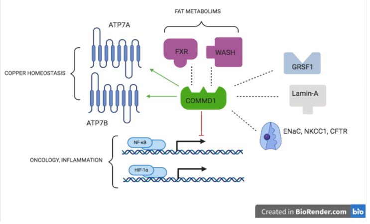
Figure 2. COMMD1Figureworks
2. COMMD1 worksOrganizing
as a Central as a CentralMolecule
Organizingin Molecule
Metabolism in Metabolism and
and Disease. Disease. Interac‐
Interactions of COMMD1
tions of COMMD1 protein with other proteins explain its role in copper homeostasis,
protein with other proteins explain its role in copper homeostasis, inflammation, fat metabolism, and inflamma‐
some hallmarks
tion, fat metabolism, and some hallmarks of cancer. Dashed lines indicate an interaction, an arrow
of cancer. Dashed lines indicate an interaction, an arrow indicates an activation, blunted line indicates an inhibition.
indicates an activation, blunted line indicates an inhibition. For details on the interactions with
For details on the interactions with ATP7A/ATP7B see [77–79], ENaC [89], NKCC1 [12], CFTR [92], WASH [100,101],
ATP7A/ATP7B see [77–79], ENaC [89], NKCC1 [12], CFTR [92], WASH [100,101], NFκB [71,96,97],
NFκB [71,96,97],FXR
FXR [102–104],
[102–104], lamin
lamin A [105],
A [105], GRSF1
GRSF1 [106],
[106], and and HIF-1α
HIF‐1α [98]. Abbreviations:
[98]. Abbreviations: CFTR, CFTR, Cystic Fibrosis
Cystic Fibrosis
TransmembraneTransmembrane
Conductance Regulator; ENaC,
Conductance EpithelialENaC,
Regulator; Sodium Channel;Sodium
Epithelial FXR, Farnesoid
Channel;X-activated Receptor;
FXR, Farnesoid X‐ GRSF1,
guanine-rich RNA sequence
activated bindingGRSF1,
Receptor; factor 1; HIF-1α, Hypoxia-Induced
guanine‐rich RNA sequencefactor
binding1α;factor
NFkB,1;Nuclear
HIF‐1α,Factor kappa-B; NKCC1,
Hypoxia‐In‐
duced factor 1α;
Sodium-Potassium-Chloride NFkB, Nuclear
Transporter Factor
1; WASH, kappa‐B; NKCC1,
Wiskott-Aldrich Sodium‐Potassium‐Chloride
syndrome Transporter
protein and SCAR Homologue.
1; WASH, Wiskott‐Aldrich syndrome protein and SCAR Homologue.
5.2. ATP7A and ATP7B Mutations in Labrador Retrievers and Dobermann Dogs
Labrador retrievers are among the most popular dog breeds worldwide. To emphasize
the power of canine genetics to dissect genetic causes in complex genetic diseases, as little
as 235 dogs were enrolled in a canine-specific genome-wide association study (GWAS).
This study revealed that an Arg1453Gln substitution in the ATP7B protein was related
to increased hepatic copper levels, whereas a Thr327Ile mutation in the ATP7A protein
partially rescued the ATP7B phenotype [107]. Similarly, an ATP7B mutation in Dutch and
USA Dobermanns increased hepatic copper levels. A mutation in ATP7A was also found.
However, there were too few cases to draw conclusions for the US cohort [108]. Beauty is in
the eye of the beholder, but the fact that ATP7A is a modifier gene for ATP7B is astonishing,
given their high level of homology. In this breed, the high prevalence of mutations in the
ATP7A gene showed the power of canine genetics to find modifier genes. The fact that
these mutations are in a gene that is causative for MD, further highlights the beauty of
canine research. It is unlikely that this would have been discovered in men where MD
is affecting 1 in 300,000 people. It turned out that COMMD1 mutations were involved
in neither Labrador nor Dobermann copper toxicosis [109]. Recently, RETN (coding for
protein RESISTIN) was discovered as a novel modifier gene in copper toxicosis in Labrador
retrievers [110]. RESISTIN is involved in hepatic fat storage and mitochondrial defects,
however, its low expression in the liver makes it difficult to directly associated RETN
mutations with hepatic copper accumulation.
6. Dogs and Cats Are Clearly Different with Regard to Hepatic Copper Accumulation
Although less stringent, inbreeding occurs in the feline pet population. Much less
is known about the genetic background of hepatic copper accumulation in cats. The first
description was in 1995 in Siamese cats [111] (Hayes Wade 1995). Copper-induced chronic
hepatitis in a European short was presented a decade later [112] (Meertens 2005). TheAnimals 2021, 11, 601 8 of 13
levels of copper, and some other trace elements in feline livers were described by various
groups [113–116] (Andreani 2010; Whittemore 2012; Passlack 2014; Yamkate 2020). In
addition to house cats (Felis catus), copper levels in other felidae were measured, including
bobcats, tigers, cougars, coyotes, and lions [117–119] (Bernard 2015; Thomason 2016),
Hough 2020. It was only in the last few years that two publications found mutations in
the ATP7B gene [120,121] (Asada 2019; Asada 2020). To our knowledge, no cats have been
described with mutations in the COMMD1 gene, not even in a survey of over 800 cats from
Finland [122].
7. The Mutual Benefits for Men and Dogs in Rare Copper Storage Diseases
Copper-related diseases in humans are often rare diseases but account for about
one-third of the chronic hepatitis cases in dogs [123]. This review has focused on copper
dyshomeostasis and described the potential power that genetic research in dog breeds could
have to benefit human biomedicine. Dogs turned out to become powerful model animals
for men [18,61,124]. Almost 20 years after the discovery of COMMD1 as a causative protein
in inherited copper toxicosis in a specific dog breed, a wealth of novel functions related to
COMMD1 were described. This novel gene product was not involved in copper overload
diseases with thus far unknown genetic causes, like ICC, ETIC, and ICT. Genetic studies in
dogs are promising, thus it is hopeful that for several other dog breeds, the genetic cause
for inherited copper toxicosis will be elucidated in the near future. We do not know if this
will initiate similar anxiety as did the discovery of COMMD1, but it also provides hope for
people suffering from rare copper-related diseases that research tools to help solving the
genetic background are just around the corner, or sometimes even closer. Therefore, the
European Union (www.lupa.eu (accessed on 1 December 2020)) and the Dutch government
provided us with a grant to exploit the downside of genetic inbreeding in dogs to the
mutual benefit of men and their best friend. In addition to the likelihood that novel
(modifier) genes involved in rare diseases, such as copper accumulation, will be found, this
work will also create a genetic base for more realistic large animal models [76,82,124].
Current deep sequencing strategies that are exploited in men will find numerous
genetic variants. Whether this will lead to a causality of the novel variants with the
phenotype will remain difficult in view of the limited sample size in the case of (very) rare
diseases. In addition, taking into account that canine inbreeding is tremendously higher
than in humans, much fewer variants will be detected in dogs. The causal association
between a novel variant and a specific phenotype will, therefore, be much more easily
established. This strategy and the implementation of state-of-art sequencing technology in
men and dogs has been nicely described [60].
8. Dogs as a Test Bed for Humans with Copper Storage Diseases:
Copper-Zinc Interactions
Dogs are ideal for testing nutritional interventions, as, opposed to people, we can keep
their dietary intake the same for prolonged periods of time. As already mentioned, copper
transporters also transport zinc. By increasing dietary zinc, the absorption of copper will,
therefore, be reduced. In dogs, a commercially available diet with low levels of copper
(5 mg per kg food, instead of 25 mg per kg in general dog food) and high levels of zinc
(250 mg per kg food, instead of 50 mg per kg in general dog food) was able to reduce
hepatic copper levels in Labrador retrievers with a history of copper storage disease and
kept them clinically healthy up till 43 months after starting dietary treatment [125]. As an
alternative, by adding 7.5 mg (twice daily) elemental zinc per kg bodyweight for 3 months,
metallothionein is induced, which acts as a chelator for copper both in the intestine as in the
liver, which has been demonstrated to reduce copper toxicity in dogs [126]. This could also
benefit people with copper storage diseases [127]. While realizing that owners allow their
pets access to sweets or to left-overs of their own food, generally speaking, the variation in
copper uptake is likely far less than in humans. Food that contains high levels of copper
includes liver, hazelnut, Brazil nut (yet low in almond), and cacao-based products. This
list clearly shows that the uptake of copper in men will be extremely variable, furtherAnimals 2021, 11, 601 9 of 13
limiting clear food-related (intervention) studies. Think twice if you find a dog with copper
storage disease.
Author Contributions: R.J.C. was responsible for the review of nutrition-related paragraphs and
corrected this paper; L.C.P. drafted this paper and guided the studies on COMMD1 in dogs. All
authors have read and agreed to the published version of the manuscript.
Funding: Both R.J.C. and L.C.P. are employees of Utrecht University and did not receive specific
grants for the manuscript.
Institutional Review Board Statement: No ethical approval for this manuscript was required.
Data Availability Statement: No new data were created or analyzed in this study. Data sharing is
not applicable to this article.
Acknowledgments: Figures were created in Biorender.com by Marloes M. Penning, medical sdent at
the University of Amsterdam (The Netherlands).
Conflicts of Interest: The authors declare no conflict of interest. The funders had no role in the
decision to publish this review.
References
1. Wakap, S.N.; Deborah, M.; Lambert, D.M.; Olry, A.; Rodwell, C.; Gueydan, C.; Lanneau, V.; Daniel, M.; Le Cam, Y.; Rath, A.
Estimating cumulative point prevalence of rare diseases: Analysis of the Orphanet database. Eur. J. Hum. Genet. 2020, 28, 165–173.
[CrossRef] [PubMed]
2. Lee, C.E.; Kaela, S.; Singleton, W.M.; Faundez, V. Rare Genetic Diseases: Nature’s Experiments on Human Development. IScience
2020, 23, 101123. [CrossRef]
3. Inesi, G. Molecular features of copper binding proteins involved in copper homeostasis. Crit. Rev. IUBMB Life 2017, 69, 211–217.
[CrossRef] [PubMed]
4. Kim, B.E.; Nevitt, T.; Thiele, D.J. Mechanisms for copper acquisition, distribution and regulation. Nat. Chem. Biol. 2008, 4, 176–185.
[CrossRef] [PubMed]
5. Zhou, B.; Gitschier, J. hCTR1: A human gene for copper uptake identified by complementation in yeast. Proc. Natl. Acad. Sci.
USA 1997, 94, 7481–7486. [CrossRef] [PubMed]
6. Lutsenko, S.; Barnes, N.L.; Bartee, M.Y.; Dmitriev, O.Y. Function and regulation of human copper-transporting ATPases. Physiol.
Rev. 2007, 87, 1011–1046. [CrossRef] [PubMed]
7. Trocello, J.-M.; Brousolle, E.; Girardot-Tinant, N.; Pelosse, M.; Lachaux, A.; Lloyd, C.; Woimant, F. Wilson’s disease, 100 years later.
Rev. Neurol. 2013, 169, 936–943. [CrossRef] [PubMed]
8. Xie, J.-J.; Wu, Z.-Y. Wilson’s disease in China. Neurosci. Bull. 2017, 33, 323–330. [CrossRef]
9. Członkowska, A.; Litwin, T.; Dusek, P.; Ferenci, P.; Lutsenko, S.; Medici, V.; Rybakowski, J.K.; Weiss, K.H.; Schilsky, M.L. Wilson
disease. Nat. Rev. Dis. Primers 2018, 4, 21. [CrossRef]
10. Lo, C.; Bandmann, O. Epidemiology and introduction to the clinical presentation of Wilson disease. Handb. Clin. Neurol. 2017,
142, 7–17.
11. Sandahl, T.D.; Laursen, T.L.; Munk, D.E.; Vilstrup, H.; Wiess, K.H.; Ott, P. The prevalence of Wilson’s disease: An update.
Hepatology 2020, 71, 722–732. [CrossRef]
12. Bull, P.C.; Thomas, G.R.; Rommens, J.M.; Forbers, J.R.; Cox, D.W. The Wilson disease gene is a putative copper transporting
P-type ATPase similar to the Menkes gene. Nat. Genet. 1993, 5, 327–337. [CrossRef]
13. Tanzi, R.E.; Petrukhin, K.; Chernov, I.; Pelleguer, J.L.; Wasco, W.; Ross, B.; Romano, D.M.; Parano, E.; Pavone, L.; Bzustowicz,
L.M.; et al. The Wilson disease gene is a copper transporting ATPase with homology to the Menkes disease gene. Nat. Genet.
1993, 5, 344–350. [CrossRef] [PubMed]
14. Pierson, H.; Nuchenditsi, A.; Byung-Eun, K.; Ralle, M.; Zachos, N.; Huster, D.; Lutsenko, S. The function of ATPase copper
transporter ATP7B in intestine. Gastroenterology 2018, 154, 168–180. [CrossRef] [PubMed]
15. Pfeiffer, R. Wilson’s disease. Semin. Neurol. 2007, 27, 123–132. [CrossRef] [PubMed]
16. Hermann, W. Classification and differential diagnosis of Wilson’s disease. Ann. Transl. Med. 2019, 7, S63. [CrossRef] [PubMed]
17. Buiakova, O.I.; Xu, J.; Lutsenko, S.; Zeitlin, S.; Das, K.; Das, S.; Ross, B.M.; Mekios, C.; Scheinberg, I.H.; Gilliam, T.C. Null Mutation
of the Murine ATP7B (Wilson Disease) Gene Results in Intracellular Copper Accumulation and Late-Onset Hepatic Nodular
Transformation. Hum. Mol. Genet. 1999, 8, 1665–1671. [CrossRef]
18. Reed, E.; Lutsenko, S.; Bandmann, O. Animal models of Wilson disease. J. Neurochem. 2018, 146, 356–373. [CrossRef] [PubMed]
19. Ferenci, P. Phenotype-genotype correlations in patients with Wilson’s disease. Ann. N. Y. Acad. Sci. 2014, 1315, 1–5. [CrossRef]
20. Lutsenko, S. Modifying factors and phenotypic diversity in Wilson’s disease. Ann. N. Y. Acad. Sci. 2014, 1315, 56–63. [CrossRef]
21. Medici, V.; Weiss, K.H. Genetic and environmental modifiers of Wilson disease. Handb. Clin. Neurol. 2017, 142, 35–41.
22. Stremmel, W.; Merle, U.; Weiskirchen, R. Clinical features of Wilson disease. Ann. Transl. Med. 2019, 7, S61. [CrossRef] [PubMed]Animals 2021, 11, 601 10 of 13
23. Simon, I.; Schaefer, M.; Reichert, J.; Stremmel, W. Analysis of the Human Atox 1 Homologue in Wilson Patients. World J.
Gastroenterol. 2008, 14, 2383–2387. [CrossRef]
24. Weiss, K.H.; Runz, H.; Noe, B.; Gotthardt, D.N.; Merle, U.; Ferenci, P.; Stremmel, W.; Füllekrug, J. Genetic Analysis of BIRC4/XIAP
as a Putative Modifier Gene of Wilson Disease. J. Inherit. Metab. Dis. 2010, 33 (Suppl. 3), S233–S240. [CrossRef]
25. Stuehler, B.; Reichert, J.; Stremmel, W.; Schaefer, M. Analysis of the Human Homologue of the Canine Copper Toxicosis Gene
MURR1 in Wilson Disease Patients. J. Mol. Med. 2004, 82, 629–634. [CrossRef] [PubMed]
26. Gromadzka, G.; Rudnicka, M.; Chabik, G.; Przybylkowski, A.; Czlonkowska, A. Genetic variability in the methylenetetrahy-
drofolate reductase gene (MTHFR) affects clinical expression of Wilson’s disease. J. Hepatol. 2011, 55, 913–919. [CrossRef]
[PubMed]
27. Bost, M.; Piguit-Lacroix, G.; Parant, F.; Wilson, C.M.R. Molecualr analysis of Wilson patients: Direct sequencing and MLPA
analysis in the ATP7B gene and Atox1 and COMMD1 gene analysis. J. Trace Elem. Med. Biol. 2012, 26, 97–101. [CrossRef]
28. Menkes, J.H.; Alter, M.; Steigleder, G.K.; Weakley, D.R.; Sung, J.H. A sex-linked recessive disorder with retardation of growth,
peculiar hair, and focal cerebral and cerebral degeneration. Pediatrics 1962, 29, 764–779. [PubMed]
29. Danks, D.M.; Campbell, P.E.; Stevens, B.J.; Mayne, V.; Cartwright, E. Menkes’s kinky hair syndrome. An inherited defect in
copper absorption with widespread effects. Pediatrics 1972, 59, 188–201.
30. Chelly, J.; Tumer, Z.; Tonnesen, T.; Petteson, A.; Ishikawa-Brush, Y.; Tommerup, N.; Norn, N.; Monaco, A.P. Isolation of a candidate
gene for Menkes disease that encodes a potential heavy metal binding protein. Nat. Genet. 1993, 3, 14–19. [CrossRef] [PubMed]
31. Mercer, J.F.B.; Livingston, J.; Hall, B.; Paynter, J.A.; Begy, C.; Chandrasekharappa, S.; Lockhart, P.; Grimes, A.; Bhave, M.;
Siemieniak, D.; et al. Isolation of a partial candidate gene for Menkes disease by positional cloning. Nat. Genet. 1993, 3, 20–25.
32. Vulpe, C.; Levinson, B.; Whitney, S.; Packman, S.; Gitschier, J. Isolation of a candidate gene for Menkes disease and evidence that
it encodes a copper-transporting ATPase. Nat. Genet. 1993, 3, 7–13. [CrossRef]
33. Gourdon, P.; Sitsel, O.; Lykkegaard Karlsen, J.; Møller, L.B.; Niassen, P. Structural models of the human copper-type ATPases
ATP7A and ATP7B. Biol. Chem. 2012, 393, 205–216. [CrossRef] [PubMed]
34. Tümer, Z.; Møller, L.B. Menkes disease. Eur. J. Hum. Genet. 2010, 18, 511–518. [CrossRef] [PubMed]
35. Tümer, Z. An overview and update of ATP7A mutations leading to Menkes disease and occipital horn syndrome. Hum. Mutat.
2013, 34, 417–429. [CrossRef]
36. Kaler, S.G. Translational research investigations on ATP7A: An important human copper ATPase. Ann. N. Y. Acad. Sci. 2014, 1314,
64–68. [CrossRef] [PubMed]
37. Smpokou, P.; Samanta, M.; Berry, G.T.; Hecht, L.; Engle, E.C.; Lichter-Konecki, U. Menkes disease in affected females: The clinical
disease spectrum. Am. J. Med. Genet. 2015, 167A, 417–420. [CrossRef]
38. Møller, L.B.; Mogensen, M.; Horn, N. Molecular diagnosis of Menkes disease: Genotype-phenotype correlation. Biochimie 2009,
91, 1273–1277. [CrossRef] [PubMed]
39. Tanner, M.S. Role of copper in Indian Childhood Cirrhosis. Am. J. Clin. Nutr. 1998, 67, 1074S–1081S. [CrossRef] [PubMed]
40. Müller, T.; Feichtinger, H.; Berger, H.; Muller, W. Endemic Tyrolean Infantile Cirrhosis: An ecogenetic disorder. Lancet 1996, 347,
877–880. [CrossRef]
41. Scheinberg, I.C.; Sternlieb, I. Wilson Disease and Idiopathic Copper Toxicosis. Am. J. Cin. Nutr. 1996, 63, 842S–845S. [CrossRef]
42. Wijmenga, C.; Müller, T.; Murli, I.S.; Brunt, T.; Feichtinger, H.; Schönitzer, D.; Houwen, R.H.; Müller, W.; Sandkuijl, L.A.; Pearson,
P.L. Endemic Tyrolean infantile cirrhosis is not an allelic variant of Wilson’s disease. Eur. J. Hum. Genet. 1998, 6, 624–628.
[CrossRef]
43. Müller, T.; van de Sluis, B.; Müller, W.; Pearson, P.; Wijmenga, C. Non-Indian childhood cirrhosis. Eur. J. Med. Res. 1999, 4,
293–297.
44. Müller, T.; van de Sluis, B.; Zhernakova, A.; van Binsbergen, E.; Janecke, A.R.; Bavdekar, A.; Pandit, A.; Weirich-Schwaiger, H.;
Witt, H.; Ellemunter, H.; et al. The canine copper toxicosis gene MURR1 does not cause non-Wilsonian hepatic copper toxicosis. J.
Hepatol. 2003, 38, 164–168. [CrossRef]
45. Ferreira, C.R.; Gahl, W.A. Disorders of metal metabolism. Transl. Sci. Rare Dis. 2017, 2, 101–139. [CrossRef] [PubMed]
46. Seachrist, L. Man’s best friend may be companion in cancer research. J. Natl. Cancer Inst. 1993, 85, 1455–1456. [CrossRef]
47. Hitte, C.; Madeoy, J.; Kirkness, E.F.; Priat, C.; Lorentzen, T.D.; Senger, F.; Thomas, D.; Derrien, T.; Ramirez, C.; Scott, C.; et al.
Facilitating genome navigation: Survey sequencing and dense radiation-hybrid gene mapping. Nat. Rev. Genet. 2005, 6, 643–648.
[CrossRef] [PubMed]
48. Parker, H.G.; Ostrander, E.A. Canine genomics and genetics: Running with the pack. PLoS Genet. 2005, 1, e58. [CrossRef]
49. Ostrander, E.A.; Wayne, R.K. The canine genome. Genome Res. 2005, 15, 1706–1716. [CrossRef] [PubMed]
50. Lindblad-Toh, K.; Wade, C.M.; Mikkelsen, T.S.; Karlsson, E.K.; Jaffe, D.B.; Kamal, M.; Clamp, M.; Chang, J.L.; Kulbokas, E.J.,
3rd; Zody, M.C.; et al. Genome sequence, comparative analysis and haplotype structure of the domestic dog. Nature 2005, 438,
803–809. [CrossRef]
51. Lequarre, A.-S.; Andersson, L.; Andre, C.; Fredholm, M.; Hitte, C.; Leeb, T.; Lohi, H.; Lindblad-Toh, K.; Georges, M. LUPA: A
European initiative taking advantage of the canine genome architecture for unravelling complex disorders in both human and
dogs. Vet. J. 2011, 189, 155–159. [CrossRef]Animals 2021, 11, 601 11 of 13
52. Vaysse, A.; Ratnakumaer, A.; Derrien, T.; Axelsson, E.; Pielberg, G.R.; Sigurdsson, S.; Fall, T.; Seppala, E.H.; Hanse, M.S.T.; Lawley,
C.T.; et al. Identification of genomic regions associated with phenotype variation between dog breeds using selection mapping.
PLoS Genet. 2011, 7, e1002316. [CrossRef] [PubMed]
53. Hoeppner, M.P.; Lundquies, A. An improved canine genome and a comprehensive catalogue of coding genes and non-coding
transcripts. PLoS ONE 2014, 9, e91172. [CrossRef] [PubMed]
54. Penso-Dolfin, L.; Swofford, R.; Johnson, J.; Alfoedi, J.; Loindblad-Toh, K.; Swarbreck, D.; Moxon, S.; Di Palma, F. An improved
microRNA annotation of the canine genome. PLoS ONE 2016, 11, e0153453. [CrossRef]
55. Wucher, V.; Legeai, F.; Hedan, B.; Rizk, G.; Lagoutee, L.; Leeb, T.; Jagannathan, V.; Cadieu, E.; David, A.; Lohi, H.; et al. FEELnc: A
tool for long non-codong RNA annotation and its pplication to the dog transcriptome. Nucl. Acid Res. 2017, 45, e57. [CrossRef]
[PubMed]
56. Wang, G.D.; Larson, G.; Kidd, J.M.; vonHoldt, B.M.; Ostrander, E.A.; Zhang, Y.P. Dog10K: The international consortium of canine
genome sequencing. Natl. Sci. Rev. 2019, 6, 611–613. [CrossRef]
57. Fieten, H.; Penning, L.C.; Leegwater, P.A.; Rothuizen, J. New canine models of copper toxicosis: Diagnosis, treatment, and
genetics. Ann. N. Y. Acad. Sci. 2014, 1314, 42–48. [CrossRef]
58. Parker, H.G.; Shearin, A.L.; Ostrander, E.A. Man’s best friend becomes biology’s best in show: Genome analyses in the domestic
dog. Annu. Rev. Genet. 2010, 44, 309–336. [CrossRef] [PubMed]
59. Larson, G.; Karlsson, E.K.; Perri, A.; Webster, M.T.; Ho, S.Y.; Peters, J.; Stahl, P.W.; Lingaas, F.; Fredholm, M.; Comstock, K.E.; et al.
Rethinking dog domestication by integrating genetics, archeology, and biogeography. Proc. Natl. Acad. Sci. USA 2012, 109,
8878–8883. [CrossRef]
60. van Steenbeek, F.G.; Hytoenen, M.K.; Leegwater, P.A.J.; Lohi, H. The canine era: The rise of a biomedical model. Anim. Genet.
2016, 47, 519–527. [CrossRef]
61. Fuentalba, I.C.; Aburto, E.M. Animal models of copper-associated liver disease. Comp. Hepatol. 2003, 2, 5. [CrossRef]
62. Twedt, D.C.; Sternlieb, I.; Gilbertson, S.R. Clinical, morphologic, and chemical studies on copper toxicosis of Bedlington Terriers.
J. Am. Vet. Med. Assoc. 1979, 175, 269–275. [PubMed]
63. Haywood, S.; Rutgers, H.C.; Christian, M.K. Hepatitis and copper accumulation in Skye terriers. Vet. Pathol. 1988, 25, 408–414.
[CrossRef]
64. Thornburg, L.P.; Rottinghaus, G.; Dennis, G.; Crawford, S. The relationship between hepatic copper content and morphologic
changes in the liver of West Highland White Terriers. Vet. Pathol. 1996, 33, 656–661. [CrossRef] [PubMed]
65. Thornburg, L.P. Histomorphological and immunohistochemical studies of chronic active hepatitis in Doberman Pinschers. Vet.
Pathol. 1998, 35, 380–385. [CrossRef]
66. Webb, C.B.; Twedt, D.C.; Meyer, D.J. Copper-associated liver disease in Dalmatians: A review of 10 dogs (1998–2001). J. Vet.
Intern. Med. 2002, 16, 665–668. [PubMed]
67. Hoffmann, G.; van den Ingh, T.S.; Bode, P.; Rothuizen, J. Copper-associated chronic hepatitis in Labrador Retrievers. J. Vet. Intern.
Med. 2006, 20, 856–861. [CrossRef] [PubMed]
68. van de Sluis, B.J.; Breen, M.; Nanji, M.; van Wolferen, M.; de Jong, P.; Binns, M.M.; Pearson, P.L.; Kuipers, J.; Rothuizen, J.; Cox,
D.W.; et al. Genetic mapping of the copper toxicosis locus in Bedlington terriers to dog chromosome 10, in a region syntenic to
human chromosome region 2p13-p16. Hum. Mol. Genet. 1999, 8, 501–507. [CrossRef]
69. van de Sluis, B.; Rothuizen, J.; Pearson, P.L.; van Oost, B.A.; Wijmenga, C. Identification of a new copper metabolism gene by
positional cloning in a purebred dog population. Hum. Mol. Genet. 2002, 11, 165–173. [CrossRef]
70. Forman, O.P.; Boursnell, M.E.G.; Dunmore, B.J.; Stendall, N.; van de Sluis, B.; Frettwell, N.; Jones, N.; Wijmenga, C.; Rothuizen, J.;
van Oost, B.A.; et al. Characterization of the COMMD1 (MURR1) Mutation Causing Copper Toxicosis in Bedlington Terriers.
Anim. Genet. 2005, 36, 497–501. [CrossRef]
71. Ganesh, L.; Burstein, E.; Guha-Niyogi, A.; Louder, M.K.; Mascola, J.R.; Klomp, L.W.; Wijmenga, C.; Duckett, C.S.; Nabel, G.J. The
gene product Murr1 restricts HIV-1 replication in resting CD4+ lymphocytes. Nature 2003, 426, 853–857. [CrossRef]
72. Spee, B.; Arends, B.; van Wees, A.M.; Bode, P.; Penning, L.C.; Rothuizen, J. Functional consequences of RNA interference targeting
COMMD1 in a canine hepatic cell line in relation to copper toxicosis. Anim. Genet. 2007, 38, 168–170. [CrossRef]
73. Vonk, W.I.; Bartuzi, P.; de Bie, P.; Kloosterhuis, N.; Wichers, C.G.; Berger, R.; Haywood, S.; Klomp, L.W.; Wijmenga, C.; van
de Sluis, B. Liver-specific Commd1 knockout mice are susceptible to hepatic copper accumulation. PLoS ONE 2011, 6, e29183.
[CrossRef]
74. Nantasanti, S.; Spee, B.; Kruitwagen, H.S.; Chen, C.; Geijsen, N.; Oosterhoff, L.A.; van Wolferen, M.E.; Palaez, N.; Fieten, H.;
Wubbolts, R.W.; et al. Disease Modeling and Gene Therapy of Copper Storage Disease in Canine Hepatic Organoids. Stem Cell
Rep. 2015, 5, 895–907. [CrossRef]
75. Su, L.C.; Ravanshad, S.; Owen, C.A., Jr.; McCall, J.T.; Zollman, P.E.; Hardy, R.M. A comparison of copper-loading disease in
Bedlington terriers and Wilson’s disease in humans. Am. J. Physiol. 1982, 243, G226–G230. [CrossRef]
76. Kruitwagen, H.S.; Penning, L.C. Precilinical models of Wilson’s disease, why dogs are cathy alternatives. Ann. Transl. Med. 2019,
7, S71. [CrossRef] [PubMed]
77. de Bie, P.; van de Sluis, B.; Burstein, E.; van den Berghe, P.V.; Muller, P.; Berger, R.; Gitlin, J.D.; Wijmenga, C.; Klomp, L.W.
Distinct Wilson’s disease mutations in ATP7B are associated with enhanced binding to COMMD1 and reduced stability of ATP7B.
Gastroenterology 2007, 133, 1316–1326. [CrossRef]Animals 2021, 11, 601 12 of 13
78. Weiss, K.H.; Lozoya, J.C.; Tuma, S.; Gotthardt, D.; Reichert, J.; Ehehalt, R.; Stremmel, W.; Fullekrug, J. Copper-induced
translocation of the Wilson disease protein ATP7B independent of Murr1/COMMD1 and Rab7. Am. J. Pathol. 2008, 173,
1783–1794. [CrossRef]
79. Vonk, W.I.; de Bie, P.; Wichers, C.G.; van den Berghe, P.V.; van der Plaats, R.; Berger, R.; Wijmenga, C.; Klomp, L.W.; van de Sluis,
B. The copper-transporting capacity of ATP7A mutants associated with Menkes disease is ameliorated by COMMD1 as a result of
improved protein expression. Cell. Mol. Life Sci. 2012, 69, 149–163. [CrossRef] [PubMed]
80. Favier, R.P.; Spee, B.; Schotanus, B.A.; van den Ingh, T.S.; Fieten, H.; Brinkhof, B.; Viebahn, C.S.; Penning, L.C.; Rothuizen, J.
COMMD1-deficient dogs accumulate copper in hepatocytes and provide a good model for chronic hepatitis and fibrosis. PLoS
ONE 2012, 7, e42158. [CrossRef] [PubMed]
81. Favier, R.P.; Spee, B.; Fieten, H.; van den Ingh, T.S.; Schotanus, B.A.; Brinkhof, B.; Rothuizen, J.; Penning, L.C. Aberrant expression
of copper associated genes after copper accumulation in COMMD1-deficient dogs. J. Trace Elem. Med. Biol. 2015, 29, 347–353.
[CrossRef]
82. Kruitwagen, H.S.; Oosterhoff, L.A.; van Wolferen, M.E.; Chen, C.; Assawarachan, S.N.; Schneeberger, K.; Kummeling, A.; van
Straten, G.; Akkerdaas, I.C.; Vinke, C.R.; et al. Long-term survival of transplanted autologous canine liver organoids in a
COMMD1-deficient dog model of metabolic liver disease. Cells 2020, 9, 410. [CrossRef] [PubMed]
83. de Bie, P.; van de Sluis, B.; Klomp, L.; Wijmenga, C. The many faces of the copper metabolism protein MURR1/COMMD1. J.
Hered. 2005, 97, 803–811. [CrossRef] [PubMed]
84. Maine, G.N.; Burstein, E. COMMD proteins: COMMing to the scene. Cell. Mol. Life Sci. 2007, 64, 1997–2005. [CrossRef]
85. Fedoseienko, A.; Bartuzi, P.; van de Sluis, B. Functional understanding of the versatile protein copper metabolism MURR1 domain
1 (COMMD1) in copper homeostasis. Ann. N. Y. Acad. Sci. 2014, 1314, 6–14. [CrossRef] [PubMed]
86. Bartuzi, P.; Hofker, M.H.; van de Sluis, B. Tuning NF-κB activity: A touch of COMMD proteins. Biochim. Biophys. Acta. 2013, 1832,
2315–2321. [CrossRef]
87. Riera-Romo, M. COMMD1: A Multifunctional Regulatory Protein. J. Cell. Biochem. 2018, 119, 34–51. [CrossRef]
88. Weiskirchen, R.; Penning, L.C. COMMD1, a multi-potent intracellular protein involved in copper homeostasis, protein trafficking,
inflammation, and cancer. J. Trace Elem. Med. Biol. 2021, 65, 126712.
89. Biasio, W.T.; Chang, T.; McIntosh, C.J.; McDonald, F.J. Identification of Murr1 as a regulator of the human delta epithelial sodium
channel. J. Biol. Chem. 2004, 279, 5429–5434. [CrossRef] [PubMed]
90. Ke, Y.; Butt, A.G.; Swart, M.; Liu, Y.F.; McDonald, F.J. COMMD1 downregulates the epithelial sodium channel through Nedd4-2.
Am. J. Physiol. Renal Physiol. 2010, 298, F1445–F1456. [CrossRef]
91. Smith, L.; Litman, P.; Liedtke, C.M. COMMD1 Interacts with the COOH Terminus of NKCC1 in Calu-3 Airway Epithelial Cells to
Modulate NKCC1 Ubiquitination. J. Physiol. Cell. Physiol. 2013, 305, C133–C146. [CrossRef] [PubMed]
92. Drévillon, L.; Tanguy, G.; Hinzpeter, A.; Arous, N.; de Becdelièvre, A.; Aissat, A.; Tarze, A.; Goossens, M.; Fanen, P. COMMD1-
mediated Ubiquitination Regulates CFTR Trafficking. PLoS ONE 2011, 6, e18334. [CrossRef]
93. Wang, J.; Fedoseienko, A.; Chen, B.; Burstein, E.; Jia, D.; Billadeau, D.D. Endosomal receptor trafficking: Retromer and beyond.
Traffic 2018, 19, 578–590. [CrossRef]
94. McNally, K.E.; Cullen, P.J. Endosomal Retrieval of Cargo: Retromer Is Not Alone. Trends Cell Biol. 2018, 28, 807–822. [CrossRef]
[PubMed]
95. Burstein, E.; Ganesh, L.; Dick, R.D.; van de Sluis, B.; Wilkinson, J.C.; Klomp, L.W.; Wijmenga, C.; Brewer, G.J.; Nabel, G.J.; Duckett,
C.S. A novel role for XIAP in copper homeostasis through regulation of Murr1. EMBO J. 2004, 23, 244–254. [CrossRef]
96. Maine, G.N.; Burstein, E. COMMD Proteins and the Control of the NF Kappa B Pathway. Cell Cycle 2007, 6, 672–776. [CrossRef]
[PubMed]
97. Mufti, A.R.; Burstein, E.; Duckett, C.S. XIAP: Cell Death Regulation Meets Copper Homeostasis. Arch. Biochem. Biophys. 2007, 463,
168–174. [CrossRef]
98. van de Sluis, B.; Mao, X.; Zhai, Y.; Groot, A.J.; Vermeulen, J.F.; van der Wall, E.; van Diest, P.J.; Hofker, M.H.; Wijmenga, C.; Klomp,
L.W.; et al. COMMD1 Disrupts HIF-1alpha/beta Dimerization and Inhibits Human Tumor Cell Invasion. J. Clin. Investig. 2010,
120, 2119–2130. [CrossRef] [PubMed]
99. Taskinen, M.; Louhimo, R.; Koivula, S.; Chen, P.; Rantanen, V.; Holte, H.; Delabie, J.; Karjalainen-Lindsberg, M.-L.; Björkholm, M.
Deregulation of COMMD1 Is Associated with Poor Prognosis in Diffuse Large B-cell Lymphoma. PLoS ONE 2014, 9, e91031.
[CrossRef]
100. Bartuzi, P.; Billadeau, D.D.; Favier, R.P.; Rong, S.; Dekker, D.; Fedoseienko, A.; Fieten, H.; Wijers, M.; Levels, H.; Huijkman,
N.; et al. CCC- And WASH-mediated Endosomal Sorting of LDLR Is Required for Normal Clearance of Circulating LDL. Nat.
Commun. 2016, 7, 10961. [CrossRef] [PubMed]
101. Phillips-Krawczak, C.A.; Singla, A.; Starokadomskyy, P.; Deng, Z.; Osborne, D.G.; Li, H.; Dick, C.J.; Gomez, T.S.; Koenecke, M.;
Zhang, J.-S.; et al. COMMD1 Is Linked to the WASH Complex and Regulates Endosomal Trafficking of the Copper Transporter
ATP7A. Mol. Biol. Cell 2015, 26, 91–103. [CrossRef]
102. Wooton-Kee, C.R.; Jian, A.K.; Wagner, M.; Grusak, M.A.; Finegold, M.J.; Lutsenko, S.; Moore, D.D. Elevated copper impairs
hepatic nuclear receptor function in Wilson’s diseases. J. Clin. Investig. 2015, 125, 3449–3460. [CrossRef]You can also read


