AIN COMMUNICATIONS Diffusion models reveal white matter microstructural changes with ageing, pathology and cognition - Oxford Academic Journals
←
→
Page content transcription
If your browser does not render page correctly, please read the page content below
doi:10.1093/braincomms/fcab106                                                                                   BRAIN COMMUNICATIONS 2021: Page 1 of 15                          | 1

BRAIN
  AIN COMMUNICATIONS

Diffusion models reveal white matter
microstructural changes with ageing,

                                                                                                                                                                                          Downloaded from https://academic.oup.com/braincomms/article/3/2/fcab106/6278287 by guest on 14 September 2021
pathology and cognition
Sheelakumari Raghavan,1 Robert I. Reid,2 Scott A. Przybelski,3 Timothy G. Lesnick,3
 Jonathan Graff-Radford,4 Christopher G. Schwarz,1 David S. Knopman,4
 Michelle M. Mielke,3,4 Mary M. Machulda,5 Ronald C. Petersen,4 Clifford R. JackJr 1 and
 Prashanthi Vemuri1

White matter microstructure undergoes progressive changes during the lifespan, but the neurobiological underpinnings related to
ageing and disease remains unclear. We used an advanced diffusion MRI, Neurite Orientation Dispersion and Density Imaging, to
investigate the microstructural alterations due to demographics, common age-related pathological processes (amyloid, tau and
white matter hyperintensities) and cognition. We also compared Neurite Orientation Dispersion and Density Imaging findings to
the older Diffusion Tensor Imaging model-based findings. Three hundred and twenty-eight participants (264 cognitively unim-
paired, 57 mild cognitive impairment and 7 dementia with a mean age of 68.3 6 13.1 years) from the Mayo Clinic Study of Aging
with multi-shell diffusion imaging, fluid attenuated inversion recovery MRI as well as amyloid and tau PET scans were included in
this study. White matter tract level diffusion measures were calculated from Diffusion Tensor Imaging and Neurite Orientation
Dispersion and Density Imaging. Pearson correlation and multiple linear regression analyses were performed with diffusion meas-
ures as the outcome and age, sex, education/occupation, white matter hyperintensities, amyloid and tau as predictors. Analyses
were also performed with each diffusion MRI measure as a predictor of cognitive outcomes. Age and white matter hyperintensities
were the strongest predictors of all white matter diffusion measures with low associations with amyloid and tau. However, neurite
density decrease from Neurite Orientation Dispersion and Density Imaging was observed with amyloidosis specifically in the tem-
poral lobes. White matter integrity (mean diffusivity and free water) in the corpus callosum showed the greatest associations with
cognitive measures. All diffusion measures provided information about white matter ageing and white matter changes due to age-
related pathological processes and were associated with cognition. Neurite orientation dispersion and density imaging and diffusion
tensor imaging are two different diffusion models that provide distinct information about variation in white matter microstructural
integrity. Neurite Orientation Dispersion and Density Imaging provides additional information about synaptic density, organiza-
tion and free water content which may aid in providing mechanistic insights into disease progression.

1   Department of Radiology, Mayo Clinic, Rochester, MN 55905, USA
2   Department of Information Technology, Mayo Clinic, Rochester, MN 55905, USA
3   Department of Quantitative Health Sciences, Mayo Clinic, Rochester, MN 55905, USA
4   Department of Neurology, Mayo Clinic, Rochester, MN 55905, USA
5   Department of Psychology, Mayo Clinic, Rochester, MN 55905, USA

Correspondence to: Prashanthi Vemuri, PhD
Mayo Clinic and Foundation
200 First Street SW, Rochester, MN 55905, USA
E-mail: vemuri.prashanthi@mayo.edu

Received February 17, 2021. Revised March 24, 2021. Accepted April 12, 2021. Advance Access publication May 19, 2021
C The Author(s) (2021). Published by Oxford University Press on behalf of the Guarantors of Brain.
V
This is an Open Access article distributed under the terms of the Creative Commons Attribution License (http://creativecommons.org/licenses/by/4.0/), which permits unrestricted reuse,
distribution, and reproduction in any medium, provided the original work is properly cited.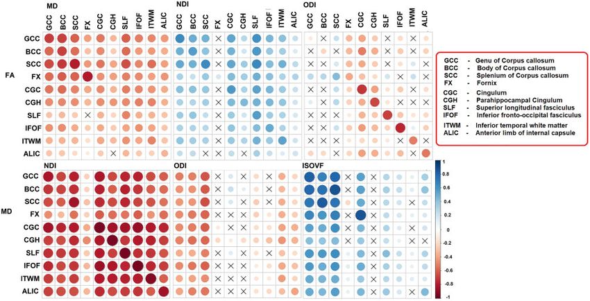
2   | BRAIN COMMUNICATIONS 2021: Page 2 of 15                                                                     S. Raghavan et al.

Keywords: diffusion tensor imaging; neurite dispersion density imaging; cerebrovascular disease
Abbreviations: BCC ¼ body of corpus callosum; CGC ¼ cingulum; CGH ¼ parahippocampal cingulum; CU ¼ cognitively unim-
paired; DTI ¼ diffusion tensor imaging; FA ¼ fractional anisotropy; FLAIR ¼ fluid attenuated inversion recovery; FX ¼ fornix;
GCC ¼ genu of corpus callosum; GM ¼ grey matter; ISOVF ¼ isotropic volume fraction; ITWM ¼ inferior temporal white matter;
MCI ¼ mild cognitive impairment; MCSA ¼ Mayo Clinic Study of Aging; MD ¼ mean diffusivity; NDI ¼ neurite density index;
NODDI ¼ neurite orientation dispersion and density Imaging; ODI ¼ orientation dispersion index; SCC ¼ splenium of corpus cal-
losum; SLF ¼ superior longitudinal fasciculus; WM ¼ white matter; WMH ¼ white matter hyperintensity

Graphical Abstract

                                                                                                                                       Downloaded from https://academic.oup.com/braincomms/article/3/2/fcab106/6278287 by guest on 14 September 2021
Introduction                                                      the microstructural environments inside the axons, between
                                                                  them, and in the extracellular water. In addition, these mod-
The white matter (WM) architecture of the human brain             els can ideally handle the ‘crossing fibre problem’ better than
undergoes substantial changes across the life span. There         DTI.7 Neurite orientation dispersion and density imaging
is clear evidence for the association between WM changes          (NODDI) is an advanced dMRI technique that uses the add-
and age as well as neuropathological processes that will          itional degrees of freedom from multi-shell data to probe the
lead to cognitive decline.1–3 Diffusion MRI is a versatile        microstructural complexity of neurites (dendrites and
method that allows us to study these WM microstructur-            axons),10 separately from CSF and to a large degree also
al details. Previous findings based on diffusion tensor           separately from each other. This biophysical modelling
imaging (DTI) revealed reduced fractional anisotropy              method divides water diffusion in the brain into three micro-
(FA) and increased mean diffusivity (MD) in association           structural compartments: intracellular space through the
with amyloid deposition, a hallmark of Alzheimer’s dis-           Neurite Density Index (NDI), which measures the signal frac-
ease,4 and cerebrovascular disease.5                              tion that is due to axons and dendrites; Orientation
   Despite its sensitivity, the clinical utility of DTI is con-   Dispersion Index (ODI), which measures angular variation or
strained by its inherent limitation in specificity of identi-     dispersion of the neurites; and the Isotropic Volume Fraction
fying the different diffusion environments6 that exist            (ISOVF), which measures free water (FW) fraction. More re-
within most individual voxels. Characterizing the different       cently, a number of studies have demonstrated the efficiency
water pools within a voxel with a single diffusion tensor         of NODDI to provide finer granularity, in comparison to
is well known to be problematic in crossing fibre regions         DTI metrics, to decipher the intra and extracellular micro-
of WM,7 and also confounds the macroscopically isotrop-           scopic features of age- and sex-specific diffusion trajecto-
ic diffusion of grey matter (GM)8,9 with that of CSF. The         ries.11,12 In addition, NODDI has been found to be useful
growing availability of multiband excitation allows the           for the early detection of neurodegenerative changes13–15 and
acquisition of roughly three times as much data in the            its association with cognitive deficits.14,16
same time as a standard DTI scan, making multiple b                  Recent findings have suggested that amyloid affects
value (diffusion weighting) shells clinically practical.          DTI measures4,17 and also the greater effect of cerebro-
Distributing the diffusion samples over >2 b values               vascular disease on diffusion alteration than Alzheimer’s
allows the use of more sophisticated and biologically             disease in memory clinic patients.17 It is also well known
plausible models to characterize the general properties of        that WM plays an important role in normal cognition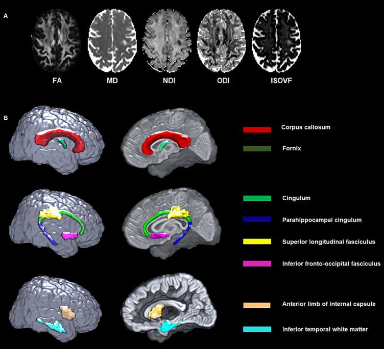
Basis of microstructural changes                                            BRAIN COMMUNICATIONS 2021: Page 3 of 15      | 3

and age-related cognitive decline.18,19 However, the effi-     angle 9 , 0.8 mm isotropic resolution), and a diffusion
ciency of NODDI models over DTI models to detect               scan using the product VE11 Simultaneous Multi-Slice ac-
Alzheimer’s disease and cerebrovascular disease patholo-       celeration with adaptive coil combination. For the diffu-
gies, and their contribution to cognitive performance, in      sion scan the field of view was 232 mm in X and Y and
population-based studies remains unclear. Given the            162 mm in the Z direction, with 2.0 mm isotropic voxels.
detailed quantification of biological processes by NODDI       The echo and repetition times were 71 and 3400 ms re-
models,6,20,21 we hypothesized that NODDI measures             spectively. Data consisted of 127 volumes with 13 non-dif-
would provide more sensitive features of microstructural       fusion-weighted images (b ¼ 0 s/mm2), and 114 diffusion-
brain changes than conventional FA and MD10,22 and             encoding gradient directions (6 b ¼ 500, 48 b ¼ 1000 and
would be more sensitive in capturing disease related proc-     60 b ¼ 2000 s/mm2), evenly spread over the entire spherical
esses. The overall goal of the study was to identify the       shells using an electrostatic repulsion model,26 and inter-

                                                                                                                                 Downloaded from https://academic.oup.com/braincomms/article/3/2/fcab106/6278287 by guest on 14 September 2021
relationships between demographics (age, sex and educa-        spersed in time to minimize gradient heating.
tion/occupation), neuroimaging measures of Alzheimer’s           The diffusion data were preprocessed using the in-
disease and cerebrovascular disease, and cognition with        house developed pipeline. After visual inspection, an
diffusion MRI (NODDI and conventional DTI) in partici-         intracranial mask was made for the diffusion MRI scan27
pants from Mayo Clinic Study of Aging (MCSA).                  and the noise in the raw diffusion images was estimated
                                                               and removed using random matrix theory.28 Then FSL’s
                                                               eddy_cuda was used to correct for head motion and eddy
Materials and methods                                          current distortion,29 followed by the correction of Gibbs
                                                               ringing30 and Rician bias.31 Diffusion tensors were fitted
Selection of participants                                      for both the multi-shell and extracted b ¼ 1000 data
We identified 328 participants consisting of 264 cogni-        using a non-linear least-squares fitting algorithm imple-
tively unimpaired (CU), 57 mild cognitive impairment           mented in dipy,32 from which FA and MD images were
(MCI) and 7 dementia from the MCSA, an epidemio-               generated. The NODDI model was fit by the Accelerated
logical cohort designed to investigate the prevalence, inci-   Microstructure Imaging via Convex Optimization
dence and risk factors for MCI and dementia among the          (AMICO) implementation33 in Python. FA, MD, NDI,
residents of Olmsted County, Minnesota. The Rochester          ODI and ISOVF maps generated from a cognitively un-
Epidemiology Project (REP) medical records-linkage sys-        impaired subject are shown in Fig. 1A.
tem23,24 was used to enumerate the MCSA sample popu-
                                                               Amyloid and tau assessment from PET scans
lation. The MCI and dementia participants were
diagnosed based on the previously published consensus          The acquisition and processing were described previous-
criteria.25 Our inclusion criteria were participants who       ly.34 From amyloid PET scans, a global amyloid load
had multi-shell diffusion data, fluid attenuated inversion     measure for each subject [standardized uptake value ratio
recovery (FLAIR)-MRI, amyloid and tau PET scans. The           (SUVR)] was computed by calculating the median uptake
clinical diagnosis was made at the time of MRI assess-         in the prefrontal, orbitofrontal, parietal, temporal, anter-
ment and almost all clinical and imaging visits were with-     ior cingulate and posterior cingulate/precuneus regions of
in 60–70 days (median was 63 days with a range of 0–           interest (ROIs) normalized by the median amyloid PET
124 days). The amyloid negative/positive (A/Aþ) and           uptake in the cerebellar crus grey matter. From tau PET
tau negative/positive (T/Tþ) proportions in the sample        scans, a composite ratio for each subject was computed
were CU (A  T  n ¼ 171, A  T þ n ¼ 23, A þ T               by calculating median tau PET uptake in the entorhinal,
n ¼ 41 and A þ T þ n ¼ 29), MCI (A  T  n ¼ 16,               amygdala, parahippocampal, fusiform, inferior temporal
A  T þ n ¼ 3, A þ T  n ¼ 12 and A þ T þ n ¼ 26) and          and middle temporal ROIs normalized by the median tau
dementia (A þ T  n ¼ 1 and A þ T þ n ¼ 6).                    PET uptake in the cerebellar crus grey matter.
   Standard protocol approvals, registrations and patient
                                                               WMH assessment from FLAIR scans
consents: The study was approved by the Mayo Clinic in-
stitutional review board and written informed consent          The 3D MPRAGE and 3D T2-weighted FLAIR images
was obtained from all participants or their surrogates.        were used to calculate WMH volume via a fully auto-
                                                               mated algorithm, updated from a previously described in-
                                                               house semi-automated method.35 Briefly, 3D FLAIR
Imaging                                                        images were preprocessed for intensity inhomogeneity
MRI acquisition and processing                                 correction36 and de-noising using a non-local means fil-
All participants underwent a 3 T head MRI protocol on          ter.37 Then, WMH were segmented based on location
one of two 3 T Siemens Prisma scanners running VE11            (spatial priors), intensity relative to the global distribution
software with 64 channel receiver head coils. The proto-       of GM intensity values, and intensity relative to the local
col included a magnetization prepared rapid gradient echo      neighbourhood of WM voxels. False-positive WMH seg-
(MPRAGE) sequence (TR/TE/TI ¼ 2300/3.14/945 ms, flip           mentations were reduced by applying a white matter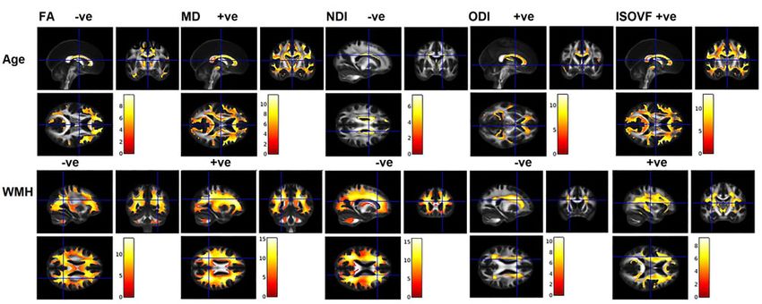
4   | BRAIN COMMUNICATIONS 2021: Page 4 of 15                                                                                          S. Raghavan et al.

                                                                                                                                                            Downloaded from https://academic.oup.com/braincomms/article/3/2/fcab106/6278287 by guest on 14 September 2021
    Figure 1 DTI and NODDI maps from a participant and white matter tracts of interest from the JHU atlas. (A) FA, MD, NDI,
    ODI and ISOVF maps generated from a 67-year-old cognitively unimpaired female participant. (B) White matter tracts of interest from JHU
    atlas. FA, fractional anisotropy; ISOVF, isotropic volume fraction; MD, mean diffusivity; NDI, neurite density index; ODI, orientation dispersion
    index.

mask derived from the 3D MPRAGE segmentation, and                              association with cognition.1,14,16,40–42: commissural fibres:
by removing isolated single-voxel detections.                                  genu (GCC), body (BCC), and splenium (SCC) of corpus
                                                                               callosum, and fornix (FX); association fibres: cingulum
Cognitive performance measures                                                 (CGC), parahippocampal cingulum (CGH), superior lon-
                                                                               gitudinal fasciculus (SLF), inferior fronto-occipital fascic-
All MCSA participants underwent a detailed neuro-                              ulus; and other relevant tracts: inferior temporal WM
psychological test battery that consisted of 9 tests cover-                    (ITWM) and anterior limb of internal capsule (Fig. 1B).
ing 4 cognitive subdomains.25,38 The present study                             The median values of FA, MD, NDI, ODI and ISOVF
utilized a global cognitive z-score that was derived as the                    were computed in these tracts by non-linearly registering
z-transformation of the average of all nine tests across                       an in-house modified version of the JHU ‘Eve’ WM
the 4 cognitive domains (memory, language, attention/ex-                       atlas43 to each subject’s image using Advanced
ecutive and visuo-spatial function).39 Individual and com-                     Normalization Tools–Symmetric Normalization (ANTS-
pound scores from Trail Making Test (Trails) A and B                           SyN).44 In this analysis, we excluded the cuneus, precu-
(time to complete the test) were used as a sensitive test                      neus, fusiform and lingual WM regions since they are
for processing speed. The raw scores were transformed                          too small for reliable registration. The median values of
into z-scores and averaged to create a composite score.                        bilateral regions were then averaged, weighting by region
                                                                               size, to produce a single measure for each bilateral
Image analysis                                                                 structure.

ROI-based analysis                                                             Voxel-based analysis of diffusion metrics
We performed an ROI analysis in ten WM tracts which                            Diffusion images were analysed using an in house devel-
were selected based on literature suggesting their                             oped voxel-based analysis (VBA) pipeline for SPM12 in
Basis of microstructural changes BRAIN COMMUNICATIONS 2021: Page 5 of 15 | 5
MATLAB to identify the global brain changes in associ- sex, education/occupation, cycle number (the number of
ation with demographics and disease pathologies. times the cognitive battery was administered to each spe-
Briefly, each subjects FA, MD, NDI, ODI and ISOVF cific subject to adjust for practice effects), and amyloid
maps were non-linearly registered to a custom-made and tau PET. We repeated the analyses for subdomain
study-specific template using ANTs-SyN. To reduce par- scores (memory, attention, language and visuospatial) and
tial volume effects and understand the regional results processing speed (Trail A, Trail B, composite score) with
based on tissue classes, additional mask images were regional WM microstructural integrity measures. We
made using GM, WM and GMþWM masks from each computed partial Pearson correlations with 95% confi-
subject’s segmented T1-weighted image. The masks were dence intervals and report the beta coefficients from the
registered to the study template using an ANTS-calcu- multiple regression analyses.
lated warp from the subject’s T1-weighted image to a
Downloaded from https://academic.oup.com/braincomms/article/3/2/fcab106/6278287 by guest on 14 September 2021
T1-like target synthesized from the FA and MD tem- Data availability
plates. The GM, WM and GMþWM masks were
Data used in this study are available upon reasonable re-
thresholded to include voxels with respective tissue type
quest via MCSA/ADRC data sharing website.
fractions >0.5. Each of the normalized diffusion images
was then smoothed with an 8-mm FWHM isotropic
Gaussian kernel and analysed per-voxel within each tis-
sue-class mask, using SPM12. Results
The characteristics of the participants are summarized in
Statistical analyses Table 1. The mean (standard deviation) age was 68.3
(13.1) years, 52% were men, 30% were APOE4 carriers,
Characteristics of the participants were summarized as
35% were amyloid positive and 27% were tau positive.
mean (standard deviation) for the continuous variables
Cognitively unimpaired individuals comprised 80% of
and count (%) for the categorical variables. WMH was
this sample.
presented and analysed as a percentage of total intracra-
nial volume (TIV). The distributions of WMH and amyl-
oid were skewed, and hence log transformed to obtain a Association between DTI and
more normal distribution. To describe the relationships NODDI metrics in different WM
between NODDI and DTI parameters, we performed a
tracts
series of unadjusted Pearson correlation analyses associat-
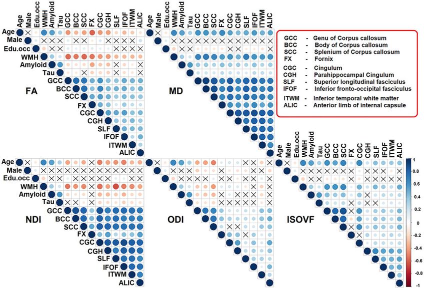
ing FA with NDI, FA with ODI, MD with NDI, MD Pearson correlations between FA, MD, NDI, ODI and
with ODI, and MD with ISOVF across subjects within ISOVF are shown in Fig. 2. Correlations within the same
each WM tract. We also used unadjusted Pearson correl- regions between measures: across the WM tracts, MD
ation analyses to describe associations between demo- and NDI showed the strongest association with each
graphics (age, sex and education/occupation), other (r 0.676) except for in the fornix. In contrast,
cerebrovascular disease (WMH) biomarkers, Alzheimer’s a modest association was observed between MD and
disease (amyloid and tau) biomarkers, and ROI-based dif- ODI in half of the regions (fornix and association tracts).
fusion (FA, MD, NDI, ODI and ISOVF) measures (corr- FA and NDI were associated modestly in most of the
plot package 0.84). WM tracts, while FA and ODI (indicators of dispersion)
To assess the contributions of cerebrovascular disease had strong associations in the association tracts, anterior
(WMH) and Alzheimer’s disease (amyloid and tau) bio- limb of internal capsule and inferior temporal WM (r
markers on the WM integrity changes, we fit multiple lin- 0.51). Similarly, MD was associated strongly with
ear regression models with each ROI diffusion measure ISOVF in the corpus callosum.
as the outcome variable, and with age, sex, education/oc-
cupation scores, WMH, amyloid and tau as predictor Associations with demographics and
variables. All the imaging variables were standardized. biomarkers of cerebrovascular
We also repeated the above analyses using voxel-wise
multiple regression analyses on the smoothed DTI and disease and Alzheimer’s disease
NODDI images with age, sex, education/occupation, Univariate associations: the univariate associations using
WMH, amyloid and tau as predictor variables. The gen- unadjusted Pearson correlations between tract measures
erated SPM-T maps were corrected for multiple compari- and age, sex, education/occupation, WMH, amyloid and
sons using family-wise error (FWE) with PFWE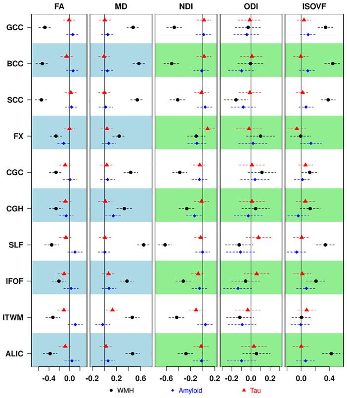
6     | BRAIN COMMUNICATIONS 2021: Page 6 of 15                                                                                                           S. Raghavan et al.

Table 1 Characteristics table with the mean (SD) listed for the continuous variables and count (%) for the categorical
variables

                                                 CU n 5 264                          MCI n 5 57                           Dementia n 5 7                           P-
                                                                                                                                                                   value
    Male, n (%)                                   135 (51%)                            30 (53%)                                4 (57%)                              0.94
    Age, years                                    65.9 (12.8)                         77.2 (9.8)                             84.7 (4.2)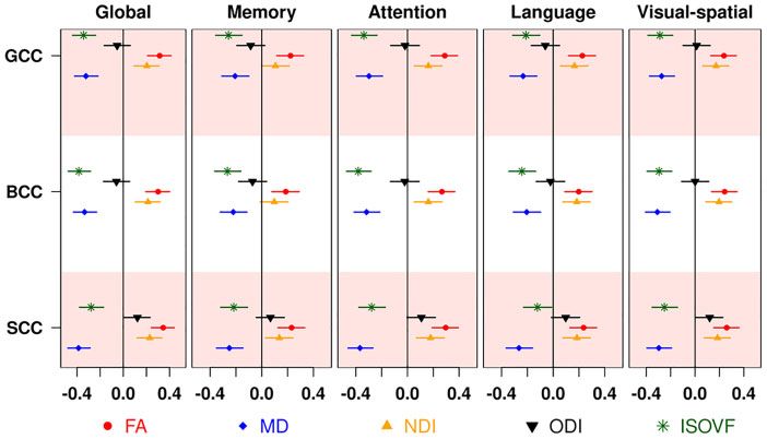
Basis of microstructural changes                                                            BRAIN COMMUNICATIONS 2021: Page 7 of 15                | 7

                                                                                                                                                         Downloaded from https://academic.oup.com/braincomms/article/3/2/fcab106/6278287 by guest on 14 September 2021
   Figure 3 Correlation matrix. Association between demographics (age, sex, education/occupation) or white matter hyperintensity
   (WMH) or amyloid or tau and diffusion measures. Edu.occ represents education/occupation. Colour legend indicates the range of
   correlations, the size of the circle indicates the strength of the correlation, and the symbol “X” indicates the non-significant P-value. FA,
   fractional anisotropy; ISOVF, isotropic volume fraction; MD, mean diffusivity; NDI, neurite density index; ODI, orientation dispersion index.

Multiple regression models with                                              was significantly associated with greater tau in inferior
focus on disease pathologies                                                 temporal WM (P ¼ 0.014).

The regression models with standardized disease patholo-
gies (WMH, amyloid and tau) as predictors and standar-
                                                                             Voxel level associations for
dized WM integrity measures of FA, MD, NDI, ODI and                          confirmation of ROI analyses
ISOVF as outcomes are shown in Fig. 4 and                                    Similar to the ROI analysis, the voxel-wise analyses
Supplementary Table 1. Associations with WMH: Across                         found the strongest associations for age and WMH with
all models, WMH had the strongest associations with all                      all diffusion metrics as displayed in Fig. 5. Modest asso-
dMRI metrics from all tracts. Higher WMH (a surrogate                        ciations were found with amyloid for both DTI and
of cerebrovascular disease) was significantly associated                     NODDI in the medial temporal lobe regions, specifically
with lower FA, higher MD, lower NDI and higher                               at the grey and WM junctions (Supplementary Fig. 1A).
ISOVF. Splenium was the only region where WMH                                The extent and strength of tau associations with dMRI
showed a statistically significant association with ODI.                     measures was minimal (Supplementary Fig. 1B).

                                                                             Association of diffusion measures
Associations with amyloid and tau                                            with cognition
Higher amyloid was significantly associated with higher                      Association results from multiple linear regression models
MD in parahippocampal cingulum (P ¼ 0.026). Higher                           of the global cognition and cognitive subdomain z-scores
amyloid was associated with lower NDI in the same re-                        with DTI and NODDI metrics after controlling for age,
gion, but the P-value was 0.053. In addition, higher MD                      sex, education/occupation, cycle visit, amyloid and tau
8 | BRAIN COMMUNICATIONS 2021: Page 8 of 15 S. Raghavan et al.
Downloaded from https://academic.oup.com/braincomms/article/3/2/fcab106/6278287 by guest on 14 September 2021
Figure 4 Association of diffusion metrics with white matter hyperintensity (WMH), amyloid and tau after controlling for
age, sex and education/occupation. Different symbols below are used for each of the primary predictors. FA, fractional anisotropy;
ISOVF, isotropic volume fraction; MD, mean diffusivity; NDI, neurite density index; ODI, orientation dispersion index.
are shown in Fig. 6 for the corpus callosum tracts (where Association of diffusion measures with
the correlations were highest). The regression coefficients disease pathologies and cognition in
for all tracts with global cognition are shown in
Supplementary Table 2. Corpus callosum generally had non-demented participants
the most significant findings except for analyses of NDI We also performed sensitivity analyses after excluding de-
and ODI where superior longitudinal fasciculus and cin- mentia participants. As described above, we evaluated (i)
gulum respectively had the greatest impact. The associa- the contribution of WMH, amyloid and tau on WM
tions between subdomain scores and diffusion metrics changes (after adjusting for age, sex, education/occupa-
had a similar pattern to that of global cognition with tion) and (ii) association of global cognition, subdomain
stronger associations with attention. Further analyses and processing speed scores with corpus callosum WM
revealed significant associations between diffusion metrics measures (after adjusting for age, sex, education/occupa-
and speed scores (Supplementary Fig. 2). As expected, the tion, and cycle number and amyloid and tau PET). There
strongest associations were observed for corpus callosum were no significant differences observed in these sensitiv-
fibres with MD and ISOVF and these changes were ity analyses as shown in the supplemental material
greatest with Trail B as an outcome. (Supplementary Figs 3–5).Basis of microstructural changes BRAIN COMMUNICATIONS 2021: Page 9 of 15 | 9
Downloaded from https://academic.oup.com/braincomms/article/3/2/fcab106/6278287 by guest on 14 September 2021
Figure 5 Association between age or white matter hyper intensity (WMH) and diffusion measures. Significance level set at
P < 0.05, FWE corrected with an extend threshold K ¼ 100. (þve and –ve represents the kind of association between variables). FA, fractional
anisotropy; ISOVF, isotropic volume fraction; MD, mean diffusivity; NDI, neurite density index; ODI, orientation dispersion index.
Figure 6 Association of diffusion metrics with cognition after controlling for age, sex and education/occupation, cycle visit,
amyloid and tau. Different symbols below are used for each of the diffusion measures. BCC, body of corpus callosum; FA, fractional
anisotropy; GCC, genu of corpus callosum; ISOVF, isotropic volume fraction; MD, mean diffusivity; NDI, neurite density index; ODI,
orientation dispersion index; SCC, splenium of corpus callosum.
Discussion about variation in WM health. There was complementary
information such that only MD and NDI had the stron-
We investigated the performance of DTI and NODDI gest correlations with each other across the tracts; (ii)
models in capturing the microstructural brain changes Age and WMH had the strongest associations with DTI
associated with demographics and pathological processes and NODDI measures among the measured WM tracts;
and their association with cognition in 328 MCSA sub- (iii) After adjusting for demographics, WMH was the
jects aged 33–98 years. The major findings of the study strongest predictor of diffusion measures; (iv) Both dMRI
were as follows: (i) NODDI and DTI are two different measures were able to detect subtle Alzheimer’s disease
biophysical models that provide distinct information related WM changes mainly at the medial temporal grey-10 | BRAIN COMMUNICATIONS 2021: Page 10 of 15 S. Raghavan et al.
white matter junctions and also WM tracts in the tem- Interestingly, ODI did not show a close association with
poral lobes; and (v) MD and ISOVF in the corpus cal- FA in the corpus callosum, which may be due to their
losum were strongest predictors of cognitive function. different responses to degeneration when most of the
Taken together, NODDI and traditional DTI measures fibres are strongly aligned.10,54 Specifically, if the callosal
are comparable in their predictive ability of WMH and boundary retreats due to atrophy, the edge voxels will be
cognition, but the non-overlapping information provided filled in by more CSF, affecting FA but not ODI.
by each may aid in providing mechanistic insights into Outside the corpus callosum, the associations between
disease progression. NODDI and DTI measures were inconsistent in the asso-
ciation and temporal WM tracts, which may be explained
Advanced biophysical models versus by the differing sensitivity of neurites to growth/matur-
ational trajectories.55,56 Across the regions, the correl-
traditional models
Downloaded from https://academic.oup.com/braincomms/article/3/2/fcab106/6278287 by guest on 14 September 2021
ation between DTI and NODDI measures was smallest in
An advanced biophysical model such as NODDI lever- the fornix. The fornix is part of the limbic system that
ages richer multi-shell diffusion gradients to examine the connects the hippocampus to the subcortical structures
physiological alterations in neurites. In Fig. 2, we directly and is also well known for partial volume contamination
compared variation in DTI with variation in NODDI sig- by CSF. This selective weakened association indicates the
nal. The idea is that DTI signals are sensitive to gross correction for CSF-contamination effects in the NODDI
anatomical and neuropathological changes associated method.
with WM,45 but they are inherently non-specific to disen-
tangle the complex tissue properties of a given voxel with Diffusion measures and age, sex and
crossing, kissing and fanning fibres.10,46 On the other
hand, NODDI measures demonstrated more putative cell
education/occupation
microstructure associations across studies10,47–51 and have Age-associated WM changes in imaging have been widely
been found to be strongly correlated with neurobiological reported. Past studies demonstrated NODDI as a key
underpinnings.7,52,53 While decreases in NDI and marker for studying ageing,47,57–59 and the association of
increases in ISOVF are straightforward to understand, age and sex.1 Consistent with the ageing literature, age
ODI changes have been hard to interpret because there is was associated with a decrease in FA and NDI, increase
no simple physical mechanism that directly relates them in MD ISOVF, and increase or decrease in ODI (which
to disease processes, such as demyelination, inflammation, depends on the tract tortuosity and the presence of cross-
or atrophy. Also, the direction of the correlation between ing fibres). Among the diffusion parameters, MD had the
axonal loss and ODI changes depends on region. most sensitive age effects across the majority of tracts,1,60
Consider a hypothetical axon that runs parallel to a bun- while the unconstrained diffusivity metric ISOVF demon-
dle for a few centimetres and then perpendicularly to a strated the greatest age effect in corpus callosum (genu
different bundle. The loss of the axon would increase the and splenium) and cingulum. The overall widespread in-
ODI in the parallel region and decrease it in the perpen- crease in ISOVF with age suggests the increase in FW
dicular region (ODI ¼ 0 for a perfectly aligned bundle concentration in specific brain regions. However, the key
and goes to 1 where fibres spread out equally in all drivers of this increased FW are largely unknown. In
directions). addition to cerebrovascular disease and neurodegenerative
Unfortunately, at the macroscopic scale of a voxel the pathologies, other possible underlying neuropathological
NODDI measures by themselves do not specify which factors include an influx of CSF or other factors like cell
axons changed. FA also suffers from this ambiguity, but shrinkage,41 edoema,61 and neuroinflammation.62
unlike ODI is also directly coupled to demyelination and Interestingly, past MRI and histology evidence clarified
atrophy. Therefore, contrasting the relationship between this as age- related increase in interstitial water.63,64
DTI (FA and MD) and NODDI (NDI, ODI and ISOVF) While most tracts had strong correlations with age,
can help understand the regional variations in these asso- there were some subtle differences. With NDI, the associ-
ciations, which are largely unknown in the population. ation tracts (especially cingulum, superior longitudinal
The most consistent relationship between NODDI and fasciculus and inferior fronto-occipital fasciculus) had the
DTI was seen with MD and NDI (but not with ODI and greatest age associations suggesting the presence of higher
ISOVF), implicating that rather than orientation and neurite density fibres in more metabolically active brain
geometry of tracts, a higher density may drive more dif- regions65,66 that may be vulnerable to detrimental system-
fusion restriction.50 We found a positive association of ic age effects. ODI exhibited heterogeneous regional var-
FA with NDI across various WM tracts with primarily a iations with age. Although there is reduced tract
stronger relationship in corpus callosum fibres. This fibre complexity in the corpus callosum fibres, the higher dis-
pathway connects the two hemispheres, and the observed persion in fornix, cingulum and parahippocampal cingu-
positive association between NDI and FA suggests the ex- lum suggests the greater loosening, fanning and possibly
istence of the same underlying physiological processes bending of axonal bundles with the advancement of
(reduced axonal packing and demyelination).10,50 age.1,49 As stated above, this could be explained asBasis of microstructural changes BRAIN COMMUNICATIONS 2021: Page 11 of 15 | 11
evidence of continuous remodelling of WM during the and MD to explain the underlying histological changes
life span which is more evident after the sixth decade. associated with WMH. The NDI finding in the genu of
Notably, the age-related changes in the hippocampal con- the corpus callosum was in accordance with a prior
nections might explain the amnestic changes in the elderly NODDI study that explored the diabetic encephalopathy
population.16,49 in subjects with cognitive impairment.72 The only meas-
The sex-specific WM integrity association of DTI and ure that did not show consistent associations with WMH
NODDI is sparsely covered in the literature. A few DTI was ODI. We believe this is due to ODI nominally being
studies evaluated sex differences and reported inconsistent a property of only healthy neurites, and thus being more
findings. The inconsistency across these findings may be orthogonal to neuronal decay than the other NODDI
due to the heterogeneity of populations and differences in measures or DTI.
the analysis methods. In this study, we observed small Though the correlations with vascular risk were not a
Downloaded from https://academic.oup.com/braincomms/article/3/2/fcab106/6278287 by guest on 14 September 2021
sex differences in eight tracts, with higher FA in males
focus of the manuscript, as previously reported74 we
and greater ODI and ISOVF in females which is consist-
found that WM measures from traditional DTI (FA and
ent with previous studies in healthy adults.1,49,67
MD) and NODDI (NDI specifically) were significantly
Reserve and resilience factors are important modulating
associated with worsening vascular risk (Results not
parameters between brain injury and cognitive outcomes.
shown). Recent researchers focussed on using global dif-
Past studies showed education/occupation as a ‘proxy’ for
cognitive reserve and related differences in fibre tract in- fusion MRI as a cerebrovascular disease marker.71,76,77
tegrity.68,69 Similarly, a previous study in the MCSA However, this work sheds light on the variability in re-
population demonstrated a significant association of intel- gional associations suggesting a greater sensitivity and
lectual enrichment on FA of the genu.70 Although this as- specificity of regional markers. Future work should be
sociation was modest, the present study showed a undertaken to widely validate and compare diffusion out-
positive association of DTI and NODDI measures with comes as cerebrovascular disease measures.
corpus callosum, cingulum, parahippocampal cingulum,
superior longitudinal fasciculus, inferior fronto-occipital
fasciculus and inferior temporal WM, which is consistent Diffusion measures and
with prior DTI studies.70,71 The anatomical localization neuroimaging Alzheimer’s disease
provides insights into the associations between resilience
mechanisms and brain maturation and plasticity.
measures
The association between amyloid deposition and WM
Diffusion measures as markers of microstructure is still a matter of debate. A non-mono-
tonic behaviour was found between both measures in
cerebrovascular disease GM78 and WM79,80 in human studies. Consistent with
Although WMH is the most commonly used biomarker our region level findings, prior DTI studies reported
for cerebrovascular disease,2 it only represents extensive reduced FA in corpus callosum and fornix81,82 in cogni-
(and structurally visible) WM damage and fails to meas- tively unimpaired individuals and increased axial diffusiv-
ure the disruption or subtle changes of the underlying ity83 and accelerated FA decrease84 in the
WM tracts. There is growing evidence supporting the parahippocampal cingulum of amyloid positive individu-
utility of DTI to characterize the WM changes in cerebro- als. As expected, the current study identified a significant
vascular disease40,72,73 even before the appearance of global association between Ab deposition and increased
WMH and cognitive decline. Notably, the observed de- MD and ISOVF along with decreased NDI in medial
crease in diffusion directionality and an increase in the
temporal lobe grey-white matter junctions, which are con-
extent of water diffusion in conjunction with WMH are
sistent with a more recent study that reported lower neu-
consistent with prior findings.40,67,73 Consistent with this
rite density in limbic and association fibres and higher
idea, a recent study using FW imaging demonstrated a
medial temporal FW.85 The medial temporal lobe is an
greater contribution of cerebrovascular disease markers
early region of neuronal changes in Alzheimer’s disease,
than Alzheimer’s disease biomarkers (CSF and PET) in
memory clinic patients.17 so the parahippocampal cingulum findings were as
Similar to the age effect, we found a strong association expected. We also found associations between tau and
of WMH with corpus callosum and association fibres.5 non-specific MD and ISOVF association in the inferior
Although there is evidence for more vascular damage in temporal WM (Fig. 4). These results are supported by a
the thinly myelinated anterior corpus callosum,5,74,75 the study of tau and NODDI in a transgenic Alzheimer’s dis-
present study showed slight variations across the meas- ease model.86 Our findings in the temporal lobe (hippo-
ures. Importantly, conventional DTI performed as well as campal and parahippocampal regions) and the temporo-
NDI in detecting cerebrovascular disease changes.76 The occipital fusiform gyrus suggest that NODDI may be able
decreased density and dispersion of the neurites and to provide more detailed information about neurite health
increased FW might contrast the lack of specificity in FA in the presence of Alzheimer’s disease pathology.12 | BRAIN COMMUNICATIONS 2021: Page 12 of 15 S. Raghavan et al.
Diffusion measures with cognition Diffusion metrics are suggested to be most strongly
Association between WM DTI alterations and cognitive de- associated with processing speed.71,93 Therefore, we also
cline in the CU, MCI and Alzheimer’s disease populations tested these hypotheses in the supplemental material and
have been reported previously.40,67,70,75 Although there was found that both DTI and NODDI strongly predicted
decreased FA and increased MD in association with cogni- processing speed (Trail B and combined). As expected,
tive decline, the exact sources of DTI signal were not commissural fibres had the greatest effect size.
studied. The present study is one of the earliest studies to The present study has several strengths and limitations.
compare DTI and NODDI based on their association with The main strength was the extensive analyses of single
cognitive performance after accounting for amyloid and tau, and multi-shell diffusion data on WM health and cogni-
which allows us to evaluate its utility as a cerebrovascular tion. Also, this is the first study to assess the relationship
disease marker. As expected, both DTI and NODDI were between NODDI metrics and cerebrovascular disease and
Downloaded from https://academic.oup.com/braincomms/article/3/2/fcab106/6278287 by guest on 14 September 2021
significantly associated with global cognition and cognitive Alzheimer’s disease biomarkers together along with asso-
subdomain scores after adjusting for age, sex, cycle visit ciations with cognitive performance. The inclusion of a
and Alzheimer’s disease biomarkers. The overall pattern representative sample population strengthens the general-
suggests that higher coherence and density, and lower FW izability of the findings. Our voxel-wise and regional
concentration and tract complexity, both correlate with bet- findings mostly corroborated each other, and the slight
ter cognitive performance.16,87 Across the tracts, the stron- differences may be due to partial volume effects, smooth-
gest association of reduced WM integrity and worse global ing, and more stringent FWE corrections. The major limi-
cognitive performance was observed in the corpus callosum. tations are the cross-sectional nature of the study and the
This is consistent with a previous DTI study in cerebrovas- lack of histological confirmation of the observed associa-
cular disease that reported highly significant correlations of tions. Another limitation is the smaller number of sub-
genu and splenium with global cognitive performance.40 jects in the dementia group, but the results remain the
Impaired interhemispheric connection pathways contribute same after excluding them. Furthermore, the regulariza-
to multiple impaired cognitive functions, such as impaired tion scheme used by the AMICO implementation of
memory, psychomotor speed, frontal lobe mediated atten- NODDI acts like a prior that gives a mild preference to
tion and executive function.88,89 Additionally, these observa- some values of NDI, ISOVF, and especially ODI, which
tions replicated our recent study in MCI that showed could be obscuring some differences between subjects.
greater predictability of high FA of genu on better cognitive Future longitudinal research with multiple biophysical
performance,75 even after controlling for amyloid and tau models11 may provide more sensitive and conclusive
PET. findings.
Although there are contributions from other domains, In summary, the present study provides evidence of
we found that the associations with WM integrity and microstructural WM alterations due to ageing and age-
cognitive performance were mainly driven by attention. related pathological processes, and their impact on cogni-
Importantly, our detailed investigation indicated that FW tion. Although NODDI-derived indices perform similar to
fraction in the corpus callosum predicted cognitive de- traditional FA and MD in predicting cognitive perform-
cline. In general, reduced neurite density correlated with ance, NODDI provides additional insights into the under-
worse cognitive performance with most of the tracts in lying synaptic density, organization and FW content
all domains. Among these, the stronger association of which are biological processes that cannot be separated
NDI than FA in superior longitudinal fasciculus may be with DTI. Among DTI and NODDI indices, MD and
due to its proximity to the crossing fibres in the centrum FW fraction provided by ISOVF were the key parameters
semiovale,90 which corresponds to higher FA and lower in predicting cognition. This study also highlights the spa-
tract complexity. Another speculation may be that the su- tial heterogeneity of tracts across the metrics and which
perior longitudinal fasciculus is connecting lateral pre- highlights the importance of looking at each diffusion
frontal to parietal brain areas, which are responsible for metrics to investigate changes in each WM region of the
the multifaceted processes we studied here. Notably, cin- brain as a function of disease progression.
gulum performed uniformly well across all domains and
diffusion metrics to predict cognition. This bundle is the
prominent WM tract that interconnects frontal, parietal, Supplementary material
and medial temporal lobe and the posterior cingulate cor- Supplementary material is available at Brain
tex. Surprisingly, the parahippocampal cingulum bundle, Communication online.
which connects the hippocampus to the rest of the brain
areas, emerged as an important tract in visuospatial func-
tion. In contrast, deteriorations in parahippocampal cin-
gulum have previously been implicated in association
Acknowledgements
with episodic memory in older subjects91 and Alzheimer’s We would like to thank Lorraine Vassallo for her help with
disease.92 editing the manuscript. We thank all the study participantsBasis of microstructural changes BRAIN COMMUNICATIONS 2021: Page 13 of 15 | 13
and staff in the Mayo Clinic Study of Aging, Mayo Communications, Inc. and receives research support from
Alzheimer’s Disease Research Center, and Aging Dementia the NIH.
Imaging Research laboratory at the Mayo Clinic for making
this study possible. We gratefully acknowledge the support
of NVIDIA Corporation for the donation of the Quadro References
P5000 GPU used in this research. 1. Cox SR, Ritchie SJ, Tucker-Drob EM, et al. Ageing and brain
white matter structure in 3,513 UK Biobank participants. Nat
Commun. 2016;7:13629.
Funding 2. Wardlaw JM, Smith EE, Biessels GJ, et al.; STandards for
ReportIng Vascular changes on nEuroimaging (STRIVE v1).
This work was supported by National Institute of Health Neuroimaging standards for research into small vessel disease and
its contribution to ageing and neurodegeneration. Lancet Neurol.
Downloaded from https://academic.oup.com/braincomms/article/3/2/fcab106/6278287 by guest on 14 September 2021
[grants R01 NS097495 (PI: Vemuri), R01 AG56366 (PI:
2013;12(8):822-838.
Vemuri), U01 AG06786 (PI: Petersen/Mielke/Jack), P50 3. Nasrabady SE, Rizvi B, Goldman JE, Brickman AM. White matter
AG16574 (PI: Petersen), R37 AG11378 (PI: Jack), R01 changes in Alzheimer’s disease: A focus on myelin and oligoden-
AG41851 (PIs: Jack and Knopman)]; the Gerald and drocytes. Acta Neuropathol Commun. 2018;6(1):22.
Henrietta Rauenhorst Foundation grant, Alzheimer’s Drug 4. Caballero MÁA, Song Z, Rubinski A, et al. Age-dependent amyl-
oid deposition is associated with white matter alterations in cogni-
Discovery Foundation (ADDF), the Alexander Family
tively normal adults during the adult life span. Alzheimers
Alzheimer’s Disease Research Professorship of the Mayo Dement. 2020;16(4):651-661.
Foundation, Liston Award, Elsie and Marvin Dekelboum 5. Cox SR, Lyall DM, Ritchie SJ, et al. Associations between vascular
Family Foundation, Schuler Foundation, Opus building risk factors and brain MRI indices in UK Biobank. Eur Heart J.
Jul 21 2019;40(28):2290-2300.
National Institute of Health (grant C06 RR018898), and
6. Pines AR, Cieslak M, Larsen B, et al. Leveraging multi-shell diffu-
was made possible by Rochester Epidemiology Project (R01 sion for studies of brain development in youth and young adult-
AG34676). hood. Dev Cogn Neurosci. 2020;43:100788.
7. Schilling KG, Janve V, Gao Y, Stepniewska I, Landman BA,
Anderson AW. Histological validation of diffusion MRI fiber
Competing interests orientation distributions and dispersion. Neuroimage. 2018;165:
200-221.
8. Jensen JH, Helpern JA, Ramani A, Lu H, Kaczynski K. Diffusional
The authors report no competing interests related to the sub-
kurtosis imaging: The quantification of non-gaussian water diffu-
mitted manuscript. However, these are additional conflicts sion by means of magnetic resonance imaging. Magn Reson Med.
unrelated to this manuscript. Dr Raghavan reports no rele- 2005;53(6):1432-1440.
vant financial disclosures. Dr Reid reports no relevant finan- 9. Jensen JH, Helpern JA. MRI quantification of non-Gaussian water
diffusion by kurtosis analysis. NMR Biomed. Aug 2010;23(7):
cial disclosures. Mr Przybelski reports no relevant financial
698-710.
disclosures. Mr Lesnick reports no relevant financial disclo- 10. Zhang H, Schneider T, Wheeler-Kingshott CA, Alexander DC.
sures. Dr Graff-Radford serves as an assistant editor for NODDI: Practical in vivo neurite orientation dispersion and dens-
Neurology and receives research support from the NIH. Dr ity imaging of the human brain. Neuroimage. 2012;61(4):1000-16.
Mielke serves as a consultant for Biogen and Brain 11. Beck D, de Lange AG, Maximov II, et al. White matter microstruc-
ture across the adult lifespan: A mixed longitudinal and cross-sec-
Protection Company and receives research funds from NIH tional study using advanced diffusion models and brain-age
and DOD. Dr Machulda receives research support from prediction. Neuroimage. 2020;224:117441.
NIH. Dr Knopman serves on a Data Safety Monitoring 12. Toschi N, Gisbert RA, Passamonti L, Canals S, De Santis S.
Board for the DIAN study. He serves on a Data Safety moni- Multishell diffusion imaging reveals sex-specific trajectories of
early white matter degeneration in normal aging. Neurobiol
toring Board for Biogen but receives no personal compensa-
Aging. 2020;86:191-200.
tion. He is an investigator in clinical trials sponsored by 13. Parker TD, Slattery CF, Zhang J, et al. Cortical microstructure in
Biogen, Lilly Pharmaceuticals and the University of Southern young onset Alzheimer’s disease using neurite orientation disper-
California. He serves as a consultant for Roche, Samus sion and density imaging. Hum Brain Mapp. 2018;39(7):
3005-3017.
Therapeutics, Third Rock and Alzeca Biosciences but
14. Slattery CF, Zhang J, Paterson RW, et al. ApoE influences regional
receives no personal compensation. Dr Petersen serves as a white-matter axonal density loss in Alzheimer’s disease. Neurobiol
consultant for Roche Inc., Merck Inc., and Biogen, Inc. He Aging. 2017;57:8-17.
serves on the Data Safety monitoring Board for Genentech, 15. Vogt NM, Hunt JF, Adluru N, et al. Cortical microstructural alter-
Inc and receives royalty from Oxford University Press and ations in mild cognitive impairment and Alzheimer’s disease de-
mentia. Cereb Cortex (New York, NY: 1991). 2020;30(5):
UpToDate. Dr Jack serves on an independent data monitor- 2948-2960.
ing board for Roche, has served as a speaker for Eisai, and 16. Wen Q, Mustafi SM, Li J, et al. White matter alterations in early-
consulted for Biogen, but he receives no personal compensa- stage Alzheimer’s disease: A tract-specific study. Alzheimers
tion from any commercial entity. He receives research sup- Dement (Amsterdam, Netherlands). 2019;11:576-587.
17. Finsterwalder S, Vlegels N, Gesierich B, et al.; Utrecht VCI study
port from NIH and the Alexander Family Alzheimer’s group. Small vessel disease more than Alzheimer’s disease deter-
Disease Research Professorship of the Mayo Clinic. Dr mines diffusion MRI alterations in memory clinic patients.
Vemuri received speaker fees from Miller Medical Alzheimers Dement. 2020;16(11):1504-1514.14 | BRAIN COMMUNICATIONS 2021: Page 14 of 15 S. Raghavan et al.
18. Bells S, Lefebvre J, Prescott SA, et al. Changes in white matter 39. Vemuri P, Lesnick TG, Przybelski SA, et al. Association of lifetime
microstructure impact cognition by disrupting the ability of neural intellectual enrichment with cognitive decline in the older popula-
assemblies to synchronize. J Neurosci. 2017;37(34):8227-8238. tion. JAMA Neurol. 2014;71(8):1017-1024.
19. Filley CM, Fields RD. White matter and cognition: Making the 40. Tuladhar AM, van Norden AG, de Laat KF, et al. White matter
connection. J Neurophysiol. 2016;116(5):2093-2104. integrity in small vessel disease is related to cognition. Neuroimage
20. Jelescu IO, Budde MD. Design and validation of diffusion MRI Clin. 2015;7:518-524.
models of white matter. Front Phys. 2017;28:61. 41. Merluzzi AP, Dean DC 3rd, Adluru N, et al. Age-dependent differ-
21. Novikov DS, Veraart J, Jelescu IO, Fieremans E. Rotationally-in- ences in brain tissue microstructure assessed with neurite orienta-
variant mapping of scalar and orientational metrics of neuronal tion dispersion and density imaging. Neurobiol Aging. 2016;43:
microstructure with diffusion MRI. Neuroimage. 2018;174: 79-88.
518-538. 42. Bendlin BB, Fitzgerald ME, Ries ML, et al. White matter in aging
22. Zhang YZ, Chang C, Wei XE, Fu JL, Li WB. Comparison of diffu- and cognition: A cross-sectional study of microstructure in adults
sion tensor image study in association fiber tracts among normal, aged eighteen to eighty-three. Dev Neuropsychol. 2010;35(3):
amnestic mild cognitive impairment, and Alzheimer’s patients. 257-277.
Downloaded from https://academic.oup.com/braincomms/article/3/2/fcab106/6278287 by guest on 14 September 2021
Neurology India. 2011;59(2):168-173. 43. Oishi K, Faria A, Jiang H, et al. Atlas-based whole brain white
23. Rocca WA, Yawn BP, St Sauver JL, Grossardt BR, Melton LJ 3rd. matter analysis using large deformation diffeomorphic metric map-
History of the Rochester Epidemiology Project: Half a century of ping: Application to normal elderly and Alzheimer’s disease partic-
medical records linkage in a US population. Mayo Clinic Proc. ipants. Neuroimage. 2009;46(2):486-499.
2012;87(12):1202-1213. 44. Avants BB, Tustison NJ, Song G, Cook PA, Klein A, Gee JC. A re-
24. St Sauver JL, Grossardt BR, Yawn BP, et al. Data resource profile: producible evaluation of ANTs similarity metric performance in
The Rochester Epidemiology Project (REP) medical records-linkage brain image registration. Neuroimage. 2011;54(3):2033-2044.
system. Int J Epidemiol. 2012;41(6):1614-1624. 45. Alexander AL, Lee JE, Lazar M, Field AS. Diffusion tensor imag-
25. Petersen RC, Roberts RO, Knopman DS, et al. Prevalence of mild ing of the brain. Neurotherapeutics. 2007;4(3):316-329.
cognitive impairment is higher in men. The Mayo Clinic Study of 46. Jeurissen B, Leemans A, Tournier JD, Jones DK, Sijbers J.
Aging. Neurology. 2010;75(10):889-897. Investigating the prevalence of complex fiber configurations in
26. Caruyer E, Lenglet C, Sapiro G, Deriche R. Design of multishell white matter tissue with diffusion magnetic resonance imaging.
sampling schemes with uniform coverage in diffusion MRI. Magn Hum Brain Mapp. 2013;34(11):2747-2766.
Reson Med. 2013;69(6):1534-1540. 47. Chang YS, Owen JP, Pojman NJ, et al. White matter changes of
27. Reid RI, Nedelska Z, Schwarz CG, et al. Diffusion specific seg- neurite density and fiber orientation dispersion during human
mentation: Skull stripping with diffusion MRI data alone. brain maturation. PloS One. 2015;10(6):e0123656.
Computational diffusion MRI mathematics and visualization. 48. Eaton-Rosen Z, Melbourne A, Orasanu E, et al. Longitudinal
Cham: Springer; 2018. measurement of the developing grey matter in preterm subjects
28. Veraart J, Novikov DS, Christiaens D, Ades-Aron B, Sijbers J, using multi-modal MRI. Neuroimage. 2015;111:580-589.
Fieremans E. Denoising of diffusion MRI using random matrix 49. Kodiweera C, Alexander AL, Harezlak J, McAllister TW, Wu YC.
theory. Neuroimage. 2016;142:394-406. Age effects and sex differences in human brain white matter of
29. Andersson JLR, Sotiropoulos SN. An integrated approach to cor- young to middle-aged adults: A DTI, NODDI, and q-space study.
rection for off-resonance effects and subject movement in diffusion Neuroimage. 2016;128:180-192.
MR imaging. Neuroimage. 2016;125:1063-1078. 50. Mah A, Geeraert B, Lebel C. Detailing neuroanatomical develop-
30. Kellner E, Dhital B, Kiselev VG, Reisert M. Gibbs-ringing artifact ment in late childhood and early adolescence using NODDI. PloS
removal based on local subvoxel-shifts. Magn Reson Med. 2016; One. 2017;12(8):e0182340.
76(5):1574-1581. 51. Timmers I, Zhang H, Bastiani M, Jansma BM, Roebroeck A,
31. Koay CG, Ozarslan E, Basser PJ. A signal transformational frame- Rubio-Gozalbo ME. White matter microstructure pathology in
work for breaking the noise floor and its applications in MRI. J classic galactosemia revealed by neurite orientation dispersion and
Magn Reson (San Diego, Calif: 1997). 2009;197(2):108-119. density imaging. J Inherit Metab Dis. 2015;38(2):295-304.
32. Garyfallidis E, Brett M, Amirbekian B, et al.; Dipy Contributors. 52. Sato K, Kerever A, Kamagata K, et al. Understanding microstruc-
Dipy, a library for the analysis of diffusion MRI data. Front ture of the brain by comparison of neurite orientation dispersion
Neuroinform. 2014;8:8. and density imaging (NODDI) with transparent mouse brain. Acta
33. Daducci A, Canales-Rodrı́guez EJ, Zhang H, Dyrby TB, Radiol Open. 2017;6(4):2058460117703816.
Alexander DC, Thiran JP. Accelerated Microstructure Imaging via 53. Sepehrband F, Clark KA, Ullmann JF, et al. Brain tissue compart-
Convex Optimization (AMICO) from diffusion MRI data. ment density estimated using diffusion-weighted MRI yields tissue
Neuroimage. 2015;105:32-44. parameters consistent with histology. Hum Brain Mapp. 2015;
34. Jack CR Jr., Wiste HJ, Weigand SD, et al. Defining imaging bio- 36(9):3687-3702.
marker cut points for brain aging and Alzheimer’s disease. 54. Genc S, Malpas CB, Holland SK, Beare R, Silk TJ. Neurite density
Alzheimers Dement. 2017;13(3):205-216. index is sensitive to age related differences in the developing brain.
35. Graff-Radford J, Arenaza-Urquijo EM, Knopman DS, et al. White Neuroimage. 2017;148:373-380.
matter hyperintensities: Relationship to amyloid and tau burden. 55. Lebel C, Gee M, Camicioli R, Wieler M, Martin W, Beaulieu C.
Brain. 2019;142(8):2483-2491. Diffusion tensor imaging of white matter tract evolution over the
36. Zhang Y, Brady M, Smith S. Segmentation of brain MR images lifespan. Neuroimage. 2012;60(1):340-352.
through a hidden Markov random field model and the expect- 56. Huang H, Zhang J, Wakana S, et al. White and gray matter devel-
ation-maximization algorithm. IEEE Trans Med Imaging. 2001; opment in human fetal, newborn and pediatric brains.
20(1):45-57. Neuroimage. 2006;33(1):27-38.
37. Manjón JV, Coupé P, Martı́-Bonmatı́ L, Collins DL, Robles M. 57. Billiet T, Vandenbulcke M, Mädler B, et al. Age-related micro-
Adaptive non-local means denoising of MR images with spatially structural differences quantified using myelin water imaging and
varying noise levels. J Magn Reson Imaging. 2010;31(1):192-203. advanced diffusion MRI. Neurobiol Aging. 2015;36(6):
38. Roberts RO, Geda YE, Knopman DS, et al. The Mayo Clinic 2107-2121.
Study of Aging: Design and sampling, participation, baseline meas- 58. Kunz N, Zhang H, Vasung L, et al. Assessing white matter micro-
ures and sample characteristics. Neuroepidemiology. 2008;30(1): structure of the newborn with multi-shell diffusion MRI and bio-
58-69. physical compartment models. Neuroimage. 2014;96:288-299.You can also read