Diversity of Adult Neural Stem and Progenitor Cells in Physiology and Disease - MDPI
←
→
Page content transcription
If your browser does not render page correctly, please read the page content below
cells
Review
Diversity of Adult Neural Stem and Progenitor Cells in
Physiology and Disease
Zachary Finkel, Fatima Esteban, Brianna Rodriguez, Tianyue Fu, Xin Ai and Li Cai *

                                          Department of Biomedical Engineering, Rutgers University, Piscataway, NJ 08854, USA; zf99@rutgers.edu (Z.F.);
                                          fce10@scarletmail.rutgers.edu (F.E.); br444@scarletmail.rutgers.edu (B.R.); tf274@scarletmail.rutgers.edu (T.F.);
                                          xa16@rutgers.edu (X.A.)
                                          * Correspondence: lcai@rutgers.edu

                                          Abstract: Adult neural stem and progenitor cells (NSPCs) contribute to learning, memory, main-
                                          tenance of homeostasis, energy metabolism and many other essential processes. They are highly
                                          heterogeneous populations that require input from a regionally distinct microenvironment including
                                          a mix of neurons, oligodendrocytes, astrocytes, ependymal cells, NG2+ glia, vasculature, cere-
                                          brospinal fluid (CSF), and others. The diversity of NSPCs is present in all three major parts of the
                                          CNS, i.e., the brain, spinal cord, and retina. Intrinsic and extrinsic signals, e.g., neurotrophic and
                                          growth factors, master transcription factors, and mechanical properties of the extracellular matrix
                                          (ECM), collectively regulate activities and characteristics of NSPCs: quiescence/survival, prolifer-
                                          ation, migration, differentiation, and integration. This review discusses the heterogeneous NSPC
                                          populations in the normal physiology and highlights their potentials and roles in injured/diseased
                                states for regenerative medicine.


Citation: Finkel, Z.; Esteban, F.;        Keywords: central nervous system (CNS); ependymal cells; neural stem and progenitor cells (NSPC);
Rodriguez, B.; Fu, T.; Ai, X.; Cai, L.    NG2+ cells; neurodegenerative diseases; regenerative medicine; retina injury; spinal cord injury
Diversity of Adult Neural Stem and        (SCI); traumatic brain injury (TBI)
Progenitor Cells in Physiology and
Disease. Cells 2021, 10, 2045. https://
doi.org/10.3390/cells10082045

                                          1. Introduction
Academic Editors:
                                               During development, neural stem cells (NSCs) are responsible for the formation
Gabriella Minchiotti
                                          of the central nervous system (CNS). Initially, NSCs, also called neuroepithelial cells,
and Annalisa Fico
                                          differentiate into radial glial cells and proliferate into pools of neural progenitor cells
Received: 15 June 2021
                                          (NPCs) [1]. NSC refers to an uncommitted cell with differentiation potential into the
Accepted: 3 August 2021
                                          neurons and glia of the CNS. NSC is defined by two essential characteristics: self-renewal
Published: 10 August 2021                 and multipotency [2]. These neural stem and progenitor cells (NSPCs) represent both
                                          populations and are established as the only self-renewing cell type in the adult CNS. NSPCs
Publisher’s Note: MDPI stays neutral      migrate and differentiate into highly specified networks of neurons via neurogenesis, and
with regard to jurisdictional claims in   oligodendrocytes and astrocytes are generated via gliogenesis [3,4] (Figure 1). Thus, NSPCs
published maps and institutional affil-   are a major research thrust in the field of regenerative medicine. Extrinsic and intrinsic
iations.                                  factors such as neurotrophic/growth factors, transcription factors, and canonical pathways
                                          guide neurogenesis and gliogenesis during development and adulthood.
                                               For the past 50 years, the topic of endogenous adult neurogenesis has been highly
                                          debated. This began with the initial discovery of adult mammalian neurogenesis in 1962 by
Copyright: © 2021 by the authors.
                                          Joseph Altman and has continued with noteworthy publications supporting the existence
Licensee MDPI, Basel, Switzerland.        or non-existence of adult neurogenesis in mammals [5]. In the adult CNS, neurogenesis
This article is an open access article    plays a primary role in essential processes such as learning, memory, maintenance of tissue
distributed under the terms and           homeostasis, and many others.
conditions of the Creative Commons
Attribution (CC BY) license (https://
creativecommons.org/licenses/by/
4.0/).

Cells 2021, 10, 2045. https://doi.org/10.3390/cells10082045                                                       https://www.mdpi.com/journal/cells
tricles. Damage to these regions can result in consequences including aberrant migration
                                  of NSPC progeny cells, incorrect dendritic branching, enhanced progenitor cell prolifera-
                                  tion, ineffective integration of cells into networks of tissue, and many others. Spinal cord
                                  injury (SCI) may affect the neurogenic niche of the central canal resulting in differing con-
Cells 2021, 10, 2045              tributions of NSPC populations to the glial scar. In addition, large differences in injury
                                                                                                                          2 of 23
                                  pathophysiology occur as a direct result of injury-mediated proliferation and altered dif-
                                  ferentiation.

      Figure 1.
      Figure 1. NSPC
                NSPC characteristics
                      characteristics in
                                      in adult
                                         adult mammals.
                                               mammals. (A)
                                                         (A) Self
                                                             Self renewal
                                                                  renewal requires
                                                                          requires input
                                                                                    input via
                                                                                          via extrinsic
                                                                                              extrinsic and
                                                                                                        and intrinsic
                                                                                                             intrinsic factors.
                                                                                                                       factors. These
                                                                                                                                These
      include signaling
      include signaling pathways
                        pathways Notch,
                                   Notch, Wnt,
                                           Wnt, and
                                                and Shh,
                                                    Shh, and
                                                         and transcription
                                                              transcription factors
                                                                            factors Sox2,
                                                                                    Sox2, Ascl1,
                                                                                          Ascl1, Bmi1,
                                                                                                 Bmi1, Tlx,
                                                                                                        Tlx, and
                                                                                                             and neurotransmitters
                                                                                                                  neurotransmitters
      and neurotrophic/trophic growth factors. (B) Multipotency allows NSPCs to differentiate into a variety of cell fates such
      and neurotrophic/trophic growth factors. (B) Multipotency allows NSPCs to differentiate into a variety of cell fates such as
      as Neurons, Astrocytes, and Oligodendrocytes. Adapted from Navarro Quiroz et al., 2018 [6].
      Neurons, Astrocytes, and Oligodendrocytes. Adapted from Navarro Quiroz et al., 2018 [6].

                                        In the eye, retinal
                                        Heterogeneous       injury results
                                                         populations       fromexist
                                                                     of NSPCs   chemical
                                                                                     in theor mechanical
                                                                                           neurogenic     damage
                                                                                                       niches of theand  is highly
                                                                                                                     brain, spinal
                                  dependent    on NSPC    activity. Traumatic  mechanical    injury of the eye results  in
                                  cord, and retina. Primary NSPCs are found in the subventricular zone (SVZ) and subgran-   severe
                                  ular zone (SGZ) of the brain and include radial glial-like cells, NG2+/oligodendrocyte
                                  progenitor cells (OPCs), and Foxj1+ ependymal cells. Both OPCs and ependymal cell
                                  populations can be found in the spinal cord. In the adult retina, potential sources of NSPCs
                                  include Müller glia cells and the ciliary epithelium (CE).
                                        NSPC response to CNS injury is extraordinarily complex and dependent upon the
                                  extent and location of injury. Injuries are most often contusion or blunt force-based and
                                  primarily result from sporting or vehicular accidents. Traumatic brain injuries (TBI) habitu-
                                  ally damage two central niches: SGZ of the hippocampus and SVZ of the lateral ventricles.
                                  Damage to these regions can result in consequences including aberrant migration of NSPC
                                  progeny cells, incorrect dendritic branching, enhanced progenitor cell proliferation, ineffec-
                                  tive integration of cells into networks of tissue, and many others. Spinal cord injury (SCI)
                                  may affect the neurogenic niche of the central canal resulting in differing contributions of
                                  NSPC populations to the glial scar. In addition, large differences in injury pathophysiology
                                  occur as a direct result of injury-mediated proliferation and altered differentiation.
Cells 2021, 10, 2045                                                                                           3 of 23

                             In the eye, retinal injury results from chemical or mechanical damage and is highly
                       dependent on NSPC activity. Traumatic mechanical injury of the eye results in severe
                       morphological and functional changes in the eye structure including retinal detachment
                       in humans [7]. Common retinal degenerative diseases include retinitis pigmentosa (RP),
                       age-related macular degeneration (AMD) and glaucoma. Retinal degeneration affects
                       photoreceptors, retinal ganglion cells and retinal pigment epithelium (RPE) to cause vision
                       loss at varying degrees and eventual blindness.
                             Adult neurogenesis in heterogeneous NSPC populations has been implicated in de-
                       myelinating, inflammatory, and neurodegenerative conditions such as Alzheimer’s disease
                       (AD), Parkinson’s disease (PD), multiple sclerosis (MS), and schizophrenia [8–10]. Early on-
                       set AD has been largely attributed to two genetic mutations, APP and presenilins (PS) [11].
                       Gene knock-out and knock-in mouse models show decreased neurogenesis, learning, and
                       memory associated with upregulation of PS genes [12]. PD is characterized by progressive
                       degeneration of dopaminergic neurons and PD-associated transgenic animal models have
                       shown increased neurogenesis in dopaminergic neurons [13]. MS is defined by oligo-
                       dendrocyte loss and axonal degeneration/demyelination [14]. A reduction in progenitor
                       proliferation in the SVZ was observed in the lesion model of MS. Neuronal loss or axonal
                       damage is characteristic of these conditions, thus modulation of adult neurogenesis, the
                       generation of new neurons, has been proposed as a prospective treatment.
                             Neurogenic activity of the brain, spinal cord, and retina may facilitate the generation
                       of functional networks of integrated tissue in damaged or diseased areas. Overall, NSPCs
                       play an essential role in injuries or degenerative disorders that are largely affected by
                       neurogenesis and disruptions in cell behavior such as traumatic brain injury (TBI), spinal
                       cord injury (SCI), retinal injury, multiple sclerosis (MS), and schizophrenia [8–10]. Thus, an
                       in-depth understanding of neurogenesis throughout the CNS will facilitate effective stem
                       cell oriented therapeutic development.

                       2. Neural Stem/Progenitor Cells
                            The adult NSPCs (e.g., progenitor cells, neuroblasts, ependymal cells, NG2+ glia)
                       are present in the stem cell niches of the brain, spinal cord, and retina. Major cell types
                       present in the general NSPC niche include neurons, oligodendrocytes, astrocytes, pericytes,
                       and endothelial cells. Neural stem cells primarily reside in the neural niches of the CNS,
                       whereas progenitor cells can be found throughout the CNS due to increased migratory
                       capacity [15–18].
                            Additional contributors to the microenvironment of NSPCs in CNS niches include
                       cerebrospinal fluid (CSF), the extracellular matrix (ECM), and vasculature. The CSF consists
                       of neurotrophic/growth factors, transcription factors, and ECM molecules required for
                       NSPC guidance and is important for cell migration, morphogenesis, growth, and develop-
                       ment [19]. The ECM provides mechanical support and regulates extracellular signaling
                       environments. Moreover, proteoglycan and glycoprotein composition varies to influence
                       signaling and bioavailability, motivating NSPC behavior within the stem cell niche [20].
                            Cellular cross talk between the stem cells and specified cell types contribute to the
                       symphony of cascading signals regulating NSPC behavior. NSPC populations in the
                       stem cell niche are highly regulated to produce neuronal or glial lineage cell types [21].
                       The vasculature also regulates neurogenesis in the adult CNS by transport of infiltrating
                       biochemical signals to interact with NSPCs [22]. In this way, intrinsic and extrinsic signals
                       regulate neurogenesis, generated via cross talk with cells, vasculature, ECM via external
                       forces, and CSF in the neural niche. Intrinsic signals include master transcription factors
                       such as Sox2 and REST [23]. Extrinsic signals include neurotrophic/trophic and growth
                       factors, neurotransmitters, and signaling pathways such as Wnt and Notch.
                            When networks of neural cell types responsible for a regulated signaling microen-
                       vironment are damaged, NSPCs exhibit extreme behavior [24]. This is due to distinctly
                       different signals or lack of signals required to regulate pools of active or quiescent NSPCs.
                       Traumatic injury stimulates NSPCs to proliferate rapidly and produce cells which con-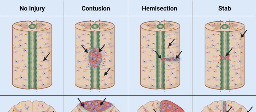
Cells 2021, 10, 2045                                                                                                                     4 of 23

       Cells 2021, 10, x                                                                                                 4 of 23
                           tribute to the glial scar border and upregulate angiogenesis in addition to neurogenic
                           activities [10]. Preferential survival of transplanted NSCs was observed in geographical
                            tributeoftohigh-density
                           areas        the glial scar border  and upregulate
                                                         vasculature,   which angiogenesis
                                                                                 is said to play in addition   to neurogenic
                                                                                                    an essential     role in theac- survival and
                            tivities [10]. Preferential
                           maintenance        of NSPCs   survival
                                                           in theofinjured
                                                                     transplanted
                                                                             spinalNSCscordwas   observed in geographical ar-
                                                                                             [22].
                            eas of high-density vasculature, which is said to play an essential role in the survival and
                                  NSPCs often generate new non-functional networks of cells in response to injury which
                            maintenance of NSPCs in the injured spinal cord [22].
                           inhibits    neural regeneration [25]. Altered niche activity may contribute to segregation of
                                  NSPCs often generate new non-functional networks of cells in response to injury
                           the
                            whichinjury    butneural
                                    inhibits   does regeneration
                                                      not lead to [25].
                                                                    regeneration
                                                                          Altered nicheof activity
                                                                                          functional
                                                                                                   may tissue.
                                                                                                         contributeDifferences
                                                                                                                       to segrega- in traumatic
                           injury
                            tion of type    and but
                                     the injury   grade   innot
                                                       does  thelead
                                                                  CNS  to result  in significant
                                                                          regeneration               changes
                                                                                         of functional    tissue.in  neurogenesis
                                                                                                                   Differences   in    in one or
                           more    niches
                            traumatic        [24].
                                         injury typeThe
                                                      andheterogeneity
                                                           grade in the CNS of result
                                                                               cell populations
                                                                                      in significant affected
                                                                                                       changes in byneurogenesis
                                                                                                                      traumatic injury result
                            in clinical
                           in  one or more     niches [24]. The
                                          inconsistencies         heterogeneity
                                                             between              of cell populations
                                                                         cases. Further,                 affectedniches
                                                                                            the neurogenic          by traumatic
                                                                                                                            of the brain, spinal
                            injury or
                           cord,    result  in clinical
                                        retina   exhibitinconsistencies  between cases.
                                                           regionally distinct      nicheFurther,     the neurogenic
                                                                                            composition         before niches    of traumatic
                                                                                                                          and after
                            the brain, spinal cord, or retina exhibit regionally distinct niche composition before and
                           injury (Figure 2).
                           after traumatic injury (Figure 2).

                           Figure 2. NSPC Niche in mammals: the SVZ and SGZ in the brain (A); the ependymal cells and
                           NG2 cells in the spinal cord (B); and the base of the optic nerve, the Müller glia, and the pig-
                           ment epithelium in the retina (C). AC, anterior chamber; CSF, cerebrospinal fluid; PC, posterior
                           chamber; SVZ, subventricular zone; SGZ, subgranular zone. Adapted from Cutler and Koko-
                           vay, 2020 [26] (A); Sabelström et al., 2014 [27], Andreotti et al., 2019; Picoli et al., 2019 [28,29] (B);
                           Yoshida et al., 2000 [30] (C).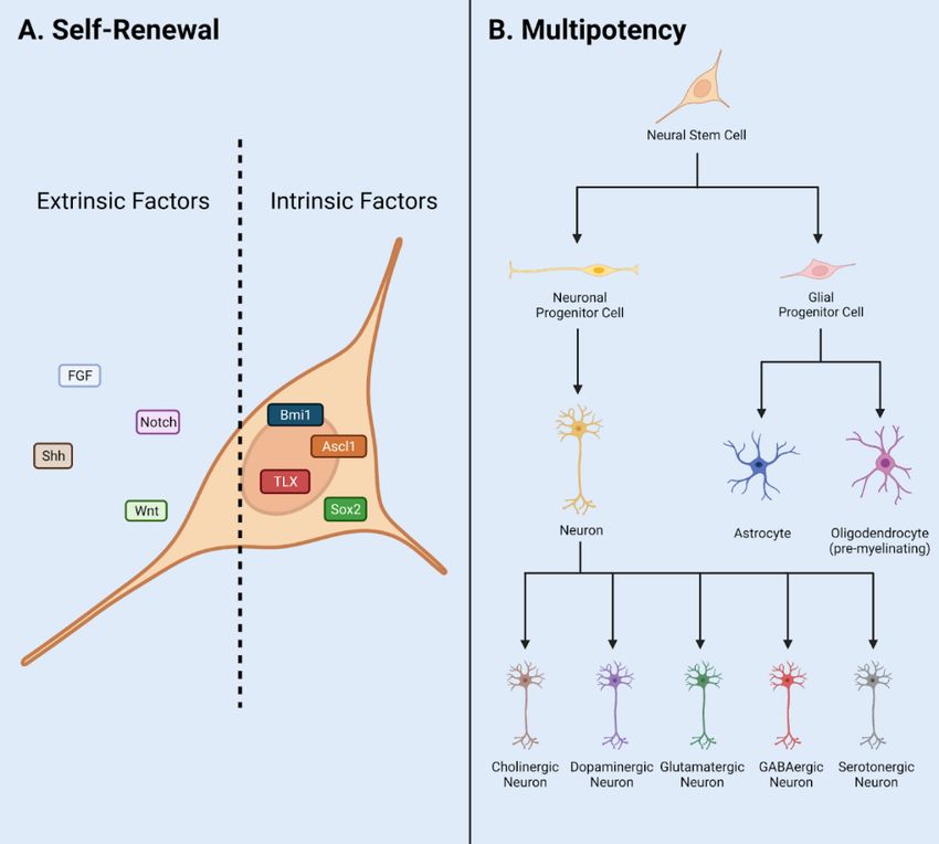
Cells 2021, 10, 2045                                                                                           5 of 23

                       2.1. Adult NSPCs in the Brain
                             The mammalian brain contains two primary neurogenic niches, i.e., the SGZ of the
                       hippocampus and the SVZ of the lateral ventricles [31]. The hypothalamus serves as a
                       third neurogenic niche conserved in some species but is nonexistent in humans [32]. Each
                       distinct niche contains specific populations of NSPCs and differing functions.
                             The hippocampal neurogenic niche is present at the base of the hippocampus within
                       the dentate gyrus (DG) in the SGZ (Figure 2A). In this niche, NSPCs are required for main-
                       tenance of the hippocampal tissue homeostasis, learning, and memory. Major stem cells in
                       this neurogenic niche are radial glial-like cells (RGLs) which maintain neurogenic activity
                       into adulthood [33]. Key cell types include OPCs, neuroblasts, immature/mature neurons,
                       and oligodendrocytes. As a note, OPC populations in this niche include NG2+ cells.
                             The neurogenic niche along the walls of the lateral ventricles is located in the SVZ
                       (Figure 2A). The lateral ventricle niche can be separated into two different geographical
                       regions in the tissue: 1. dorsal, 2. lateral. Both dorsal and lateral components are in direct
                       contact with pools of CSF, where the ependymal cell layer serves as a border between CSF
                       and niche NSPCs [34]. This allows regulated contact between the ventricular cavities and
                       undifferentiated progeny. Internal mechanisms direct NSPC behavior via fluid flow of
                       CSF in the lateral ventricles [19]. NSPCs include astrocytes, neuronal/and glial progenitor
                       subtypes, and neuroblasts [35–37]. Progenitors can be subdivided into further populations
                       based on gene mapping analysis in both domains. Transcriptional patterning in temporal
                       and spatial arrangements shows distinct NSPC populations [38]. Differential gene expres-
                       sion is driven by cell niche based signaling. Major signals include the Wnt/B-catenin and
                       sonic hedgehog (Shh) pathway and are important to maintain regulatory behavior [39].
                             Adult neurogenesis in the hippocampus is dictated by intrinsic and extrinsic cues [40].
                       Signaling is initiated by surrounding cell types and vasculature in addition to master
                       transcription factors Oct4, Sox2, and CREB. Signals from the Notch, Wnt, Shh, and other
                       pathways direct neurogenesis in the SGZ.
                             The rostral migratory stream is a migration pathway for neuroblasts from the SVZ
                       to the olfactory bulb and is present in some mammalian species. Conserved signaling
                       pathways direct differentiation and integration of specified neurons and glia into the
                       olfactory bulb. However, this is present to a lesser extent in larger mammalian species such
                       as humans. Migrating neuroblasts from the hippocampal niche have been documented in
                       rodent models to contribute to olfactory bulb mature cell types [41]. However, in human
                       and primate models these cells are instead generated in the striatum. Damage to the
                       neural niche of the hippocampus has been associated with cognitive deficits in learning
                       and memory.
                             The hypothalamus neurogenic niche is located near the lateral ventricles below the
                       SVZ, also called the periventricular zone [42]. Major cell types in this niche include hy-
                       pothalamic ribbon cells lining the outer wall and monocytes which may present neurogenic
                       potential. Three populations of NSCs have been found in the hypothalamus of animal
                       models including mouse, rat, and monkey including tanycytes, ependymal cells, and small
                       stellate cells [15]. These populations generate neurons and glia throughout life in the
                       hypothalamic parenchyma. Neurogenesis in this region occurs at a lesser incidence in com-
                       parison with the two classic niches, hippocampal SGZ and lateral ventricles SVZ. This may
                       translate into functional significance in murine models via control of energy metabolism.
                             In the injured brain, specific regulation of quiescence/survival in NSPCs has been
                       attributed to the small glycoprotein lactadherin, growth factors vascular endothelial growth
                       factor (VEGF), fibroblast growth factor-2 (FGF2), and Notch and Wnt pathways [39,43,44].
                       Proliferation is regulated by lactadherin, amyloid precursor protein, neurotrophic factor
                       Tumor necrosis factor alpha (TNFa), growth factors FGF2, and VEGF, chemokine CX3CL1,
                       and pathways Shh, Notch, and Wnt [45]. Migration is regulated by growth factor VEGF,
                       chemokines CCR2 and CX3CL1, and the Wnt pathway [44]. Differentiation is regulated by
                       growth factors FGF2 and VEGF, chemokines CCR2 and CX3CL1, as well as Notch and Shh
                       pathways. Integration is regulated by growth factor VEGF and chemokine CX3CL1 [43].
Cells 2021, 10, 2045 6 of 23
Injury-induced or altered signals contribute to the enhanced proliferation, aberrant progen-
itor migration, ineffective integration, and reduced dendritic branching observed in TBI
and SCI.
Using a combination of transgenic mouse model and single-cell RNA-seq analysis,
distinct adult NSPC populations were identified in the SVZ [46]. In this study, GFP+ cells
represent Nestin+ stem cell populations in the adult. Four groups of NSCs and three groups
of progenitor cells were characterized with in vivo and in vitro RNA-seq studies of the SVZ
neurogenic niche [46]. Immunostaining and imaging analysis revealed distinct subgroups
of cells separated by signal intensity: high GFP, low GFP and no GFP, and co-labeled
with specific markers such as DCX and GLAST. Further, RNA-seq analysis isolated cells
into profiles of quiescent and active stem cells in addition to stem cell markers, e.g., Sox2,
Ascl1, and DCX. Groups of cells are also separated anatomically, further supporting the
existence of distinct populations. NSPC heterogeneity has also been demonstrated using
stem cell markers including Gli1 and Ascl1 in both dividing and nondividing NSPCs [47].
The utility of these NSPC populations is unknown, but clear differences exist in gene
expression profile.
2.2. Adult NPSCs in the Spinal Cord
The mammalian spinal cord contains one neurogenic niche in the ependyma of the
central canal in which stem cells are present in an undifferentiated and self-renewable
state (Figure 2B). The central canal serves as a continuation of the lateral ventricles into the
spinal cord, while the ependymal cells serve as the bridge and a major regulatory element
between the CSF and the stem cell niche [48]. The central canal neurogenic niche is lined
with multiple populations of ependymal cells and CSF contacting neurons [49]. Ependymal
cell populations can be further characterized into cells with short basal processes and cells
with long extended processes. Other major components of the niche include NG2+ cells,
vasculature, astroglial cells, and oligodendrocytes. Populations of progenitors in the
spinal cord are indicated by markers Olig2, PDGFRa, and NG2 [50]. In addition, the
ependymal cell layer is surrounded by supporting mature cell types, while the layer itself
contains astroglial cells, NG2+ cells, and Nestin+ undifferentiated stem cells [49]. In
normal physiology, NSPC proliferation is observed in this stem cell niche, indicated by
Ki67 antibody staining in numerous studies [51,52].
Extrinsic signals guiding adult neurogenesis in the spinal cord include connexin,
Notch and Wnt signaling pathways [18,53]. Intrinsic signals include neural progenitor
transcription factors Nkx6.1, Pax6, and Olig6 [54–56]. These signals cohesively create an
environment to control NSPC activity and maintain normal pools of immature and mature
cell types in quiescent or active states. During injury or disease, NSPCs are subject to
altered specific niche-based signals and exhibit skewed behavior. Thus, the neural niche
in the central canal of the spinal cord is incredibly unique and maintained by a delicate
balance of intrinsic and extrinsic signals.
SCI affects the NSPC stem cell niche in models of contusive, surgical stab, and slice
injury at any anatomical level of the spinal cord [53]. Common clinical SCI disturbs the
niche due to equidistant dorsal and ventral positioning of the central canal [24]. NSPCs
proliferate after injury and interact with inflammatory signals to produce the glial scar
border, a chemical/physical barrier which segregates the injury and prevents additional
damage [57]. However, this scar also prevents axonal outgrowth into the site of injury and
generation of new cell types within the neural lesion. NSPCs proliferate and differentiate
into reactive astrocytes in the injured spinal cord and contribute to the glial scar border. In
addition to newly generated progeny, resident astrocytes transition to reactive gliosis state
and are recruited to the site of injury, lengthen their processes, and fatten to become the
scar border [58]. A multitude of NSPCs in the spinal cord produce progeny of differing
lineages to contribute to the glial scar after SCI and TBI.
Two major cell types have been controversially implicated in the NSPC response to
injury and pose high therapeutic potential: NG2+ and ependymal cells. Many publishedCells 2021, 10, 2045 7 of 23
studies are in support of the stem-like character or non-stem-like character of these cells.
Both NG2+ cells and ependymal cells have been reported to contribute to the formation of
the scar border. More recently, NG2+ cells have been shown to contribute to the generation
of neurons in the injured spinal cord [57,59]. We will discuss the heterogeneity of NSPCs
after injury with a focus on the activity of NG2+ and ependymal cells in Section 4.
2.3. Adult Retinal Stem Cells
Cells from regions of the adult retina such as the retinal pigment epithelium (RPE) [60,61],
CE [62–66], Müller glia cells [64,67–69], iris pigment epithelium [70,71] and optic nerve [61]
show stem cell characteristics to varying degrees in humans and rodents (Figure 2C).
Among them, the CE and Müller glia are identified as two main retinal stem cell sources.
A subpopulation of adult human RPE cells is capable of being activated to become RPE
retinal stem cells in vitro and differentiated into multipotent stable RPE or mesenchymal
lineages [60]. The optic nerve lamina region (ONLR) in both humans and mice contains a
retinal NPC niche [61]. Adult NPCs in the ONLR exhibit multipotency and generate two
types of glia: astrocytes and oligodendrocytes. These populations contribute to enable
glial replacement and remyelination in adulthood [61]. The derived adult rat iris pigment
epithelium (IPE) cells have NSPC properties and can differentiate into rod photoreceptor
cells under CRX expression [71]. NeuroD induces human iris cells into rod photoreceptor
cells. Moreover, Yuko et al. observed the combination of CRX, RX and NeuroD induces the
generation of photoreceptor cells from the derived human IPE cells [70].
Non-pigmented CE cells show stem cell markers and actively proliferate after photore-
ceptor cell degeneration or retinal ganglion cell injury in the mouse model [62,72]. In the
human CE, non-pigmented CE cells are labeled with stem cell markers, e.g., Sox2, Chx10
and Notch1. Non-pigmented CE cells showed proliferative ability under epidermal growth
factor (EGF) induction using explants of the human retina [63]. CE cells including the pig-
mented cells and non-pigmented cells from human and mouse express NSPC cell markers
and characteristics in vitro [64]. CE cells can be induced into photoreceptor cells, bipolar
cells, retinal ganglion cells and Müller glia cells in the mouse model [65]. In addition,
human CE cells can be induced into many types of retinal cells in vitro [66].
Müller glial cells are also considered as a primary source of retinal stem cells. Bhatia et al.
concluded that retinal Müller glia may perform similar functions ascribed to astrocytes,
ependymal cells and oligodendrocytes in other regions of the CNS [64]. Das et al. also
stated that Müller glia are the NSCs of the adult retina [67]. They demonstrated that
rat Müller glia have potential to generate retinal neurons in vitro and in vivo. Moreover,
they proved the role of Notch and Wnt pathways in regulating this activity. Similarly, in
mouse models, Müller glia can be reprogrammed into photoreceptors and retinal ganglion
cells under certain culture conditions [68]. In adult human eyes, no evidence has been
found to suggest that Müller glia possess the retinal neuronal regeneration ability in vivo.
However, in vitro, these progenitor-type glia can be induced to proliferate and differentiate
into retinal neurons and RPE cells [69]. Human Müller glia-derived stem cells can be
differentiated toward the fate of retinal ganglion cell (RGC) precursors using FGF-2 and
Notch inhibition [69]. In summary, Müller glia-derived stem cells can function as NSCs
and serve as a potential target of therapy for retinal degenerative disease.
Common retinal diseases/injuries such as retinitis pigmentosa (RP) and age-related
macular degeneration (AMD) cause the photoreceptor cell loss and damaged RPE. How-
ever, no enhanced differentiation or proliferation was observed after injury [65]. Damaged
cells release growth factors and cytokines which cause the Müller glia cell to differentiate,
proliferate and express progenitor cell markers [73]. The ability of these proliferating Müller
cells to regenerate new neurons and repair the injured retina appears to be extremely lim-
ited. Regardless, the multipotent stem cells may generate more functional photoreceptor
cells and help with the recovery of vision loss in the RP and AMD via transplantation
method [74].Cells 2021, 10, 2045 8 of 23
2.4. Heterogeneity between CNS Niches
The perivascular stem cell niche is not technically a NSPC niche, but it interacts with
cell types and influences NSPC behavior in all niches, thus contributing to the diversity of
NSPC behavior observed in the mammalian CNS. In particular, the retina contains sources
of NSPCs such as Müller glia and CE. Major factors unique to the retinal niche include
CRX, RX and NeuroD. Interestingly, the retina does not contain ependymal cells, a major
controversial stem type cell in the brain and spinal cord. However, NG2+ cells can be
found in the retina [75]. The brain contains NSPC populations such as radial glial-like cells,
OPCs, and ependymal cells. However, these populations and their characteristics vary
throughout distinct NSPC niches. Major signals unique to the SGZ and SVZ include Shh
pathway and transcription factors CREB and Oct4 [76]. The spinal cord stem cell niche
contains both ependymal cells and NG2+ cells. Signals unique to the spinal cord include
connexin signaling. The activity and consistency of NG2+ populations vary significantly
between the niches of the brain, spinal cord, and retina. Specifically, NG2+ cells in the brain
and spinal cord generate oligodendrocyte cell types and consist of glia and pericytes [77].
However, NG2+ cells in the retina consist of microglia and pericytes [75]. Ependymal cells
also exhibit a variety of diverse behaviors in neurogenic niches of the brain and spinal cord.
These controversial stem-like cells will be discussed in the following sections.
Understanding the heterogeneity of these stem cell populations and neurogenic niches
is necessary to effectively design therapeutics for SCI, TBI, mechanical/chemical injury,
and diseased states such as Glaucoma, Retinitis Pigmentosa, demyelinating diseases, and
inflammatory conditions.
3. Notch1CR2-GFP+ NSPCs in Development and Injury
The canonical Notch signaling pathway is required to regulate the quiescence, prolif-
eration, and differentiation of NSPCs in the CNS [56,78–80]. The Cai lab identified a 399-bp
cis-element in the second intron of the Notch1 locus (CR2) [81]. In the Notch1CR2-GFP
transgenic mouse, CR2 directs the reporter GFP expression in the interneuron progenitor
cells. The activities of Notch pathway and NSPCs can be traced by the reporter GFP
expression (Figure 3A). The cell fate of GFP tagged interneuron progenitors have been
characterized in both normal development and neurological disease/injury conditions,
which facilitate the study of the potentials of NSPCs in regenerative medicine [79–82]. In
these studies, the Cai lab has demonstrated that GFP+ NSPCs preferentially differentiate
into interneurons of the brain and spinal cord during embryonic development and in
adulthood [56,80]. Injury increased the number of GFP+ NSPCs and interneurons at the
injury site in a closed head injury model [80]. These results demonstrate that the endoge-
nous NSPCs in the brain proliferate after injury and differentiate into specific cell fates
(Figure 3B).
In a more recent study, virus-mediated Gsx1 expression in NSPCs displayed an
increased rate of cell proliferation with increased number of GFP+ NSPCs. Gsx1 further
promoted neuronal differentiation over glial lineage in the injured spinal cord (Figure 3C).
This resulted in an increased number of neurons, reduced reactive astrocytes and glial scar
formation, and improved functional recovery [79]. Genetic manipulation of NSPCs is a
primary therapeutic approach in the field of regenerative medicine [83]. Many conditions
are defined by major cell loss and accompanied by decreased neurogenesis, e.g., SCI, TBI,
MS, PD. Engineering NSPCs to increase proliferation and differentiation presents a viable
option to promote effective regeneration of lost tissue in the CNS [84]. Gene/cell therapy
can be used to express target genes in host cells, e.g., neurons, astrocytes, NSPCs, and
oligodendrocytes [85,86]. NSPC specificity can be accomplished via choice of promotor,
enhancer, and viral serotype. Common promoters target NSPCs including Nestin, Notch1,
NG2, and Sox2. In recent years, forced expression of neurogenic genes (e.g., Ascl1, Gsx1,
and Sox11) in stem cell populations promotes cell/tissue regeneration [79,87,88]. Many
NSPC subpopulations have been identified, but functional and mechanistic understanding
is limited [89]. Transgenic animal models such as the Notch1CR2-GFP allow in vivoMany NSPC subpopulations have been identified, but functional and mechanistic under-
standing is limited [89]. Transgenic animal models such as the Notch1CR2-GFP allow in
vivo investigation of specific NSPC populations and are vital to develop effective thera-
peutics in the future [56,79–82].
Cells 2021, 10, 2045 The Notch1CR2-GFP transgenic animal model serves as a valuable tool to study 9 ofen-
23
dogenous NSPCs following traumatic CNS injury [56,79–82]. Further, NSPCs represent
an important cell source for neural regeneration in the adult mammalian CNS [56,79–82].
For this reason, diversity of NSPC populations (e.g., Nestin+, Notch1+, NG2+, Foxj1+ cells)
investigation
have become anof specific
intenselyNSPC populations
focused and are in
area of research vital to developmedicine
regenerative effective for
therapeutics
CNS dis-
in the future [56,79–82].
eases and injuries. Several controversial issues arise and are discussed in the next section.
Figure 3.
Figure 3. Utilities
Utilities of
of the
the Notch1CR2-GFP
Notch1CR2-GFP transgenic
transgenic mouse
mouse line
line in
in SCI
SCI and
and TBI
TBI models.
models. (A)(A) Notch1CR2-GFP
Notch1CR2-GFP transgenic
transgenic
mouse model labels NSPCs in the CNS. (B) Adult NSPCs in the brain proliferate in the acute phase of TBI and differentiate
mouse model labels NSPCs in the CNS. (B) Adult NSPCs in the brain proliferate in the acute phase of TBI and differentiate
into neurons in the chronic phase of TBI. (C) In the injured spinal cord, Gsx1 expression promotes adult NSPC proliferation
into neurons in the chronic phase of TBI. (C) In the injured spinal cord, Gsx1 expression promotes adult NSPC proliferation
and preferential differentiation into excitatory interneurons and inhibits astrocytes and glial scar formation after injury.
and preferential differentiation into excitatory interneurons and inhibits astrocytes and glial scar formation after injury.
Adapted from Tzatzalos, et al., 2012 [81] (A), Anderson et al., 2020 [80] (B) and Patel et al., 2021 [79] (C).
Adapted from Tzatzalos, et al., 2012 [81] (A), Anderson et al., 2020 [80] (B) and Patel et al., 2021 [79] (C).
The Notch1CR2-GFP transgenic animal model serves as a valuable tool to study en-
dogenous NSPCs following traumatic CNS injury [56,79–82]. Further, NSPCs represent an
important cell source for neural regeneration in the adult mammalian CNS [56,79–82]. For
this reason, diversity of NSPC populations (e.g., Nestin+, Notch1+, NG2+, Foxj1+ cells)
have become an intensely focused area of research in regenerative medicine for CNS dis-
eases and injuries. Several controversial issues arise and are discussed in the next section.
4. Controversial NSPC Populations in Injury/Disease
Adult NSPC populations are composed of diverse cell types in the mammalian CNS,
which contribute to growth and regeneration after injury and disease. Several cell types,
e.g., ependymal and NG2+ cells have been controversially proposed as stem cells. The
stem cell behavior of these populations has been implicated in the injury response and
neurodegenerative/demyelinating disorders [90]. These populations are primarily glial
producing cells; however, extrinsic and intrinsic factors have been used to modulate the
glial fate to neuronal fate for functional recovery after traumatic injury [59,80]. Both
populations have also been polemically associated with the glial scar formation after TBICells 2021, 10, 2045 10 of 23
and SCI in mammals [91,92]. In the following sections, we will explore the stemness of
these populations in normal physiology and the utility of these populations as a treatment
for CNS injury/disease. To appropriately assess ependymal and NG2+ cells as a stem cell,
we will use the basic criteria: self-renewal and multipotency [2,93] (Figure 1).
4.1. Ependymal Cells
Ependymal cells are neuroglia which line the central canal of the spinal cord and the
lateral ventricles of the brain. The epithelial layer (ependyma) acts as a barrier between
the CSF and stem cell niche and regulates CSF balance and NSPC activity in central
CNS niches [49]. In this layer, ependymal cells project differing length cilia into the CSF
and aid in motility, production, and absorption of the CSF [94]. Ependymal cells have
been controversially proposed as stem cells in the brain and spinal cord. Contradicting
results have been reported regarding the appropriate contribution of ependymal cells to
the pathophysiology of SCI and TBI [91,95,96]. This discrepancy is attributed to major
differences in CNS injury models, animal models, and quantification techniques [95,97].
Differences in injury models range from damage to the ependymal layer, gray, and white
matter to no damage to the ependymal layer but exposure to injury mediators such as
glutamate. Further, age of animals used in research may also account for discrepancies in
reported ependymal cell ability, as younger animals maintain populations with increased
neurogenic activity in comparison with older animals [95].
During development, the ependymal and neuronal cell fates are decided by numerous
transcription factors, e.g., Ascl1, Sox10. Non-differentiated pools of progenitors generated
by NSCs produce ependymal and neural cell types in a finely tuned spatiotemporal
manner [98]. By embryonic day 15.5 (E15.5), the ependymal cell populations can be
fully distinguished [99]. In the adult, ependymal cells express Foxj1 and proliferate actively
to produce multipotent glial fated cell types such as astrocytes and oligodendrocytes [91].
This does not occur at a high rate or contribute to tumorigenesis in the spinal cord or
brain [51]. Pools of ependymal cells also have displayed self-renewal capability, but this
capability decreases with age of animal [95].
Extrinsic factors guide ependymal cell activity in the adult including neurotrophic
factors and Notch and Wnt signaling pathways. Intrinsic factors include transcription factor
Foxj1 and nuclear factor IX (NFIX) [94]. Quiescence/survival is regulated by DNA-binding
protein inhibitor (Id3) and HES family transcription factor 5 (Hes5) [97]. Proliferation is
regulated by Wnt signaling and growth factors [100]. Differentiation is directed by the
Geminin superfamily, an antagonist of DNA replication, and NFIX [52,101]. Migration is
regulated by NFIX and non-muscle myosin II [48]. Ependymal cells also secrete factors to
produce chemical gradients promoting migration of neuroblasts in the SVZ of the lateral
ventricles [102] (Table 1). Ependymal cell activity is consistent with the consensus that
adult neurogenesis is present in the adult mammalian CNS but decreases with age and
development. In addition, the neurogenic activity in many stem-like cell types decreases
with age [46].
After CNS injury, NSPCs become activated and display enhanced proliferation and
differentiation potential outside of the normal lineage programming [96]. Ependymal
cell activity also varies significantly between major SCI models such as contusion model,
hemisection/transection model, and stab model (Figure 4).Cells 2021, 10, x 11 of 23
Cells 2021, 10, 2045 11 of 23
activity also varies significantly between major SCI models such as contusion model, hem-
isection/transection model, and stab model (Figure 4).
Figure 4.
Figure 4. Behavior
Behavior of
of ependymal
ependymal cells
cells and
and NG2+
NG2+ cells
cells in
in animal
animal models
models of
of SCI.
SCI. In
In normal
normal physiology,
physiology, the
the ependymal
ependymal cells
cells
lining the wall of the central canal are largely quiescent, while NG2+ cells are ubiquitously distributed throughout the
lining the wall of the central canal are largely quiescent, while NG2+ cells are ubiquitously distributed throughout the
grey and white matter of the spinal cord. In contusion SCI, the ependymal cell layer is not damaged, but may increase
grey and white matter of the spinal cord. In contusion SCI, the ependymal cell layer is not damaged, but may increase
proliferation and differentiation potential. In the hemisection model, the ependymal cell layer is damaged and ependymal
proliferation and differentiation potential. In the hemisection model, the ependymal cell layer is damaged and ependymal
cells/NG2+ cells are activated by injury. In stab SCI, the ependymal cell layer is damaged and contributes greatly to glial
cells/NG2+ cells are activated by injury. In stab SCI, the ependymal cell layer is damaged and contributes greatly to glial
scar formation. Adapted from Sabelström et al., 2014 [27], Hackett et al., 2016 [103], and Picoli et al., 2019 [29].
scar formation. Adapted from Sabelström et al., 2014 [27], Hackett et al., 2016 [103], and Picoli et al., 2019 [29].
In the stab SCI model, a thin blade penetrates the central canal, disturbing the epen-
In the stab SCI model, a thin blade penetrates the central canal, disturbing the ependy-
dymal cell layer and damaging ependymal cells. This results in high proliferation and
mal cell layer and damaging ependymal cells. This results in high proliferation and
contribution to both the glial scar content and lesion by ependymal progeny [51,104]. Im-
contribution to both the glial scar content and lesion by ependymal progeny [51,104]. Im-
ages and quantification
ages and quantificationofofthis this scar
scar border
border directly
directly within
within the NSC
the NSC nicheniche
of theofcentral
the central
canal
canal
may have overinflated the regenerative capacity of ependymal cells in adult mammals.mam-
may have overinflated the regenerative capacity of ependymal cells in adult Due
mals.
to the Due to the
location of location
the injury,ofthe
thesegregation
injury, the segregation
of the injuredofcells
the injured cells and
and formation formation
of scar tissue
of scar tissue
consists highlyconsists highly ofcell
of ependymal ependymal cell progeny
progeny which which
can easily can easily
integrate intointegrate into
the network
the network
of scar tissueofinscar tissue
their in their vicinity.
immediate immediateIn vicinity.
addition,Inaaddition, a greater percentage
greater percentage of damaged of
damaged ependymal cells in this injury model result in increased proliferation.
ependymal cells in this injury model result in increased proliferation. As a note, the little As a note,
migration necessary from the NSPC niche to the entire lesion primarily differentiates this
injury model from many others and results in high ependymal contribution.Cells 2021, 10, 2045 12 of 23
In the dorsal hemisection or full transection SCI model, a surgical blade is used to
slice half, or the entire spinal cord and the central canal ependymal cell populations are
damaged. However, these cells still contribute little to the glial scar border due to the limited
migratory capacity of ependymal progeny following injury [105]. Resident astrocytes and
NG2+ cells in these models contribute to glial scar border, with lesser ependymal progeny
recruitment to the injury [106]. The anatomical location of the damaged cells in this model
explains the small ependymal contribution to the scar border and neural lesion. Populations
primarily line the central canal, and the slice injury minimizes cellular/niche damage. Thus,
consistency of damaged or injury-stimulated cell types is little in comparison with stab
model (Figure 4).
In the contusion SCI model, the most clinically relevant injury model, a pneumatically
driven rod is dropped onto the cord and raised immediately to create the injury. The
ependymal cell layer is disturbed, but not penetrated and thus minimal ependymal cell
progeny migrate to the site of injury and contribute to the glial scar [107] (Figure 4). While
ependymal cells are multipotent and produce oligodendrocyte and astrocyte fated cells,
the limited migratory capacity of ependymal cells after injury results in little contribution
to the injury itself. The contusion injury does not directly damage ependymal cells but
does damage tissue in proximity with the NSPC niche of the central canal.
Ependymal contribution to glial scar has been associated with age, as younger ependy-
mal cells in the spinal cord retain more proliferative and migratory capacity, thus increased
contribution to glial scarring [51]. This is significant in clinically relevant contusion SCI
model, where the lesion is not in direct contact with the central canal ependymal layer but
may be subject to molecular signals from damaged cells.
The ependymal cell populations in the brain and spinal cord are highly heterogeneous
and can generate neuroblasts and glia in response to stroke, elicit aberrant NSPC activity
following SCI and TBI. Within ependymal populations, subpopulations have been identi-
fied by gene expression studies [108]. However, the function of these subpopulations is
still under investigation, but a thorough understanding will support the development of
treatments for stroke, TBI, SCI, schizophrenia, and many other injured or diseased states.
Table 1. Ependymal and NG2+ cell activity: normal physiology vs. injury.
Ependymal No Injury Contusion Hemisection Stab
No: Ependyma
not injured [91]
Yes, Medium Yes [95]
Yes, Low:
Proliferation [52] Yes, High [109] Yes, Medium
Ependyma
Yes [51] [51]
injured [91]
Yes [110]
Yes [95] Yes, Low [91]
Differentiation No [51] Yes [109]
Yes [51] Yes [110]
Yes, Low [91]
Migration Yes, Low [52] Yes, Low [109] Yes, High [95]
Yes [110]
Yes [111]
Quiescence No No No
Yes [51]
Glial Scar Yes: Ependyma
N/A Yes [109] No [51]
Formation injured [91]
Neural Lesion N/A Yes [109] Yes [51] Yes [91]Cells 2021, 10, 2045 13 of 23
Table 1. Cont.
NG2 No Injury Contusion Hemisection Stab
Yes, Gradual Yes, Low [109]
Yes, High [57]
Proliferation Decline [112] Yes, High [57] Yes, High [115]
Yes, High [114]
Yes, High [111] Yes, High [113]
Yes, Medium
Yes, Medium Yes, Medium [116] Yes, Low [106]
Differentiation
[112] [106] Yes, Medium Yes [115]
[57]
Yes, Medium Yes [116]
Migration Yes [113] Yes [57]
[112] Yes [115]
Yes, Low [116]
Quiescence No [113] No [114] Decrease [117]
Possibly [21]
Glial Scar Yes [57] Yes [116] Yes, 5–8% [58]
N/A
Formation Yes, 25% [58] Yes, 5% [58] Yes [115]
Yes, Delayed
Yes [118]
Neural Lesion N/A Yes, High [113] increase [118]
Yes [115]
Yes [114]
Ependymal and NG2+ cell stem-like behaviors in the normal physiology and after different types of SCI.
Ependymal cells in the adult mammalian CNS are stem-like cells, as demonstrated
by their ability of self-renewal and multipotency (Tables 1 and 2). They contribute to glial
populations in the normal physiology and injury. In the stab SCI model, a blade penetrates
the ependymal cell layer in the central canal. In this case, ependymal cells contribute
greatly to glial scar border formation and migrate into the neural lesion. However, in all
other validated SCI models these cells do not contribute a significant number of astroglial
progeny to the glial scar. These cells serve as a major source of NSPCs in the spinal cord
but do not provide a suitable therapeutic target in contusion SCI. Regardless, these cells
may serve as a viable therapeutic target for regeneration in stab wound type clinical SCI
and TBI.
Table 2. Literature supporting or refuting NG2+ and ependymal cells as stem cells.
Ependymal For Against
Capable of Division or
[95,119–121] [97,122]
Self-Renewal
Capable of Giving Rise to Specialized Cells [95,108,119,121] [91,97]
Expression of Stem Cell Markers [97,108,119] [59]
NG2 For Against
Caple of Division or
[106,111,123] [50]
Self-Renewal
Capable of Giving Rise to Specialized Cells [59,106,111,123] N/A
Expression of Stem Cell Markers [57,59] [50]
References in support and against NG2+ cells and Ependymal cells as stem cells in the CNS.
4.2. NG2+ Cells
The NG2 is a type I transmembrane glycoprotein also called chondroitin sulfate pro-
teoglycan 4 or nerve glial antigen-2 [124]. The NG2+ cells are heterogeneous populations
composed of glia, pericytes and macrophages of vasculature, also regarded as polyden-
drocytes. Cell morphology varies throughout life and subpopulation, but generally can
be characterized by soma with long extended or short processes. A major percentage of
the NG2+ glia population are oligodendrocyte progenitor cells (OPCs) which actively con-
tribute to the oligodendrocyte population [116]. Populations of OPCs have been harvestedCells 2021, 10, 2045 14 of 23
and purified in vitro and approximately 95% are positive for NG2 marker [50]. NG2+ cells
have been controversially proposed as stem-like cells in the literature [58]. However, sev-
eral studies contradict each other regarding this cell type classification and contribution
to the pathophysiology of SCI, TBI, and various diseased states. These discrepancies are
largely due to differences in recombinant genetic mouse lines, animal models, injury mod-
els, and quantification techniques. Transgenic mouse lines have been established to target
NG2+ populations using promoters from PDGFRa, Olig2, and Sox10 genes; commonly
expressed in NG2+ populations [125]. Approximately 80% or more cells in NG2+ popu-
lations are targeted by these factors, but in varying ratios [111]. This fact contributes to
inconsistencies between published results on NG2+ populations and NSPC characteriza-
tion. Targeted populations may not truly represent the NG2+ cells but extend to a variety
of other cell types as well [111]. In addition, major differences in injury models and glial
scar properties contributes to controversial NG2+ stem-like nature [125]. Issues may also
result from astrocyte identification methods within the glial scar border, as GFAP is the
most common astrocyte marker used but is not expressed by all astrocyte subtypes.
During development, the NG2 marker exists in three major populations of self-
renewing cells: oligodendrocyte lineage, NG2 glia, and astrogenic glia. Within early
to mid-stages of development, NG2+ cells have high differentiation potential and sup-
ply progeny to populations such as astrocytes and oligodendrocytes [126]. In the adult,
there are three well-established glial populations, i.e., astrocytes, microglia, and oligo-
dendrocytes, and NG2+ cells make up the fourth major glial population [50,127,128].
NG2+ polydendrocytes are characterized as highly proliferative [106]. NG2+ cells are
evenly distributed throughout brain and spinal cord, often described as a checker pattern
in tissue sections of the spinal cord. These cells also interact uniquely with neurons and glia,
receiving both inhibitory and excitatory signals from areas throughout the brain indicating
the diverse functionality of the NG2+ cell populations [129]. Interestingly, NG2+ cells
generate white matter mature oligodendrocyte cells at a faster rate than grey matter mature
oligodendrocyte cells. In addition, NG2+ cells in white and grey matter have been shown to
exhibit differing morphological and electrophysiological characteristics such as the ability
to generate mature action potential spikes in white matter NG2+ cells [120].
NG2+ cells isolated from the rat optic nerve exhibited multipotency in vitro as they
differentiated into oligodendrocyte or astrocyte cell fates [130]. However, this capacity
is limited in vivo, as NG2+ contribute primarily to oligodendrocyte populations [77,123].
Interestingly, ectopic expression of Sox2 has been shown to restore multipotent lineage
capacity in the adult [59]. Self-renewal has also been observed in NG2+ cell populations
in vivo [131]. Distinct heterogeneous populations of NG2+ cells exist in the adult CNS and
may contribute to a complex variety of essential activities, e.g., maintenance of homeostasis,
glutamate signaling [124,132].
Extrinsic and intrinsic factors guide NG2+ cell activity in the adult, including ciliary
neurotrophic factor (CNF), brain derived neurotrophic factor (BDNF), astrocyte derived
growth factors, neurotransmitters, cytokines, Notch and Wnt signaling pathways, and
transcription factors Olig2 and Sox10 [106,125,133]. The interaction between these signals
maintains the delicate balance to maintain the NSPC behavior of NG2+ populations. Quies-
cence/survival of these populations is regulated by chemokine CXCL12 and CXCR4 [134].
Proliferation is directed by intrinsic gene Ascl1 [135]. Differentiation is directed by Shh
signaling in oligodendrocyte populations in the dorsal SVZ, but not the ventral SVZ, in-
dicating regionally distinct NG2+ populations in the SVZ. In addition, oligodendrocyte
lineage differentiation is directed by Wnt signaling and the kon-tiki gene. Migration is
directed by NG2 interaction, as shown in stab injuries to orient the NG2+ cells toward
the injury site [136]. Integration is directed by neurotransmitter receptor activation and
synaptic input from glutamatergic and GABAergic neurons.
NG2+ cell populations react to traumatic injury and neurodegenerative/demyelinating
conditions. Specifically, traumatic injury reactivates the differentiation potential in pop-
ulations of the brain and spinal cord. NG2+ cells transition to a stem-like state and areCells 2021, 10, 2045 15 of 23
assumed to contribute to the injury in two segregated zones, i.e., necrotic core and glial
scar. The ratio of infiltrating cells and percentage NG2+ contribution to total scar border
varies significantly between injury types. For example, in the contusion SCI model, 25%
NG2+ progeny cells were reported in the glial scar border [57]. This may be due to in-
creased inflammation and extent of injury resulting in greater differentiation potential. In
the dorsal hemisection model, 5% NG2+ astrocyte progeny content has been reported in
the glial scar border [51]. In the cortical stab model, 8% NG2+ cell progeny content has
been reported in the glial scar border [58]. Thus, the highest NG2+ contribution to the glial
scar border occurs in the contusion injury model (Figure 4). This may be due to the size
and depth of lesion formed in clinically relevant contusion SCI.
In acute injury, NG2+ cells migrate into the injury site and contribute to the formation
of the glial scar border. Specifically, the migratory capacity of NG2+ cells increase with
traumatic injury in both the brain and spinal cord [137,138]. This may be due to signals di-
recting aberrant migratory activity or regenerative abilities of NG2+ cells in the spinal cord.
In the formation of the glial scar border, Wnt signaling controls multipotent differentiation
into astrocyte and oligodendrocyte cell types [139]. In addition, NG2+ progeny cells in the
spinal cord preferentially differentiate into reactive astrocytes via Shh signaling.
NG2+ cells are a potential target for SCI, TBI, MS, and other demyelinating and de-
generative conditions due to the consistency of NG2+ cells throughout the neurogenic
niches of the CNS. NSPC genes such as Sox2, Olig2, Pax6, and PDGFRa have been force-
fully overexpressed in NG2+ populations and resulted in increases in neurogenesis and
reactivation of stem-like characteristics [126]. The clinical relevance of this approach has
recently been demonstrated, i.e., targeted overexpression of Sox2 in NG2+ populations
resulted in improved functional recovery after SCI [59]. Neurogenesis in the post injury
CNS is altered significantly leading to ineffective maintenance of homeostasis, learning,
memory, and generation of nonfunctional neuronal networks.
Overall, although the NG2+ cells are not inherently stem-like cells, they are self-
renewing during development and into adulthood (Tables 1 and 2). In addition, NG2+
cells are widely known to actively proliferate and contribute to oligodendrocyte glial
cells in the mammalian CNS. NG2+ cells are not multipotent in nature, however, the
multilineage potential is reactivated by injury and mediators such as transcription factors.
This capability of NG2 cells to acquire multipotency has great potential to contribute to
neural regeneration. Thus, these cells present a viable therapeutic route to modulate the
glial scar formation in SCI and TBI. Gene or cell therapy targeted for NG2+ cells may use
the acquired self-renewal capability of these populations to regenerate tissue in the CNS.
The broad distribution of these cells throughout the CNS may increase the application and
ability of targeted stem cell activity to treat injured and diseased states. Other feasible
therapeutic approaches include applying neurotrophic, growth, and anti-inflammatory
factors to guide the activity of NG2+ cells after injury.
5. Conclusions
A wide variety of NSPCs reside within the adult CNS. This diversity contributes to
the complex pathophysiology of clinical injured and diseased states of the CNS such as
SCI, TBI, and retinal degeneration. Adult neurogenesis in the brain, spinal cord, and retina
is necessary for maintenance of homeostasis, learning, memory, and energy metabolism.
Interestingly, variability exists between major neurogenic niches of the CNS in the retina,
brain, and spinal cord. Differences in specification of these cells, ratio of cells, and existence
at all or some of these cell types in the neurogenic individual niches varies greatly with
anatomical location [28].
NG2+ and ependymal cells are heterogeneous populations distributed distinctively
in the CNS neural niches. During development, NG2+ cells are multipotent and self-
renewing, contributing to glial cell populations including astrocytes and oligodendrocytes.
Multipotency is lost in postnatal stages but is acquired following traumatic injury and
by manipulation of gene expression, thus these cells present a highly viable target forYou can also read