Anabolic Therapies in Osteoporosis and Bone Regeneration - MDPI
←
→
Page content transcription
If your browser does not render page correctly, please read the page content below
International Journal of
           Molecular Sciences

Review
Anabolic Therapies in Osteoporosis and
Bone Regeneration
Gabriele Russow 1,2 , Denise Jahn 1,2 , Jessika Appelt 1,2 , Sven Märdian 1,2 ,
Serafeim Tsitsilonis 1,2,3 and Johannes Keller 1,2,3, *
 1    Center for Musculoskeletal Surgery, Charité—Universitätsmedizin Berlin, corporate member of Freie
      Universität Berlin, Humboldt-Universität zu Berlin, and Berlin Institute of Health, 13353 Berlin, Germany;
      gabriele.russow@charite.de (G.R.); denise.jahn@charite.de (D.J.); jessika.appelt@charite.de (J.A.);
      sven.maerdian@charite.de (S.M.); serafeim.tsitsilonis@charite.de (S.T.)
 2    Julius Wolff Institute for Biomechanics and Musculoskeletal Regeneration, Charité—Universitätsmedizin
      Berlin, corporate member of Freie Universität Berlin, Humboldt-Universität zu Berlin, and Berlin Institute of
      Health, 13353 Berlin, Germany
 3    Berlin Institute of Health, 13353 Berlin, Germany
 *    Correspondence: johannes.keller@charite.de

 Received: 16 November 2018; Accepted: 18 December 2018; Published: 26 December 2018

 Abstract: Osteoporosis represents the most common bone disease worldwide and results in a
 significantly increased fracture risk. Extrinsic and intrinsic factors implicated in the development of
 osteoporosis are also associated with delayed fracture healing and impaired bone regeneration.
 Based on a steadily increasing life expectancy in modern societies, the global implications of
 osteoporosis and impaired bone healing are substantial. Research in the last decades has revealed
 several molecular pathways that stimulate bone formation and could be targeted to treat both
 osteoporosis and impaired fracture healing. The identification and development of therapeutic
 approaches modulating bone formation, rather than bone resorption, fulfils an essential clinical
 need, as treatment options for reversing bone loss and promoting bone regeneration are limited.
 This review focuses on currently available and future approaches that may have the potential to
 achieve these aims.

 Keywords: osteoporosis; anabolic therapy; bone regeneration; parathyroid hormone; sclerostin;
 romosozumab; denosumab

1. Introduction
      Osteoporosis represents a polygenetic, environmentally modifiable bone disease, which often
results in fragility fractures and poses a high risk of fractures in low impact trauma. Furthermore,
the molecular perturbations leading to osteoporosis are also associated with delayed fracture healing
and impaired bone regeneration. Based on a steadily increasing life expectancy in modern societies,
the worldwide implications of osteoporosis and impaired bone healing are tremendous. Therefore,
the clinical need to reverse bone loss, to stimulate bone formation and to boost bone regeneration is
increasing and has become a crucial challenge for professional health care providers. A range of drugs
approved by the United States Federal Drug Administration (FDA), which work by inhibiting bone
resorption, are available for the prevention and treatment of osteoporosis. These substances including
bisphosphonates, the monoclonal antibody denosumab and selective oestrogen receptor modulators,
only inhibit the breakdown of bone but do not stimulate the formation of new bone. Research in the
last decades has revealed several pathways that stimulate bone formation and could be applied to
treat both osteoporosis and impaired fracture healing. This review focuses on currently available and

Int. J. Mol. Sci. 2019, 20, 83; doi:10.3390/ijms20010083                               www.mdpi.com/journal/ijms
Int. J. Mol. Sci. 2019, 20, 83 2 of 17
future approaches that may be employed to target bone formation and bone regeneration in every day
clinical practice.
2. Bone Turnover—Osteoporosis
Skeletal tissue represents a highly dynamic tissue that continues to change throughout a lifespan.
This process of skeletal turnover is called bone remodelling and is required to protect the structural
integrity of bone tissue and to contribute metabolically to the body’s balance of calcium and phosphate.
Remodelling includes the resorption of old or damaged bone (bone resorption), which is followed by
the formation of new bone (bone formation). In bone tissue, three different and highly specialized cell
types are thought to be responsible for the resorption and formation phases of bone remodelling.
First, osteoclasts, originating from the hematopoietic/monocyte-macrophage lineage, are the
only cells within the organism capable of bone resorption. Under the influence of specific
cytokines, including receptor activator of nuclear factor kappa-B ligand (RANKL) and macrophage
colony-stimulating factor, osteoclast progenitors fuse to form multinucleated osteoclasts, which attach
to the bone surface and commence resorption [1]. A combination of lysosomal enzymes and hydrogen
ions is used to break down the organic and the mineral phase of the bone matrix, respectively, resulting
in resorption pits called Howship Lacunae [2]. Second, bone-forming osteoblasts are derived from
mesenchymal stem cells through the activation of specific transcription factors including activating
transcription factor 4, osterix and runt-related transcription factor 2 (Runx2) [3]. The differentiating
osteoblasts migrate to the site of bone resorption and fill the Howship Lacunae by first depositing
primarily new collagen. This non-mineralized bone matrix later mineralizes to form woven bone
which is subsequently remodelled to yield mature, biomechanically stable lamellar bone [4]. Thereafter,
osteoblasts either undergo apoptosis, flatten and become a bone-lining cell or further differentiate
into osteocytes. Osteoblast-osteoclast communication is enabled through cell-cell contact, cytokines
and extracellular matrix interaction [5,6]. Osteoblasts are capable of modulating bone resorption,
whereas osteoclasts can affect the formation of new bone. Finally, osteocytes represent the most
abundant cell type in mature bone. These cell types are embedded within the bone matrix and are
considered to play a role in bone remodelling by transmitting signals to other bone cells regarding
mechanical stress. One of most important osteocyte-derived signals is the peptide sclerostin [7]. In the
bone microenvironment sclerostin inhibits Wnt/β-catenin signals, which is known to promote bone
formation and to suppress bone resorption. In this way, sclerostin is thought to inhibit bone apposition
and to activate bone resorption. Mechanistically, sclerostin inhibits the binding of Wnt ligands to their
respective receptor complexes and therefore leads to decreased intracellular β-catenin, the key effector
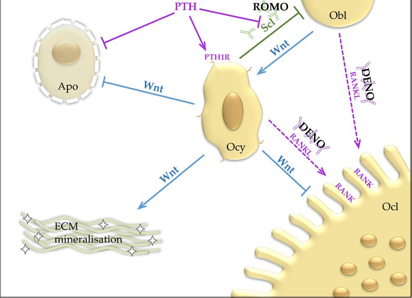
mediator of the Wnt pathway (Figure 1).Int. J. Mol. Sci. 2019, 20, 83 3 of 17
Int. J. Mol. Sci. 2018, 19, x FOR PEER REVIEW 3 of 17
Figure
Figure 1. Top
1. Top left:left:
Wnt Wnt bindstotothe
binds theFrizzled
Frizzled receptor
receptor(Fz)
(Fz)and
and the LRP5/6
the co-receptor.
LRP5/6 LRP5/6
co-receptor. and Fzand
LRP5/6
deactivate the β-catenin destruction complex, which leads to accumulation of β-catenin. β-catenin
Fz deactivate the β-catenin destruction complex, which leads to accumulation of β-catenin. β-catenin
translocates into the nucleus, where it regulates transcription of Wnt target genes with TCF/LEF.
translocates into the nucleus, where it regulates transcription of Wnt target genes with TCF/LEF.
Sclerostin inhibits binding of Wnt to LRP5/6. PTH binds to LRP6 and causes an Wnt-independent
Sclerostin inhibits binding of Wnt to LRP5/6. PTH binds to LRP6 and causes an Wnt-independent
deactivation of the β-catenin destruction complex. bottom: Wnt promotes the osteoblastic lineage and
deactivation of the β-catenin destruction complex. bottom: Wnt promotes the osteoblastic lineage and
inhibits osteoclastogenesis and apoptosis. BMP is a strong promoter of osteoblastic differentiation.
inhibits
PTHosteoclastogenesis
acts through the PTH1R and apoptosis.
receptor inBMP is a stronglineage
the osteoblastic promoter andof osteoblastic
has differentiation.
an inhibiting effect on
PTHsclerostin
acts through the PTH1R receptor in the osteoblastic lineage and has an
expression. APT and TPT work through selective activation of PTH1R activation. ROMOinhibiting effect on
sclerostin expression. APT and TPT work through selective activation of
binds sclerostin. DENO binds RANKL and prevents RANK activation. Apo, Apoptosis; APT, PTH1R activation. ROMO
binds
Abaloparatide, parathyroid hormone-related protein analogue; β-cat. DC, β-catenin destructionAPT,
sclerostin. DENO binds RANKL and prevents RANK activation. Apo, Apoptosis;
Abaloparatide,
complex, targetsparathyroid hormone-related
β-catenin for protein
ubiquitination and analogue;
subsequent β-cat.in DC,
degradation destruction
β-catenin BMP2/7,
the proteasome;
complex,
Bone targets
morphogenetic
β-catenin for ubiquitination
protein 2 and 7; DENO,andDenosumab,
subsequent monoclonal
degradationantibody
in the proteasome; BMP2/7,
against RANKL;
BoneECM, Extracellular protein
morphogenetic matrix; Fz, Frizzled
2 and receptor,
7; DENO, G-protein coupled
Denosumab, receptor,antibody
monoclonal target foragainst
Wnt; LRP5/6,
RANKL;
ECM,Low-density
Extracellular lipoprotein receptor-related
matrix; Fz, protein
Frizzled receptor, 5 or 6; LRP6,
G-protein coupledLow-density lipoprotein
receptor, target receptor-
for Wnt; LRP5/6,
related protein
Low-density 6; MSC,receptor-related
lipoprotein Mesenchymal stem cell; Obl,
protein 5 or Osteoblast; Ocl, Osteoclast;
6; LRP6, Low-density Ocy, Osteocyte;
lipoprotein PTH,
receptor-related
Parathyroid
protein 6; MSC,hormone; TPT, Teriparatide,
Mesenchymal stem cell; peptide Fragment ofOcl,
Obl, Osteoblast; PTH;Osteoclast;
PTH1R, parathyroid hormone PTH,
Ocy, Osteocyte; 1
receptor; RANK, Receptor Activator of NF-κB; RANKL, Receptor Activator of NF-κB Ligand; ROMO,
Parathyroid hormone; TPT, Teriparatide, peptide Fragment of PTH; PTH1R, parathyroid hormone
Romosozumab, monoclonal antibody against sclerostin; Scl, Sclerostin; TCF/LEF, T cell
1 receptor; RANK, Receptor Activator of NF-κB; RANKL, Receptor Activator of NF-κB Ligand;
factor/lymphoid enhancer factor; Wnt, Wingless-related integration site/Wnt signalling pathway.
ROMO, Romosozumab, monoclonal antibody against sclerostin; Scl, Sclerostin; TCF/LEF, T cell
factor/lymphoid enhancer factor; Wnt, Wingless-related integration site/Wnt signalling pathway.Int. J. Mol. Sci. 2019, 20, 83 4 of 17
The activity of bone cells is influenced directly or indirectly by a large variety of different factors.
Local factors including cytokines, chemokines and growth factors among others, are expressed and
secreted by cells within the bone microenvironment and exert auto- and/or paracrine effects governing
bone turnover. A large array of different systemic factors including hormonal signals have been
demonstrated to regulate bone metabolism, for example parathyroid hormone and oestrogen which
play a crucial role in the balance between bone formation and bone resorption [1].
In a healthy organism, the processes of bone resorption and formation are tightly regulated,
resulting in the maintenance of sufficient bone mass with adequate structure and mechanical quality.
If this balance is disturbed, osteoporosis may develop, which represents the most prevalent bone
disease worldwide [8]. In most cases, osteoporosis is caused by increased bone resorption with
insufficient bone formation, resulting in an increased fracture risk with high socioeconomic costs.
The term osteoporosis was first used in the 19th century to describe abnormally hollow bones in
cadavers [9]. Osteoporosis, as it is defined by the World Health Organization today, is a decrease of
bone mineral density (BMD) measured at the lumbar spine or hip of at least 2.5 standard deviations
from the mean of a healthy reference population. Additionally, a clinical method of diagnosis has been
proposed by the National Bone Health Alliance Group not solely relying on BMD measurement [10,11]
but also including the recommended criteria of specific fracture occurrence and fracture risk score (i.e.,
FRAX, see below), providing an alternative basis for osteoporosis diagnosis.
Patients with osteoporosis have a disrupted bone architecture, a lower quality of bone tissue
and, as a result, compromised bone strength and increased risk of fracture [8,12]. Osteoporosis
affects an ever-increasing number of people in the aging population of modern society. According
to the United States Centre for Disease Control, approximately 16.2% of adults over the age of 65
have osteoporosis and 48.3% of the same population exhibit a low bone mass (decrease of BMD
between 1.5 and 2.5 standard deviations). Women over the age of 65 have a 5-times higher prevalence
of osteoporosis than men, while only showing a much smaller increase in the prevalence of low
bone mass. Aside from postmenopausal osteoporosis, caused by a decrease in oestrogen and senile
osteoporosis, there are multiple causes for secondary osteoporosis. The most common cause of
secondary osteoporosis is represented by glucocorticoid-induced osteoporosis (GIOP). Continuously
increased glucocorticoid levels result in a decrease in osteoblast differentiation and function and an
increase in osteoclastogenesis [13]. Importantly, the sole evaluation of BMD is not sufficient to assess
fracture risk in GIOP, as it fails to reflect the disruption of bone architecture and increased risk of falls.
As stated above, a major complication of osteoporosis is an increase in fracture risk. Every fifth
man and every other woman over the age of 50 will sustain a fracture due to increased bone fragility in
their lifetime [8]. Fractures in elderly patients, depending on localization, morphology, comorbidities
and healing potential, can lead to lasting disability and death. Fractures which are attributable to
osteoporosis, are most commonly femoral neck fractures, vertebral fractures, distal radius fractures
and pelvic fractures, followed by femur shaft fractures, humerus fractures and rib fractures [14].
Factors that increase fracture risk in osteoporotic patients include but are not limited to age, history
of fall, previous fracture, diabetes, smoking, rheumatoid arthritis, long-term glucocorticoid use and
alcohol use [8,15,16]. Scores have been developed to evaluate the fracture risk in osteoporotic patients,
for example the most widely known Fracture Risk Assessment Score (FRAX), which takes a selection of
nine risk factors into account [9,17]. Although widely used, the benefit of these scores is controversial
and thus has not been established into general guidelines [18]. The mortality after osteoporotic
fracture is dependent on the type of fracture, treatment and postoperative mobility, as well as BMI
and comorbidities [19]. In the case of hip fractures, fewer than half of the hospitalized patients recover
pre-fracture competence in their activities and mortality is as high as 36% within the first year following
fracture [20]. Based on the high prevalence of osteoporosis in modern society, a 50-year-old woman’s
lifetime risk of dying from a hip fracture was reported equal to her risk of dying from breast cancer [21].
In the light of these facts it is apparent that osteoporosis requires effective treatment.
The foundation of treatment and prevention of osteoporosis has been reviewed elsewhere and includesInt. J. Mol. Sci. 2019, 20, 83 5 of 17
weight-bearing exercises, fall avoidance and adequate nutrition to ensure sufficient calcium, vitamin D
and protein intake [22]. These general measures, however, are not effective in all patients, especially
in geriatric patients confined to nursing homes and in patients who have previously experienced an
osteoporotic fracture and thus may require additional pharmacologic treatment regimes. The current
pharmacological therapy aims at correcting the imbalance between bone resorption and formation at
the level of osteoclasts and osteoblasts, thereby decreasing the risk of fracture events. A number
of pharmacologic agents have been identified to lower fracture risk in both experimental and
clinical studies. These pharmacological agents can be broadly subdivided into two principal groups:
those decreasing bone resorption (by inhibiting osteoclast activity) and those increasing bone formation
(by enhancing osteoblast activity).
2.1. Osteoporosis—Antiresorptive Therapy
It appears evident that the inhibition of bone resorption prevents loss of bone mass and
architecture, explaining the fact that antiresorptive drugs represent a widely used substance
class. Antiresorptive agents including bisphosphonates and the monoclonal antibody to RANKL
(denosumab) target the generation, function and survival of osteoclasts and thus reduce the rate of
bone resorption. As bone formation is coupled to bone resorption, inhibition of bone resorption is
followed by a decrease in osteoblast activity. While this is initially associated with an increase in bone
mineral density and some improvement of structural and material properties of bone tissue, increasing
evidence points towards an association of long-term suppression of osteoclast activity with increased
microdamage accumulation and an alteration in both bone mineralization and collagen formation [23].
Although antiresorptive drugs in general display a low rate of adverse effects, the suppression of bone
turnover may explain necrosis of the jaw and the occurrence of atypical fractures of the femur which
can be observed in patients with high-dose or long-term bisphosphonate usage, respectively [24,25].
Therefore, as antiresorptive agents fail to adequately restore bone mass and bone quality, there is a
continued interest in the identification of molecular targets which stimulate osteoblast activity and
result in an increased bone mass with restored skeletal architecture.
2.2. Osteoporosis—Anabolic Therapy
In principle, stimulating bone formation by pharmacologic means (anabolic therapy) can increase
bone mass to a greater extent than antiresorptive drugs. While there is a variety of different
antiresorptive agents employed in every day clinical practice (e.g., oestrogen, selective oestrogen
receptor modulators, bisphopshonates, denosumab), the only currently available treatment regimen to
stimulate bone formation is represented by daily injections of parathyroid hormone (PTH) or one of its
analogues such as teriparatide and abaloparatide.
2.2.1. PTH—Teriparatide and Abaloparatide
In a healthy organism, PTH functions as an essential endocrine regulator of calcium and phosphate
concentrations in the extracellular space, which is crucial for maintaining serum and urinary calcium
levels within the physiological range. Chronically elevated PTH levels, as observed in primary and
secondary hyperparathyroidism, cause a high bone-turnover state with bone resorption exceeding bone
formation, ultimately resulting in osteoporosis [26]. However, daily injections of PTH (intermittent
PTH or iPTH) or its peptide fragment PTH1–34 (teriparatide) with recurrent, temporary rises in serum
concentration, primarily stimulate bone formation and only to a minor extent bone resorption [27].
This results in a net effect of increased bone mass, improved bone microarchitecture and increased
mechanical strength.
In skeletal tissue, PTH primarily binds to and exerts its biologic effects through the parathyroid
hormone 1 receptor (PTH1R). Among other cell types, this G protein-coupled receptor is expressed
in mesenchymal stem cells, osteoblasts and osteocytes but not in osteoclasts (Figure 1). It is now
understood that the catabolic (i.e., pro-resorptive) effect of PTH is primarily mediated through anInt. J. Mol. Sci. 2019, 20, 83 6 of 17
increased expression of RANKL and the decreased production of its decoy receptor osteoprotegerin
(OPG) in osteoblasts and their precursors and possibly also in osteocytes [26]. Although the precise
molecular mechanism by which PTH stimulates bone formation is not entirely clear to date, previous
studies demonstrated that PTH increased the proliferation and differentiation of osteoblasts and their
precursors both in vitro and in vivo. Moreover, PTH was shown to inhibit osteoblast apoptosis and to
activate bone lining cells. Mechanistically, transactivation of Runx2, the transcription factor crucial
for osteoblast differentiation, is activated by PTH through cAMP/protein kinase A [28]. Moreover,
ERK1/2-mitogen-activated protein kinase and phosphatidylinositol phosphate signalling pathways
are also activated by PTH, which results in an enhanced osteoblast proliferation [29].
Another significant effect of PTH is the activation of the Wnt signalling pathway in cells of the
osteoblast lineage, including osteoblasts and their precursors, as well as osteocytes [30]. Wnt ligands
bind to receptors of the Frizzled family together with co-receptors of the low-density lipoprotein
receptor-related protein (LRP) family, LRP5 and LRP6 [31]. This results in the activation of canonical
signalling cascades and the stabilization of cytosolic β-catenin, a key effector mediator of the Wnt
pathway. After translation into the nucleus, β-catenin forms a complex with the T cell factor/lymphoid
enhancer factor (TCF/LEF) family of transcription factors and proceeds to interact with the genomic
DNA to regulate the transcription of Wnt target genes [31]. PTH was shown to increase β-catenin levels
in cells of the osteoblast lineage and thus stimulate osteoblast proliferation and differentiation [32].
Another study found that PTH, once bound to PTH1R, is also capable of directly complexing with
LRP6, resulting in Wnt ligand-independent activation of β-catenin activation [23,33].
PTH may not only stimulate bone formation through a direct effect on Wnt signalling in osteoblasts
but also indirectly through reducing sclerostin production by osteocytes [7,34]. Sclerostin represents an
osteocyte-specific protein, which potently antagonizes Wnt signalling in bone cells [35]. This hypothesis
results from the observations that PTH suppresses the expression of sclerostin in bone tissue, that PTH
levels inversely correlate with sclerostin levels in healthy women and that women treated with iPTH
display decreased serum concentrations of sclerostin [36,37]. Initial experimental studies revealed no
increase in bone mass in the distal femur of both sclerostin-deficient and sclerostin-overexpressing
mice receiving iPTH [38]. However, other studies showed that iPTH increases both bone formation
and resorption in both wildtype and sclerostin-deficient mice [39]. Furthermore, iPTH significantly
increased the trabecular thickness and mineral apposition rate in sclerostin-deficient mice, indicating
that iPTH stimulates bone formation independently of sclerostin suppression [39]. This uncertainty
regarding the role of sclerostin in the osteoanabolic effect of iPTH lies within the altered baseline bone
density, which is characteristic of mice either lacking or overexpressing sclerostin and further studies
are warranted to dissect the exact molecular mechanism responsible for the therapeutic effect of iPTH.
Although iPTH or teriparatide primarily stimulate bone formation through its high affinity for
the R0 conformation of the PTH1R, a gradual increase in bone resorption can be observed during
prolonged usage [40]. Therefore, the clinical use of iPTH and teriparatide action is based on its effect
of stimulating bone formation before it enhances bone resorption, the period when they are maximally
anabolic (anabolic window). In the case of PTH, the anabolic window lasts approximately 18 to
24 months, before bone resorption exceeds bone formation and no net increase in bone mass can be
achieved, limiting its therapeutic use to a maximum of 2 years [41].
In order to possibly prolong the anabolic window, abaloparatide, a structurally related agent has
been developed and recently approved by the FDA for the treatment of postmenopausal osteoporosis.
Abaloparatide is a synthetic analogue of parathyroid hormone-related protein (PTHrP) which binds
transiently to the RG conformation of PTH1R and also requires daily subcutaneous injections.
Experimental studies demonstrated that abaloparatide increases trabecular thickness and improves
trabecular microstructure [42]. In a phase 3 clinical trial with 2463 ambulatory postmenopausal women,
of which 1901 completed the study, abaloparatide was shown to reduce vertebral and non-vertebral
fractures compared to placebo or teriparatide [43]. According to currently available data, abaloparatide
reduces the number needed to treat for prevention of non-vertebral, clinical and major osteoporoticInt. J. Mol. Sci. 2019, 20, 83 7 of 17
fractures compared to teriparatide [44]. Nonetheless, the claim that the anabolic effect is accompanied
by less bone resorption with abaloparatide than teriparatide, thus widening the anabolic window,
still requires further evidence [45]. Abaloparatide was approved by the FDA in April 2017. However,
a higher risk of select adverse effects including cardiovascular events when compared to teriparatide
have resulted in the refusal of the marketing authorization by the European Medicines Agencies so
far [46].
2.2.2. Sclerostin-Neutralizing Antibody—Romosozumab
Searching for novel targets to increase bone formation, researchers soon became interested in a
rare, autosomal-recessive form of a high bone mass disorder, which resulted in the identification of
sclerostin as a key regulator of osteoblast activity. Patients with sclerosteosis—a loss of function
mutation—or Van Buchem disease—a genetic mutation affecting sclerostin expression—display
high bone mass with excellent biomechanical stability due to an excessive osteoblast activity [47].
Similarly, mice lacking functional sclerostin protein display a striking high bone mass phenotype,
whereas transgenic mice over-expressing sclerostin are osteoporotic [48]. Further mechanistic studies
demonstrated that sclerostin, secreted primarily from osteocytes within the bone microenvironment,
reaches the bone surface through osteocyte canaliculi, where it inhibits co-receptor localization with
Frizzled receptors through binding LRP5 and/or LRP6 [7,35,49]. Activation of Wnt signalling is thus
inhibited, resulting in decreased osteoblastogenesis and bone formation. In addition, sclerostin was
demonstrated to promote bone resorption by increasing the production of RANKL in osteocytes [50].
Although the exact mechanism of action is still not fully clarified to date, it is undoubted that sclerostin
is primarily produced by osteocytes and that it acts as an anti-osteoanabolic molecule (Figure 1).
As a rational consequence of these observations, the therapeutic effect of inhibiting sclerostin
with neutralizing antibodies in various animal models was subsequently tested. Data from these
experimental studies showed a consistent effect of sclerostin immunoneutralization to increase bone
formation, bone mass and biomechanical stability at various skeletal sites [51,52]. These results led to
the development of romosozumab, a highly specific, monoclonal antibody against human sclerostin
which is applied subcutaneously once every month.
Phase III clinical trials (FRAME and STRUCTURE) in female patients suffering from
postmenopausal osteoporosis have shown that romosozumab increases bone mineral density at
the lumbar spine and hip and reduces the risk of vertebral and clinical fractures in comparison with
placebo [53,54]. Romosozumab reduced the risk of vertebral, non-vertebral and clinical fractures in
comparison with the bisphosphonate alendronate in women with severe osteoporosis (ARCH) [55].
This was accompanied by an increase in the markers of bone formation, whereas the markers of bone
resorption decreased, indicating dual action (i.e., stimulation of bone formation and inhibition of bone
resorption) of romosozumab. At present, the approval of romosozumab by the authorities is awaiting
further investigations of a potential increased risk of serious adverse effects including cardiovascular
events, which has been associated with romosozumab treatment in the ARCH study [55].
2.2.3. Future Perspectives
Apart from the agents discussed above, various cytokines, chemokines, growth-factors and
other signalling molecules have been identified to be of crucial importance in regulating bone
formation [56–58] and may thus represent suitable targets to augment osteoblast function. Their use
as bone-anabolic agents, however, is often hindered by the fact that tissue-specific delivery at sufficient
dosage cannot be achieved [58]. As an alternative, gene therapy or transfer offers an attractive
technology, which could potentially overcome these limitations. Although not tested in humans,
several experimental studies with animal models have proven the potential efficacy of this novel
approach. Exogenous genetic material is introduced in order to modify or correct cell differentiation or
function. Targeted delivery and transcription of genes encoding critical regulators in bone remodelling
including BMPs, PTH or OPG has proven efficient to treat experimental osteoporosis [59–67].Int. J. Mol. Sci. 2019, 20, 83 8 of 17
The protective effect was not limited to the bones which were intramedullary injected with the
respective vectors but also in other bones of the same animal. Moreover, based on the growing
understanding of the role of microRNA (miRNA) in the epigenetic regulation of osteoporosis and
bone metabolism [68], targeted activation or inactivation of bone-specific miRNA could represent yet
another molecular therapy to boost osteoanabolic responses in the skeleton. Although further work
is required to fully comprehend the potential clinical implications and to exclude potential serious
adverse effects, this encourages the further development of gene therapy as a novel approach to
stimulate bone formation in osteoporosis.
3. Fracture Healing—Impaired Bone Regeneration
Bone tissue is not only continually remodelled by the combined and tightly regulated activity of
bone cells but also has the remarkable capacity for scar-free repair following fracture. The processes
governing bone turnover in health and disease are also effective during bone regeneration, as fracture
healing can be regarded to represent a juxtaposition of tissue formation (anabolism) and tissue
resorption (catabolism or remodelling). These concepts are useful for understanding bone repair
and have led to the evaluation of osteoporosis drugs for the treatment of impaired fracture healing.
Fracture healing or bone regeneration, results from a complex interplay of cellular and molecular
signalling events that reiterate embryonic skeletal development. Traditionally, fracture healing is
subdivided into four main phases that show a significant degree of overlap: (1) inflammatory phase,
(2) soft callus phase, (3) hard callus phase and (4) remodelling phase [69]. Bone regeneration starts with
an inflammatory response and hematoma formation caused by the disruption and leakage of the bone
marrow and damage to the vascular and soft tissue. A hypoxic sub-phase promotes revascularization.
This is followed by the formation of a soft fibro-cartilaginous matrix, consisting primarily of fibroblasts
and chondrocytes, which provides a certain degree of mechanical stability at the fracture site and acts
as a template for the hard callus. Due to the combined activity of osteoclasts and osteoblasts, the soft
callus is gradually replaced by hard callus during the osteogenic phase, resulting in irregular woven
bone with high vascularization. Finally, the woven callus is replaced by lamellar bone which resembles
the original cortical and trabecular form of mature bone.
Fracture healing is an evolutionary highly conserved process which functions effectively and
efficiently without significant complications in the majority of affected patients. However, in up to
10–20% of patients with fractures, impaired bone regeneration including fracture non-union can be
observed, despite the considerable progress in the advance and optimization of surgical fracture
care [70]. Non-union is defined as a fractured bone, for which a minimum of nine months has elapsed
since the injury and for which there have been no signs of healing for three months. Aside from
the high medical costs associated with the treatment of non-unions, patients suffering from delayed-
or non-union are frequently unable to follow their occupation during the treatment process [71].
A large range of different factors has been identified to be associated with impaired bone regeneration,
including intrinsic factors, such as the age and gender of the patient and extrinsic factors, such as
the location and extent of displacement of the fracture. Non-union presents an ongoing therapeutic
challenge and, similar to osteoporosis, is often associated with significant morbidity, resulting in
decreased quality of life in affected patients and high socioeconomic costs.
3.1. Impaired Fracture Healing—Antiresorptive Therapy
The use of bisphosphonates in osteoporosis for the prevention of fragility fractures is well
established, their value in promoting fracture healing and in preventing and treating non-union
much less so [72–74]. In animal studies, bisphosphonates were shown to cause an increase in callus
volume and bone mineral content during primary enchondral ossification, while causing delayed
remodelling of the fracture callus [75,76]. They increase the bone-implant contact after surgical fixation
of the fracture, however they do not appear to affect the healing rate or time [77,78]. In clinical
studies bisphosphonates have been shown to increase overall BMD and time to union after distalInt. J. Mol. Sci. 2019, 20, 83 9 of 17
radius fracture [71,79]. Similar to animal studies however, bisphosphonates do not reduce time
to consolidation of the fracture or the rate of healing and bolus bisphosphonate therapy 2 weeks
after surgery has been demonstrated to increase BMD in the hip and to significantly reduce overall
mortality [80].
The monoclonal antibody denosumab binds to RANKL, prevents it from binding to its receptor
RANK on the cell surface and therefore inhibits osteoclast recruitment and differentiation. Similar to
bisphosphonates, denusomab has been shown to increase callus formation and delay remodelling in
animal studies, however the formed callus seems to have better biomechanical properties compared
to bisphosphonate treatment [79]. In the clinical trials conducted to date, denusomab did not delay
fracture healing in patients primarily receiving antiresorptive therapy for osteoporosis [81]. However,
clinical studies on the effect of denusomab on impaired fracture healing including delayed or non-union
are insufficient to allow for clinically relevant conclusions and warrant further studies.
3.2. Impaired Fracture Healing—Anabolic Therapy
As osteoclast function is required to remove necrotic bone fragments and the cartilaginous tissue
intermediate during bone regeneration, it is assumed that the stimulation of bone formation is more
favourable to improve bone regeneration than the inhibition of bone resorption. Based on the anabolic
effect of iPTH, teriparatide and abaloparatide in intact bone, this has led researchers to investigate
their use for the prevention and treatment of impaired fracture healing.
3.2.1. PTH
Both iPTH and teriparatide have been shown to promote fracture healing in animal studies
employing various species [26]. Callus developing under iPTH treatment has been shown to mature
faster and to exhibit superior biomechanical properties compared to controls [82]. iPTH promoted
accelerated bone formation in a murine open fracture model, although there was no increase in the
rate of bone union [83]. Teriparatide has also been shown to increase chondrocyte differentiation and
recruitment and therefore to enhance enchondral ossification [30]. Furthermore, iPTH caused a 2
to 3-fold increase in regulatory T-cell populations in mice, which in turn were previously shown to
promote callus formation by balancing the excessive inflammatory reaction observed during the early
stages of fracture repair [84,85]. A recent study comparing the effects of teriparatide and abaloparatide
on bone healing in rats found both drugs to improve fracture healing but in the employed models the
potency per µg of abaloparatide seemed lower than the relation reported from the human osteoporosis
trial (ACTIVE) [45,86].
Because most animal studies used PTH or its analogues in supraphysiological doses, associated
with the potential risk of osteosarcoma development following long-term application, there were
significant concerns that clinical studies using only physiological doses would not show the desired
results for treatment efficacy [87]. Available clinical studies employing varying protocols of dosing,
timing and duration of application for fracture treatment have provided conflicting results [88]. In this
regard, it is worth mentioning that PTH may not only be applied systemically but also locally. Animal
studies investigating the local delivery of PTH or teriparatide via various scaffolds implanted into
bone defects have shown promising results and reported a superior rate and degree of ossification [89].
However, similar to the systemic route of application, insufficient understanding regarding optimal
dosing and timing has prevented the use of locally applied PTH or its derivatives in clinical practice
so far.
3.2.2. Bone Morphogenetic Proteins
One family of peptides, on which more profound information regarding pharmacologic
application and clinical value to boost bone regeneration is available, is represented by bone
morphogenetic proteins (BMP). BMPs are a family of cytokines pertaining to the TGF-β superfamily
and function as key regulators of tissue development in embryonic and adult animals. BMPs were firstInt. J. Mol. Sci. 2019, 20, 83 10 of 17
discovered in 1965 for their capacity to induce ectopic bone formation [90]. To date, over 30 different
BMPs have been described and associated with pleiotrophic functions in regulating a wide range of
different cell types, including mesenchymal stem cells and cells of the osteoblast and chondroblast
lineage required for bone regeneration. The concentration of BMPs and their function varies greatly
throughout the process of fracture healing. BMP-2, -4 and -7 were found to be expressed at high levels
during the early stages of fracture repair around the periosteum and to potentiate the differentiation
of mesenchymal stem cells into chondroblasts and osteoblasts [91,92]. In contrast, BMP-3 is one of
the few BMPs expressed in osteoclasts and can be considered to function as an antagonist of most
osteogenic BMPs [92]. Local delivery of BMPs has shown promising results in animal studies for spinal
fusion and fracture healing. Recombinant human BMP-2 (rhBMP-2) was subsequently approved by
the FDA in the early 2000s for open tibial fractures, anterior interbody fusion in the lumbar spine and
subsequently maxillary sinus and alveolar ridge augmentation after tooth extraction to fill resulting
defects; rhBMP-7 was approved for open tibia fractures. Multiple series of off-label use randomized
clinical trials were published, including cervical spinal fusions, radius fractures and non-union [92].
In bone defects, BMPs promoted healing when used in combination with a variety of scaffolds and
autologous or allogenic grafts. This was shown in both small and large animal models with cranial and
maxillary defects, as well as with segmental bone defects otherwise resulting in non-unions [93–95].
However, clinical testing of locally applied rhBMP has revealed potential detrimental side effects,
such as heterotopic ossification, inflammation and oedema, in addition to osteolysis when used in
high concentrations. Severe clinical complications like swelling in cervical spinal fusion causing
airway obstruction and segmental spinal collapse due to increased bone resorption, have caused a
re-evaluation of the use of BMPs for enhancing bone fracture healing [96–98]. Glaeser et al. have
however recently managed to reduce inflammation and swelling while causing a stimulation in BMP-2
mediated bone formation through application of the NEMO binding domain peptide (NBD) with
BMP-2, opening the possibility for reduction of the complications associated with clinical use of
BMP-2 [99]. NBD inhibits the activation of NF-κB, a central regulator to the inflammatory response.
The combination of adjuncts with lesser doses of BMPs may provide a future perspective for clinical
applications. It is noteworthy that none of the hitherto tested BMPs is approved for systemic application
or osteoporosis therapy, based on their short half-life and the aforementioned adverse effect.
3.2.3. Sclerostin-Neutralizing Antibodies
In conjunction with rhBMP-2 anti-sclerostin antibodies were reported to improve bone
regeneration in a rat femoral defect model when compared to rhBMP-2 alone [100]. However, in a
study on segmental defects in rats without additional BMP, the application of anti-sclerostin antibody
did not enable bony bridging and solely induced an osteoanabolic response in the surrounding intact
bone, which is explained by its lack of osteoinductive potential [101]. A recent study demonstrated
that Sost-deficient mice, which do not express sclerostin protein, are capable of bridging critical-size
calvarial bone defects, which otherwise fail to heal in wild-type mice [102]. Based on the currently
available data, the anti-sclerostin antibody romosozumab developed for the treatment of osteoporosis,
may have possible applications in the treatment of skeletal defects in bones with intramembranous
ossification such as the skull. Similar to PTH and its related analogues, further clinical studies
employing different pharmacologic timing and dosing are required in order to evaluate the clinical
value of inhibiting sclerostin during fracture repair.
3.2.4. Future Perspectives
Due to the potential side effects associated with the systemic application of a number of substances
with high potential for bone regeneration, some research groups have focused on establishing local
delivery methods to defect sites. Both non-genetic methods, such as conjugating oligoaspartic acid,
which has a high affinity for hydroxyapatite in fracture sites and promotes elevated concentrations
of the chosen agent within the fracture site, and methods using gene therapy for enhancing localInt. J. Mol. Sci. 2019, 20, 83 11 of 17
transcription of growth factors have been described [103]. For example, a number of research groups
have tried to find alternative methods to modulate the BMP-2 signalling pathway within the fracture
site using viral vectors or copolymer-protected gene vectors [104,105]. However, these approaches are
purely experimental at this stage and warrant further investigation for their clinical use to promote
bone regeneration.
4. Conclusions
Based on the current demographic development, the number of patients with diseases of the
musculoskeletal system including osteoporosis and impaired fracture healing is expected to rise
steadily. The understanding of the complex cellular and molecular interactions that govern bone
metabolism and bone regeneration in health and disease has given rise to novel compounds with
high therapeutic potency and a potential low risk for adverse effects. The nature of osteoporosis and
impaired bone regeneration, as well as the presence of different comorbidities in affected patients,
may require individualized treatment regimens employing more than just one bone drug to achieve
the best possible outcomes. The further development and study of therapeutic approaches targeting
bone formation, rather than bone resorption, fulfills an essential clinical need, as treatment options for
reversing bone loss and promoting bone regeneration are currently limited.
Author Contributions: Conceptualization, G.R. and J.K.; writing—original draft preparation, G.R., D.J., J.A. and
J.K.; writing—review and editing, G.R., D.J., J.A., S.T., S.M. and J.K.; visualization, G.R, S.M., S.T.
Funding: This work was funded in part by the German Research Foundation (DFG KE 2179/2-1 and
TS 303/2-1), the Else-Kröner-Fresenius Stiftung (EKFS 2017_A22) and the Berlin Institute of Health.
We acknowledge support from the German Research Foundation (DFG) and the Open Access Publication Fund of
Charité—Universitätsmedizin Berlin.
Conflicts of Interest: The authors do not have any conflict of interest.
Abbreviations
ACTIVE Abaloparatide Comparator Trial In Vertebral Endpoints Trial
ARCH Active-Controlled Fracture Study in Postmenopausal Women with Osteoporosis at
High Risk
BMD Bone mineral density
BMI Body mass index
BMP Bone morphogenetic protein
cAMP cyclic Adenosine monophosphate
ERK1/2 Extracellular-signal Regulated Kinase 1 and 2
FDA United States Food and Drug Administration
FRAME Fracture Study in Postmenopausal Women with Osteoporosis
FRAX Fracture Risk Assessment Score
GIOP Glucocorticoid-induced osteoporosis
iPTH Intermittent PTH
LRP5/6 Low-density lipoprotein receptor-related protein 5 or 6
LRP6 Low-density lipoprotein receptor-related protein 6
miRNA Micro RNA
NBD NEMO binding domain peptide
NEMO NF-kappa-B essential modulator
NF-κB Nuclear factor “kappa-light-chain-enhancer” of activated B-cells
OPG osteoprotegerin
PTH Parathyroid hormone
PTH1-34 Teriparatide, peptide Fragment of PTH
PTH1R parathyroid hormone 1 receptor
PTHrP parathyroid hormone-related protein
RANK Receptor Activator of NF-κBInt. J. Mol. Sci. 2019, 20, 83 12 of 17
RANKL Receptor Activator of NF-κB Ligand
rhBMP Recombinant BMP
Runx2 Runt-related transcription factor 2
Scl Sclerostin
STRUCTURE An Open-label Study to Evaluate the Effect of Treatment With Romosozumab or
Teriparatide in Postmenopausal Women
TCF/LEF T cell factor/lymphoid enhancer factor
Wnt Wingless-related integration site/Wnt signalling pathway
WT Wild type
References
1. Teitelbaum, S.L. Bone resorption by osteoclasts. Science 2000, 289, 1504–1508. [CrossRef] [PubMed]
2. Howship, J. Microscopic Observations on the Structure of Bone. Med. Chir. Trans. 1816, 7, 382–592.11.
[CrossRef] [PubMed]
3. Luo, Y.; Zhang, Y.; Miao, G.; Zhang, Y.; Liu, Y.; Huang, Y. Runx1 regulates osteogenic differentiation of
BMSCs by inhibiting adipogenesis through Wnt/beta-catenin pathway. Arch. Oral Biol. 2018, 97, 176–184.
[CrossRef] [PubMed]
4. Bahney, C.S.; Zondervan, R.L.; Allison, P.; Theologis, A.; Ashley, J.; Ahn, J.; Miclau, T.; Marcucio, R.;
Hankenson, K.D. The Cellular Biology of Fracture Healing. J. Orthop. Res. 2018. [CrossRef] [PubMed]
5. Abdelgawad, M.E.; Delaisse, J.M.; Hinge, M.; Jensen, P.R.; Alnaimi, R.W.; Rolighed, L.; Engelholm, L.H.;
Marcussen, N.; Andersen, T.L. Early reversal cells in adult human bone remodeling: OSTEOBLASTIC nature,
catabolic functions and interactions with osteoclasts. Histochem. Cell Biol. 2016, 145, 603–615. [CrossRef]
[PubMed]
6. Chen, X.; Wang, Z.; Duan, N.; Zhu, G.; Schwarz, E.M.; Xie, C. Osteoblast-osteoclast interactions.
Connect. Tissue Res. 2018, 59, 99–107. [CrossRef] [PubMed]
7. Koide, M.; Kobayashi, Y. Regulatory mechanisms of sclerostin expression during bone remodeling. J. Bone
Miner. Metab. 2018. [CrossRef]
8. Lorentzon, M.; Cummings, S.R. Osteoporosis: THE evolution of a diagnosis. J. Intern. Med. 2015, 277,
650–661. [CrossRef]
9. Kanis, J.A.; Johansson, H.; Harvey, N.C.; McCloskey, E.V. A brief history of FRAX. Arch. Osteoporos. 2018,
13, 118. [CrossRef]
10. Siris, E.S.; Adler, R.; Bilezikian, J.; Bolognese, M.; Dawson-Hughes, B.; Favus, M.J.; Harris, S.T.;
Jan de Beur, S.M.; Khosla, S.; Lane, N.E.; et al. The clinical diagnosis of osteoporosis: A position statement
from the National Bone Health Alliance Working Group. Osteoporos. Int. 2014, 25, 1439–1443. [CrossRef]
11. Papaioannou, A.; Kennedy, C. Diagnostic criteria for osteoporosis should be expanded. Lancet Diabetes
Endocrinol. 2015, 3, 234–236. [CrossRef]
12. Cosman, F.; de Beur, S.J.; LeBoff, M.S.; Lewiecki, E.M.; Tanner, B.; Randall, S.; Lindsay, R. Clinician’s Guide
to Prevention and Treatment of Osteoporosis. Osteoporos. Int. 2014, 25, 2359–2381. [CrossRef] [PubMed]
13. Canalis, E.; Mazziotti, G.; Giustina, A.; Bilezikian, J.P. Glucocorticoid-induced osteoporosis:
PATHOPHYSIOLOGY and therapy. Osteoporos. Int. 2007, 18, 1319–1328. [CrossRef] [PubMed]
14. Warriner, A.H.; Patkar, N.M.; Curtis, J.R.; Delzell, E.; Gary, L.; Kilgore, M.; Saag, K. Which fractures are most
attributable to osteoporosis? J. Clin. Epidemiol. 2011, 64, 46–53. [CrossRef] [PubMed]
15. Deloumeau, A.; Molto, A.; Roux, C.; Briot, K. Determinants of short term fracture risk in patients with a
recent history of low-trauma non-vertebral fracture. Bone 2017, 105, 287–291. [CrossRef] [PubMed]
16. Ferrari, S.L.; Abrahamsen, B.; Napoli, N.; Akesson, K.; Chandran, M.; Eastell, R.; El-Hajj Fuleihan, G.;
Josse, R.; Kendler, D.L.; Kraenzlin, M.; et al. Diagnosis and management of bone fragility in diabetes:
AN emerging challenge. Osteoporos. Int. 2018, 29, 2585–2596. [CrossRef] [PubMed]
17. Kanis, J.A.; Johnell, O.; Oden, A.; Johansson, H.; McCloskey, E. FRAX and the assessment of fracture
probability in men and women from the UK. Osteoporos. Int. 2008, 19, 385–397. [CrossRef]Int. J. Mol. Sci. 2019, 20, 83 13 of 17
18. Crandall, C.J.; Larson, J.; LaCroix, A.; Cauley, J.A.; LeBoff, M.S.; Li, W.; LeBlanc, E.S.; Edwards, B.J.;
Manson, J.E.; Ensrud, K. Predicting Fracture Risk in Younger Postmenopausal Women: Comparison of
the Garvan and FRAX Risk Calculators in the Women’s Health Initiative Study. J. Gen. Intern Med. 2018.
[CrossRef]
19. Akinleye, S.D.; Garofolo, G.; Culbertson, M.D.; Homel, P.; Erez, O. The Role of BMI in Hip Fracture Surgery.
Geriatr. Orthop. Surg. Rehabil. 2018, 9. [CrossRef]
20. Abrahamsen, B.; van Staa, T.; Ariely, R.; Olson, M.; Cooper, C. Excess mortality following hip fracture:
A systematic epidemiological review. Osteoporos. Int. 2009, 20, 1633–1650. [CrossRef]
21. Cummings, S.R.; Black, D.M.; Rubin, S.M. Lifetime risks of hip, Colles’, or vertebral fracture and coronary
heart disease among white postmenopausal women. Arch. Intern Med. 1989, 149, 2445–2448. [CrossRef]
[PubMed]
22. Khan, A.Z.; Rames, R.D.; Miller, A.N. Clinical Management of Osteoporotic Fractures. Curr. Osteoporos. Rep.
2018, 16, 299–311. [CrossRef] [PubMed]
23. Sims, N.A.; Ng, K.W. Implications of osteoblast-osteoclast interactions in the management of osteoporosis
by antiresorptive agents denosumab and odanacatib. Curr. Osteoporos. Rep. 2014, 12, 98–106. [CrossRef]
[PubMed]
24. Larsen, M.S.; Schmal, H. The enigma of atypical femoral fractures: A summary of current knowledge.
EFORT Open Rev. 2018, 3, 494–500. [CrossRef]
25. Lim, S.J.; Yeo, I.; Yoon, P.W.; Yoo, J.J.; Rhyu, K.H.; Han, S.B.; Lee, W.S.; Song, J.H.; Min, B.W.; Park, Y.S.
Incidence, risk factors, and fracture healing of atypical femoral fractures: A multicenter case-control study.
Osteoporos. Int. 2018, 29, 2427–2435. [CrossRef] [PubMed]
26. Wojda, S.J.; Donahue, S.W. Parathyroid hormone for bone regeneration. J. Orthop. Res. 2018, 36, 2586–2594.
[CrossRef]
27. Langdahl, B.L.; Silverman, S.; Fujiwara, S.; Saag, K.; Napoli, N.; Soen, S.; Enomoto, H.; Melby, T.E.;
Disch, D.P.; Marin, F.; Krege, J.H. Real-world effectiveness of teriparatide on fracture reduction in patients
with osteoporosis and comorbidities or risk factors for fractures: Integrated analysis of 4 prospective
observational studies. Bone 2018, 116, 58–66. [CrossRef] [PubMed]
28. Swarthout, J.T.; D’Alonzo, R.C.; Selvamurugan, N.; Partridge, N.C. Parathyroid hormone-dependent
signaling pathways regulating genes in bone cells. Gene 2002, 282, 1–17. [CrossRef]
29. Cheng, Z.Y.; Ye, T.; Ling, Q.Y.; Wu, T.; Wu, G.Y.; Zong, G.J. Parathyroid hormone promotes osteoblastic
differentiation of endothelial cells via the extracellular signal-regulated protein kinase 1/2 and nuclear
factor-kappaB signaling pathways. Exp. Ther. Med. 2018, 15, 1754–1760. [CrossRef]
30. Kakar, S.; Einhorn, T.A.; Vora, S.; Miara, L.J.; Hon, G.; Wigner, N.A.; Toben, D.; Jacobsen, K.A.; Al-Sebaei, M.O.;
Song, M.; Trackman, P.C.; et al. Enhanced chondrogenesis and Wnt signaling in PTH-treated fractures.
J. Bone Miner. Res. 2007, 22, 1903–1912. [CrossRef]
31. Krishnan, V.; Bryant, H.U.; Macdougald, O.A. Regulation of bone mass by Wnt signaling. J. Clin. Investig.
2006, 116, 1202–1209. [CrossRef] [PubMed]
32. Tobimatsu, T.; Kaji, H.; Sowa, H.; Naito, J.; Canaff, L.; Hendy, G.N.; Sugimoto, T.; Chihara, K. Parathyroid
hormone increases beta-catenin levels through Smad3 in mouse osteoblastic cells. Endocrinology 2006, 147,
2583–2590. [CrossRef] [PubMed]
33. Wan, M.; Yang, C.; Li, J.; Wu, X.; Yuan, H.; Ma, H.; He, X.; Nie, S.; Chang, C.; Cao, X. Parathyroid hormone
signaling through low-density lipoprotein-related protein 6. Genes Dev. 2008, 22, 2968–2979. [CrossRef]
[PubMed]
34. Keller, H.; Kneissel, M. SOST is a target gene for PTH in bone. Bone 2005, 37, 148–158. [CrossRef] [PubMed]
35. Li, X.; Zhang, Y.; Kang, H.; Liu, W.; Liu, P.; Zhang, J.; Harris, S.E.; Wu, D. Sclerostin binds to LRP5/6 and
antagonizes canonical Wnt signaling. J. Biol. Chem. 2005, 280, 19883–19887. [CrossRef] [PubMed]
36. Drake, M.T.; Srinivasan, B.; Modder, U.I.; Peterson, J.M.; McCready, L.K.; Riggs, B.L.; Dwyer, D.; Stolina, M.;
Kostenuik, P.; Khosla, S. Effects of parathyroid hormone treatment on circulating sclerostin levels in
postmenopausal women. J. Clin. Endocrinol. Metab. 2010, 95, 5056–5062. [CrossRef] [PubMed]
37. Bellido, T.; Ali, A.A.; Gubrij, I.; Plotkin, L.I.; Fu, Q.; O’Brien, C.A.; Manolagas, S.C.; Jilka, R.L. Chronic
elevation of parathyroid hormone in mice reduces expression of sclerostin by osteocytes: A novel mechanism
for hormonal control of osteoblastogenesis. Endocrinology 2005, 146, 4577–4583. [CrossRef]Int. J. Mol. Sci. 2019, 20, 83 14 of 17
38. Kramer, I.; Loots, G.G.; Studer, A.; Keller, H.; Kneissel, M. Parathyroid hormone (PTH)-induced bone gain is
blunted in SOST overexpressing and deficient mice. J. Bone Miner. Res. 2010, 25, 178–189. [CrossRef]
39. Robling, A.G.; Kedlaya, R.; Ellis, S.N.; Childress, P.J.; Bidwell, J.P.; Bellido, T.; Turner, C.H. Anabolic and
catabolic regimens of human parathyroid hormone 1-34 elicit bone- and envelope-specific attenuation of
skeletal effects in Sost-deficient mice. Endocrinology 2011, 152, 2963–2975. [CrossRef]
40. Cheloha, R.W.; Gellman, S.H.; Vilardaga, J.P.; Gardella, T.J. PTH receptor-1 signalling-mechanistic insights
and therapeutic prospects. Nat. Rev. Endocrinol. 2015, 11, 712–724. [CrossRef]
41. Pazianas, M. Anabolic effects of PTH and the ‘anabolic window’. Trends Endocrinol. Metab. 2015, 26, 111–113.
[CrossRef] [PubMed]
42. Chandler, H.; Lanske, B.; Varela, A.; Guillot, M.; Boyer, M.; Brown, J.; Pierce, A.; Ominsky, M.; Mitlak, B.;
Baron, R.; Kostenuik, P.; Hattersley, G. Abaloparatide, a novel osteoanabolic PTHrP analog, increases cortical
and trabecular bone mass and architecture in orchiectomized rats by increasing bone formation without
increasing bone resorption. Bone 2018, 120, 148–155. [CrossRef] [PubMed]
43. Miller, P.D.; Hattersley, G.; Riis, B.J.; Williams, G.C.; Lau, E.; Russo, L.A.; Alexandersen, P.; Zerbini, C.A.;
Hu, M.Y.; Harris, A.G.; et al. Effect of Abaloparatide vs Placebo on New Vertebral Fractures in
Postmenopausal Women with Osteoporosis: A Randomized Clinical Trial. JAMA 2016, 316, 722–733.
[CrossRef] [PubMed]
44. Reginster, J.Y.; Hattersley, G.; Williams, G.C.; Hu, M.Y.; Fitzpatrick, L.A.; Lewiecki, E.M. Abaloparatide is
an Effective Treatment Option for Postmenopausal Osteoporosis: Review of the Number Needed to Treat
Compared with Teriparatide. Calcif. Tissue Int. 2018, 103, 540–545. [CrossRef] [PubMed]
45. Miller, P.D.; Hattersley, G.; Lau, E.; Fitzpatrick, L.A.; Harris, A.G.; Williams, G.C.; Hu, M.Y.; Riis, B.J.; Russo, L.;
Christiansen, C. Bone mineral density response rates are greater in patients treated with abaloparatide
compared with those treated with placebo or teriparatide: Results from the ACTIVE phase 3 trial. Bone 2018,
120, 137–140. [CrossRef] [PubMed]
46. Boyce, E.G.; Mai, Y.; Pham, C. Abaloparatide: Review of a Next-Generation Parathyroid Hormone Agonist.
Ann. Pharmacother. 2018, 52, 462–472. [CrossRef] [PubMed]
47. Van Lierop, A.H.; Appelman-Dijkstra, N.M.; Papapoulos, S.E. Sclerostin deficiency in humans. Bone 2017, 96,
51–62. [CrossRef]
48. Li, X.; Ominsky, M.S.; Niu, Q.T.; Sun, N.; Daugherty, B.; D’Agostin, D.; Kurahara, C.; Gao, Y.; Cao, J.;
Gong, J.; et al. Targeted deletion of the sclerostin gene in mice results in increased bone formation and bone
strength. J. Bone Miner. Res. 2008, 23, 860–869. [CrossRef]
49. Shi, C.; Li, J.; Wang, W.; Cao, W.; Cao, X.; Wan, M. Antagonists of LRP6 regulate PTH-induced cAMP
generation. Ann. N. Y. Acad. Sci. 2011, 1237, 39–46. [CrossRef]
50. Wijenayaka, A.R.; Kogawa, M.; Lim, H.P.; Bonewald, L.F.; Findlay, D.M.; Atkins, G.J. Sclerostin stimulates
osteocyte support of osteoclast activity by a RANKL-dependent pathway. PLoS ONE 2011, 6, e25900.
[CrossRef]
51. Alaee, F.; Virk, M.S.; Tang, H.; Sugiyama, O.; Adams, D.J.; Stolina, M.; Dwyer, D.; Ominsky, M.S.; Ke, H.Z.;
Lieberman, J.R. Evaluation of the effects of systemic treatment with a sclerostin neutralizing antibody on
bone repair in a rat femoral defect model. J. Orthop. Res. 2014, 32, 197–203. [CrossRef] [PubMed]
52. Ominsky, M.S.; Brown, D.L.; Van, G.; Cordover, D.; Pacheco, E.; Frazier, E.; Cherepow, L.; Higgins-Garn, M.;
Aguirre, J.I.; Wronski, T.J.; et al. Differential temporal effects of sclerostin antibody and parathyroid hormone
on cancellous and cortical bone and quantitative differences in effects on the osteoblast lineage in young
intact rats. Bone 2015, 81, 380–391. [CrossRef] [PubMed]
53. Cosman, F.; Crittenden, D.B.; Ferrari, S.; Khan, A.; Lane, N.E.; Lippuner, K.; Matsumoto, T.; Milmont, C.E.;
Libanati, C.; Grauer, A. FRAME Study: The Foundation Effect of Building Bone With 1 Year of Romosozumab
Leads to Continued Lower Fracture Risk After Transition to Denosumab. J. Bone Miner. Res. 2018, 33,
1219–1226. [CrossRef] [PubMed]
54. Graeff, C.; Campbell, G.M.; Pena, J.; Borggrefe, J.; Padhi, D.; Kaufman, A.; Chang, S.; Libanati, C.;
Gluer, C.C. Administration of romosozumab improves vertebral trabecular and cortical bone as assessed
with quantitative computed tomography and finite element analysis. Bone 2015, 81, 364–369. [CrossRef]
[PubMed]You can also read