Glucocorticoid receptor haploinsufficiency causes hypertension and attenuates hypothalamic-pituitary-adrenal axis and blood pressure adaptions to ...
←
→
Page content transcription
If your browser does not render page correctly, please read the page content below
Edinburgh Research Explorer
Glucocorticoid receptor haploinsufficiency causes hypertension
and attenuates hypothalamic-pituitary-adrenal axis and blood
pressure adaptions to high-fat diet
Citation for published version:
Michailidou, Z, Carter, RN, Marshall, E, Sutherland, HG, Brownstein, DG, Owen, E, Cockett, K, Kelly, V,
Ramage, L, Al-Dujaili, EAS, Ross, M, Maraki, I, Newton, K, Holmes, MC, Seckl, JR, Morton, NM, Kenyon,
CJ & Chapman, KE 2008, 'Glucocorticoid receptor haploinsufficiency causes hypertension and attenuates
hypothalamic-pituitary-adrenal axis and blood pressure adaptions to high-fat diet' The FASEB Journal, vol.
22, no. 11, pp. 3896-3907. DOI: 10.1096/fj.08-111914

Digital Object Identifier (DOI):
10.1096/fj.08-111914

Link:
Link to publication record in Edinburgh Research Explorer

Document Version:
Publisher's PDF, also known as Version of record

Published In:
The FASEB Journal

Publisher Rights Statement:
This is an Open Access article distributed under the terms of the Creative Commons Attribution Non-Commercial
License (http://creativecommons.org/licenses/by-nc/3.0/us/) which permits unrestricted non-commercial use,
distribution, and reproduction in any medium, provided the original work is properly cited.

General rights
Copyright for the publications made accessible via the Edinburgh Research Explorer is retained by the author(s)
and / or other copyright owners and it is a condition of accessing these publications that users recognise and
abide by the legal requirements associated with these rights.

Take down policy
The University of Edinburgh has made every reasonable effort to ensure that Edinburgh Research Explorer
content complies with UK legislation. If you believe that the public display of this file breaches copyright please
contact openaccess@ed.ac.uk providing details, and we will remove access to the work immediately and
investigate your claim.

Download date: 14. Oct. 2018

The FASEB Journal • Research Communication
Glucocorticoid receptor haploinsufficiency causes
hypertension and attenuates hypothalamic-pituitary-
adrenal axis and blood pressure adaptions
to high-fat diet
Z. Michailidou,* R. N. Carter,* E. Marshall,* H. G. Sutherland,† D. G. Brownstein,*
E. Owen,* K. Cockett,* V. Kelly,* L. Ramage,* E. A. S. Al-Dujaili,‡ M. Ross,*
I. Maraki,* K. Newton,† M. C. Holmes,* J. R. Seckl,* N. M. Morton,* C. J. Kenyon,*
and K. E. Chapman*,1
*Centre for Cardiovascular Sciences, Queen’s Medical Research, Institute, University of Edinburgh,
Edinburgh, UK; †Medical Research Council Human Genetics Unit, Edinburgh, UK; and ‡Queen
Margaret University, Musselburgh, East Lothian, UK
ABSTRACT Glucocorticoid hormones are critical to Glucocorticoids coordinately regulate gene
respond and adapt to stress. Genetic variations in the pathways in response to external (stress) and internal
glucocorticoid receptor (GR) gene alter hypothalamic- (circadian) cues and form part of important homeo-
pituitary-adrenal (HPA) axis activity and associate with static control mechanisms, critical in adaption to envi-
hypertension and susceptibility to metabolic disease. Here ronmental stressors. They are required for the integrity
we test the hypothesis that reduced GR density alters of central nervous system function, for the response to
blood pressure and glucose and lipid homeostasis and infection and injury, and for cardiovascular and meta-
limits adaption to obesogenic diet. Heterozygous GRgeo/ⴙ bolic homeostasis. They regulate blood pressure (1),
mice were generated from embryonic stem (ES) cells with energy intake and expenditure, and glucose and lipid
a gene trap integration of a -galactosidase-neomycin homeostasis (2). In excess, glucocorticoids cause hyper-
phosphotransferase (geo) cassette into the GR gene tension, visceral obesity, insulin resistance/diabetes,
creating a transcriptionally inactive GR fusion protein. and disordered mood and cognition (Cushing’s syn-
Although GRgeo/ⴙ mice have 50% less functional GR, drome), whereas glucocorticoid deficiency causes hy-
they have normal lipid and glucose homeostasis due to potension, fatigue, weight loss, and anorexia.
compensatory HPA axis activation but are hypertensive Most actions of glucocorticoids are mediated
due to activation of the renin-angiotensin-aldosterone
through the widely distributed glucocorticoid receptor
system (RAAS). When challenged with a high-fat diet,
(GR; refs. 3–5), which belongs to the superfamily of
weight gain, adiposity, and glucose intolerance were sim-
nuclear receptor transcription factors (6). Studies in
ilarly increased in control and GRgeo/ⴙ mice, suggesting
vitro (7, 8) and in vivo (9, 10) have shown the impor-
preserved control of intermediary metabolism and energy
balance. However, whereas a high-fat diet caused HPA tance of receptor density in determining cellular glu-
activation and increased blood pressure in control mice, cocorticoid sensitivity. Complete loss of GR is incom-
these adaptions were attenuated or abolished in GRgeo/ⴙ patible with postnatal survival (11, 12), but partial loss
mice. Thus, reduced GR density balanced by HPA activa- of GR function in humans causes the rare familial/
tion leaves glucocorticoid functions unaffected but min- sporadic glucocorticoid resistance syndrome (13), char-
eralocorticoid functions increased, causing hypertension. acterized by hypercortisolism without other features of
Importantly, reduced GR limits HPA and blood pressure Cushing’s syndrome. Polymorphisms in the human
adaptions to obesogenic diet.—Michailidou, Z., Carter, Nr3c1 gene (encoding GR; here abbreviated to GR) are
R. N., Marshall, E., Sutherland, H. G., Brownstein, D. G.,
Owen, E., Cockett, K., Kelly, V., Ramage, L., Al-Dujaili, 1
Correspondence: Endocrinology Unit, Centre for Cardio-
E. A. S., Ross, M., Maraki, I., Newton, K., Holmes, M. C., vascular Sciences, The Queen’s Medical Research Institute,
Seckl, J. R., Morton, N. M., Kenyon, C. J., Chapman, K. E. 47 Little France Crescent, Edinburgh, EH16 4TJ, UK. E-mail:
Glucocorticoid receptor haploinsufficiency causes hyper- karen.chapman@ed.ac.uk
tension and attenuates hypothalamic-pituitary-adrenal axis This is an Open Access article distributed under the terms
and blood pressure adaptions to high-fat diet. FASEB J. 22, of the Creative Commons Attribution Non-Commercial Li-
3896 –3907 (2008). www.fasebj.org cense (http://creativecommons.org/licenses/by-nc/3.0/us/)
which permits unrestricted non-commercial use, distribution,
and reproduction in any medium, provided the original work
Key Words: HPA axis 䡠 diet-induced obesity 䡠 NR3C1 䡠 glu- is properly cited.
cose and lipid homeostasis doi: 10.1096/fj.08-111914
3896 0892-6638/08/0022-3896 © The Author(s)associated with hyper- or hyposensitivity to exogenous sidase-neomycin phosphotransferase (geo) reporter cassette
glucocorticoid (dexamethasone) suppression of the between exons 3 and 4 of the GR gene. Fluorescence in situ
hypothalamic-pituitary-adrenal (HPA) axis and a range hybridization (FISH) confirmed a single integration site on
chromosome 18 at the GR locus. Chimeric mice were gener-
of metabolic and cardiovascular parameters. Thus, ated by injection of ESKN92 cells into C57BL/6J blastocysts.
polymorphisms associated with glucocorticoid hyper- Chimeras were mated to C57BL/6J female mice to achieve
sensitivity (BclI, N363S) are typically (dependent on germ line transmission and further backcrossed to generate a
age, gender, and ethnic background) associated with congenic line (full name, Nr3c1gtESK92MRCHGU; here abbrevi-
hypertension, obesity, decreased lean mass, insulin ated to GRgeo). Heterozygotes were identified by polymerase
resistance, and increased risk of cardiovascular disease chain reaction (PCR) for the presence of lacZ (in the geo
cassette) using the following primers: forward, 5⬘-GTTGC-
despite reduced plasma cortisol levels, whereas the
GCAGCCTGAATGGCG-3⬘; and reverse, 5⬘-GCCGTCACTC-
ER22/23EK polymorphism, associated with glucocorti- CAACGCAGCA-3⬘. All the experiments described were per-
coid hyposensitivity, is associated with beneficial meta- formed on adult male (5– 6 months) GRgeo/⫹ and GR⫹/⫹
bolic outcomes (reviewed in ref. 14). littermates, backcrossed for five generations (F5) to C57BL/
Several mouse lines with altered GR expression or 6J. Experimental groups were 8 to 9 per group, unless
function have been generated to investigate the role of otherwise stated. To generate homozygous GRgeo/geo mice,
GR in HPA axis regulation and its role in mood F7 GRgeo/⫹ mice were intercrossed.
regulation and in the immune system (reviewed in refs.
15–17). Fewer studies have investigated the effects of Animal maintenance and diets
altered GR density on cardiovascular and metabolic
outcomes. Reduced GR density through antisense GR All animal experimentation was conducted in strict accord
transgene expression (central nervous system targeted, with the accepted standards of humane animal care under
the auspices of the Animal (Scientific Procedures) Act UK
although widely expressed) lowered energy intake but
1986 after prior approval by the local ethical committee. Mice
also caused obesity (18, 19). However, neuron-specific were housed in standard cages under controlled lighting
deletion of GR caused a small-lean phenotype post- (12:12-h light-dark cycle, lights on at 7 AM) and were fed
weaning and increased energy expenditure, due to the standard chow from weaning unless specified otherwise. For
peripheral effects of high glucocorticoid levels coupled measurement of unstressed plasma corticosterone ACTH and
with normal levels of peripheral GR (20). Liver-specific glucose in tail nick blood samples, animals were acclimatized
GR gene deletion led to hypoglycemia, but only after to single housing and tail nicks were performed within 1 min
of disturbing the cage. For diet-induced obesity experiments,
prolonged starvation, and ameliorated hyperglycemia mice were weaned onto either HF/low carbohydrate diet
in streptozotocin-induced diabetes (21). Recently, trans- (58% kcal as fat, D12331; Research Diets, New Brunswick, NJ,
genic mice have been described with cardiomyocyte- USA) or low-fat (LF)/high-carbohydrate diet (11% kcal as fat,
specific GR overexpression. These mice display conduc- D12328; Research Diets) and remained on the diet for 22 wk
tion defects, reduced heart rate, atrioventricular block, with ad libitum access to water and diet. Body weight and food
altered calcium homeostasis, and ion channel remod- intake were monitored weekly and for 3 wk, respectively.
eling in isolated cardiomyocytes (22). Although these
studies have provided important information on the Tissue collection, metabolic parameters, and liver
tissue-specific functions of GR, the cardiovascular and triglyceride levels
metabolic consequences of globally altered GR density,
and hence glucocorticoid sensitivity, have not been de- Experimental mice were killed by decapitation between 8 and 10
AM. Trunk blood samples were collected into EDTA-coated
scribed, although this is the clinically relevant situation.
tubes (Sarstedt, Nümbrecht, Germany) and centrifuged (6000 g,
Here we have generated a novel line of mice with 10 min), and plasma was stored at ⫺80°C before assay. Tissues
reduced GR density, heterozygous for a null mutation were rapidly frozen in dry ice for RNA or Western blot analysis
of the GR gene (GRgeo/⫹), to investigate whether global or fixed in formalin (left adrenal, kidney) for histology. Evening
reduction in GR density alters cardiovascular (blood plasma corticosterone levels were determined from blood sam-
pressure), fat distribution, and metabolic (glucose and pled (by tail nick) at 7 PM. Plasma corticosterone was measured
lipid homeostasis) parameters. A high-fat (HF) diet was by an in-house radioimmunoassay as described previously (24).
Plasma ACTH levels were measured by ELISA (Biomerica,
introduced to determine whether GR haploinsuffi- Newport Beach, CA, USA). For the glucose tolerance test
ciency altered the adaptive hormonal and metabolic (GTT), animals were deprived of food for 6 h, 2 mg/g body
changes that accompany dietary-induced obesity. weight of 25% glucose was injected intraperitoneally, and tail
nick blood sampling was performed at time 0 (before injection)
and 15, 30, 60, and 120 min after injection. Plasma glucose was
measured using a glucose monitoring system (One Touch Ultra,
MATERIALS AND METHODS
Lifescan, Johnson & Johnson, Langhorne, PA, USA), insulin by
ELISA (Crystalchem, Downers Grove, IL, USA), nonesterified
Generation of GRgeo/ⴙ mice fatty acids (NEFAs) with a NEFA C kit (Wako Chemicals GmbH,
Nuess, Germany), and triglycerides with an L-type triglyceride
A mouse ES cell line (ESKN92) generated by gene-trap kit (Wako Chemicals GmbH). Insulin, NEFAs, and triglycerides
mutagenesis in which a -galactosidase-neomycin phospho- were measured after 24 h of food deprivation. Hepatic triglyc-
transferase (geo) reporter cassette integrated within the GR erides were extracted after homogenization of 100 mg liver in
gene, generating a translational fusion between GR and geo, isopropanol (10 vol) and then incubation at 37°C for 45 min.
has been described previously (23). 5⬘-Rapid amplification of After centrifugation (3000 g, 10 min), 10 l supernatant was
cDNA ends (RACE) confirmed integration of the -galacto- incubated at 37°C for 5 min with 1 ml Thermotrace triglyceride
HYPERTENSION AND ALTERED ADAPTION IN GRgeo/⫹ MICE 3897reagent (Alpha Laboratories, Eastleigh, UK) and absorbance at Santa Cruz Biotechnology, Santa Cruz, CA, USA). Immuno-
500 nm was measured. Plasma renin activity and angiotensino- reactive bands were visualized by chemiluminesense (ECL kit;
gen concentration were measured by radioimmunoassay as Amersham Biosciences, Little Chalfont, UK). An anti--tubu-
described previously (25). Plasma aldosterone concentration lin monoclonal antibody (Sigma Aldrich, St. Louis, MO,
was measured by in-house ELISA as described previously (26, USA) or membrane staining with Ponceau red were used to
27). Formalin-fixed tissues were processed for histopathology, verify equal protein loading between samples.
sectioned (4 m), and stained with hematoxylin and eosin for
histopathological examination. X-gal staining
X-gal (5-bromo-4-chloro-3-indolyl--d-galactopyranoside) staining
Blood pressure measurements
to detect -galactosidase activity was carried out on frozen
coronal brain sections (10 m). Sections were transferred
Systolic blood pressure was measured on 2 separate days in directly from ⫺80°C into fixative (4% paraformaldehyde, 0.02%
conscious mice by tail cuff plethysmography (Harvard Appa- Nonidet P-40, 0.01% sodium deoxycholate, 5 mM EGTA, and 2
ratus, Edenbridge, UK) as described previously (28). Before mM MgCl2) for 15 min at 4°C; washed twice in PBS containing
measurements were recorded, all mice underwent three 2 mM MgCl2, 0.02% Nonidet P-40, and 0.01% sodium deoxy-
periods of training to acclimatize them to the procedure. cholate; and stained for 6 h in PBS containing 2 mM MgCl2, 0.02%
Mice were kept at 37°C for 10 min before measurements were Nonidet P-40, 0.01% sodium deoxycholate, 5 mM potassium ferri-
initiated. Mean systolic blood pressure was calculated from cyanide, 5 mM potassium ferrocyanide, and 1 mg/ml X-gal.
the mean of 12 measurements per mouse.
Transfection assays
RNA extraction and real-time PCR
Human embryonic kidney (HEK293) cells were maintained
in Dulbecco modified Eagle medium (DMEM) supple-
Total RNA was extracted from frozen tissues as described mented with 10% fetal calf serum, 100 U/ml penicillin,
previously (25). One microgram of RNA was pretreated and 100 g/ml streptomycin, at 37°C, 5% CO2. For trans-
with DNaseI (Invitrogen, Paisley, UK) and then reverse fection, 2.5 ⫻ 105 cells were seeded per well in 6-well plates
transcribed into cDNA using oligo(dT) primer and Super- coated with poly-d-lysine in DMEM supplemented with
script III first strand cDNA synthesis kit (Invitrogen). 10% charcoal-stripped fetal calf serum and transfected the
Real-time PCR was carried out on cDNA using a Light- following day using Lipofectamine 2000 (Invitrogen) with
Cycler 480 (Roche Diagnostics, Burgess Hill, UK) with a 500 ng reporter plasmid, 200 ng expression plasmid
commercial master mix (FAM-hydrolysis probe, Roche and/or empty vector, pcDNA3.1(⫺) (Invitrogen), and 50
Diagnostics) and the following primer/probe sets (Applied ng pRL-CMV (Promega, Southampton, UK) encoding re-
Biosystems, Foster City, CA, USA): -actin, Mm00607939_s1; nilla luciferase, used as internal control. Dexamethasone
GR, Mm01260497_m1 (amplicon spanning exons 5– 6) and (1 M) was added 1 h after transfection, and the cells were
Mm00433832_m1 (amplicon spanning exons 2–3); 11- harvested 48 h later for luciferase assays. Reporter plasmids
HSD1, Mm00476182_m1; angiotensinogen, Mm00599662; were MMTV-LTR-luciferase (30) or rPNMT-998/-466 Luc
11-HSD2, Mm00492541_g1; and mineralocorticoid receptor (31). Expression plasmids encoded wild-type (WT) mouse
(MR), Mm01241597_m1. Renin primers were in-house prim- GR or GR-geo. WT GR was subcloned from pSV2wrec
ers as follows: forward, 5⬘-GGTGCCCTCCACCAAGTG-3⬘; re- (32). A polymorphism within the DNA binding domain of
verse 5⬘-TCAGAGGACTCATAGAGGCTGTGA-3⬘; and probe, the pSV2wrec-encoded GR (encoding Val437) (33) was
5⬘-AGCCGCCTCTACCTTGCTTGTGGG-3⬘. Negative con- changed to Gly437, to be identical to the C57BL/6J and
trols omitting Superscript III or RNA were included. Anneal- 129 mouse strain-encoded GR, by site-directed mutagene-
ing temperature was 60°C. Data were analyzed using the sis. The GR-geo cDNA was assembled from the WT GR
second derivative maximum method. The ratio of levels of the cDNA, the 5⬘-RACE product, and the pGT3 vector in the
transcript of interest to levels of -actin mRNA was deter- gene trap (23) by subcloning of appropriate DNA frag-
mined for each sample. ments. All constructs were verified by DNA sequencing.
In situ mRNA hybridization Statistical analysis
The effects of genotype and diet were assessed by two-way
Brain and pituitary GR mRNA levels were determined by in
ANOVA followed by post hoc Tukey’s tests for group differ-
situ mRNA hybridization histochemistry, as described previ-
ences. Significance was set at P ⬍ 0.05. To evaluate significant
ously (29). Briefly, coronal brain sections (10 m) were
differences in GR mRNA levels between GR⫹/⫹ and GRgeo/⫹
hybridized overnight at 55°C to 35S-labeled GR cRNA probe
mice, a Student’s t test was used. For GTT comparisons, the
complementary to exons 5–9 of GR (29; absent from ESKN92-
area under the curve was calculated for each animal and then
encoded GR-geo mRNA). Sections were then treated with
group means compared with 2-way ANOVA. For transfection
RNase at 37°C for 1 h and washed at 60 –70°C. Hybridized
assays, data were analyzed by ANOVA, followed by post hoc
sections were exposed to autoradiographic film for 7 days. For
tests. Values are means ⫾ se.
densitometry, autoradiographs were imaged on a lightbox
fitted with a coolsnap photometrics camera and analyzed
using MCID software (InterFocus Imaging, Cambridge, UK). RESULTS
Western blotting GR-geo allele is a null allele that decreases
functional GR levels in heterozygous mice
Fifty milligrams of epididymal fat tissue was homogenized in
600 l protein extraction buffer (Invitrogen). Proteins (25 g Generation of GRgeo/⫹ mice
homogenate) were resolved on 4 –12% Bris-Tris Novex pre-
cast Gels (Invitrogen), transferred to a nitrocellulose mem- GRgeo/⫹ mutant mice were generated from an ES cell
brane, and then incubated with a GR-specific antibody (M-20, line (ESKN92) in which a geo reporter cassette had
3898 Vol. 22 November 2008 The FASEB Journal 䡠 www.fasebj.org MICHAILIDOU ET AL.Figure 1. The GR-geo fusion protein is tran-
scriptionally inactive. A) Schematic view of
the functional domains of GR (top), exon
structure of GR cDNA (middle), and predicted
structure of the GR-geo chimeric mRNA (bot-
tom). The N-terminal domain (NTD) of GR is
encoded by exon 2 (exon 1 is noncoding), the
DNA binding domain (DBD) by exons 3– 4, and
the ligand binding domain (LBD) by exons
5–9. The geo cassette replaces exons 4 –9. B,
C) HEK293 cells were transiently transfected
with WT mouse GR, GR-geo, or “empty” vector
together with MMTV LTR-luciferase (B) or
PMNT-998/-466-luciferase (C) reporters and
then incubated without (white bars) or with
(black bars) 1 M dexamethasone. Data are
means ⫾ se of 3 independent experiments, each performed in triplicate. Values are expressed relative to vector, arbitrarily set
to 1. ***P ⬍ 0.001 vs. untreated mice. Note the log scale for promoter activity. A.U., arbitrary units.
integrated into the GR gene, generating a transcrip- Functional GR levels are reduced in GRgeo/⫹ mice
tional and translational fusion (23). 5⬘-RACE carried
out on ESKN92 RNA and PCR analysis of genomic DNA X-gal staining of brain sections from GRgeo/⫹ mice
demonstrated the site of integration between exons 3 showed expression of GR-geo throughout the brain,
and 4 (data not shown). The resulting fusion protein mirroring normal GR mRNA expression (Fig. 2A, B).
lacks part of the DNA binding domain and the entire Moreover, quantitative PCR (qPCR) measurements of
ligand binding domain (Fig. 1A). GR mRNA levels in GR⫹/⫹ and GRgeo/⫹ mice using
primers that span exons 2–3 (present in both normal
The encoded protein is transcriptionally inactive GR and GR-geo alleles) did not differ between geno-
types (data not shown). However, in situ mRNA hybrid-
ization histochemistry of GR mRNA, using a probe
The ability of the GR-geo fusion protein to activate complementary to exons 5–9 of GR (absent from
transcription was tested in transfected HEK293 cells, mRNA encoding GR-geo; Fig. 1A) showed a halving in
which lack functional endogenous GR. In contrast to the full-length GR mRNA levels in the brain (hip-
WT GR, the GR-geo fusion protein had no effect on pocampus and paraventricular nucleus of the hypothal-
promoter activity either in the presence or absence of amus) and pituitary of GRgeo/⫹ mice compared with
dexamethasone (Fig. 1B, C), demonstrating that it is GR⫹/⫹ littermates (Fig. 2B–D; Table 1). Similarly, qPCR
transcriptionally inactive and that the exon 2–3 en- to detect exons 5– 6 showed that GRgeo/⫹ mice had
coded portion of GR, present in the fusion protein, has ⬃50% reduced full-length GR mRNA levels in adipose
no constitutive activity. Importantly, cotransfection of tissue, muscle, liver, and adrenal gland (Table 1).
GR-geo with WT GR had no effect on dexamethasone- Interestingly, the decrease measured in the adrenal
mediated WT GR transactivation of either MMTV-LTR gland was significantly greater than that in adipose
(Fig. 1B) or the PNMT promoter (Fig. 1C), demonstrat- tissue and liver (P⬍0.05; repeated one-way ANOVA),
ing that the fusion protein does not exert a dominant- possibly reflecting developmental effects. Western blot
negative activity. Thus, although the fusion protein is analysis showed similar reductions in a 95 kDa GR
expressed in GRgeo/⫹ mice (e.g., Fig. 2), these proper- protein in GRgeo/⫹ mice (Fig. 3). A 191 kDa immuno-
ties predict that the GR-geo allele is a null allele. reactive protein, corresponding to the predicted mass
of the GR-geo fusion protein, was detected in GRgeo/⫹
Homozygous GRgeo/geo mice die postnatally but not in GR⫹/⫹ mouse tissues (Fig. 3).
GRgeo/ⴙ mice show altered HPA adaption to HF diet
Previous data have shown that homozygosity for a null
allele of GR is lethal (34), whereas ⬃10 –15% of mice
homozygous for a hypomorphic allele survive (11). To Differing HPA axis alterations in response to HF diet in
test the lethality of the GR-geo allele, heterozygous GRgeo/⫹ mice
GRgeo/⫹ mice were intercrossed. Of 145 offspring,
none were homozygous for the GR-geo allele, suggest- HF diets stimulate the HPA axis (35) and induce
ing that homozygous mice die before adulthood, con- hypertension (36, 37). It is well known that GR manip-
sistent with previous data showing the lethality of a null ulations affect the HPA axis (15). To establish that HPA
allele around birth (11, 34). In contrast, examination of axis activity is increased in GRgeo/⫹ mice and to deter-
embryos from heterozygous intercrosses showed the mine whether reduced GR levels alter the metabolic
presence of homozygous GRgeo/geo embryos at the adaption to an HF diet, GRgeo/⫹ mice were weaned
expected frequency (26.6%). onto a defined HF or LF diet and maintained on the
HYPERTENSION AND ALTERED ADAPTION IN GRgeo/⫹ MICE 3899Figure 2. GRgeo/⫹ mice have reduced GR mRNA levels in the brain, with normal
distribution of GR-geo expression. A) Representative X-gal stained GRgeo/⫹ brain
section showing strong staining in a pattern identical to GR mRNA distribution in
hippocampal subfields CA1/2, dentate gyrus (DG), and paraventricular nucleus of the
hypothalamus (PVN), with weaker staining in cortex and thalamus. B, C) Representa-
tive autoradiographs showing in situ hybridization of GR mRNA in brain coronal
sections of GR⫹/⫹ mice (B) and GRgeo/⫹ mice (C). D) Quantification of GR mRNA
levels [optical density (OD)] in the PVN of GR⫹/⫹ (white bars) and GRgeo/⫹ mice
(black bars) (n⫽6/group; *P⬍0.05).
diets for 22 wk. Although on the LF diet GRgeo/⫹ mice circulating corticosterone levels remained higher in
had elevated plasma corticosterone levels (basal and GRgeo/⫹ mice compared with their GR⫹/⫹ littermates
peak) compared with GR⫹/⫹ mice (Fig. 4), consistent and were unaffected by diet (Fig. 4B). There was no
with the predicted increase in basal HPA axis activity in significant difference between genotypes in basal
GR deficiency (15), they resisted the HF diet-induced (morning) plasma ACTH levels in either LF-fed (data
increase in basal (morning) plasma corticosterone lev- not shown) or HF-fed mice (GR⫹/⫹ vs. GRgeo/⫹: 38⫾8
els (Fig. 4). Thus, while the HF diet increased basal and 19⫾7 pg/ml, respectively), consistent with the
corticosterone levels in GR⫹/⫹ mice by ⬃2-fold, it had similar basal plasma corticosterone levels in the latter.
no significant effect on basal corticosterone levels in Similarly, there were no significant effects of either
GRgeo/⫹ mice, resulting in no difference between mice genotype or diet on pituitary POMC mRNA levels,
of the two genotypes fed the HF diet (Fig. 4). Peak although there was a trend for the HF diet to increase
TABLE 1. GRgeo/⫹ mice have half of the normal GR mRNA levels in the brain and peripheral tissues
GR mRNA level (AU)
Tissue GR⫹/⫹ GRgeo/⫹ Reduction (%) P value
Brain
Hippocampus
CA1 0.4 ⫾ 0.05 0.2 ⫾ 0.03 50 0.002
CA2 0.5 ⫾ 0.06 0.2 ⫾ 0.05 60 0.003
CA3 0.14 ⫾ 0.01 0.06 ⫾ 0.02 57 0.002
DG 0.4 ⫾ 0.03 0.2 ⫾ 0.03 50 0.002
PVN 0.4 ⫾ 0.1 0.2 ⫾ 0.05 50 0.05
Pituitary 0.5 ⫾ 0.07 0.2 ⫾ 0.04 60 0.002
Periphery
Adrenal gland 1.7 ⫾ 0.3 0.6 ⫾ 0.08 65 0.002
Liver 1.0 ⫾ 0.05 0.7 ⫾ 0.06 30 0.002
Epididymal fat 1.3 ⫾ 0.09 0.7 ⫾ 0.05 46 0.0002
Inguinal fat 1.1 ⫾ 0.06 0.6 ⫾ 0.1 45 0.006
Mesenteric fat 0.6 ⫾ 0.07 0.3 ⫾ 0.08 50 0.05
EDL muscle 1.4 ⫾ 0.1 0.6 ⫾ 0.1 57 0.004
Soleus muscle 1.0 ⫾ 0.1 0.5 ⫾ 0.04 50 0.002
Levels of GR mRNA in brain were measured by in situ mRNA hybridization using a cRNA probe complementary to exons 5–9 of GR, absent
from mRNA encoding the GR-geo fusion protein. Levels of full-length GR mRNA in peripheral tissues were measured by qPCR using a
primer-probe set spanning exons 5– 6. Student’s t test was performed for comparisons between genotypes (n⫽6/group). Significance was set at
P ⬍ 0.05. AU, arbitrary units; CA, cornu ammonis; DG, dentate gyrus; PVN, paraventricular nucleus of the hypothalamus; EDL, extensor longus
digitalis.
3900 Vol. 22 November 2008 The FASEB Journal 䡠 www.fasebj.org MICHAILIDOU ET AL.Figure 3. GRgeo/⫹ mice have reduced functional GR protein
levels. Representative Western blot analysis showing de-
creased normal GR (95 kDa) in epididymal fat of GRgeo/⫹
(⫹/⫺) mice compared with GR⫹/⫹ (⫹/⫹) mice (top). The
GR-geo fusion protein (191 kDa) was apparent only in
GRgeo/⫹ mice. Tubulin (middle) and ponceau red staining
(bottom) demonstrate equivalent protein loading.
POMC mRNA levels, only in GR⫹/⫹ mice (LF- vs.
HF-fed GR⫹/⫹ mice: 0.7⫾0.1 and 1.3⫾0.3, n⫽2 and 5,
respectively, P⫽0.3; LF- vs. HF-fed GRgeo/⫹ mice:
0.9⫾0.2 and 0.9⫾0.2, respectively, n⫽5/group). Strik-
ingly, and similar to basal plasma corticosterone levels,
adrenal weight was significantly higher in LF-fed
GRgeo/⫹ mice compared with their GR⫹/⫹ littermates,
but, in contrast to GR⫹/⫹ mice, in which adrenal weight
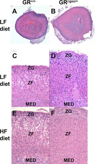
Figure 5. GRgeo/⫹ mice have larger adrenal glands. Repre-
was increased by the HF diet, the HF diet had no effect sentative images of hematoxylin and eosin stained sections of
on adrenal weight in GRgeo/⫹ mice (Fig. 4C). Adrenal adrenal glands from LF-fed GR⫹/⫹ mice (A, C); LF-fed
morphology also differed between genotypes, and this GRgeo/⫹ mice (B, D); HF-fed GR⫹/⫹ mice (E); and HF-fed
was particularly apparent on the LF diet (Fig. 5). The GRgeo/⫹ mice (F). ZF, zona fasciculate; ZG, zona glomeru-
morphology suggests stimulation of corticosterone- and losa; MED, medulla. n ⫽ 8 –9/group. Scale bars ⫽ 25 m.
aldosterone-producing cells of GRgeo/⫹ mice in the
zona fasciculata and glomerulosa, respectively. In adre-
nal glands from GRgeo/⫹ mice, the columns of endo- diet (Fig. 5E, F), but the morphological differences
crine cells in the zona fasciculata are longer than in between LF and HF groups were more pronounced in
GR⫹/⫹ mice, with individual cells having homogeneous GR⫹/⫹ mice than in GRgeo/⫹ mice (Fig. 5). Measure-
eosinophilic cytoplasm (Fig. 5A–D). The zona glomeru- ments of adrenal cell size in the different zones (glo-
losa of GRgeo/⫹ adrenal glands appears thicker than in merulosa, fascilulata, and reticularis) did not differ
GR⫹/⫹ mice, with more glomeruli evident and contain- between genotypes (data not shown), indicating that
ing hypertrophied epithelium (Fig. 5C, D). This adre- the larger adrenal glands in GRgeo/⫹ mice reflect
nal phenotype was also apparent in mice fed the HF hyperplasia rather than cellular hypertrophy.
Figure 4. HPA hyperactivity in GRgeo/⫹ mice. A, B) Plasma corticosterone (cort) levels in LF-fed GR⫹/⫹ (white bars), HF-fed
GR⫹/⫹ (hatched bars), LF-fed GRgeo/⫹ (black bars) and HF-fed GRgeo/⫹ mice (gray bars) in the morning (A) and evening (B).
C) Left adrenal weights in LF or HF-fed GR⫹/⫹ and GRgeo/⫹ mice (n⫽8 –9/group). *,†P ⬍ 0.05; **P ⬍ 0.01; †††P ⬍ 0.001. ‡P ⬍
0.01 vs. HF-fed GR⫹/⫹ mice.
HYPERTENSION AND ALTERED ADAPTION IN GRgeo/⫹ MICE 3901Figure 6. Unaltered body composition and glucose homeostasis in GRgeo/⫹ mice after HF diet. Body weight (A) and plasma
glucose levels (B) after GTT in LF-fed GR⫹/⫹ mice (open squares, dashed line); HF-fed GR⫹/⫹ mice (open circles, dashed line);
LF-fed GRgeo/⫹ mice (black squares, solid line) and HF-fed GRgeo/⫹ mice (black circles, solid line) (n⫽6/group). ***P ⬍ 0.001.
GRgeo/⫹ mice have normal adipose tissue distribution genotypes fed the LF diet showed similar responses in
and glucose homeostasis but higher liver triglyceride levels GTTs with an identical impaired response in mice fed
on HF diet HF diet (Fig. 6B).
Although GR polymorphisms in humans are associated
GRgeo/ⴙ mice are hypertensive and show an
with alterations in body weight and/or composition
attenuated blood pressure response to HF diet
(reviewed in ref. 14), there were no differences in body
weight (Fig. 6A) or food intake (data not shown)
between GRgeo/⫹ mice and their GR⫹/⫹ littermates, on In LF-fed mice, systolic blood pressure was significantly
either the LF or HF diet. There were also no differences elevated (8 mmHg) in GRgeo/⫹ mice compared with
between genotypes on either diet in weights of adipose their GR⫹/⫹ littermates (Fig. 7). Furthermore, blood
tissue (epididymal, inguinal, and mesenteric) and mus- pressure was increased in both genotypes by the HF
cle (extensor digitorum longus) or in plasma levels of diet, although interestingly the magnitude of the in-
insulin, NEFAs, and triglyceride (Table 2). However, crease was lower in GRgeo/⫹ mice (8 mmHg) than in
while the HF diet caused the expected increase in liver GR⫹/⫹ mice (14 mmHg), resulting in no difference in
triglyceride levels in both genotypes, levels were higher blood pressure between genotypes in mice fed the HF
in HF-fed GRgeo/⫹ than in GR⫹/⫹ mice (Table 2). Both diet (Fig. 7).
TABLE 2. Similar regional adiposity, lean mass, and plasma insulin and lipid levels in GRgeo/⫹ and GR⫹/⫹ mice
LF diet HF diet
Variable GR⫹/⫹ GRgeo/⫹ GR⫹/⫹ GRgeo/⫹
Epididymal fat mass
% Body weight 2.08 ⫾ 0.36 1.66 ⫾ 0.38 4.42 ⫾ 0.22*** 4.90 ⫾ 0.38***
Absolute wt (mg) 657 ⫾ 140 489 ⫾ 126 1972 ⫾ 110 2476 ⫾ 285
Inguinal fat mass
% Body weight 1.79 ⫾ 0.31 1.36 ⫾ 0.07 4.22 ⫾ 0.27*** 4.5 ⫾ 0.3.5***
Absolute wt (mg) 559 ⫾ 119 387 ⫾ 23 1914 ⫾ 186 2230 ⫾ 274
Mesenteric fat mass
% Body weight 1.31 ⫾ 0.15 1.07 ⫾ 0.07 2.16 ⫾ 0.13** 2.00 ⫾ 0.17**
Absolute wt (mg) 408 ⫾ 67 301 ⫾ 31 979 ⫾ 92 984 ⫾ 121
Brown adipose mass
% Body weight 0.38 ⫾ 0.03 0.38 ⫾ 0.03 0.62 ⫾ 0.05** 0.79 ⫾ 0.05**
Absolute wt (mg) 118 ⫾ 15 107 ⫾ 8 282 ⫾ 31 381 ⫾ 34
Muscle (EDL) mass
% Body weight 0.24 ⫾ 0.02 0.22 ⫾ 0.02 0.16 ⫾ 0.01** 0.15 ⫾ 0.01**
Absolute wt (mg) 70.3 ⫾ 4 60.8 ⫾ 5 68.6 ⫾ 3 68.8 ⫾ 3
Plasma insulin (ng/ml) 0.2 ⫾ 0.01 0.3 ⫾ 0.1 0.9 ⫾ 0.2* 1.0 ⫾ 0.2*
Plasma NEFA (mEq/l) 0.8 ⫾ 0.1 0.6 ⫾ 0.1 0.65 ⫾ 0.1 0.8 ⫾ 0.1
Plasma TG (mmol/l) 0.44 ⫾ 0.05 0.37 ⫾ 0.06 0.52 ⫾ 0.03 0.47 ⫾ 0.04
Liver TG (mol/g) 5.4 ⫾ 0.7 8.0 ⫾ 1.6 14.8 ⫾ 0.7*** 18⫾0.8***
Plasma NEFA, plasma triglyceride (TG), and plasma insulin levels were measured after 24 h fast. *P ⬍ 0.05; **P ⬍ 0.01; ***P ⬍ 0.001 vs.
LF diet. Values in italics indicate a significant difference between genotypes. n ⫽ 8/group.
3902 Vol. 22 November 2008 The FASEB Journal 䡠 www.fasebj.org MICHAILIDOU ET AL.composition in humans, few animal studies have ad-
dressed the effects of altered GR density on body weight
and composition and none have examined the effect
on metabolic adaption to the HF diet. Here we found
that reduced tissue GR density has no effect on body
weight, adipose tissue distribution or glucose homeosta-
sis, basally, on a chow diet (unpublished observations)
or an LF diet or after an HF diet. However, like most
humans heterozygous for mutations in the GR gene,
GRgeo/⫹ mice have an activated HPA axis and, as we
have now shown, elevated blood pressure. Interestingly,
GRgeo/⫹ mice failed to show the full extent of the HPA
and blood pressure adaptions to the HF diet seen in
control mice, suggesting that tissue GR density limits
the adaptive response to chronic dietary stress.
Figure 7. GRgeo/⫹ mice have elevated blood pressure. Systolic As expected, GRgeo/⫹ mice display a hyperactive HPA
blood pressure (SBP) in LF-fed GR⫹/⫹ mice (white bars),
HF-fed GR⫹/⫹ mice (hatched bars), LF-fed GRgeo/⫹ mice axis, with elevated plasma corticosterone and larger
(black bars), and HF-fed GRgeo/⫹ mice (gray bars). *P ⬍ 0.05; adrenal glands. The latter results from hyperplasia
**,††P ⬍ 0.01. rather than cellular hypertrophy, since adrenal cell size
was not different in GRgeo/⫹ mice in any of the zones.
GRgeo/ⴙ mice have an activated RAAS This is consistent with previous data showing increased
plasma corticosterone levels in mice with reduced GR
Increased systolic blood pressure in GRgeo/⫹ mice was density (11, 18), although measurements in these pre-
associated with a 2-fold increase in plasma renin activ- vious studies are likely to have been stress levels. It
ity, which was unaffected by diet in either genotype, differs, however, from a previous study in mice het-
remaining higher in GRgeo/⫹ mice (Fig. 8A). Similarly, erozygous for a null allele of GR (GRnull/⫹) in which
plasma aldosterone levels were increased in GRgeo/⫹ nonstressed plasma corticosterone levels, both morn-
mice, irrespective of diet (Fig. 8B). Plasma angio- ing and evening, were the same as in control mice,
tensinogen levels were comparable between LF diet-fed although the mice did show an increased plasma corti-
GRgeo/⫹ and GR⫹/⫹ mice but were markedly increased costerone response to stress (40), similar to GRgeo/⫹
(3-fold) by the HF diet only in GRgeo/⫹ mice (Fig. 8C). mice (our unpublished data). The discrepancy in non-
Hepatic angiotensinogen mRNA levels mirrored the stressed plasma corticosterone levels between the two
pattern of plasma angiotensinogen (Fig. 8E). Increased models may be related to environmental or strain
adipose tissue expression of angiotensinogen, although differences: here, backcrossed to C57BL/6J for five
much lower than hepatic expression, has been postu- generations, whereas Ridder et al. (40) examined F1
lated to contribute to or even underlie hypertension in offspring of a C57BL/6J X FVB/N cross. We (41) have
diet-induced obesity (38). However, although we ob- noted higher basal plasma corticosterone levels in
served higher expression of angiotensinogen mRNA in FVB/N mice.
epididymal adipose tissue of LF-fed GRgeo/⫹ mice com- GRgeo/⫹ mice have completely normal body weight,
pared with GR⫹/⫹ mice, levels were lower in HF-fed regional adiposity, and lean mass. Glucose homeostasis
mice and did not differ between genotypes (Fig. 8E). (measured by fasting plasma glucose) and insulin lev-
Neither kidney nor adrenal renin mRNA levels differed els, as well as glucose tolerance, were also entirely
between genotypes on the LF diet nor were they altered normal in GRgeo/⫹ mice. This metabolic phenotype
by the HF diet (Table 3). Similarly, renal 11-HSD2 contrasts with the obesity seen in chow-fed transgenic
and MR mRNA levels, both critical determinants of mice with globally reduced GR density because of
blood pressure (39), were identical between genotypes antisense GR RNA expression (18, 19). These mice,
and they did not differ with diet (Table 3). Thus, similar to GRgeo/⫹ mice, also show an activated HPA
activation of the RAAS in GRgeo/⫹ mice is likely to axis (18). The reason for the discrepancy between these
underlie their hypertension but does not account for two models, each with reduced GR and elevated corti-
changes in blood pressure after the HF diet. The costerone, is not clear but may relate to the unknown
activation of RAAS in GRgeo/⫹ mice was not accompa- effects of the GR transgene in the antisense GR-express-
nied by grossly abnormal kidney function, since kidney ing mice in which knockdown of the GR is achieved
morphology was similar in both genotypes (data not through use of a neurofilament gene promoter, which
shown). may be differentially expressed within the central ner-
vous system. Mice with a conditional deletion of the GR
gene in neurons have very high plasma corticosterone
DISCUSSION levels due to a lack of negative feedback at the hypo-
thalamus (42). However, these mice have normal pe-
Although GR polymorphisms are associated with hyper- ripheral GR levels, are growth retarded, and have
tension and differences in body mass index and body proportionately more body fat before weaning, but less
HYPERTENSION AND ALTERED ADAPTION IN GRgeo/⫹ MICE 3903Figure 8. Activation of the RAAS in GRgeo/⫹ mice. Plasma renin activity (A), plasma aldosterone concentration (B), plasma
angiotensinogen concentration (C), epididymal fat angiotensinogen (AGT) mRNA levels (D), and hepatic AGT mRNA levels
(E) in LF-fed GR⫹/⫹ mice (white bars), HF-fed GR⫹/⫹ mice (hatched bars), LF-fed GRgeo/⫹ mice (black bars), and HF-fed
GRgeo/⫹ mice (gray bars). For plasma measurements, n ⫽ 5/group. For mRNA levels, n ⫽ 8 –9/group. *,†P ⬍ 0.05; **,††P ⬍ 0.01;
***,†††P ⬍ 0.001.
after (20). In GRgeo/⫹ mice, reduced peripheral GR levels has previously been reported and postulated to
density may compensate for the increased plasma cor- be due to the more rapid secretion and turnover of
ticosterone levels, normalizing body fat distribution. newly synthesized renin compared with the long half-
The lack of effect of genotype on glucose homeostasis life of renin mRNA (44). Alternatively, renin clearance
is not surprising, as selective GR gene deletion in may be increased in GRgeo/⫹ mice. Despite reduced GR
hepatocytes showed that GR is essential for glucose density and elevated plasma corticosteroid levels in
homeostasis only after prolonged fasting or in a dia- GRgeo/⫹ mice, there were no compensatory changes, at
betic state (21). Moreover, antisense GR mice also show least in the kidney, in the expression of MR or 11-
normal glucose homeostasis (19). HSD2, which protects MR from illicit occupation by
Like most humans heterozygous for inactivating mu- glucocorticoids (39). Similarly, MR levels in the brain
tations in the GR gene (43), GRgeo/⫹ mice are hyper- are normal in mice with forebrain-specific deletion of
tensive. In GRgeo/⫹ mice, the hypertension results from GR (45), suggesting that MR density is not altered to
elevated activity of the renin-angiotensin-aldosterone compensate for reduced GR expression. The increased
system. Plasma aldosterone levels were increased in plasma aldosterone levels in GRgeo/⫹ mice were consis-
GRgeo/⫹ mice, as was plasma renin activity. The latter, tent with the adrenal histopathology, suggesting high
however, was not associated with increased expression synthetic activity of aldosterone producing cells. This
of renin mRNA in either the kidney or adrenal gland. A was predicted from the phenotype of GRhypo/hypo mice,
similar discrepancy between plasma renin and mRNA which have elevated levels of aldosterone synthase in
TABLE 3. Similar expression of mRNAs encoding renin, 11-HSD2, and MR in GRgeo/⫹ and GR⫹/⫹ mice
LF diet HF diet
mRNA Tissue GR⫹/⫹ GRgeo/⫹ GR⫹/⫹ GRgeo/⫹
Renin Kidney 0.87 ⫾ 0.07 0.95 ⫾ 0.20 0.83 ⫾ 0.09 0.75 ⫾ 0.06
Adrenal 0.98 ⫾ 0.20 1.0 ⫾ 0.10 ND ND
11-HSD2 Kidney 0.85 ⫾ 0.03 0.86 ⫾ 0.09 0.94 ⫾ 0.04 0.77 ⫾ 0.09
MR Kidney 0.80 ⫾ 0.08 0.96 ⫾ 0.20 0.98 ⫾ 0.07 0.99 ⫾ 0.10
Levels of specific mRNAs were measured by qPCR and are expressed in arbitrary units. There were no significant effects of either genotype
or diet upon levels of any of the mRNAs measured (n⫽6/group).
3904 Vol. 22 November 2008 The FASEB Journal 䡠 www.fasebj.org MICHAILIDOU ET AL.the zona glomerulosa (11), and is probably a conse- ute. Plasma angiotensinogen levels were increased by quence of elevated plasma ACTH levels in these mice. the HF diet in GRgeo/⫹ mice, probably as a result of Indeed, Pomc⫺/⫺ mice, which lack circulating ACTH, increased hepatic expression of angiotensinogen, but have small adrenal glands and dramatically reduced were unaffected in GR⫹/⫹ mice in which the diet- plasma aldosterone levels (46). induced increase in blood pressure was greater. The HF HF diets chronically stress the HPA axis in rodents, diet had no effect on plasma aldosterone levels or increasing basal corticosterone levels and causing adre- plasma renin activity in either genotype. Thus, activa- nal enlargement (35, 47), although negative feedback tion of the systemic RAAS by the HF diet in GR⫹/⫹ mice efficiency is unaffected by HF diet (35). Consistent with is ruled out. Elevated plasma glucocorticoid levels drive this, in GR⫹/⫹ mice, the HF diet from weaning in- hypertension in both humans and rodents (1, 54). creased adrenal size and increased morning plasma However, the underlying mechanisms remain contro- corticosterone levels. In contrast, in GRgeo/⫹ mice, the versial but are likely to include changes in the sympa- HF diet had no effect on either of these parameters. thetic nervous system. In this regard, it is of note that Thus, differences between genotypes in these parame- adrenal catecholamines, implicated in the HF diet- ters were eliminated by the diet. Evening (nonstressed) induced hypertension (53), are reduced in GR⫹/⫺ mice plasma corticosterone levels were unaffected by the HF (11, 34). It will be of interest, in future work, to diet and remained higher in GRgeo/⫹ mice. These determine whether sympathetic nervous system activity results suggest that the HPA axis response to the HF is altered in GRgeo/⫹ mice. diet is limited by tissue GR density, this adaptive re- In humans, polymorphisms in the GR gene associ- sponse being abolished by a 50% reduction in GR ated with hypersensitivity or hyposensitivity to glucocor- levels. ticoids in a dexamethasone suppression test have been Surprisingly, given the role of glucocorticoids in variably associated with differences in body mass index, energy homeostasis (2), GRgeo/⫹ mice showed an en- waist-hip ratio, altered body composition, and meta- tirely normal body weight gain, altered glucose ho- bolic parameters (reviewed in ref. 14) as well as differ- meostasis and insulin sensitivity on the HF diet, and did ences in blood pressure (55–59). However, the associ- not differ from GR⫹/⫹ controls. Similarly, mice of both ation studies (14) are controversial and may depend on genotypes fed the HF diet doubled their hepatic accu- age, gender, and ethnic background. The data re- mulation of triglyceride compared with mice on the ported here provide insights into that heterogeneity. In control diet, although hepatic triglyceride levels were our model of glucocorticoid resistance due to ⬃50% significantly higher in HF-fed GRgeo/⫹ mice than in reduction in GR density, the observed physiological GR⫹/⫹ mice. Although plasma NEFAs did not differ differences between genotypes depend on environ- between LF- and HF-fed mice, these were measured ment. Thus, several of the differences between geno- after a 24 h fast, when NEFAs release from adipose types are abolished or reduced after the HF diet. If the tissue constitutes the major energy source and is max- same applies in humans, then (for example) obesity or imal. Glucocorticoids increase hepatic lipogenesis and other chronic stress may mask differences in blood secretion (48 –50). Furthermore, the consequences of pressure between populations with different GR poly- altered glucocorticoid status on hepatic triglyceride morphisms. metabolism are dependent on diet composition (51), Glucocorticoids form part of important homeostatic but only when the diet is fed ad libitum (52). Thus, if control mechanisms that are critical in adapting to lipid flux is low, when animals are maintained on an LF environmental challenges. Thus, they maintain stability diet or with the restricted HF diet, then glucocorticoids through change over time (allostasis; reviewed in refs. minimally affect hepatic triglyceride storage and secre- 60, 61). GRgeo/⫹ mice do not show the normal adap- tion (52). Whether the increased hepatic triglyceride tions to the HF diet; they have an attenuated blood levels in GRgeo/⫹ mice are a consequence of the pressure response and do not show the alterations in elevated peak corticosterone levels in these mice adrenal size and HPA axis activity seen in WT mice. (morning levels did not differ after the HF diet) or a Consequently, basal corticosterone levels are normal- direct consequence of reduced hepatic GR density ized between genotypes after the HF diet. Moreover, remains to be determined. 11-HSD1 levels are un- GRgeo/⫹ mice accumulated more hepatic triglyceride, likely to contribute to elevated hepatic triglycerides, possibly because of inadequate adaption to excessive since adipose or liver mRNA levels were unaltered dietary fat/energy intake and the need to protect vital (unpublished observations). organs. Continued excessive lipid accumulation in the Blood pressure was increased by the HF diet in both liver may, in turn, lead to insulin resistance and meta- genotypes, consistent with previous data (36, 37, 53). bolic disease. GR density is likely to be limiting in other However, the magnitude of the HF-induced increase in systems in adaption to environmental stressors. Mice blood pressure was smaller in GRgeo/⫹ mice so that heterozygous for inactivating mutations in GR show blood pressure in HF-fed GRgeo/⫹ mice did not differ reduced coping behavior in a mouse correlate of from that in HF-fed GR⫹/⫹ mice. The mechanism depression, with compromised indicators of neural underlying the blood pressure increase with the HF plasticity (40), whereas overexpression of GR results in diet remains unclear but may differ between genotypes. resistance to stress and robust protection against endo- In GRgeo/⫹ mice, further RAAS activation may contrib- toxins. These adaptive or “remodeling” effects of glu- HYPERTENSION AND ALTERED ADAPTION IN GRgeo/⫹ MICE 3905
cocorticoids in the adult response to the HF diet are                         11.   Cole, T. J., Blendy, J. A., Monaghan, A. P., Krieglstein, K.,
reminiscent of the prenatal “programming” effects of                                Schmid, W., Aguzzi, A., Fantuzzi, G., Hummler, E., Unsicker, K.,
                                                                                    and Schutz, G. (1995) Targeted disruption of the glucocorticoid
glucocorticoids (reviewed in ref. 62) where exposure of                             receptor gene blocks adrenergic chromaffin cell development
the late fetus to glucocorticoids permanently programs                              and severely retards lung maturation. Genes Dev. 9, 1608 –1621
tissue systems throughout life, including blood pres-                         12.   Tronche, F., Kellendonk, C., Reichardt, H. M., and Schütz, G.
                                                                                    (1998) Genetic dissection of glucocorticoid receptor function in
sure, glucose/lipid homeostasis, and tissue GR density                              mice. Curr. Opin. Genet. Dev. 8, 532–538
itself. It will be of great interest to determine whether                     13.   Kino, T., and Chrousos, G. P. (2001) Glucocorticoid and
GR density influences the prenatal programming ef-                                  mineralocorticoid resistance/hypersensitivity syndromes. J. En-
fects of glucocorticoids in the same way that it alters the                         docrinol. 169, 437– 445
                                                                              14.   van Rossum, E. F., and Lamberts, S. W. (2004) Polymorphisms
postnatal blood pressure and HPA adaptions to a                                     in the glucocorticoid receptor gene and their associations with
chronic HF diet.                                                                    metabolic parameters and body composition. Recent Prog. Horm.
                                                                                    Res. 59, 333–357
  We thank Mark Danielsen (Georgetown University                              15.   Gass, P., Reichardt, H. M., Strekalova, T., Henn, F., and
                                                                                    Tronche, F. (2001) Mice with targeted mutations of glucocorti-
School of Medicine, Washington, DC, USA) for kindly
                                                                                    coid and mineralocorticoid receptors: models for depression
providing the SV2wrec plasmid encoding mouse GR cDNA                                and anxiety? Physiol. Behav. 73, 811– 825
and David Pearce (University of California, San Francisco,                    16.   Howell, M. P., and Muglia, L. J. (2006) Effects of genetically altered
Department of Medicine, San Francisco, CA, USA) for                                 brain glucocorticoid receptor action on behavior and adrenal axis
providing the rPNMT-998/-466 Luc plasmid. We thank the                              regulation in mice. Front. Neuroendocrinol. 27, 275–284
Biomedical Research Resources facility, University of Edin-                   17.   Kleiman, A., and Tuckermann, J. P. (2007) Glucocorticoid
burgh, facility for assistance with animal care and members                         receptor action in beneficial and side effects of steroid therapy:
of the Endocrinology Unit for helpful discussions and                               lessons from conditional knockout mice. Mol. Cell. Endocrinol.
advice. This work was supported by a Wellcome Trust PhD                             275, 98 –108
Studentship (to Z.M.), a British Heart Foundation Project                     18.   Pepin, M.-C., Pothier, F., and Barden, N. (1992) Impaired type
                                                                                    II glucocorticoid receptor function in mice bearing antisense
Grant (to K.E.C., C.J.K., N.M.M., and J.R.S.), a Wellcome                           RNA transgene. Nature 355, 725–728
Trust Programme Grant (to J.R.S. and K.E.C.), a Wellcome                      19.   Richard, D., Chapdelaine, S., Deshaies, Y., Pepin, M. C., and
Trust Project Grant (to M.C.H and J.R.S.), and a Medical                            Barden, N. (1993) Energy balance and lipid metabolism in
Research Council Programme Grant (to C.J.K.). N.M.M. is                             transgenic mice bearing an antisense GCR gene construct.
supported by a Wellcome Trust Research Career Develop-                              Am. J. Physiol. Regul. Integr. Comp. Physiol. 265, R146 –R150
ment Fellowship.                                                              20.   Kellendonk, C., Eiden, S., Kretz, O., Schutz, G., Schmidt, I.,
                                                                                    Tronche, F., and Simon, E. (2002) Inactivation of the GR in the
                                                                                    nervous system affects energy accumulation. Endocrinology 143,
                                                                                    2333–2340
                                                                              21.   Opherk, C., Tronche, F., Kellendonk, C., Kohlmuller, D.,
REFERENCES                                                                          Schulze, A., Schmid, W., and Schutz, G. (2004) Inactivation of
                                                                                    the glucocorticoid receptor in hepatocytes leads to fasting
 1.    Whitworth, J. A., Brown, M. A., Kelly, J. J., and Williamson, P. M.          hypoglycemia and ameliorates hyperglycemia in streptozotocin-
       (1995) Mechanisms of cortisol-induced hypertension in hu-                    induced diabetes mellitus. Mol. Endocrinol. 18, 1346 –1353
       mans. Steroids 60, 76 – 80                                             22.   Sainte-Marie, Y., Nguyen Dinh Cat, A., Perrier, R., Mangin, L.,
 2.    Dallman, M. F., Strack, A. M., Akana, S. F., Bradbury, M. J.,                Soukaseum, C., Peuchmaur, M., Tronche, F., Farman, N.,
       Hanson, E. S., Scribner, K. A., and Smith, M. (1993) Feast and               Escoubet, B., Benitah, J. P., and Jaisser, F. (2007) Conditional
       famine– critical role of glucocorticoids with insulin in daily               glucocorticoid receptor expression in the heart induces atrio-
       energy-flow. Front. Neuroendocrinol. 14, 303–347                             ventricular block. FASEB J. 21, 3133–3141
 3.    Ballard, P. L., Baxter, J. D., Higgins, S. J., Rousseau, G. G., and    23.   Sutherland, H. G., Mumford, G. K., Newton, K., Ford, L. V.,
       Tomkins, G. M. (1974) General presence of glucocorticoid                     Farrall, R., Dellaire, G., Caceres, J. F., and Bickmore, W. A.
       receptors in mammalian tissues. Endocrinology 94, 998 –1002                  (2001) Large-scale identification of mammalian proteins local-
 4.    Hollenberg, S. M., Weinberger, C., Ong, E. S., Cerelli, G., Oro,             ized to nuclear sub-compartments. Hum. Mol. Genet. 10, 1995–
       A., Lebo, R., Thompson, E. B., Rosenfeld, M. G., and Evans,                  2011
       R. M. (1985) Primary structure and expression of a functional          24.   Harris, H. J., Kotelevtsev, Y., Mullins, J. J., Seckl, J. R., and
       human glucocorticoid receptor cDNA. Nature 318, 635– 641                     Holmes, M. C. (2001) Intracellular regeneration of glucocor-
 5.    Miesfeld, R., Rusconi, S., Godowski, P. J., Maler, B. A., Okret, S.,         ticoids by 11b-hydroxysteroid dehydrogenase (11b-HSD)-1
       Wikström, A. C., Gustafsson, J.-Å., and Yamamoto, K. R. (1986)              plays a key role in regulation of the hypothalamic-pituitary-
       Genetic complementation of a glucocorticoid receptor defi-                   adrenal axis: analysis of 11b-HSD-1 deficient mice. Endocrinol-
       ciency by expression of cloned receptor cdna. Cell 46, 389 –399              ogy 142, 114 –120
 6.    Mangelsdorf, D. J., Thummel, C., Beato, M., Herrlich, P.,              25.   Morton, N. M., Densmore, V., Wamil, M., Ramage, L., Nichol,
       Schutz, G., Umesono, K., Blumberg, B., Kastner, P., Mark, M.,                K., Bunger, L., Seckl, J. R., and Kenyon, C. J. (2005) A polygenic
       Chambon, P., and Evans, R. M. (1995) The nuclear receptor                    model of the metabolic syndrome with reduced circulating and
       superfamily: the second decade. Cell 83, 835– 839                            intra-adipose glucocorticoid action. Diabetes 54, 3371–3378
 7.    Vanderbilt, J. N., Miesfeld, R., Maler, B. A., and Yamamoto, K. R.     26.   Al-Dujaili, E. A., and Edwards, C. R. (1981) Development and
       (1987) Intracellular receptor concentration limits glucocorti-               application of a simple radioimmunoassay for urinary aldoste-
       coid-dependent enhancer activity. Mol. Endocrinol. 1, 68 –74                 rone. Clin. Chim. Acta 116, 277–287
 8.    Ramdas, J., Liu, W., and Harmon, J. M. (1999) Glucocorticoid-          27.   Al-Dujaili, E. A. (2006) Development and validation of a simple
       induced cell death requires autoinduction of glucocorticoid                  and direct ELISA method for the determination of conjugated
       receptor expression in human leukemic T cells. Cancer Res. 59,               (glucuronide) and non-conjugated testosterone excretion in
       1378 –1385                                                                   urine. Clin. Chim. Acta 364, 172–179
 9.    Reichardt, H. M., Umland, T., Bauer, A., Kretz, O., and Schütz,       28.   Evans, A. L., Brown, W., Kenyon, C. J., Maxted, K. J., and Smith,
       G. (2000) Mice with an increased glucocorticoid receptor gene                D. C. (1994) Improved system for measuring systolic blood
       dosage show enhanced resistance to stress and endotoxic shock.               pressure in the conscious rat. Med. Biol. Eng. Comput. 32,
       Mol. Cell. Biol. 20, 9009 –9017                                              101–102
10.    Pazirandeh, A., Xue, Y., Prestegaard, T., Jondal, M., and Okret,       29.   Seckl, J. R., Dickson, K. L., and Fink, G. (1990) Central
       S. (2002) Effects of altered glucocorticoid sensitivity in the               5,7-dihydroxytryptamine lesions decrease hippocampal glu-
       T-cell lineage on thymocyte and T-cell homeostasis. FASEB J. 16,             cocorticoid and mineralocorticoid receptor messenger ribonu-
       727–729                                                                      cleic acid expression. J. Neuroendocrinol. 2, 911–916

3906      Vol. 22   November 2008                         The FASEB Journal 䡠 www.fasebj.org                                    MICHAILIDOU ET AL.
30. Lefebvre, P., Barard, D. S., Cordingley, M. G., and Hager, G. L. 46. Coll, A. P., Challis, B. G., Yeo, G. S., Snell, K., Piper, S. J., Halsall,
(1991) Two regions of the mouse mammary tumor virus long D., Thresher, R. R., and O’Rahilly, S. (2004) The effects of
terminal repeat regulate the activity of its promoter in mam- proopiomelanocortin deficiency on murine adrenal develop-
mary cell lines. Mol. Cell. Biol. 11, 2529 –2537 ment and responsiveness to adrenocorticotropin. Endocrinology
31. Adams, M., Meijer, O. C., Wang, J., Bhargava, A., and Pearce, D. 145, 4721– 4727
(2003) Homodimerization of the glucocorticoid receptor is not 47. Carroll, K. K., and Noble, R. L. (1952) Effects of feeding rape oil
essential for response element binding: activation of the phe- on some endocrine functions of the rat. Endocrinology 51,
nylethanolamine N-methyltransferase gene by dimerization-de- 476 – 486
fective mutants. Mol. Endocrinol. 17, 2583–2592 48. Klausner, H., and Heimberg, M. (1967) Effect of adrenalcortical
32. Danielsen, M., Northrop, J. P., and Ringold, G. M. (1986) The hormones on release of triglycerides and glucose by liver. Am. J.
mouse glucocorticoid receptor: mapping of functional domains Physiol. 212, 1236 –1246
by cloning, sequencing and expression of wild-type and mutant 49. Kirk, C. J., Verrinder, T. R., and Hems, D. A. (1976) Fatty acid
receptor proteins. EMBO J. 5, 2513–2522 synthesis in the perfused liver of adrenalectomized rats. Biochem.
33. Kasai, Y. (1990) Two naturally-occurring isoforms and their J. 156, 593– 602
expression of a glucocorticoid receptor gene from an androgen- 50. Bartlett, S. M., and Gibbons, G. F. (1988) Short- and longer-
dependent mouse tumor. FEBS Lett. 274, 99 –102 term regulation of very-low-density lipoprotein secretion by
34. Finotto, S., Krieglstein, K., Schober, A., Deimling, F., Lindner, insulin, dexamethasone and lipogenic substrates in cultured
K., Bruhl, B., Beier, K., Metz, J., Garcia-Arraras, J. E., Roig- hepatocytes. A biphasic effect of insulin. Biochem. J. 249, 37– 43
Lopez, J. L., Monaghan, P., Schmid, W., Cole, T. J., Kellendonk, 51. Mantha, L., Palacios, E., and Deshaies, Y. (1999) Modulation of
C., Tronche, F., Schutz, G., and Unsicker, K. (1999) Analysis of triglyceride metabolism by glucocorticoids in diet-induced obe-
mice carrying targeted mutations of the glucocorticoid receptor sity. Am. J. Physiol. Regul. Integr. Comp. Physiol. 277, R455–R464
gene argues against an essential role of glucocorticoid signalling 52. Mantha, L., and Deshaies, Y. (2000) Energy intake-independent
for generating adrenal chromaffin cells. Development 126, 2935– modulation of triglyceride metabolism by glucocorticoids in the
2944 rat. Am. J. Physiol. Regul. Integr. Comp. Physiol. 278, R1424 –R1432
35. Tannenbaum, B. M., Brindley, D. N., Tannenbaum, G. S., 53. Kaufman, L. N., Li, H. Y., Peterson, M. M., and Gilardy, A. K.
Dallman, M. F., McArthur, M. D., and Meaney, M. J. (1997) (1993) Adrenal medulla as a mediator of diet-induced hyper-
High-fat feeding alters both basal and stress-induced hypotha- tension. Am. J. Physiol. Regul. Integr. Comp. Physiol. 265, R1–R6
lamic- pituitary-adrenal activity in the rat. Am. J. Physiol. Endocri- 54. Magiakou, M. A., Smyrnaki, P., and Chrousos, G. P. (2006)
nol. Metab. 273, E1168 –E1177 Hypertension in Cushing’s syndrome. Best Pract. Res. Clin.
36. Mills, E., Kuhn, C. M., Feinglos, M. N., and Surwit, R. (1993) Endocrinol. Metab. 20, 467– 482
Hypertension in CB57BL/6J mouse model of non-insulin-de- 55. Watt, G. C., Harrap, S. B., Foy, C. J., Holton, D. W., Edwards,
pendent diabetes mellitus. Am. J. Physiol. Regul. Integr. Comp. H. V., Davidson, H. R., Connor, J. M., Lever, A. F., and Fraser,
Physiol. 264, R73–R78 R. (1992) Abnormalities of glucocorticoid metabolism and the
37. Williams, T. D., Chambers, J. B., Roberts, L. M., Henderson, renin-angiotensin system: a four-corners approach to the iden-
R. P., and Overton, J. M. (2003) Diet-induced obesity and tification of genetic determinants of blood pressure. J. Hypertens.
cardiovascular regulation in C57BL/6J mice. Clin. Exp. Pharma- 10, 473– 482
col. Physiol. 30, 769 –778 56. Panarelli, M., Holloway, C. D., Fraser, R., Connell, J. M. C.,
38. Frederich, R. C., Jr., Kahn, B. B., Peach, M. J., and Flier, J. S. Ingram, M. C., Anderson, N. H., and Kenyon, C. J. (1998)
(1992) Tissue-specific nutritional regulation of angiotensinogen Glucocorticoid receptor polymorphism, skin vasoconstriction,
in adipose tissue. Hypertension 19, 339 –344 and other metabolic intermediate phenotypes in normal human
39. Seckl, J. R., and Brown, R. W. (1994) 11-hydroxysteroid subjects. J. Clin. Endocrinol. Metab. 83, 1846 –1852
dehydrogenase: on several roads to hypertension. J. Hypertension 57. Takami, S., Wong, Z. Y., Stebbing, M., and Harrap, S. B. (1999)
12, 105–112 Linkage analysis of glucocorticoid and beta2-adrenergic recep-
40. Ridder, S., Chourbaji, S., Hellweg, R., Urani, A., Zacher, C., tor genes with blood pressure and body mass index. Am. J.
Schmid, W., Zink, M., Hortnagl, H., Flor, H., Henn, F. A., Physiol. Heart Circ. Physiol. 276, H1379 –H1384
Schutz, G., and Gass, P. (2005) Mice with genetically altered 58. Rosmond, R., Chagnon, Y. C., Holm, G., Chagnon, M., Pérusse,
glucocorticoid receptor expression show altered sensitivity for L., Lindell, K., Carlsson, B., Bouchard, C., and Björntorp, P.
stress-induced depressive reactions. J. Neurosci. 25, 6243– 6250 (2000) A glucocorticoid receptor gene marker is associated with
41. Yau, J. L., Noble, J., Thomas, S., Kerwin, R., Morgan, P. E., abdominal obesity, leptin, and dysregulation of the hypotha-
Lightman, S., Seckl, J. R., and Pariante, C. M. (2007) The lamic-pituitary- adrenal axis. Obes. Res. 8, 211–218
antidepressant desipramine requires the ABCB1 (Mdr1)-type 59. Di Blasio, A. M., van Rossum, E. F., Maestrini, S., Berselli, M. E.,
p-glycoprotein to upregulate the glucocorticoid receptor in Tagliaferri, M., Podesta, F., Koper, J. W., Liuzzi, A., and Lam-
mice. Neuropsychopharmacology 32, 2520 –2529 berts, S. W. (2003) The relation between two polymorphisms in
42. Tronche, F., Kellendonk, C., Kretz, O., Gass, P., Anlag, K., the glucocorticoid receptor gene and body mass index, blood
Orban, P. C., Bock, R., Klein, R., and Schutz, G. (1999) pressure and cholesterol in obese patients. Clin. Endocrinol.
Disruption of the glucocorticoid receptor gene in the nervous (Oxf.) 59, 68 –74
system results in reduced anxiety. Nat. Genet. 23, 99 –103 60. Korte, S. M., Koolhaas, J. M., Wingfield, J. C., and McEwen, B. S.
43. Kino, T., Vottero, A., Charmandari, E., and Chrousos, G. P. (2005) The Darwinian concept of stress: benefits of allostasis
(2002) Familial/sporadic glucocorticoid resistance syndrome and costs of allostatic load and the trade-offs in health and
and hypertension. Ann. N. Y. Acad. Sci. 970, 101–111 disease. Neurosci. Biobehav. Rev. 29, 3–38
44. Nakamura, N., Soubrier, F., Menard, J., Panthier, J. J., Rougeon, 61. Chapman, K. E., and Seckl, J. R. (2007) 11beta-HSD1, inflam-
F., and Corvol, P. (1985) Nonproportional changes in plasma mation, metabolic disease and age-related cognitive (dys)func-
renin concentration, renal renin content, and rat renin messen- tion. Neurochem Res
ger RNA. Hypertension 7, 855– 859 62. Seckl, J. R. (2001) Glucocorticoid programming of the fetus;
45. Boyle, M. P., Brewer, J. A., Funatsu, M., Wozniak, D. F., Tsien, adult phenotypes and molecular mechanisms. Mol. Cell. Endocri-
J. Z., Izumi, Y., and Muglia, L. J. (2005) Acquired deficit of nol. 185, 61–71
forebrain glucocorticoid receptor produces depression-like
changes in adrenal axis regulation and behavior. Proc. Natl. Received for publication April 21, 2008.
Acad. Sci. U. S. A. 102, 473– 478 Accepted for publication July 10, 2008.
HYPERTENSION AND ALTERED ADAPTION IN GRgeo/⫹ MICE 3907You can also read