Formation and immunomodulatory function of meningeal B cell aggregates in progressive CNS autoimmunity
←
→
Page content transcription
If your browser does not render page correctly, please read the page content below
doi:10.1093/brain/awab093                                                                                        BRAIN 2021: 144; 1697–1710   | 1697

Formation and immunomodulatory function
of meningeal B cell aggregates in
progressive CNS autoimmunity

                                                                                                                                                       Downloaded from https://academic.oup.com/brain/article/144/6/1697/6164962 by guest on 14 October 2021
Meike Mitsdoerffer,1,2 Giovanni Di Liberto,3 Sarah Dötsch,4 Christopher Sie,2
Ingrid Wagner,3 Monika Pfaller,2 Mario Kreutzfeldt,3 Simon Fräßle,4 Lilian Aly,1
Benjamin Knier,1 Dirk H. Busch,4,5 Doron Merkler3 and Thomas Korn1,2,6

Meningeal B lymphocyte aggregates have been described in autopsy material of patients with chronic mul-
tiple sclerosis. The presence of meningeal B cell aggregates has been correlated with worse disease.
However, the functional role of these meningeal B cell aggregates is not understood.
Here, we use a mouse model of multiple sclerosis, the spontaneous opticospinal encephalomyelitis model,
which is built on the double transgenic expression of myelin oligodendrocyte glycoprotein-specific T-cell
and B-cell receptors, to show that the formation of meningeal B cell aggregates is dependent on the
expression of a4 integrins by antigen-specific T cells. T cell-conditional genetic ablation of a4 integrins in
opticospinal encephalomyelitis mice impaired the formation of meningeal B cell aggregates, and surprising-
ly, led to a higher disease incidence as compared to opticospinal encephalomyelitis mice with a4 integrin-
sufficient T cells. B cell-conditional ablation of a4 integrins in opticospinal encephalomyelitis mice
resulted in the entire abrogation of the formation of meningeal B cell aggregates, and opticospinal
encephalomyelitis mice with a4 integrin-deficient B cells suffered from a higher disease burden than regular
opticospinal encephalomyelitis mice. While anti-CD20 antibody-mediated systemic depletion of B cells in
opticospinal encephalomyelitis mice after onset of disease failed to efficiently decrease meningeal B cell
aggregates without significantly modulating disease progression, treatment with anti-CD19 chimeric antigen
receptor-T cells eliminated meningeal B cell aggregates and exacerbated clinical disease in opticospinal en-
cephalomyelitis mice. Since about 20% of B cells in organized meningeal B cell aggregates produced either
IL-10 or IL-35, we propose that meningeal B cell aggregates might also have an immunoregulatory function
as to the immunopathology in adjacent spinal cord white matter. The immunoregulatory function of menin-
geal B cell aggregates needs to be considered when designing highly efficient therapies directed against
meningeal B cell aggregates for clinical application in multiple sclerosis.

1 Klinikum rechts der Isar, Department of Neurology, Technical University of Munich, 81675 Munich, Germany
2 Klinikum rechts der Isar, Institute for Experimental Neuroimmunology, Technical University of Munich, 81675
  Munich, Germany
3 Division of Clinical Pathology, Department of Pathology and Immunology, Geneva Faculty of Medicine, Centre
  Médical Universitaire, 1211 Geneva, Switzerland
4 Institute for Medical Microbiology, Immunology, and Hygiene, Technical University of Munich, 81675 Munich,
  Germany
5 National Center for Infection Research (DZIF), Technical University of Munich, 81675 Munich, Germany
6 Munich Cluster for Systems Neurology (SyNergy), DZNE site Munich, 81377 Munich, Germany

Received October 22, 2020. Revised January 18, 2021. Accepted January 19, 2021. Advance access publication March 9, 2021
C The Author(s) (2021). Published by Oxford University Press on behalf of the Guarantors of Brain. All rights reserved.
V
For permissions, please email: journals.permissions@oup.com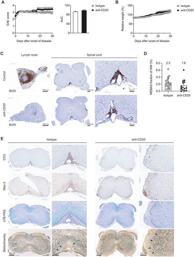
1698     | BRAIN 2021: 144; 1697–1710                                                                                  M. Mitsdoerffer et al.

Correspondence to: Thomas Korn
Klinikum rechts der Isar, Technical University of Munich
Institute for Experimental Neuroimmunology
Ismaninger Str. 22, 81675 Munich, Germany
E-mail: thomas.korn@tum.de

Keywords: B cell; meningeal inflammation; multiple sclerosis; experimental autoimmune encephalomyelitis; CAR
T cell
Abbreviations: CAR = chimeric antigen receptor; EAE = experimental autoimmune encephalomyelitis; MEBAG =
meningeal B cell aggregate; OSE = opticospinal encephalomyelitis

                                                                                                                                                    Downloaded from https://academic.oup.com/brain/article/144/6/1697/6164962 by guest on 14 October 2021
Introduction                                                              induced with recombinant MOG protein.12 In contrast, in the spon-
                                                                          taneous model of EAE, the opticospinal encephalomyelitis (OSE)
B lymphocyte aggregates have been identified in the meningeal             mouse, in which both T cells14 and B cells15 express a MOG-specific
space of patients with secondary progressive multiple sclerosis.1         antigen receptor, MEBAGs develop in abundance.16,17
CD35 + follicular dendritic cells1 and proliferating (Ki67 + ) B cells2       In this study, we exploited the OSE mouse model to investigate
were identified in these B cell aggregates. Therefore, they have          the role of a4 integrins in T cells and B cells for the generation of
been considered as tertiary lymphoid tissue with bona fide B cell         MEBAGs in the subarachnoid space. Conditional genetic ablation
follicles.1,2 B cell follicles are highly organized structures where      of Itga4, which encodes a4 integrins, in T cells led to the reduced
class switch recombination and somatic hypermutation is induced           formation of MEBAGs in OSE mice. Yet, the incidence of spontan-
in specific B cell clones by repeated rounds of cognate interaction       eous EAE was increased in these mice as compared to regular OSE
with T follicular helper cells.3 Eventually, germinal centre (GC)         mice. B cell-conditional Itga4–/– OSE mice lacked MEBAGs and
B cells develop into plasma blasts that produce affinity matured          developed a higher disease burden than their wild-type OSE litter-
antibodies. It is possible that meningeal B cell aggregates               mates. While B cell depletion in presymptomatic OSE mice using a
(MEBAGs) contribute to immunopathology in multiple sclerosis.             monoclonal anti-CD20 antibody alleviated disease development,
For instance, by secretion of autoantibodies, demyelination in ad-        the same treatment did not significantly modulate disease pro-
jacent cortical areas could be promoted, which was inferred by the        gression when started in symptomatic OSE mice with already
finding that demyelinated areas of the cortex were identified in          established MEBAGs. Anti-CD20 antibodies only partially reduced
close proximity with MEBAGs.2 However, while the compilation of           MEBAGs. In contrast, anti-CD19 chimeric antigen receptor (CAR)-T
these observations is intriguing, only few mechanistic studies            cell application essentially eliminated MEBAGs, and significantly
have addressed the function of MEBAGs for the promotion of                enhanced a chronic progressive disease course in OSE mice.
immunopathology in CNS autoimmunity.                                      Together, these data indicated that MEBAGs might support immu-
    In fact, it is possible that MEBAGs develop in the meningeal          noregulatory processes in compartmentalized CNS inflammation,
compartment in a counterregulatory process in response to on-             challenging the view that efficient meningeal B cell depletion
going inflammation. Intriguingly, the combined neutralization of          might be a uniformly beneficial therapeutic intervention to halt
the B cell growth and maturation factors BAFF and APRIL4 exacer-          chronic progression in multiple sclerosis.
bated multiple sclerosis in a dose dependent manner.5 Moreover,
B cells were shown to contribute to dampening inflammatory
responses by secretion of regulatory cytokines including IL-106 and       Materials and methods
IL-35,7 and IgA producing plasma cells mitigated the severity of ex-
                                                                          Mice
perimental autoimmune encephalomyelitis (EAE) by acting within
the CNS compartment.8                                                     TCRMOG (2D2) mice,14 BCRMOG, KI/KI (TH) mice,15 Itga4flox/flox mice,18
    Th17 cells were shown to contribute to the creation of a stromal      Cd4-Cre mice,19 and Cd19-Cre mice20 have been previously
niche in the meninges to support the development of MEBAGs,               reported. The OSE mouse has also been described previously.16,17
and the lymphotoxin (LT)bR ligands expressed by Th17 cells ap-            In addition to regular OSE mice (2D2; TH), we bred T cell-condition-
pear to license meningeal stromal cells for this task in SJL/J mice.9     al a4 integrin deficient OSE mice (2D2; TH; Itga4flox/flox; Cd4-Cre) as
Also, IL-17 and podoplanin produced by Th17 cells were suggested          well as B cell-conditional a4 integrin deficient OSE mice (2D2; TH;
to induce MEBAGs during EAE.10 Notably, during the development            Itga4flox/flox; Cd19-Crewt/KI) for this study and termed them OSE-
of secondary lymphoid tissue in ontogenesis, lymphoid tissue in-          Itga4DT and OSE-Itga4DB mice, respectively. All mice were on pure
ducer cells rely on the expression of a4 integrins for entering the       C57BL/6 background and were kept in the facilities of the
stromal niche that later accommodates secondary lymphoid                  Technical University of Munich according to the local guidelines
tissue.11                                                                 for animal welfare and experimentation (TVA AZ: ROB-55.2-
    The analysis of MEBAGs has been hampered because MOG(35–              2532.Vet_02-13-29,         ROB-55.2-2532.Vet_02-17-69,       ROB-55.2-
55)-induced EAE, the most widely used animal model for multiple           2532.Vet_02-17-234,        ROB-55.2-2532.Vet_02-14-95,       ROB-55.2-
sclerosis, essentially lacks B cell aggregates in the meninges.12         2532.Vet_03-18-53).
Immunization of mice with recombinant MOG protein instead of
MOG(35–55) peptide engages B cells as antigen presenting cells
(APCs), and has been used to show that MHC class II expression
                                                                          Retroviral vector construction
and interaction with T cells rather than secretion of antibodies is       The previously described second-generation murine m1928E CAR
essential for the generation of immunopathology in EAE.13                 construct21 was used for retroviral transduction of primary murine
However, the abundance of MEBAGs is very low even in EAE                  splenocytes. The construct contains a murine anti-murine CD19
Meningeal B cell aggregates in CNS autoimmunity                                                  BRAIN 2021: 144; 1697–1710          | 1699

single chain variable fragment (scFv) fused—via a CD8-derived            Adoptive transfer experiment
extracellular and transmembrane domain—to the intracellular
                                                                         Naı̈ve (CD4 + CD44–CD25–) 2D2 T cells were isolated by flow cyto-
CD28 costimulatory domain and the CD3 f signalling domain,
                                                                         metric sorting from lymph nodes and spleens of a4 integrin defi-
which were synthesized into a retroviral pMP71 vector (kindly pro-
                                                                         cient 2D2 mice (2D2; Itga4flox/flox; Cd4-Cre) or corresponding 2D2
vided by Wolfgang Uckert, MDB Berlin, Germany). For selection via
                                                                         control mice. T cells were then loaded with proliferation dye
cell sorting, this gene construct is connected—via a P2A linker—to             R
                                                                         eFluorV 450 (Invitrogen) according to the manufacturer’s instruc-
a truncated, functionally inert version of the epidermal growth fac-
                                                                         tions and 3  106 cells per mouse were transferred intravenously
tor receptor (EGFRt22; kindly provided by Stanley Riddell, Fred
                                                                         into BCRMOG mice on Day –1. On Days 0 and 1, mice were immu-
Hutchinson Cancer Research Center, USA).
                                                                         nized intravenously with 20 mg MOG(35–55) peptide in PBS. Control
                                                                         mice received only PBS. On Day 4, cells were isolated from the
                                                                         spleen of recipient mice, stimulated with PMA (50 ng/ml) and iono-
Cell culture and retroviral transduction                                 mycin (1 mg/ml) for 2.5 h at 37 C in the presence of monensin (1 ll/
                                                                         ml BD GolgiStop), stained for live/dead fixable dye, surface markers
Murine T cells were cultivated in RPMI 1640 (Gibco), supplemented
                                                                         (CD4, Va3.2, Vb11), and intracellular cytokines (IFN-c, IL-17) as
with 10% foetal calf serum (FCS), 0.025% L-glutamine, 0.1% HEPES,

                                                                                                                                                 Downloaded from https://academic.oup.com/brain/article/144/6/1697/6164962 by guest on 14 October 2021
                                                                         described,24 and analysed on a CytoFLEX flow cytometer (Beckman
0.001% gentamycin, 0.002% streptomycin, and 25 U/ml IL-2.
                                                                         Coulter). Flow cytometric data were analysed with FlowJo (BD,
Platinum-E cells were grown in Dulbecco’s modified Eagle medium
                                                                         Version 10.7.1).
(DMEM) (Gibco), supplemented with 10% FCS, 0.025% L-glutamine,
0.1% HEPES, 0.001% gentamycin, and 0.002% streptomycin.
    The CAR construct was retrovirally transduced into murine sple-      Histology including determination of MEBAG area
nocytes obtained from wild-type C57BL/6 mouse spleens using              and Mac-3 signal quantification
Platinum-E cells. Primary mouse splenocytes were brought into sin-
                                                                         Mice were perfused with cold PBS followed by 4% paraformalde-
gle-cell suspension, incubated in erythrocyte lysis buffer once and
                                                                         hyde fixation (pH 7.4). Brain and spinal cord were dissected and
stimulated overnight with purified anti-mouse CD3 (1:1000, clone
                                                                         embedded in paraffin. Antigen retrieval was performed on 3-mm
145-2C11, BD Pharmingen) and anti-mouse CD28 (1:3000, clone
                                                                         thick sections according to standardized protocols by heating with
37.51, BD Pharmingen) antibodies, as well as 25 U/ml IL-2. For retro-
                                                                         citrate buffer (pH 6). Endogenous peroxidases were neutralized
viral particle production, RD114 cells were transfected with pMP71
                                                                         (peroxidase blocking reagent, Dako), and non-specific binding of
expression vector (containing the CAR construct, gag/pol and
                                                                         antibodies was blocked for 5 min with PBS/1% BSA/2% FCS. For
amphotropic envelope) by calcium phosphate precipitation. Virus
                                                                         immunohistochemistry, sections were incubated with rat anti-
supernatant was filtered through 0.45-mm pore filters and spin-ocu-
                                           R
                                                                         mouse B220 (clone RA3-6B2, eBioscience), polyclonal rabbit anti-
lated onto plates coated with RetroNectinV (Takara Bio Europe SAS)
                                                                         mouse CD3 (Dako) or rat anti-mouse Mac-3 (clone M3/84).
at 3000g at 32 C for 2 h. Stimulated mouse splenocytes were added
                                                                         Horseradish peroxidase (HRP)-coupled goat anti-rat or goat anti-
to the plates and centrifuged at 800g at 32 C for 90 min. Transduced
                                                                         rabbit IgG were used as secondary antibodies, respectively, fol-
splenocytes were expanded for 2 days. Transduced EGFR + CD19–
                                                                         lowed by visualization with liquid diaminobenzidine (DAB) plus
splenocytes were then sorted by flow cytometry (CAR-T cells) and
                                                                         chromogen substrate solution (Dako). Sections were counter-
used for adoptive transfer. To produce control cells, C57BL/6 spleno-
                                                                         stained with hemalaun (Merck). For histopathological analyses,
cytes were similarly activated but left untransduced. These spleno-
                                                                         sections were stained with Luxol fast blue/periodic acid Schiff
cytes were then also sorted for CD19– cells and used as control cells
                                                                         agent (LFB/PAS) and Bielschowsky silver impregnation to assess
in the adoptive transfer experiments.
                                                                         inflammation, demyelination and axonal pathology, respectively.
                                                                         Stained sections were scanned using Pannoramic Digital Slide
                                                                         Scanner 250 FLASH II or P1000 (3DHistech) at  200 magnification
Antibody staining and flow cytometry sorting                             and in a final resolution of 0.22 mm/px.
                                                                              The meningeal area of B220 + cell aggregates (MEBAGs) and the
After retroviral transduction and expansion, transduced T cells          spinal cord white matter area of the same section were quantified
were sorted using a MoFloII Cell Sorter (Beckman Coulter) or             using the 3DHistech Pannoramic Viewer Tool. The MEBAG area was
FACSAria (BD Biosciences) on EGFRt + cells after staining with anti-     then calculated as fraction of the respective white matter area. For
EGFR antibody (BioLegend). Cells were stained with the respective        each mouse the three sections with the largest MEBAGs were
antibody panel in the dark at 4 C for 20 min. Staining with anti-       included, and at least three mice per group were analysed. For quan-
mouse CD19 monoclonal antibody (clone 1D3, PECF594, BD                   tification of the Mac-3 signal in the spinal cord parenchyma (includ-
Pharmingen) and propidium iodide (Invitrogen) was added to               ing white and grey matter but excluding the meningeal
allow for exclusion of B cells and dead cells.                           compartment), whole slide images were analysed using a custom-
                                                                         made script based on Cognition Network Language (Definiens
                                                                         Developer XD software V2.7.0, Definiens AG). Tissue was detected
                                                                         and only spinal cord sections were further processed. DAB detection
Spontaneous EAE and in vivo treatments
                                                                         was performed using a Bayesian classifier for this stain. For each
Spontaneous EAE in regular OSE mice, OSE-Itga4DT and OSE-Itga4DB         section, the total parenchymal area and the DAB + area within the
mice was monitored daily as described previously.23 To deplete B         parenchyma were calculated. Data were summarized using R-pro-
cells, mice received the anti-CD20 antibody 18B12 intraperitoneally      ject (R Core Team (2014). R: A language and environment for statis-
once a week (10 mg/g weight) or the respective control antibody          tical computing. R Foundation for Statistical Computing, Vienna,
MOPC-21 (IgG1). Treatment was either initiated before onset of dis-      Austria (http://www.R-project.org/ accessed 12 May 2021).
ease at the age of 4 weeks or after onset of disease as soon as mice
reached a score of 52. Alternatively, we used CD19-directed CAR-T
cells for depletion of B cells. In this case mice were irradiated with
                                                                         RNAscope in situ hybridization
2.5 Gy at the age of 4–5 weeks. One day later, they received 1–2        Fluorescent in situ hybridization (FISH) was done using the
106 CAR-T cells or control cells intravenously.
                                                                                   R
                                                                         RNAscopeV Fluorescent Multiplex Kit V2 (cat 323100, Advanced Cell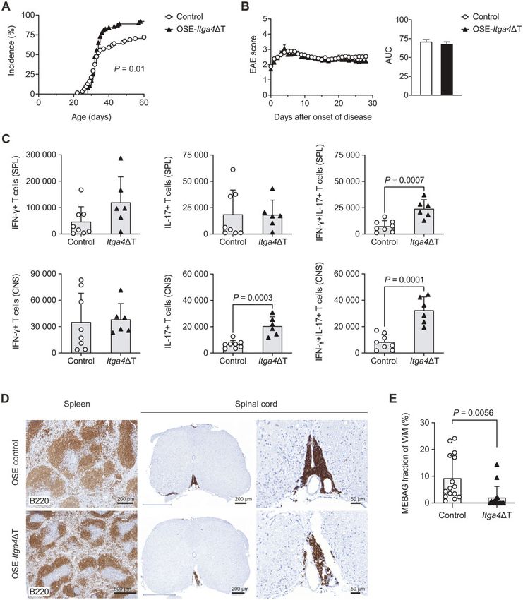
1700    | BRAIN 2021: 144; 1697–1710                                                                                 M. Mitsdoerffer et al.

Diagnostics, Inc.). The in situ hybridization protocol was performed     curve’ and compared by t-test between groups, as indicated in the
following recommended specifications for murine FFPE brain tissue.       figure legends. Body-weight loss was assessed by two-way ANOVA
Probes against murine Ebi3 (cat 448921), Il10-C3 (cat 317261-C3) and     followed by Sidak’s multiple comparisons test. Statistical analysis
Il12a-C2 (cat 414881-C2) were commercially available from Advanced       was performed in Graphpad Prism 8.4.3 (Graphpad Software, Inc).
Cell Diagnostics, Inc. RNAscope. Ebi3 was visualized by Tyramide
Signal Amplification (TSA) plus Fluorescein (Perkin Elmer,
                                                                         Data availability
NEL741001KT), Il10-C3 was visualized with TSA plus Cyanine 3
(Perkin Elmer, NEL744001KT), and Il12-C2 was visualized with TSA         The data that support the findings of this study are available from
plus Cyanine 5 (Perkin Elmer, NEL745001KT). The FISH protocol on         the corresponding author upon reasonable request.
murine brains was followed by fluorescence immunostaining with
rat anti-mouse B220 (eBioscience, clone RA3-6B2) and visualized
using a secondary donkey anti-rat IgG (H + L) conjugated with            Results
DyLight 405 (Jackson Immunoresearch, Cat. 712-475-153). As indi-         T cell-conditional ablation of Itga4 in OSE mice
cated in the manufacturer’s protocol, positive control probes (Cat.
                                                                         increases the incidence of opticospinal
320881) containing Polr2a-C1, Ppib-C2, Ubc-C3 and negative control

                                                                                                                                                 Downloaded from https://academic.oup.com/brain/article/144/6/1697/6164962 by guest on 14 October 2021
probes DapB (Cat. 320871) were used. Confocal images of B220 cell
                                                                         encephalomyelitis
clusters were acquired at 63 magnification (Zeiss LSM800). Positive     Th17 cells (but not Th1 cells) are able to access the CNS compart-
signals were quantified by a blinded experimenter using ZEN 2.3 lite     ment in the absence of Itga4 or when a blocking antibody to Itga4
(Zeiss). For representative images, contrast was linearly enhanced       is administered in the MOG(35–55)-induced EAE model.25,26 Similar
using the tools levels, curves, brightness, and contrast in Adobe        findings were reported in humans when analysing T helper cell
Photoshop CC.                                                            subsets in the blood and the CSF of patients with multiple
                                                                         sclerosis.26,27
                                                                             Here, we used an established model of opticospinal multiple
Preparation of mononuclear cells from the CNS and
                                                                         sclerosis, i.e. a double transgenic mouse line expressing a MOG-
flow cytometry                                                           specific TCR and BCR (termed OSE mice)16,17 to test the clinical
At the peak of disease, CNS-infiltrating cells were isolated after       relevance of genetic ablation of Itga4 on antigen-specific T cells in
perfusion through the left cardiac ventricle with PBS. Brain and         a model that is independent of adjuvant-mediated immune activa-
spinal cord were extracted. Tissues were digested with collagenase       tion. OSE mice were crossed with Cd4-Cre transgenic animals as
D (2.5 mg/ml) and DNase I (1 mg/ml) at 37 C for 45 min. After pass-     well as with mice bearing a floxed Itga4 allele.18 This breeding
ing the tissue through a 70-mm cell strainer, cells of the spinal cord   yielded T cell-conditional a4 integrin-deficient OSE mice (TCRMOG;
                                                                         BCRMOG, KI/KI; Itga4flox/flox; Cd4-Cre), termed OSE-Itga4DT mice. As
                                                     R
and brain were separated by discontinuous PercollV gradient (70%/
37%) centrifugation. Mononuclear cells were isolated from the            reported,17 control OSE mice (TCRMOG; BCRMOG, KI/KI; Itga4flox/flox)
interphase.                                                              developed CNS disease with an incidence of 60% at 6 weeks of age.
    Cells were stained with live/dead fixable dyes [Aqua (405 nm         In contrast, OSE-Itga4DT mice developed disease at a higher inci-
excitation), Invitrogen] and antibodies to surface markers: CD3e         dence than OSE control mice (Fig. 1A). At 6 weeks of age, more
(145-2C11), CD4 (GK1.5 or RM4-5), CD8a (53–6.7), CD11b (M1/70),          than 80% of OSE-Itga4DT mice were sick. We did not observe a pre-
CD11c (HL3), CD19 (1D3 or 6D5), CD44 (IM7), CD45 (30-F11), CD45.1        dominant ‘atypical’, i.e. atactic, disease phenotype in OSE-Itga4DT
(A20), CD45.2 (104), CD45R (B220; RA3-6B2), CD49d (a4 integrin,          mice, and the mortality and disease course of OSE-Itga4DT mice
9C10/MFR.4.B), 2D2 TCR Va3.2 (RR3-16) and Vb11 (RR3-15); all BD          and regular OSE mice were comparable (Fig. 1B and
Biosciences, eBioscience or BioLegend.                                   Supplementary Table 1). Taken together, genetic ablation of Itga4
    For intracellular cytokine staining, cells were restimulated with    in T cells increased the incidence of opticospinal EAE.
50 ng/ml PMA (Sigma-Aldrich), 1 lg/ml ionomycin (Sigma-Aldrich)
and monensin (1 ll/ml BD GolgiStop) at 37 C for 2.5 h. Subsequent
to live/dead and surface staining, cells were fixed and permeabi-
                                                                         OSE-Itga4DT mice exhibit higher numbers of Th17
lized (Cytofix/CytopermTM and Perm/Wash Buffer; BD Biosciences),         cells within the CNS parenchyma
and stained for cytokines IL-17A (TC11-18H10.1; BioLegend) and           To assess the cytokine phenotype of the CNS T cells in this OSE
IFN-c (XMG1.2, eBioscience). Cells were analysed using a CyAnTM          model, we isolated the mononuclear cell infiltrate from the CNS
ADP 9 flow cytometer (Beckmann/Coulter) and a CytoFLEX S                 compartment of sick OSE and OSE-Itga4DT mice on Day 20 after dis-
(Beckmann Coulter). Cell counting was performed by a Guava               ease onset. We found significantly higher numbers of Th17 cells in
easyCyte 5HT cytometer (Merck) together with 7-AAD (BD), Fixable         the CNS of OSE-Itga4DT mice than in the CNS of OSE control mice
Red Dead Cell Stain (ThermoFisher), or Fixable Viability Dye             (Fig. 1C, bottom). Also, the number of IL-17/IFN-c double producing
eFluorTM 520 (eBioscience) and CD45 (30-F11) or CD45.2 (104). All        CD4 + T cells was increased in OSE-Itga4DT mice (Fig. 1C, bottom)
data analysis was facilitated using FlowJo version 10 (Tree Star,        while the number of IFN-c producers was similar in the CNS of
now BD Biosciences).                                                     OSE-Itga4DT mice and OSE control mice. As the number of Th17
                                                                         cells in the spleen of OSE-Itga4DT mice and OSE mice was not sig-
                                                                         nificantly different (Fig. 1C, top), we concluded that the higher
Quantification and statistical analysis                                  number of Th17 cells in the CNS of OSE-Itga4DT mice was due to a
Statistical evaluations of cell frequency measurements and cell          preferential recruitment of Th17 cells to the CNS and not attribut-
numbers were performed by one-way ANOVA followed by Sidak’s              able to generalized differences in the commitment of T helper cells
multiple comparison test when two genotypes of multiple cell             in the peripheral immune compartment of OSE and OSE-Itga4DT
populations were compared, as indicated in the figure legends.           mice. To test the priming of MOG-specific T cells sufficient and de-
Multiplicity adjusted P-values 5 0.05 were considered significant.       ficient in a4 integrin expression, we transferred naive 2D2 T cells
EAE scores between groups were analysed as disease burden per            or Itga4–/– 2D2 T cells into BCRMOG transgenic (TH) recipients, chal-
individual day with one-way ANOVA and Dunnett’s post hoc test.           lenged them with MOG peptide (in the absence of adjuvant) and
Alternatively, the disease burden was calculated as ‘area under the      tested the indicator cells for proliferation and cytokine production.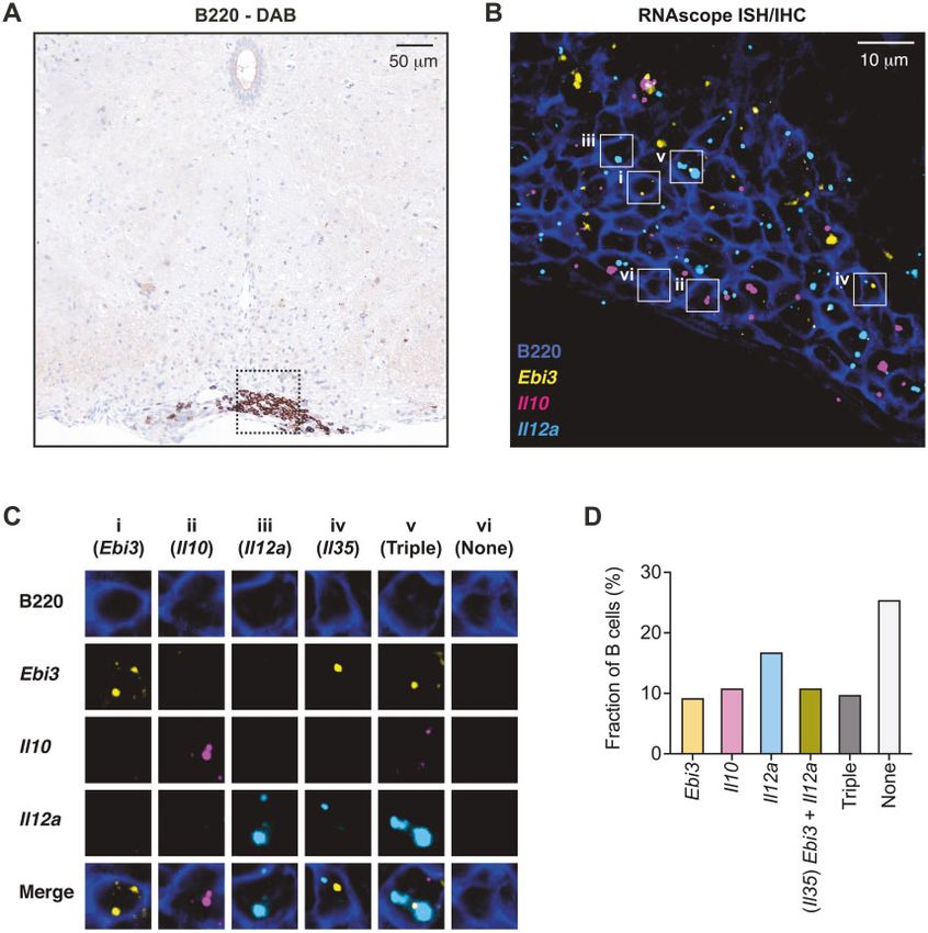
Meningeal B cell aggregates in CNS autoimmunity                                                             BRAIN 2021: 144; 1697–1710            | 1701

                                                                                                                                                               Downloaded from https://academic.oup.com/brain/article/144/6/1697/6164962 by guest on 14 October 2021

Figure 1 a4 integrin expression on T cells is necessary for the induction of MEBAGs in the OSE mouse model. (A and B) Spontaneous EAE was moni-
tored in littermate control mice (OSE; TCRMOG; BCRMOG, KI/KI; Itga4flox/flox, n = 68) and T cell-conditional a4 integrin deficient OSE mice (OSE-Itga4DT;
TCRMOG; BCRMOG, KI/KI; Itga4flox/flox, Cd4-Cre, n = 76). (A) Incidence of clinical signs of disease; survival curve, Mantel-Cox log-rank test. (B) EAE score
including area under the curve (AUC), Student’s t-test. (C) Three to 5 days after onset of disease, mononuclear cells were isolated from the spleen and
the CNS of diseased OSE and OSE-Itga4DT mice and investigated by intracellular cytokine staining. Absolute numbers of IFN-c producing CD4 + T cells,
of IL-17 producing CD4 + T cells, and of double producers in the spleen (top row) and in the CNS (bottom row) of OSE control or OSE-Itga4DT mice.
Student’s t-test, symbols indicate individual mice. (D and E) Immunohistochemical staining for B220 in OSE mice and OSE-Itga4DT mice at 4 weeks
after disease onset. (D) Representative spinal cord sections. Scale bar = 500 mm, 200 mm, 50 mm. (E) The meningeal area covered by MEBAGs was
assessed as fraction of the transversal white matter area at the respective spinal cord level. Symbols indicate individual sections. In each group five
mice were included in the analysis, and the three sections of each mouse with the largest MEBAGs were analysed; unpaired Student’s t-test. WM =
white matter.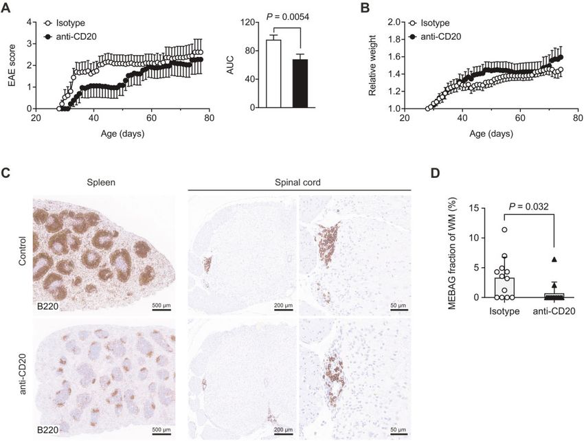
1702    | BRAIN 2021: 144; 1697–1710                                                                                     M. Mitsdoerffer et al.

Both the proliferative response as well as the production of IFN-c        effects of anti-CD20 treatment on established MEBAGs (Fig. 3C and
and IL-17 were similar in 2D2 T cells and Itga4–/– 2D2 T cells, re-iso-   D), indicating that intrathecal MEBAG-resident B cells were rela-
lated from the spleen of TH host mice, indicating that lack of a4 in-     tively resistant to depletion with anti-CD20 antibodies.
tegrin expression in T cells did not push their commitment to the             In more detailed histological analyses, we observed that post-
Th17 lineage (Supplementary Fig. 1). Rather, the higher number of         symptomatic anti-CD20 treatment of OSE mice failed to attenuate
Th17 cells in the CNS of OSE-Itga4DT mice as compared to OSE mice         inflammatory pathology in the spinal cord of OSE mice. Indeed, in-
pointed to a differential recruitment to the CNS. These results are       flammatory infiltrates of T cells and macrophages tended to be
in line with previous reports suggesting that Th17 cells are able to      more widespread into the spinal cord white matter and less
use alternative routes into the CNS independent of a4 integrin ex-        restricted to the vicinity of MEBAGs in anti-CD20 treated than in
pression.26–28 Notably, even though the OSE-Itga4DT model does            control-treated OSE mice (Fig. 3E), concomitant with an equally
not rely on immunization with a strong adjuvant that might bias           widespread demyelination and loss of axonal density (as assessed
the immune reaction towards a Th17 response, a predominance of            by LFB-PAS and Bielschowsky staining, respectively; Fig. 3E).
antigen-specific Th17 cells was an eminent feature of the CNS T           Therefore, the histopathological analysis largely mirrored the clin-
cell compartment in OSE-Itga4DT mice.                                     ical phenotype and supported the idea that established MEBAGs
                                                                          might have a role in mitigating the deep infiltration of inflamma-

                                                                                                                                                       Downloaded from https://academic.oup.com/brain/article/144/6/1697/6164962 by guest on 14 October 2021
                                                                          tory cells from the subarachnoid space into the CNS parenchyma.
MEBAGs are decreased in OSE-Itga4DT mice
Meningeal B cell aggregates (MEBAGs) have been described in the
                                                                          The ablation of MEBAGs in OSE-Itga4DB mice leads to
OSE model.16,17 These aggregates mostly consist of B cells and ex-
                                                                          a higher disease burden
hibit structural and functional features of tertiary lymphoid fol-
licles (for reviews, see Drayton et al.29 and Mitsdoerffer and            It has previously been reported that the recruitment of B cells to
Peters30). However, while correlative data have been published in         the meningeal compartment is entirely dependent on the expres-
human multiple sclerosis cases,1,31 the relevance of meningeal            sion of a4 integrins on B cells while ablation of Itga4 on B cells does
lymphocytic aggregates as to CNS immunopathology and clinical             not affect the ‘immune function’ of B cells in the systemic com-
disease course is unclear. Therefore, we analysed the extent of           partment.33 Therefore, we wanted to test whether ablation of Itga4
MEBAGs in OSE control mice and OSE-Itga4DT mice, which differed           on B cells could be used as a means to manipulate the formation
in their disease incidence. Control OSE mice showed extensive             of MEBAGs without interfering with systemic immune functions of
meningeal lymphocytic aggregates. In contrast, OSE-Itga4DT mice           B cells in the OSE model. We crossed CD19-CreKI/wt into the OSE
had significantly fewer and smaller meningeal lymphocytic infil-          background that carried floxed alleles of Itga4 (TCRMOG; BCRMOG,
                                                                          KI/KI
trates in relation to the corresponding trans-sectional white mat-              ; Itga4flox/flox; Cd19-CreKI/wt) and termed these B cell-conditional
ter area (Fig. 1D and E), despite prominent infiltration of the           a4 integrin deficient OSE animals OSE-Itga4DB mice. First, cohorts
meninges and parenchyma with a4 integrin deficient Th17 cells.            of OSE-Itga4DB and littermate control mice (TCRMOG; BCRMOG, KI/KI;
                                                                          Itga4flox/flox; Cd19-Crewt/wt) were monitored for their clinical disease
                                                                          course. Over extended observation periods, the incidence was
Systemic B cell depletion has disease phase-specific                      similar, but the disease burden and mortality were higher in OSE-
effects in OSE mice                                                       Itga4DB mice than in control animals (Fig. 4A–C). As expected, the
OSE-Itga4DT mice suffered from enhanced intrathecal inflamma-             meningeal infiltrates in OSE-Itga4DB mice were essentially devoid
tion even though the number of MEBAGs was reduced. To address             of B cells and by definition, the area of MEBAGs in these mice was
whether the presence of MEBAGs fed back on intrathecal inflam-            significantly reduced as compared to littermate controls (Fig. 4D
mation, we aimed to acutely eliminate MEBAGs in OSE mice. Since           and E). In histological analyses, we noticed that the depth of infil-
B cells are the primary constituent of MEBAGs, we decided to apply        trates of T cells and macrophages from the meningeal surface into
a B cell-depletion strategy. First, we used an anti-CD20 antibody in      the spinal cord white matter as well as the subsequent demyelin-
the presymptomatic stage of wild-type OSE mice. In accordance             ation were increased in OSE-Itga4DB mice as compared to regular
with prior studies in a spontaneous EAE model,32 B cell-depletion         OSE control mice (Fig. 4F and G). These results supported the no-
before the onset of clinical signs resulted in delayed and less se-       tion that B cells in MEBAGs might adopt an immunomodulatory
vere disease in OSE mice as compared to control antibody treat-           function during sustained CNS inflammation.
ment (Fig. 2A, B and Supplementary Table 2), consistent with the
idea that interference with the APC function of B cells in secondary      Anti-CD19 CAR-T cells mediate efficient
lymphoid tissues decreases priming of encephalitogenic T cells. In        interventional depletion of B cells in MEBAGs
this setting the formation of MEBAGs was not abrogated. However,
the area covered by MEBAGs in relation to the trans-sectional             While OSE-Itga4DB mice never developed MEBAGs, from a ‘thera-
white matter area was reduced in OSE mice after presymptomatic            peutic’ perspective the acute depletion of already established
anti-CD20 treatment as compared to control-treatment (Fig. 2C             MEBAGs is a more relevant intervention, and we have observed
and D).                                                                   that anti-CD20 antibody administration failed to efficiently ablate
   To tease apart the relevance of B cells as APCs in the peripheral      established MEBAGs in OSE mice (Fig. 3). Therefore, we explored al-
immune compartment and in MEBAGs and thus a potential role of             ternative methods to deplete MEBAG-resident B cells. Here, we
B cells in sustained intrathecal inflammation, we used anti-CD20          tested an anti-CD19 CAR-T cell approach that had been used in a
in OSE mice, only after clinical signs of disease were apparent. In       preclinical model for acute lymphoblastic leukaemia.21 Because of
this setting, administration of an anti-CD20 antibody failed to im-       their higher density of MEBAGs as compared to OSE-Itga4DT mice,
prove the clinical score in OSE mice and by tendency, even wors-          we used regular OSE mice for the CAR-T cell experimental design.
ened the disease burden on the population level (Fig. 3A, B and           Since CAR-T cell recipients needed to be sublethally irradiated in
Supplementary Table 3). While anti-CD20 treatment after onset of          order to increase the take-rate of transferred cells, we first investi-
disease led to efficient elimination of B cells from the circulation      gated the impact of this irradiation protocol (see ‘Materials and
and from peripheral lymph nodes (Supplementary Fig. 2), the area          methods’ section) on the natural disease course of OSE mice and
of MEBAGs was not significantly reduced in OSE mice with variable         confirmed that sublethal irradiation of OSE mice reduced but
Meningeal B cell aggregates in CNS autoimmunity BRAIN 2021: 144; 1697–1710 | 1703
Downloaded from https://academic.oup.com/brain/article/144/6/1697/6164962 by guest on 14 October 2021
Figure 2 Pre-onset depletion of B cells delays and attenuates spontaneous EAE but does not prevent chronic progressive disease. OSE mice were ei-
ther treated with isotype control antibody or with a depleting anti-mouse CD20 monoclonal antibody (18B12, IgG1), throughout the experiment start-
ing at 4 weeks of age. Isotype (IgG1), n = 9; anti-CD20, n = 9. (A) EAE score including AUC, Student’s t-test. (B) Relative weight. (C) At the end of the
clinical monitoring period, spleen and spinal cord sections were analysed for B cells by staining with B220. Representative sections for isotype control
or anti-CD20 treated mice, respectively. Scale bars = 500 mm, 200 mm, 50 mm. (D) The MEBAG area was assessed as fraction of the transversal white
matter area at the respective spinal cord level. Symbols indicate individual sections. In each group four mice were included in the analysis, and the
three sections of each mouse with the largest MEBAGs were analysed; unpaired Student’s t-test.
did not prevent the development of progressive disease ablated in these mice, anti-CD19 CAR-T cell recipients but not con-
(Supplementary Fig. 3). Next, sublethally irradiated OSE mice trol-treated OSE mice, showed massive inflammatory infiltrates of
received either control cells or anti-CD19 CAR-T cells. In contrast T cells and macrophages that were no longer restricted to the
to systemic anti-CD20 treatment, B cells in tissues including in meningeal compartment but were scattered deep into the spinal
MEBAGs were substantially depleted in anti-CD19 CAR-T cell recip- cord white matter throughout the spinal cord parenchyma (Fig. 5D
ients as compared to control-treated OSE mice when analysed at and E), associated with more widespread demyelination and axon-
3 weeks after anti-CD19 CAR-T cell administration (Fig. 5A and B). al loss (Fig. 5D). While these data again indicate that MEBAGs
The administration of anti-CD19 CAR-T cells was not associated might exert an immune regulatory function, this function is mech-
with a gross disruption of the blood–brain barrier in our model anistically not well understood. When we tested MEBAGs in regu-
(Supplementary Fig. 4), even though it has been reported that anti- lar OSE mice for the expression of Il10 and Il35 (Il12a plus Ebi3) by
R
CD19 CAR-T cells might have off-target effects on a small subfrac- RNAscopeV (Supplementary Fig. 5), the mRNA of these cytokines
tion of pericytes in C57BL/6 mice.34 Notably, during a latency was clearly associated with B cells in MEBAGs of OSE mice (Fig. 6A–
period of 2 weeks, anti-CD19 CAR-T cell treatment did not alter C). In fact, 10% of MEBAG B cells contained only Il10 mRNA or Il35
the clinical course in OSE mice as compared to control-treated mRNA, respectively, and an additional 10% expressed both mRNA
mice (Fig. 5C and Supplementary Table 4), suggesting that this species. Therefore, one-third of the B cells located in MEBAGs had
intervention was not associated with a substantial cytokine storm. the potential to produce these immunomodulatory cytokines (Fig.
However, as of 2 weeks post administration, anti-CD19 CAR-T cell 6D)—a feature that was entirely lost with the extensive ablation of
recipients developed a more severe disease course with marked le- MEBAGs after anti-CD19 CAR-T cell treatment.
thality as compared with their control counterparts (Fig. 5C and In summary, our data suggest that B cell depletion with a
Supplementary Table 4). While MEBAGs were strongly reduced or monoclonal antibody to CD20 will only insufficiently target1704 | BRAIN 2021: 144; 1697–1710 M. Mitsdoerffer et al.
Downloaded from https://academic.oup.com/brain/article/144/6/1697/6164962 by guest on 14 October 2021
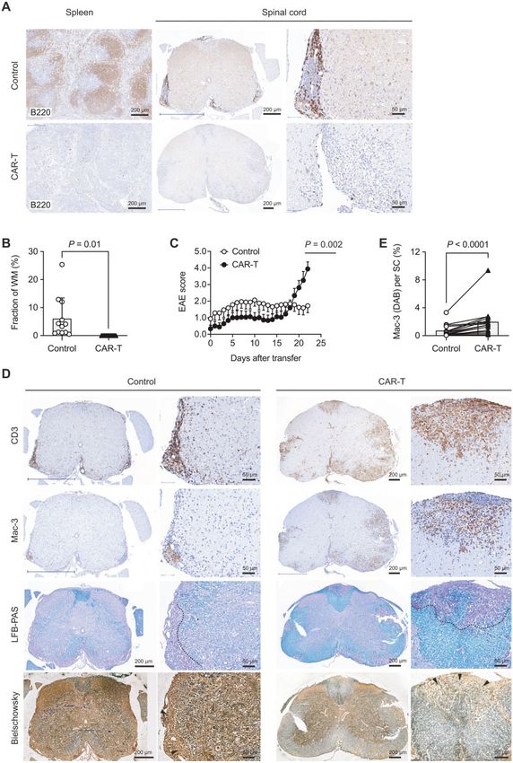
Figure 3 Post-onset depletion of B cells fails to improve clinical signs of disease. (A and B) OSE mice were treated with isotype control antibody (IgG1) or a
depleting antibody to CD20. Treatment (injection of 10 mg/g body weight of isotype antibody or anti-CD20 intraperitoneally once a week) was initiated
when an individual mouse reached a score of 2 and continued throughout the experiment. (A) EAE score including AUC, Student’s t-test. (B) Relative
weight. (C) Immunohistochemical analysis for B220 (staining B cells) in lymph node and spinal cord sections prepared at 4 weeks after start of treatment
of OSE mice as indicated. Representative sections. Scale bars = 200 mm and 50 mm. (D) The meningeal area covered by MEBAGs was assessed as fraction
of the transversal white matter area at the respective spinal cord level of OSE mice analysed at 4 weeks after start of treatment with control antibody or
anti-CD20. Multiple sections of at least three mice per group; unpaired Student’s t-test. (E) Representative spinal cord sections stained for T cells (CD3)
and macrophages (Mac-3) of OSE mice treated post-EAE-onset with control IgG1 or anti-CD20. Note that the demyelinated area (limited by dashed line in
the LFB-PAS stainings) and the area of reduced axonal density (arrow heads in the Bielschowsky stainings) largely corresponded to the intensity of the
macrophage infiltrate in each condition. Scale bars = 200 mm and 100 mm. WM = white matter.Meningeal B cell aggregates in CNS autoimmunity BRAIN 2021: 144; 1697–1710 | 1705
Downloaded from https://academic.oup.com/brain/article/144/6/1697/6164962 by guest on 14 October 2021
Figure 4 OSE-Itga4DB mice fail to build up MEBAGs in the spinal cord meningeal compartment and show a pronounced disease burden. Spontaneous EAE
was monitored in littermate control mice (OSE; TCRMOG; BCRMOG, KI/KI; Itga4flox/flox, n = 68) and B cell-conditional a4 integrin deficient OSE mice (OSE-Itga4DB;
TCRMOG; BCRMOG, KI/KI; Itga4flox/flox, Cd19-Crewt/KI, n = 76). (A) Incidence of EAE in OSE and OSE-Itga4DB mice. (B) EAE score including AUC, Student’s t-test. (C)
Mortality of OSE and OSE-Itga4DB mice; Mantel-Cox log-rank test. (D) In some animals, spleen and spinal cord sections were analysed for B cells by staining
with B220 at around 4 weeks after onset of disease. Representative sections for OSE control or OSE-Itga4DB mice, respectively. Scale bars = 200 mm and 50 mm.
(E) The MEBAG area was assessed as fraction of the transversal white matter area at the respective spinal cord level. Symbols indicate individual sections. In
each group six mice were included in the analysis, and the three sections of each mouse with the largest MEBAGs were analysed; unpaired Student’s t-test.
(F) Spinal cord sections of OSE control mice or OSE-Itga4DB mice were prepared 4 weeks after onset of disease and stained for T cells (CD3) and macrophages
(Mac-3). The amount of demyelination was assessed by LFB-PAS (demyelinated area limited by dashed line). Scale bars = 200 mm and 50 mm. (G) The macro-
phage infiltrate in the spinal cord parenchyma was quantified in littermate control mice and OSE-Itga4DB mice by assessing the fraction of the spinal cord
parenchyma (excluding meninges) that displayed a Mac-3 signal. Individual mice from the two groups were paired, and the spinal cord sections with the
largest Mac-3 infiltrates from three mice in each group were analysed. Bars represent means; t-test.1706 | BRAIN 2021: 144; 1697–1710 M. Mitsdoerffer et al.
Downloaded from https://academic.oup.com/brain/article/144/6/1697/6164962 by guest on 14 October 2021
Figure 5 Anti-CD19 CAR-T cell intervention eliminates MEBAGs and is associated with delayed worsening of disease in OSE mice. After sublethal irradiation,
OSE mice were monitored for clinical signs of EAE. At first signs of EAE, mice were either transferred with control cells or with T cells that were engineered to
express an anti-CD19 CAR construct. (A) Three weeks after transfer, the elimination of B cells in various compartments was analysed by immunohistochemis-
try. B220 staining in spleen (left) and spinal cord meninges (middle and right). Representative sections of control-treated (top row) and anti-CD19 CAR-T cell
treated (bottom row) OSE mice. Scale bar = 200 mm and 50 mm. (B) The MEBAG area in control cell-treated and CAR-T cell-treated mice was assessed as fraction
of the transversal white matter area at the respective spinal cord level. Symbols indicate individual sections. In each group at least three mice were included in
the analysis, and the three sections of each mouse with the largest MEBAGs were analysed; unpaired Student’s t-test. (C) EAE score of control-treated (n = 8)
versus anti-CD19 CAR-T cell-treated (n = 9) OSE mice. ANOVA plus Sidak’s post-test for individual days. Representative of two experiments. (D)
Immunohistochemical analysis of T cell (CD3, top row) and macrophage (Mac-3) infiltrates into the spinal cord as well as demyelination (LFB-PAS) and axonal
density (Bielschowsky) in spinal cord sections of control-treated OSE mice (left) and anti-CD19 CAR-T cell-treated OSE mice (right) on Day 23 after start of treat-
ment. Scale bar = 200 mm and 50 mm. (E) The macrophage infiltrate in the spinal cord parenchyma (excluding meninges) of control-treated and anti-CD19 CAR-
T cell-treated OSE mice was assessed by pairing individual mice of each group. The spinal cord sections with the top five largest Mac-3 infiltrates of three mice
in each group were analysed. Bars represent means; t-test. SC = spinal cord; WM = white matter.Meningeal B cell aggregates in CNS autoimmunity BRAIN 2021: 144; 1697–1710 | 1707
MEBAGs. In contrast, when MEBAGs are ablated by anti-CD19 CAR- reduced the amount of MEBAGs but aggravated disease progres-
T cell administration, the inflammatory response in the CNS com- sion. Our data suggest that (i) the formation of meningeal lympho-
partment is fundamentally exacerbated, implying a regulatory role cyte aggregates is associated with a progressive phenotype in a
of MEBAGs. Consistent with this finding, OSE-Itga4DB mice that es- spontaneous mouse model of EAE; (ii) firm establishment of men-
sentially lack MEBAGs in the first place build up an enhanced ingeal lymphocyte aggregates marks a stage of the disease where
intrathecal inflammatory load. antibody mediated immune interventions in the systemic immune
compartment fail to decrease inflammation within the CNS; and
(iii) MEBAGs might have a regulatory function during chronic in-
Discussion flammation in the meningeal compartment.
In this study, we introduce several strategies to modulate the for- Th17 cells have been shown to be inducers of ectopic lymphoid
mation of meningeal lymphocyte aggregates in a model of spon- tissue in the meningeal compartment.10 Our data appear to con-
taneous CNS autoimmunity. By ablating a4 integrins on MOG- trast this observation since OSE-Itga4DT mice had fewer and
specific T cells or on MOG-specific B cells, severe inflammation in smaller MEBAGs despite extensive Th17 driven inflammation in
the CNS was uncoupled from the formation of MEBAGs in OSE the CNS. However, in our model, Th17 cells lacked a4 integrin ex-
pression, and our data suggest that a4 integrin expression on
Downloaded from https://academic.oup.com/brain/article/144/6/1697/6164962 by guest on 14 October 2021
mice. Similarly, in wild-type OSE mice with a build-up of MEBAGs,
anti-CD19 CAR-T cell-mediated elimination of B cells efficiently T cells in the intrathecal compartment is directly involved in the
Figure 6 B cells in MEBAGs have the potential to produce immunomodulatory cytokines. Spinal cord sections of untreated OSE mice were prepared
three weeks after first manifestation of clinical signs of disease. (A) Immunohistochemical staining for B220 to show a representative MEBAG. Scale
RV
bar = 50 mm. (B) FISH was performed using the RNAscope protocol for the indicated probes, and a representative MEBAG (as outlined in A) is
depicted. Scale bar = 10 mm. (C) Magnification of individual B cells as identified by B220 staining (i–vi) as indicated in B, stained for the RNA species
VR
Ebi3, Il10, and Il12a by RNAscope . (D) Quantification of B cells positive for the indicated RNA species. Fraction of RNA positive B cells of all B cells
within MEBAGs. Analysis of n = 6 MEBAGs from three mice. ISH/IHC = in situ hybridization/immunohistochemistry.1708 | BRAIN 2021: 144; 1697–1710 M. Mitsdoerffer et al.
formation of MEBAGs. A putative role of a4 integrins has been sug- relevance of B cell aggregates in a model of chronic CNS auto-
gested in lymphoid tissue inducer cells, which are instrumental in immunity. Anti-CD19 CAR-T cell-mediated depletion of B cells in
the formation of ectopic lymphoid tissue in distinct niches in the OSE mice efficiently and (in contrast to anti-CD20 treatment) sus-
gut during development35 and in restoration of secondary lymph- tainably reduced MEBAGs, and resulted in an enhanced progres-
oid tissue architecture after infection in adults.36 Here, we sive disease course (due to unleashed inflammation) after a delay
exploited our observation of reduced MEBAG formation in the of 2 weeks. This delayed worsening is not compatible with a cyto-
CNS compartment in OSE-Itga4DT mice to investigate the function- kine release syndrome and rather argues in favour of a loss-of-
al significance of MEBAGs for immunopathology during CNS function effect of MEBAGs.
autoimmunity. While our data are consistent with the idea that MEBAGs con-
Compelling evidence suggests that CD20-targeted B cell-deplet- tribute to the local generation of regulatory immune cells, the
ing therapeutic interventions are highly efficient in patients with mechanistic underpinning and compartment-specific steps of
relapsing multiple sclerosis.37,38 Similarly, depletion of B cells be- this process need to be determined. Notably, even in human aut-
fore disease onset in a model, in which a MOG-specific TCR was opsy material, MEBAGs were associated with cortical grey matter
transgenically expressed on an SJL genetic background, prevented lesions (with strong microglia and macrophage activation) but
spontaneous EAE,32 suggesting that cognate B cells might be also with partially de- and remyelinated cortical lesions.
Downloaded from https://academic.oup.com/brain/article/144/6/1697/6164962 by guest on 14 October 2021
powerful (and perhaps indispensable) antigen presenting cells to Furthermore, the extent of cortical demyelination did not correl-
autoreactive T cells in the systemic immune compartment.39 Our ate with the presence of MEBAGs,51 and therefore, it is possible
results in the OSE model confirmed this observation because pre- that MEBAGs would not so much promote immunopathology in
emptive B cell depletion by a monoclonal antibody to CD20 grey matter as they would foster a milieu that enables reparative
delayed the onset and mitigated the clinical signs of disease. processes.
Conversely, in the OSE-Itga4DB model, in which the systemic APC A caveat in the translation of our model to the human situation
function of B cells was likely unaltered but lacked MEBAGs, the ini- is that meningeal pathology in OSE mice is focused on the spinal
tial phase of EAE was identical to regular OSE mice. However, over cord meningeal compartment. Adjacent to MEBAGs in OSE mice is
time the disease burden of OSE-Itga4DB mice was higher than in the spinal cord white matter as opposed to cortical grey matter in
OSE controls, which correlated with a more pronounced inflam- secondary progressive multiple sclerosis patients with forebrain
matory tissue damage in the absence of MEBAGs in OSE-Itga4DB meningeal tertiary lymphoid follicle-like structures. It is likely that
mice. white matter and grey matter deal differently with adjacent in-
Interestingly, in humans, where anti-CD20 therapy is highly effi- flammation. Cortical grey matter tends to support less immune
cient in limiting systemic inflammatory activity in patients with cell infiltrates, resolves inflammation faster and shows a higher
multiple sclerosis, secondary progression of disease may not be pre- propensity to remyelinate.52,53 Also, MEBAG-produced immuno-
vented40 (even though larger scale trials to investigate this observa- globulins and their complement-mediated effector functions
tion systematically are lacking). The rationale for the application of might be more harmful to grey matter than to white matter. In our
B cell targeted therapies in progressive stages of multiple sclerosis study, we have not addressed antibody secreting functions of
has been built on the observation that B cells constitute a major cel- MEBAGs and the potential contribution of intrathecally synthe-
lular component of the meningeal infiltrates that were observed in sized anti-MOG antibodies or other toxic mediators that have been
multiple sclerosis patients with chronic disease. Correlative data suggested to contribute to cortical pathology.54–56 However, by
(mostly from human autopsy material) implied that MEBAGs are applying an efficient interventional setup to eliminate MEBAGs in
associated with progressive disease.1,2,31,41,42 However, while cortical a chronic EAE model, our study challenges the view that menin-
demyelination has been observed in both multiple sclerosis and geal lymphocyte aggregates are exclusively drivers of pathogen-
anti-MOG antibody associated disease,43 the latter is characterized icity in the CNS. Further mechanistic studies need to consider that
by the lack of a progressive disease course,44 and subpial cortical de- these structures might also subserve an effort of the meningeal
myelination is also observed in early multiple sclerosis in the ab- space to confine chronic inflammation and to reduce immunopa-
sence of high degree meningeal inflammation45 (for a review see thology in the CNS parenchyma. Observations on the failure of
Lassmann46). Therefore, the causal pathogenic relevance of MEBAGs therapeutic approaches that targeted BAFF in patients with mul-
remains controversial. In addition, regulatory functions of B cells tiple sclerosis might help to better inform these studies. In fact,
and plasma cells in the meninges have been identified that rely on neutralizing the plasma cell differentiation and growth factor
the secretion of downmodulatory cytokines such as IL-10.47 IL-10 BAFF aggravated the severity of multiple sclerosis, indicating that
producing regulatory B cells and IgA + plasma cells were shown to B cells might have regulatory properties.5 The identification of IL-
be recruited into the subarachnoid space from the systemic immune 10 and IL-35 in B cells within MEBAGs supports this idea.
compartment.8 Therefore, based on the available data in human
patients, it is difficult to conclude that MEBAGs are universal drivers
Acknowledgements
of immunopathology. Apart from these pathophysiological argu-
ments, we need to consider that it is not clear whether antibody- We would like to thank Tanja Kuhlmann (Institute for
mediated depletion of B cells is at all an efficient means to target in- Neuropathology, University of Muenster) for help with initial
flammatory lymphocyte aggregates in the subarachnoid space.48,49 immunohistochemistry stainings.
CAR-T cell approaches that target CD19 + B cells are becoming
established therapies for certain hematopoietic malignancies (for
review see Neepalu et al.50). Cytokine release syndrome and the so- Funding
called immune cell associated neurotoxicity syndrome are early
T.K. is supported by the Deutsche Forschungsgemeinschaft
and late complications of this therapy, respectively. However, next
[SFB1054-B06, TRR128-A07, TRR128-A12, TRR274-A01, and EXC
generation CAR-T cell approaches will combine their efficient 2145 (SyNergy) ID 390857198], the ERC (CoG 647215), and by the
B cell depleting properties with the possibility to re-eliminate the Hertie Network of Clinical Neuroscience. M.M. was supported by
transferred CAR-T cells on demand.21 Therefore, we considered the Deutsche Forschungsgemeinschaft [EXC 2145 (Synergy)]. D.M.
this strategy in order to deplete tissue resident B cells (within
is supported by the Swiss National Science Foundation
MEBAGs). We used this approach as a tool to investigate the
(310030_185321) and the ERC (CoG 865026).Meningeal B cell aggregates in CNS autoimmunity BRAIN 2021: 144; 1697–1710 | 1709
Competing interests 16. Bettelli E, Baeten D, Jäger A, Sobel RA, Kuchroo VK. Myelin oligo-
dendrocyte glycoprotein-specific T and B cells cooperate to induce
The authors report no competing interests. a Devic-like disease in mice. J Clin Invest. 2006;116:2393–2402.
17. Krishnamoorthy G, Lassmann H, Wekerle H, Holz A.
Spontaneous opticospinal encephalomyelitis in a double-trans-
Supplementary material
genic mouse model of autoimmune T cell/B cell cooperation.
Supplementary material is available at Brain online. J Clin Invest. 2006;116:2385–2392.
18. Scott LM, Priestley GV, Papayannopoulou T. Deletion of alpha4
integrins from adult hematopoietic cells reveals roles in
References homeostasis, regeneration, and homing. Mol Cell Biol. 2003;23:
1. Magliozzi R, Howell O, Vora A, et al. Meningeal B-cell follicles in 9349–9360.
secondary progressive multiple sclerosis associate with early 19. Lee PP, Fitzpatrick DR, Beard C, et al. A critical role for Dnmt1
onset of disease and severe cortical pathology. Brain. 2007;130: and DNA methylation in T cell development, function, and
1089–1104. survival. Immunity. 2001;15:763–774.
2. Howell OW, Reeves CA, Nicholas R, et al. Meningeal inflamma- 20. Rickert RC, Roes J, Rajewsky K. B lymphocyte-specific, Cre-
Downloaded from https://academic.oup.com/brain/article/144/6/1697/6164962 by guest on 14 October 2021
tion is widespread and linked to cortical pathology in multiple mediated mutagenesis in mice. Nucleic Acids Res. 1997;25:
sclerosis. Brain. 2011;134:2755–2771. 1317–1318.
3. Takemori T, Kaji T, Takahashi Y, Shimoda M, Rajewsky K. 21. Paszkiewicz PJ, Fräßle SP, Srivastava S, et al. Targeted antibody-
Generation of memory B cells inside and outside germinal cen- mediated depletion of murine CD19 CAR T cells permanently
ters. Eur J Immunol. 2014;44:1258–1264. reverses B cell aplasia. J Clin Invest. 2016;126:4262–4272.
4. Mackay F, Browning JL. BAFF: a fundamental survival factor for 22. Wang X, Chang W-C, Wong CW, et al. A transgene-encoded cell
B cells. Nat Rev Immunol. 2002;2:465–475. surface polypeptide for selection, in vivo tracking, and ablation
5. Kappos L, Hartung H-P, Freedman MS, et al.; ATAMS Study of engineered cells. Blood. 2011;118:1255–1263.
Group. Atacicept in multiple sclerosis (ATAMS): A randomised, 23. Korn T, Mitsdoerffer M, Croxford AL, et al. IL-6 controls Th17 im-
placebo-controlled, double-blind, phase 2 trial. Lancet Neurol. munity in vivo by inhibiting the conversion of conventional
2014;13:353–363. T cells into Foxp3 + regulatory T cells. Proc Natl Acad Sci U S A.
6. Mizoguchi A, Mizoguchi E, Takedatsu H, Blumberg RS, Bhan AK. 2008;105:18460–18465.
Chronic intestinal inflammatory condition generates IL-10-pro- 24. Knier B, Hiltensperger M, Sie C, et al. Myeloid-derived suppres-
ducing regulatory B cell subset characterized by CD1d upregula- sor cells control B cell accumulation in the central nervous sys-
tion. Immunity. 2002;16:219–230. tem during autoimmunity. Nat Immunol. 2018;19:1341–1351.
7. Shen P, Roch T, Lampropoulou V, et al. IL-35-producing B cells 25. Glatigny S, Duhen R, Oukka M, Bettelli E. Cutting edge: Loss of a4
are critical regulators of immunity during autoimmune and in- integrin expression differentially affects the homing of Th1 and
fectious diseases. Nature. 2014;507:366–370. Th17 cells. J Immunol. 2011;187:6176–6179.
8. Rojas OL, Pröbstel A-K, Porfilio EA, et al. Recirculating intestinal 26. Rothhammer V, Heink S, Petermann F, et al. Th17 lymphocytes
IgA-producing cells regulate neuroinflammation via IL-10. Cell. traffic to the central nervous system independently of a4 integ-
2019;176:610–624.e18. rin expression during EAE. J Exp Med. 2011;208:2465–2476.
9. Pikor NB, Astarita JL, Summers-Deluca L, et al. Integration of 27. Schneider-Hohendorf T, Rossaint J, Mohan H, et al. VLA-4 block-
Th17- and lymphotoxin-derived signals initiates meningeal- ade promotes differential routes into human CNS involving
resident stromal cell remodeling to propagate neuroinflamma- PSGL-1 rolling of T cells and MCAM-adhesion of TH17 cells. J Exp
tion. Immunity. 2015;43:1160–1173. Med. 2014;211:1833–1846.
10. Peters A, Pitcher LA, Sullivan JM, et al. Th17 cells induce ectopic 28. Reboldi A, Coisne C, Baumjohann D, et al. C-C chemokine recep-
lymphoid follicles in central nervous system tissue inflamma- tor 6-regulated entry of TH-17 cells into the CNS through the
tion. Immunity. 2011;35:986–996. choroid plexus is required for the initiation of EAE. Nat Immunol.
11. Yoshida H, Kawamoto H, Santee SM, et al. Expression of 2009;10:514–523.
alpha(4)beta(7) integrin defines a distinct pathway of lymphoid 29. Drayton DL, Liao S, Mounzer RH, Ruddle NH. Lymphoid organ
progenitors committed to T cells, fetal intestinal lymphotoxin development: From ontogeny to neogenesis. Nat Immunol. 2006;
producer, NK, and dendritic cells. J Immunol. 2001;167: 7:344–353.
2511–2521. 30. Mitsdoerffer M, Peters A. Tertiary lymphoid organs in central
12. Magliozzi R, Columba-Cabezas S, Serafini B, Aloisi F. nervous system autoimmunity. Front. Immun. 2016;7:451.
Intracerebral expression of CXCL13 and BAFF is accompanied 31. Magliozzi R, Howell OW, Reeves C, et al. A Gradient of neuronal
by formation of lymphoid follicle-like structures in the menin- loss and meningeal inflammation in multiple sclerosis. Ann
ges of mice with relapsing experimental autoimmune enceph- Neurol. 2010;68:477–493.
alomyelitis. J Neuroimmunol. 2004;148:11–23. 32. Pöllinger B, Krishnamoorthy G, Berer K, et al. Spontaneous
13. Molnarfi N, Schulze-Topphoff U, Weber MS, et al. MHC class II- relapsing-remitting EAE in the SJL/J mouse: MOG-reactive
dependent B cell APC function is required for induction of CNS transgenic T cells recruit endogenous MOG-specific B cells. J Exp
autoimmunity independent of myelin-specific antibodies. J Exp Med. 2009;206:1303–1316.
Med. 2013;210:2921–2937. 33. Lehmann-Horn K, Sagan SA, Bernard CCA, Sobel RA, Zamvil SS.
14. Bettelli E, Pagany M, Weiner HL, Linington C, Sobel RA, Kuchroo B-cell very late antigen-4 deficiency reduces leukocyte recruit-
VK. Myelin oligodendrocyte glycoprotein-specific T cell receptor ment and susceptibility to central nervous system autoimmun-
transgenic mice develop spontaneous autoimmune optic ity. Ann Neurol. 2015;77:902–908.
neuritis. J Exp Med. 2003;197:1073–1081. 34. Parker KR, Migliorini D, Perkey E, et al. Single-cell analyses iden-
15. Litzenburger T, Fässler R, Bauer J, et al. B lymphocytes tify brain mural cells expressing CD19 as potential off-tumor
producing demyelinating autoantibodies: Development and targets for CAR-T immunotherapies. Cell. 2020;183:126–142.e17.
function in gene-targeted transgenic mice. J Exp Med. 1998;188: 35. Mebius RE. Organogenesis of lymphoid tissues. Nat Rev
169–180. Immunol. 2003;3:292–303.You can also read