Mice lacking spinal 2GABAA receptors: Altered GABAergic neurotransmission, diminished GABAergic antihyperalgesia, and potential compensatory ...
←
→
Page content transcription
If your browser does not render page correctly, please read the page content below
Zurich Open Repository and
Archive
University of Zurich
Main Library
Strickhofstrasse 39
CH-8057 Zurich
www.zora.uzh.ch
Year: 2020
Mice lacking spinal 2GABAA receptors: Altered GABAergic
neurotransmission, diminished GABAergic antihyperalgesia, and potential
compensatory mechanisms preventing a hyperalgesic phenotype
Tudeau, Laetitia ; Acuña, Mario A ; Albisetti, Gioele W ; Neumann, Elena ; Ralvenius, William T ;
Scheurer, Louis ; Poe, Michael ; Cook, James M ; Johannssen, Helge C ; Zeilhofer, Hanns Ulrich
Abstract: Diminished synaptic inhibition in the superficial spinal dorsal horn contributes to exagger-
ated pain responses that accompany peripheral inflammation and neuropathy. 2GABAA receptors
(2GABAAR) constitute the most abundant GABAAR subtype at this site and are the targets of re-
cently identified antihyperalgesic compounds. Surprisingly, hoxb8-2−/− mice that lack 2GABAAR
from the spinal cord and peripheral sensory neurons exhibit unaltered sensitivity to acute painful stim-
uli and develop normal inflammatory and neuropathic hyperalgesia. Here, we provide a comprehensive
analysis of GABAergic neurotransmission, of behavioral phenotypes and of possible compensatory mech-
anisms in hoxb8-2−/− mice. Our results confirm that hoxb8-2−/− mice show significantly diminished
GABAergic inhibitory postsynaptic currents (IPSCs) in the superficial dorsal horn but no hyperalgesic
phenotype. We also confirm that the potentiation of dorsal horn GABAergic IPSCs by the 2-preferring
GABAAR modulator HZ-166 is reduced in hoxb8-2−/− mice and that hoxb8-2−/− mice are resis-
tant to the analgesic effects of HZ-166. Tonic GABAergic currents, glycinergic IPSCs, and sensory
afferent-evoked EPSCs did not show significant changes in hoxb8-2−/− mice rendering a compensatory
up-regulation of other GABAAR subtypes or of glycine receptors unlikely. Although expression of sero-
tonin and of the serotonin producing enzyme tryptophan hydroxylase (TPH2) was significantly increased
in the dorsal horn of hoxb8-2−/− mice, ablation of serotonergic terminals from the lumbar spinal cord
failed to unmask a nociceptive phenotype. Our results are consistent with an important contribution
of 2GABAAR to spinal nociceptive control but their ablation early in development appears to activate
yet-to-be identified compensatory mechanisms that protect hoxb8-2−/− mice from hyperalgesia.
DOI: https://doi.org/10.1016/j.brainres.2020.146889
Posted at the Zurich Open Repository and Archive, University of Zurich
ZORA URL: https://doi.org/10.5167/uzh-188836
Journal Article
Published Version
The following work is licensed under a Creative Commons: Attribution-NonCommercial-NoDerivatives
4.0 International (CC BY-NC-ND 4.0) License.
Originally published at: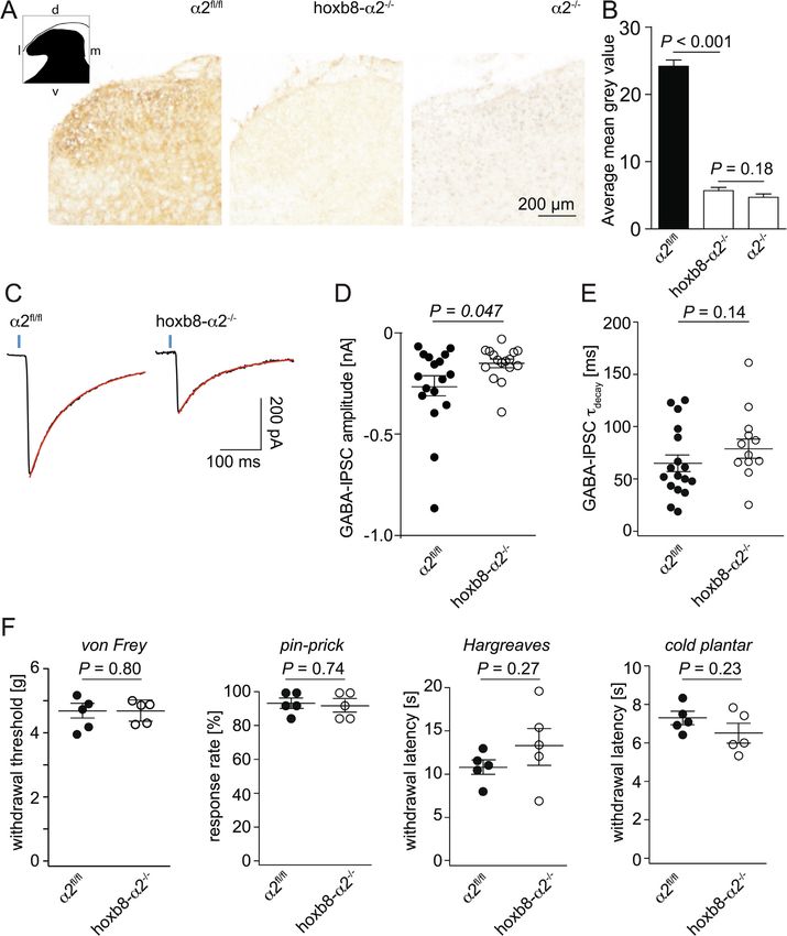
Tudeau, Laetitia; Acuña, Mario A; Albisetti, Gioele W; Neumann, Elena; Ralvenius, William T; Scheurer,
Louis; Poe, Michael; Cook, James M; Johannssen, Helge C; Zeilhofer, Hanns Ulrich (2020). Mice lacking
spinal 2GABAA receptors: Altered GABAergic neurotransmission, diminished GABAergic antihyper-
algesia, and potential compensatory mechanisms preventing a hyperalgesic phenotype. Brain Research,
1741:146889.
DOI: https://doi.org/10.1016/j.brainres.2020.146889
2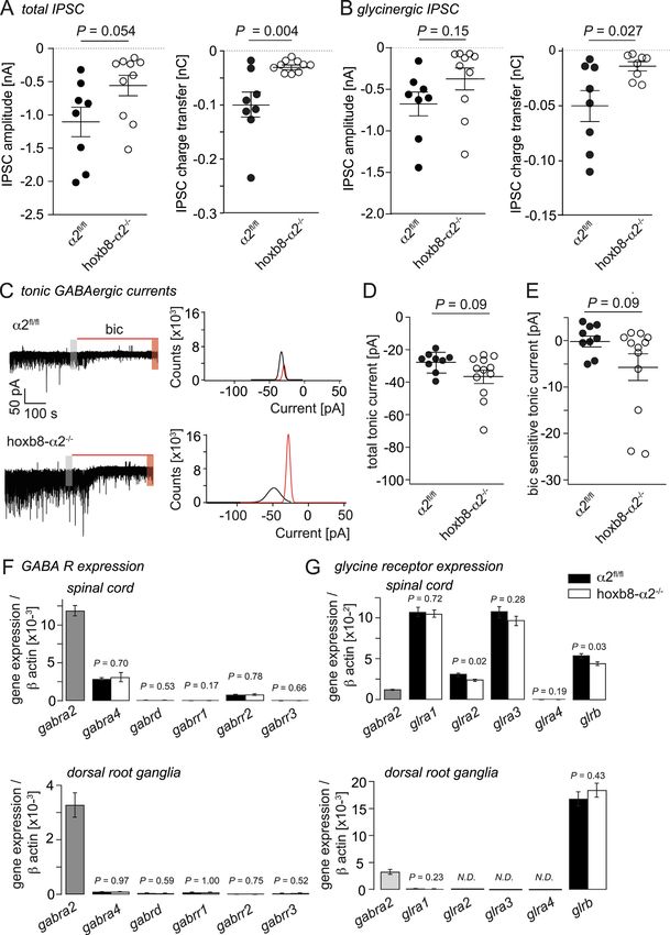
Brain Research 1741 (2020) 146889

                                                                        Contents lists available at ScienceDirect

                                                                                  Brain Research
                                                          journal homepage: www.elsevier.com/locate/brainres

Research report

                                                                                                                                                             T
Mice lacking spinal α2GABAA receptors: Altered GABAergic
neurotransmission, diminished GABAergic antihyperalgesia, and potential
compensatory mechanisms preventing a hyperalgesic phenotype
Laetitia Tudeaua, Mario A. Acuñaa, Gioele W. Albisettia, Elena Neumanna, William T. Ralveniusa,
Louis Scheurera, Michael Poeb, James M. Cookb, Helge C. Johannssena, Hanns Ulrich Zeilhofera,c,
                                                                                                ⁎

a
    Institute of Pharmacology and Toxicology, University of Zurich, CH-8057 Zurich, Switzerland
b
    Department of Chemistry and Biochemistry, University of Wisconsin-Milwaukee, Milwaukee, USA
c
    Institute of Pharmaceutical Sciences, Swiss Federal Institute of Technology (ETH) Zurich, CH-8093 Zurich, Switzerland

H I GH L IG H T S

    • α2 GABA Rs are the predominant GABA R isoform in the dorsal horn.
                  A                                   A

    • Here, we investigated mice lacking these receptors from the spinal cord.
    • We found reduced GABAergic inhibition and a lack of GABA R-mediated analgesia.
                                                                              A

    • Unexpectedly, acute nociception was not altered in these mice.
    • No compensatory   up-regulation of glycinergic inhibition was detected.

A R T I C LE I N FO                                            A B S T R A C T

Keywords:                                                      Diminished synaptic inhibition in the superficial spinal dorsal horn contributes to exaggerated pain responses
Pain                                                           that accompany peripheral inflammation and neuropathy. α2GABAA receptors (α2GABAAR) constitute the most
Disinhibition                                                  abundant GABAAR subtype at this site and are the targets of recently identified antihyperalgesic compounds.
GABA                                                           Surprisingly, hoxb8-α2−/− mice that lack α2GABAAR from the spinal cord and peripheral sensory neurons
Spinal cord
                                                               exhibit unaltered sensitivity to acute painful stimuli and develop normal inflammatory and neuropathic hy-
Benzodiazepine
Mouse mutant
                                                               peralgesia. Here, we provide a comprehensive analysis of GABAergic neurotransmission, of behavioral pheno-
Glycine                                                        types and of possible compensatory mechanisms in hoxb8-α2−/− mice. Our results confirm that hoxb8-α2−/−
                                                               mice show significantly diminished GABAergic inhibitory postsynaptic currents (IPSCs) in the superficial dorsal
                                                               horn but no hyperalgesic phenotype. We also confirm that the potentiation of dorsal horn GABAergic IPSCs by
                                                               the α2-preferring GABAAR modulator HZ-166 is reduced in hoxb8-α2−/− mice and that hoxb8-α2−/− mice are
                                                               resistant to the analgesic effects of HZ-166. Tonic GABAergic currents, glycinergic IPSCs, and sensory afferent-
                                                               evoked EPSCs did not show significant changes in hoxb8-α2−/− mice rendering a compensatory up-regulation of
                                                               other GABAAR subtypes or of glycine receptors unlikely. Although expression of serotonin and of the serotonin
                                                               producing enzyme tryptophan hydroxylase (TPH2) was significantly increased in the dorsal horn of hoxb8-α2−/
                                                               −
                                                                  mice, ablation of serotonergic terminals from the lumbar spinal cord failed to unmask a nociceptive pheno-
                                                               type. Our results are consistent with an important contribution of α2GABAAR to spinal nociceptive control but
                                                               their ablation early in development appears to activate yet-to-be identified compensatory mechanisms that
                                                               protect hoxb8-α2−/− mice from hyperalgesia.

1. Introduction                                                                                     maintaining a physiological level of pain sensitivity. Local ablation or
                                                                                                    silencing of inhibitory dorsal horn neurons through cell type-specific
   Inhibitory interneurons of the spinal dorsal horn and their fast in-                             expression of diphtheria toxin or tetanus toxin, evokes exaggerated
hibitory neurotransmitters GABA and glycine play critical roles in                                  responses to acute nociceptive stimulation, abnormal withdrawal

     ⁎
         Corresponding author at: Institute of Pharmacology and Toxicology, University of Zurich, CH 8057 Zurich, Switzerland.
         E-mail address: zeilhofer@pharma.uzh.ch (H.U. Zeilhofer).

https://doi.org/10.1016/j.brainres.2020.146889

Available online 18 May 2020
Received 12 February 2020; Received in revised form 24 April 2020; Accepted 12 May 2020

0006-8993/ © 2020 The Author(s). Published by Elsevier B.V. This is an open access article under the CC BY-NC-ND license
(http://creativecommons.org/licenses/BY-NC-ND/4.0/).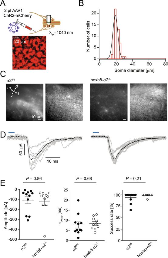
L. Tudeau, et al.                                                                                                              Brain Research 1741 (2020) 146889

responses to light touch, and spontaneous aversive behaviors (Foster               supraspinal CNS areas.
et al., 2015). Earlier studies reported similar changes with GABAA or                  In order to assess the consequences of α2GABAAR subunit ablation
glycine receptor blockers injected locally into the spinal canal of rats           for spinal inhibitory neurotransmission, we combined electro-
(Beyer et al., 1985; Roberts et al., 1986; Yaksh, 1989; Sivilotti and              physiological whole-cell recordings and optogenetics in transverse
Woolf, 1994). Work from several laboratories has shown that a loss of              slices of the lumbar spinal cord. We crossed the vGAT::ChR2-YFP allele
inhibitory neurotransmission occurs endogenously in neuropathic or                 (Zhao et al., 2011) into α2fl/fl and hoxb8-α2−/− mice. In spinal cord
inflammatory pain states (Ahmadi et al., 2002; Moore et al., 2002; Coull            slices of these double and triple transgenic mice, IPSCs were robustly
et al., 2003; Harvey et al., 2004; Zhang et al., 2011). A critical con-            evoked by brief (4 ms) wide field illumination of the dorsal horn with
tribution of diminished inhibition to exaggerated pain sensitivity in              blue light (see also Foster et al., 2015). We restricted our analyses to
inflammatory and neuropathic disease states is also supported by                    photocurrent negative, presumed excitatory, interneurons, which are
pharmacological studies that have demonstrated antihyperalgesic effi-                required for the full behavioral expression of pain behaviors (Wang
cacy of compounds that facilitate dorsal horn inhibitory neuro-                    et al., 2013) and constitute critical elements of the dorsal circuitry in-
transmission (Knabl et al., 2008; Huang et al., 2017) and by the anti-             volved in different forms of pathological pain (Peirs et al., 2015). The
hyperalgesic efficacy of GABAergic neuron precursor transplantation                  restriction of our analyses to photocurrent negative cells also allowed
into the spinal cord (Braz et al., 2012). Studies employing compounds              avoiding possible contaminations of IPSCs with photocurrents. Slices
acting on GABAARs have pointed to a particularly relevant role for                 were continuously superfused with the glycine receptor antagonist
GABAARs containing α2 subunits (α2GABAARs), which are enriched in                  strychnine (0.5 µM) to isolate the GABAergic IPSC component. GA-
the superficial dorsal horn, i.e. in the termination area of nociceptive            BAergic IPSC amplitudes were about 50% smaller in the hoxb8-α2−/−
fibers (Bohlhalter et al., 1994; Bohlhalter et al., 1996; Paul et al., 2012).       mice (-151 ± 21 pA, n = 16 cells) compared to α2fl/fl mice
To demonstrate that the antihyperalgesic effect of α2GABAAR mod-                    (-267 ± 51 pA, n = 17 cells) (Fig. 1C, D). Decay time constants (τdecay)
ulators originates from a spinal site and does not involve supraspinal             of GABAergic IPSCs showed a trend towards slower decay in neurons of
CNS areas, hoxb8-α2−/− mice have been generated, which lack                        hoxb8-α2−/− mice (83.4 ± 9.0 ms) relative to α2fl/fl mice
α2GABAARs from the spinal cord up to the spinal segment C4 (Paul                   (65.0 ± 8.2 ms) but the difference did not reach statistical significance
et al., 2014). These mice exhibited the expected reduction in GABAAR-              (Fig. 1E).
mediated membrane currents and, in line with this change, a reduced                    We also confirmed that baseline nociceptive sensitivity was un-
antihyperalgesia by the α2-preferring GABAAR modulator HZ-166                      changed in hoxb8α2−/− mice. To this end, we compared the sensitiv-
(Rivas et al., 2009; Di Lio et al., 2011). An unexpected result of these           ities of hoxb8α2-/− and α2fl/fl mice in a battery of somatosensory and
experiments was however the absence of a pronociceptive phenotype in               nociceptive tests including the von Frey test of light punctate me-
hoxb8-α2−/− mice.                                                                  chanical sensitivity, the Hargreaves plantar test for heat sensitivity, the
    To gain more insight into this paradox, we performed an in-depth               pin-prick test for noxious mechanical stimulation and the cold plantar
analysis of inhibitory GABAergic neurotransmission in the dorsal horn              test. We did not detect significant differences between hoxb8-α2−/−
of hoxb8-α2−/− mice and of potential compensatory mechanisms.                      and α2fl/fl mice in any of the four tests (Fig. 1F).
These experiments confirmed a reduction in the amplitude of
GABAergic IPSCs by about half and an almost complete loss of the                   2.2. Hoxb8-α2−/− mice exhibit reduced potentiation of GABAergic IPSCs
antihyperalgesic effects of HZ-166 in hoxb8-α2−/− mice. Our results                 by HZ-166 and diminished analgesia by HZ-166
exclude increases in glycinergic inhibition and an up-regulation of other
GABAAR subtypes as likely compensatory mechanisms. Although our                        The lack of an apparent baseline nociceptive phenotype in hoxb8-
immunohistochemical analyses revealed an increased abundance of                    α2−/− mice (Paul et al., 2014) contrasts with previous studies in GA-
serotoninergic markers in the dorsal horn, ablation of serotonergic                BAAR receptor point-mutated mice that have suggested a critical role of
terminals from the spinal dorsal horn failed to unmask a pronociceptive            α2GABAARs in spinal pain control (Knabl et al., 2008). We therefore re-
phenotype. Our results thus confirm a critical role of α2GABAARs in                 addressed whether deletion of spinal α2GABAARs would have an im-
spinal nociceptive control. However, a loss of α2GABAAR expression                 pact on the efficacy of α2-GABAAR-preferring benzodiazepine site li-
early in development appears to be compensated by a still unknown                  gands with known analgesic activity. For these experiments, we chose
process that prevents the development of a hyperalgesic phenotype.                 the α2GABAAR-preferring and less sedating benzodiazepine site agonist
                                                                                   HZ-166 (Rivas et al., 2009; Di Lio et al., 2011). As a prerequisite for
2. Results                                                                         these experiments, we analyzed the in vitro pharmacological profile of
                                                                                   HZ-166 in HEK 293 cells transiently transfected with different combi-
2.1. Hoxb8α2−/− mice lack α2GABAARs from the spinal cord and exhibit               nations of GABAAR subunits (α1β2γ2, α2β3γ2, α3β3γ2 and α5β2γ2). In
reduced GABAergic synaptic inhibition but do not show a hyperalgesic               agreement with a previous publication (Rivas et al., 2009), we found
phenotype                                                                          that both potency and efficacy of GABA current (IGABA) potentiation by
                                                                                   HZ-166 were higher for α3GABAAR and α2GABAAR than for α1GA-
    We first verified that α2GABAARs were completely lost from the                   BAAR and α5GABAAR subtypes (Fig. 2A). We then characterized the
lumbar spinal cord of hoxb8α2−/− mice. To this end, we quantified                   sensitivity of GABAergic IPSCs to HZ-166 in slices prepared from α2fl/fl
expression of the GABAAR α2 subunits in transverse sections of lumbar              and hoxb8-α2−/− mice. We used a saturating concentration of HZ-166
spinal cord of α2fl/fl, hoxb8α2−/− and global α2−/− mice (Fig. 1A,B).                (10 µM) and analyzed changes in τdecay. After application of HZ-166,
α2fl/fl mice showed the previously observed expression of α2GABAAR                   τdecay of light-evoked GABAergic currents increased by 94 ± 12%
subunits in the dorsal horn with highest density in the superficial layers,         (n = 17 cells) of control values in α2fl/fl mice. In hoxb8-α2−/− mice,
the main termination area of nociceptive fibers. α2GABAAR subunit                   τdecay increased only by 57 ± 12% (n = 13) (Fig. 2B). Finally, we
immunoreactivity in lumbar spinal cord sections from hoxb8α2−/−                    employed a mouse pain model to test whether the decreased sensitivity
mice was virtually indistinguishable from that of global α2−/− mice.               of GABAergic IPSCs to HZ-166 would translate to diminished analgesia.
This result is in line with the reported expression pattern of hoxb8-cre           In these experiments we included global α2R/R point-mutated mice in
in spinal cord neurons and astrocytes, and in DRG neurons up to about              addition to hoxb8-α2−/− and α2fl/fl mice. These mice carry a histidine
cervical level C4 (Witschi et al., 2010). The absence of any detectable            to arginine point mutation in the α2GABAAR gene (Gabra2) that ren-
α2 subunit reactivity indicates that most, if not all, α2 subunits present         ders α2GABAAR insensitive to most benzodiazepine site agonists in-
in the lumbar spinal cord are expressed by intrinsic spinal neurons or             cluding HZ-166 (Benson et al., 1998; Paul et al., 2014). For these ex-
peripheral sensory neurons and do not reside on axons descending from              periments, we used the formalin model of tonic pain and injected 4%

                                                                               2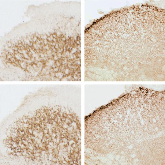
L. Tudeau, et al.                                                                                                                Brain Research 1741 (2020) 146889

Fig. 1. Baseline morphological, electrophysiological and behavioral analysis of hoxb8-α2−/− mice. A-B, α2GABAAR expression. A, Transverse lumbar spinal dorsal
horn sections stained for GABAAR α2 subunits in α2fl/fl, hoxb8-α2−/− and global α2−/− mice. d, dorsal; v, ventral; l, lateral; m, medial. B, Quantification of
immunoperoxidase intensities (n = 16 sections from 2 mice, unpaired t-test). C–E, GABAergic IPSCs. C, Example traces of light-evoked GABA-IPSCs (vertical blue
bars represent 4 ms light pulse) recorded in the presence of strychnine (500 nM). Hoxb8-α2−/− mice show reduced GABAergic IPSC amplitudes compared to α2fl/fl.
Decay time constants were determined from double exponential fits, shown in red. D, Amplitudes of light-evoked GABAergic IPSCs in hoxb8-α2−/− and α2fl/fl mice
(n = 17 cells from 14 mice, and n = 16 cells from 14 mice, for α2fl/fl and hoxb8-α2−/− mice, respectively, unpaired t-test). Data are presented as mean ± SEM. E,
Scatter plot shows no significant change in decay time constants between the two genotypes (unpaired t-test). F, Somatosensory and nociceptive response thresholds
of naïve α2fl/fl and hoxb8-α2−/− mice. Each data point indicates an individual mouse. Data are presented as mean ± SEM, n = 5 mice for all genotypes, P values
indicate α errors from unpaired t-tests.

formalin into one hind paw and examined the analgesic effect of HZ-                 vehicle instead of HZ-166, no differences were found in the formalin-
166 in the three genotypes (Fig. 2C). HZ-166 (16 mg/kg, i.p.) sig-                 induced pain behavior between the three genotypes. These experiments
nificantly reduced the number of formalin-induced nocifensive reac-                 confirm an important role of α2GABAARs in dorsal horn pain control.
tions (assessed as time spent licking of the injected paw) in α2fl/fl mice,          Since these findings contrast with the apparent lack of a hyperalgesic
but failed to do so in hoxb8α2−/− and α2R/R mice. In mice treated with             phenotype in hoxb8-α2−/− mice, we next sought to identify possible

                                                                               3
L. Tudeau, et al.                                                                                                               Brain Research 1741 (2020) 146889

Fig. 2. Diminished analgesia by HZ-166 in hoxb8-α2−/− mice. A, GABA-evoked membrane currents were measured in HEK 293 cells transiently transfected with
recombinant α1β2γ2, α2β3γ2, α3β3γ2 and α5β2γ2 GABAARs. Example traces on top show current responses evoked by GABA before (light trace) and during
application of saturating concentrations of HZ-166 (10 µM, dark trace). Bottom graph representing dose–response curves of the four different GABAAR subtypes at
increasing HZ-166 concentrations. GABA was applied at EC10 (1 µM, 5 µM, 8 µM, and 1 µM for α1β2γ2, α2β3γ2, α3β3γ2 and α5β2γ2 GABAARs, respectively). B,
Potentiating actions of HZ-166 on light-evoked GABAergic IPSCs recorded from superficial dorsal horn neurons of α2fl/fl and hoxb8-α2−/− mice. Normalized
example traces recorded in the presence of strychnine (500 nM) in α2fl/fl and hoxb8-α2−/− mice before (black) and after (green) application of HZ-166 (10 µM). HZ-
166-evoked prolongation of IPSC decay kinetics was significantly smaller in hoxb8-α2−/− than in α2fl/fl mice (n = 17 and 13, for α2fl/fl and hoxb8-α2−/− mice,
respectively, unpaired t-test). C, HZ-166-mediated analgesia. Total number of licking bouts evoked by subcutaneous formalin injection into one hindpaw of α2fl/fl,
hoxb8-α2−/− and α2R/R mice treated wither with HZ-166 (16 mg/kg, i.p.) or vehicle.

compensatory mechanisms explaining this apparent paradox.                          component (-633 ± 155 pA, n = 8, versus −374 ± 133 pA, n = 10).
                                                                                   When the total charge transfer was analyzed, a significant reduction
2.3. Potential compensatory processes preventing a hyperalgesic phenotype          was observed both of the total IPSC (-100 ± 24 pC, n = 8, versus
in hoxb8-α2−/− mice                                                                -28.0 ± 3.4 pC, n = 8; P = 0.004, unpaired t-test) and for its glyci-
                                                                                   nergic component (-49.6 ± 13.8 pC, n = 8, versus -14.0 ± 4.0 pC,
    In addition to GABA, glycine is a second fast inhibitory neuro-                n = 8, P = 0.027) clearly ruling out an increase in glycinergic synaptic
transmitter in the spinal cord. An increase in glycinergic inhibition              currents as a compensatory process.
might thus be a straightforward mechanism to compensate for dimin-                     Although α2GABAARs are primarily located at synapses (Sassoe-
ished synaptic inhibition by GABA. We therefore compared the ampli-                Pognetto et al., 2000), they are also found at extrasynaptic sites, where
tudes and charge tansfer of the total (mixed GABAergic/glycinergic)                they may form a receptor reserve (Gouzer et al., 2014). GABAARs lo-
IPSC in superficial dorsal horn neurons of hoxb8-α2−/− and α2fl/fl mice               cated at extrasynaptic sites also underlie tonic membrane currents that
(Fig. 3A,B). In these experiments, we relied again on the analysis of              are found in a subpopulation of dorsal horn neurons, where they are
light-evoked IPSCs in presumed excitatory (photocurrent-negative)                  activated by ambient GABA (for review, see (Farrant and Nusser, 2005).
neurons. After recording of the total IPSCs for 1 min, we added                    These tonic GABAergic currents are thought to be carried by high-af-
strychnine (500 nM) to the bath perfusion to determine (via subtraction            finity α5GABAAR or δ subunit-containing GABAARs (Delgado-Lezama
of the remaining amplitude) the amplitude of the glycinergic compo-                et al., 2013; Fritschy and Panzanelli, 2014). However, a possible con-
nent. In case of the amplitudes of light-evoked IPSCs, we found a trend            tribution of α2GABAAR to tonic currents, for example in situations of
towards reduced IPSC amplitudes in hoxb8-α2−/− mice both for the                   increased ambient GABA concentrations, cannot be excluded. To ex-
total IPSC (-1054 ± 200 pA, n = 8, versus −558 ± 154 pA, n = 10,                   amine tonic membrane currents in hoxb8-α2−/− mice, we performed
for α2fl/fl and hoxb8-α2−/− mice, respectively) and for its glycinergic              again whole-cell patch-clamp recordings from presumed excitatory

                                                                               4
L. Tudeau, et al.                                                                                                      Brain Research 1741 (2020) 146889

                                                                                                                                 (caption on next page)

neurons. After 5 min of baseline holding current recording, we added          (−41.2 ± 4 pA, n = 12 cells) relative to neurons in slices from α2fl/fl
bicuculline (20 µM) to the bath perfusion to determine the GABAergic          mice (-27 ± 2.0 pA, n = 9). This increase was likely due to larger
contribution (Fig. 3C). Neurons recorded in slices obtained from hoxb8-       GABAergic tonic currents as the bicuculline-sensitive component was
α2−/− mice had on average slightly larger holding currents                    also larger in hoxb8-α2−/− mice (-5.8 ± 2.8 pA, n = 12) than in α2fl/

                                                                          5
L. Tudeau, et al.                                                                                                                     Brain Research 1741 (2020) 146889

Fig. 3. No compensatory increase in glycinergic IPSCs or tonic GABAergic currents in hoxb8α2−/− mice. A, Amplitudes (left) and charge transfer (right) of total
(mixed GABAergic/glycinergic) light-evoked IPSCs in α2fl/fl and hoxb8-α2−/− mice (n = 8 cells from 6 mice and n = 10 cells from 9 mice, for α2fl/fl and hoxb8-
α2−/− mice, respectively, unpaired t-test). B, Same as A, but for the glycinergic IPSC component (n = 8 cells from 6 mice and n = 10 cells from 9 mice, for α2fl/fl and
hoxb8-α2−/− mice, respectively). For 2 cells from the hoxb8-α2−/− group, no meaningful charge transfer value was obtained for the glycinergic IPSC component. C-
E, tonic GABAergic currents. C, Example traces of tonic currents in α2fl/fl and hoxb8-α2−/− mice before and during application of bicuculline (20 µM). Grey and red
bars indicate the areas that were analyzed for the all-point histograms of control and bicuculline conditions, respectively. The all-point histograms follow a Gaussian
distribution and depict the amplitude scattering in a 30 s range analyzed from the current recorded in control and bicuculline conditions, respectively. D, Plot
showing holding currents in baseline conditions (before application of bicuculline) in hoxb8-α2−/− mice (n = 7 cells) compared to α2fl/fl mice (n = 9 cells),
unpaired t-test. E, same as D, but GABAergic component (after application of bicuculline) (n = 12 cells from 7 mice and n = 7 cells from 4 mice, for α2fl/fl and hoxb8-
α2−/− mice, respectively, unpaired t-test. F,G, Quantitative expression of mRNAs encoding for certain GABAA (F) and glycine receptor subunit genes in the spinal
cord (top) and DRGs (bottom) of α2fl/fl mice and hoxb8-α2−/− mice. Expression gabra2 (encoding for the α2 GABAA receptor subunit) in α2fl/fl mice is shown for
comparison (grey column). Data on gabra2 are taken from Paul et al., 2012. Expression of glra2, glra3 and glra4 in DRGs was below the detection threshold (N.D., not
detectable). P values were obtained using unpaired two-tailed unpaired t-test. n = 6–7 mice per group.

fl
  mice (α2fl/fl: −0.3 ± 1.1 pA, n = 9) (Fig. 3D,E).                                     2.4. Other potential compensatory mechanisms preventing a pronociceptive
    To complement these functional data with data on gene expression,                 phenotype in hoxb8-α2−/− mice
we performed quantitative RT-PCR of GABAA and glycine receptor
subunits (Fig. 3F, G). In a previous study (Paul et al., 2012), we already                Other potential mechanisms possibly counteracting a loss in
reported that the expression of GABAAR α1 and α3 - α6 subunits was                    GABAergic inhibition include a compensatory weakening of glutama-
unchanged in spinal cords and DRGs of hoxb8-α2−/−. In the present                     tergic transmission within the spinal dorsal horn or a strengthening of
study, we have complemented these previous results and investigated in                descending inhibitory pain control. Vesicular glutamate transporters of
addition the expression of the GABAAR δ subunit, which together with                  which three isoforms (vGluT1-3) exist are marker genes of glutama-
the α4 subunit is a part of many extrasynaptic GABAARs (Delgado-                      tergic neurons (El Mestikawy et al., 2011). They are localized to axon
Lezama et al., 2013), of the three GABAAR ρ subunits, which give rise to              terminals of glutamatergic neurons where they mediate glutamate up-
atypical bicuculline-insensitive GABAARs previously also called GA-                   take into presynaptic storage vesicles. In the spinal cord, vGluT1 ex-
BACRs (Bormann and Feigenspan, 1995), and of all five glycine receptor                 pression originates almost exclusively from peripheral non-nociceptive
subunits (glra1 - glra4 and glrb). Relative to the GABAAR α2 subunit, all             touch-sensitive and proprioceptive sensory neurons (Oliveira et al.,
four GABAAR subunits analyzed here were expressed at much lower                       2003). vGluT2 is expressed by nociceptive sensory neurons, but most
levels both in the spinal cord and the DRGs. No significant differences                 vGluT2 protein in the spinal cord is found in interneurons. vGluT3 is
were found between α2fl/fl and hoxb8-α2−/− mice. In the spinal cord,                    only weakly expressed in the dorsal horn but it is present in a small
all glycine receptor subunits, with the exception of the α4 subunit                   band at the border between laminae II and III where it originates from
(glra4), were expressed at higher levels than the GABAAR α2 subunit                   vGluT3 expression in the terminals of tyrosine hydroxylase positive
(gabra2). A minor, yet statistically significant, down-regulation in                   peripheral sensory neurons (Seal et al., 2009) and neurons descending
hoxb8-α2−/− mice was obtained for glra2 and glrb. In DRGs, expression                 from the brainstem (Oliveira et al., 2003). To assess potential changes
of glycine receptor subunits was below the detection limit (glra2 –                   in vGluT1-expression in the dorsal horn of hoxb8-α2−/− mice and α2fl/
glra4) or very low (glra1). In both α2fl/fl and hoxb8-α2−/− mice, a                     fl
                                                                                           mice,     we    performed    quantitative     (densitometric)    im-
relatively high expression was found for glrb, which is however in many               munohistochemical analyses (Fig. 5). Because of the differential ex-
tissues not translated into protein despite high mRNA levels (Weltzien                pression of vGluTs in the superficial and deep dorsal horn, we analyzed
et al., 2012).                                                                        expression in the superficial and deep dorsal horn separately. While we
    In addition to mediating synaptic (phasic) and extrasynaptic (tonic)              did not detect differences for vGluT1 and vGluT2, there was a small but
inhibition via dendritic and somatic GABAARs, GABAARs contribute                      statistically significant difference in vGluT3 signal intensity in the su-
also to presynaptic inhibition via receptors located on spinal axons and              perficial dorsal horn. Because many of the descending neurons in this
terminals of peripheral sensory neurons, including nociceptors                        area are serotonergic and co-express vGluT3 with markers of ser-
(Rudomin and Schmidt, 1999; Willis, 1999). We therefore also in-                      otonergic transmission (Oliveira et al., 2003), we analyzed potential
vestigated a possible impact of GABAAR α2 subunit deletion on ex-                     changes in the serotonergic system. We found a pronounced and highly
citatory neurotransmission between primary sensory fibers and dorsal                   significant increase in immunoreactivity against both serotonin and
horn second order neurons using optogenetics for presynaptic primary                  tryptophan hydroxylase 2 (TPH2) in hoxb8-α2−/− mice compared to
afferent stimulation. Bilateral sciatic nerve injections of AAV1 ChR2-                 α2fl/fl mice (Fig. 5). As serotonergic input to the dorsal horn is generally
mCherry virus were made into the sciatic nerve of α2fl/fl and hoxb8-                    believed to contribute to endogenous pain control (Fields et al., 2006),
α2−/− mice. These injections led to the infection of dorsal root gang-                we hypothesized that this up-regulation may constitute a compensatory
lion neurons of the lumbar segment L4 (Fig. 4A). Soma diameter ana-                   mechanism possibly explaining the absence of a hyperalgesic pheno-
lysis of the infected neurons indicated that the great majority of in-                type in hoxb8-α2−/− mice.
fected cells had a soma diameter of approximately 20 µm (Fig. 4B,
n = 72 cells from 3 mice), in line with a preferred infection of noci-
                                                                                      2.5. Ablation of serotonergic axon terminals from the dorsal horn of hoxb8-
ceptive (small diameter) neurons. Seven days after virus injection, we
                                                                                      α2−/− mice fails to unmask a pro-nociceptive phenotype
prepared transverse spinal cord slices of the L4 segment for electro-
physiological recordings from neurons in the superficial dorsal horn
                                                                                          To examine, whether the observed up-regulation of markers of the
(laminae I and II). After establishing a whole-cell recording, we sti-
                                                                                      dorsal horn serotonergic innervation underlies the absence of a pro-
mulated the dorsal horn with blue light (λ = 473 nm, 4 ms, power
                                                                                      nociceptive phenotype in hoxb8-α2−/− mice, we pharmacologically
density: 20.4 mW/mm2) and recorded light-evoked EPSCs (Fig. 4C,D).
                                                                                      ablated serotonergic axon terminals in the spinal cord through three
No statistically significant difference was observed between genotypes
                                                                                      intraspinal injections of 5,7 dihydroxytryptamine (DHT; 2.5 µg in
for EPSC amplitude (α2fl/fl mice: −108.2 ± 34.7 pA, n = 11; hoxb8-
                                                                                      500 nl) covering the left spinal segments L4-L6 (Fig. 6). Mice were
α2−/−: −117.8 ± 41.3 pA, n = 10), decay kinetics (α2fl/fl:
                                                                                      pretreated with desipramine (25 mg/kg, i.p.) to avoid ablation of nor-
7.6 ± 1.6 ms; hoxb8-α2−/−: 8.5 ± 0.9 ms) or success rate (α2fl/fl:
                                                                                      adrenergic terminals, which are also present in the spinal dorsal horn.
94.2 ± 3.4%, hoxb8-α2−/−: 99 ± 1.0%) (Fig. 4E).
                                                                                      Efficient ablation of serotonergic terminals was verified with serotonin
                                                                                      immunostaining for each mouse analyzed in the sensory tests (Fig. 6A).

                                                                                  6
L. Tudeau, et al. Brain Research 1741 (2020) 146889
Fig. 4. Excitatory input from primary nociceptive
fibers onto dorsal horn second order neurons. A,
Scheme illustrating the AAV1 ChR2-mCherry injec-
tion into the sciatic nerve of α2fl/fl or hoxb8-α2−/−
mice. Pseudo-coloured z-projection two-photon
image of virus-infected ganglion cells in DRGs of
lumbar segment L4. B, Histogram showing the dis-
tribution of infected dorsal root ganglion cell soma
diameters which could be fitted with a Gaussian
function (average diameter: 20.9 ± 0.4 µm; n = 72
cells from 3 mice). C, Epifluorescence (left) and
infra-red contrast images (right) of the superficial
dorsal horn in acute spinal cord slices prepared from
nerve-injected mice. Epifluorescence images show
punctate mCherry expression on the spinal terminals
of infected dorsal root ganglion neurons. D, Example
traces from the two genotypes showing the average
(black) of 7 (α2fl/fl) and 10 (hoxb8-α2−/−) EPSCs
(grey) after 4 ms blue light exposure (horizontal
blue bars) of primary afferent terminals. E,
Statistical comparisons of amplitude, success rate
and decay time constants (n = 11 cells and n = 10
cells, for α2fl/fl and hoxb8-α2−/− mice, respec-
tively, unpaired t-test).
We then tested the sensitivity of the ipsilateral hindpaw to stimulation studies have shown that specific activation (via positive allosteric
with von Frey filaments, in the pin-prick test, the Hargreaves test and modulation) of α2GABAARs reduces pain sensitivity in several rodent
the cold plantar test, to respectively assess sensitivity to innocuous and pain models (Knabl et al., 2009; Reichl et al., 2012; Ralvenius et al.,
noxious punctate mechanical stimulation, noxious heat and noxious 2015), and in human experimental pain models (van Amerongen et al.,
cold stimuli (Fig. 6B). In agreement with previous work, ablation of 2019). Based on these findings one might expect that ablation of
serotonergic terminals did not sensitize naïve wild-type mice (Carr α2GABAARs from the spinal cord would lead to an hyperalgesic phe-
et al., 2014). However, it also failed to induce sensitization in hoxb8- notype or spontaneous pain behaviors as observed after ablation or
α2−/− mice. silencing of inhibitory dorsal horn neurons (Foster et al., 2015) or after
spinal blockade of GABAARs (Roberts et al., 1986; Sivilotti and Woolf,
1994). However, in a previous study, hoxb8-α2−/− mice, which lack
3. Discussion α2GABAARs from the spinal cord, showed no increased sensitivity to
acute noxious mechanical and thermal stimuli and no signs of sponta-
α2GABAARs serve critical functions in the control of spinal noci- neous pain (Paul et al., 2014). This discrepancy may indicate the
ceptive circuits (Zeilhofer et al., 2012b; Zeilhofer et al., 2012a). Several
7L. Tudeau, et al. Brain Research 1741 (2020) 146889
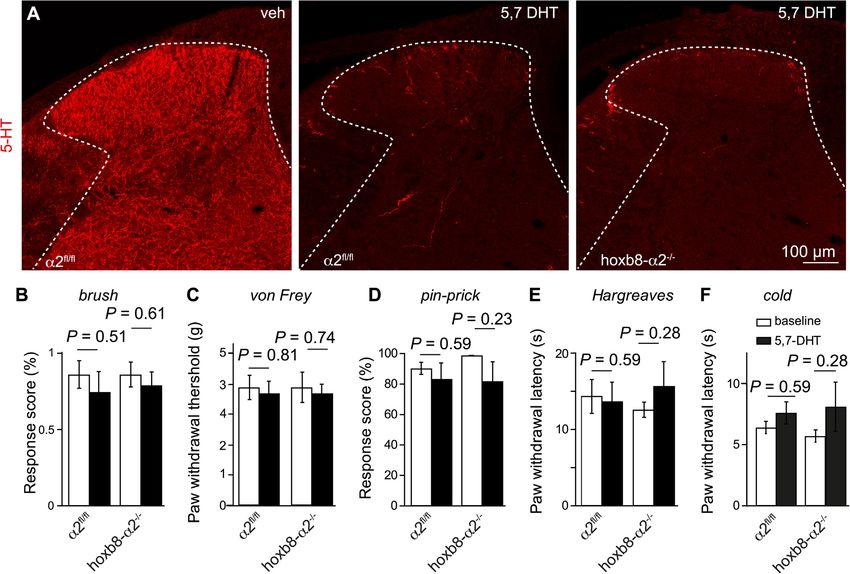
Fig. 5. vGluT1-3 and serotonergic marker expression. A, Transverse sections of lumbar spinal cord dorsal horns of α2fl/fl and hoxb-8α2−/− mice stained for vesicular
glutamate transporter vGlT1 – vGluT3 and markers of serotonergic expression (5-HT and TPH2). 1, sdh; 2, ddh. B, Quantification of immunoperoxidase intensities in
stainings for vGluT1, vGluT2, vGluT3, 5-HT, and TPH2 (in α2fl/fl and hoxb8-α2−/− mice, n = 4, each). Separate analyses for the superficial and deep dorsal horn
(sdh, superficial dorsal horn; ddh, deep dorsal horn).
presence of compensatory mechanisms at the cellular and/or molecular GABA concentrations are low outside synaptic clefts, GABAARs med-
level, which may counteract the reduction in GABAergic inhibition. iating tonic GABAergic currents should have high affinities for GABA.
Revealing such mechanisms might benefit the discovery of novel anti- Previous work has shown that α4/δ and α5GABAARs fulfill these cri-
hyperalgesic drug targets. teria (Delgado-Lezama et al., 2013; Fritschy and Panzanelli, 2014). It
In the present study, we have examined the functional consequences has been shown that α5GABAARs contribute to tonic currents in the
of GABAAR α2 subunit deletion from the spinal cord on the cellular and spinal dorsal horn, but α5 subunit-deficient mice exhibit normal acute
behavioral level. We confirmed that hoxb8-α2−/− mice show reduced nociceptive pain behavior (Perez-Sanchez et al., 2016). Similar findings
synaptic inhibition in the superficial dorsal horn but nevertheless ex- have been made in case of α4GABAARs, which typically assemble with
hibit normal acute pain sensitivities. We then continued with an in- δ GABAAR subunits to form benzodiazepine-insensitive extrasynaptic
depth analysis of GABAergic neurotransmission in hoxb8-α2−/− and GABAARs (Storustovu and Ebert, 2006). Mice lacking these δ subunits
corresponding wild-type (α2fl/fl) mice. Our electrophysiological re- exhibit reduced tonic GABAergic membrane currents but also show no
cordings revealed a reduction in the amplitudes of light-evoked changes in acute nociceptive sensitivity (Bonin et al., 2011). Further-
GABAergic IPSCs without a compensatory increase in glycinergic in- more, tonic GABAergic currents in superficial dorsal horn neurons are
hibition. In fact, the total IPSC amplitude was reduced by a similar small and are present only in a subset of neurons (Mitchell et al., 2007)
percentage as the GABAergic IPSC component, suggesting that the loss consistent with the low abundance of GABAAR α4 subunits in the spinal
of α2GABAARs also reduced the glycinergic IPSC component. This dorsal horn (Paul et al., 2012). These results argue against a major role
might suggest the presence of presynaptic α2GABAARs that facilitate of extrasynaptic GABAAR and of tonic GABAergic currents in the reg-
transmitter release (Kawaguchi and Sakaba, 2017). Alternatively, the ulation of nociception. On the other hand, gaboxadol (also known as
loss of GABAARs might impair the clustering of glycine receptors at THIP), which enhances currents through α4/δ containing GABAAR
postsynaptic sites. Direct evidence for either of the two processes is (Adkins et al., 2001), reduced acute nociception in wild-type but not in
however missing. δ subunit-deficient mice (Bonin et al., 2011). These results on acute
In subsequent analyses we found a trend towards increased ampli- nociception are hence reminiscent of what was observed in the present
tudes of tonic GABAergic membrane currents in hoxb8-α2−/− mice. study with HZ-166 in chronic pain models.
The absence of a pronociceptive phenotype in hoxb8-α2−/− mice An alternative and in our opinion more likely explanation for the
might hence be due to preserved tonic GABAergic inhibition mediated absence of a pronociceptive phenotype in hoxb8-α2−/− mice is the
by extrasynaptic GABAARs. Although this explanation cannot be fully presence of compensatory mechanisms that counteract diminished
ruled out, it appears unlikely. Tonic GABAergic currents originate from GABAergic inhibition. In this context, it is interesting to note that hy-
the activation of extrasynaptic GABAARs by ambient GABA. Since peralgesia induced by ablation or silencing of inhibitory dorsal horn
8L. Tudeau, et al. Brain Research 1741 (2020) 146889
Fig. 6. Ablation of serotonergic axon terminals in the lumbar dorsal horn. A, Transverse spinal cord sections of α2fl/fl and hoxb8-α2−/− mice prepared on day 6 after
local spinal injection of 5,7-DHT (3 injections à 500 nl of 5 µg/µl) and stained for serotonin. B-F, Somatosensory and nociceptive responses in α2fl/fl and hoxb8-α2−/
−
mice five days after 5,7-DHT injection (same doses as above; n = 7 mice per group, paired t-test).
neurons also shows some recovery over the course of a few weeks contribution of α2GABAARs to spinal nociceptive control. Their abla-
(Foster et al., 2015). As the reduction in GABAergic IPSC amplitudes tion early in development appears to induce an up-regulation of dorsal
was not compensated by an increase in glycinergic inhibition, we horn serotonergic innervation and a yet-to-be identified compensatory
speculated whether the loss in inhibition might be compensated by a mechanism that protects hoxb8-α2−/− mice from a hyperalgesic phe-
reduction in excitatory drive to the dorsal horn or an increased in- notype.
hibitory control via descending antinociceptive fiber tracts. We did not
detect a reduction in nociceptive input strength from peripheral sensory 4. Methods and materials
fibers onto dorsal horn second-order neurons, which is in line with
unaltered expression of vGluT1 and vGluT2 protein in the dorsal horn 4.1. Animals
of hoxb8-α2−/− mice. We found however a significant up-regulation of
the vGluT3 isoform which is found not only in low-threshold mechan- All experimental procedures were approved by the Cantonal
oreceptive C fibers (Seal et al., 2009) but also serotonergic neurons of Veterinary Office (licenses 126/2012, 74/2013, 86/2013, 031/2016).
the brainstem (Domonkos et al., 2016). This drew our attention to Behavioral experiments were performed in α2fl/fl; hoxb8::cre double
possible changes in descending serotonergic inhibition. Serotonin is transgenic mice (short hoxb8-α2−/− mice) and in cre-negative α2fl/fl
synthesized in neurons of the nucleus raphe magnus, located in the mice. HoxB8-cre and GABAAR α2fl/fl mice have been described pre-
hindbrain, which is the main source of spinal 5-HT release (Hornung, viously (Witschi et al., 2010; Witschi et al., 2011). For combined
2003). Although spinally released serotonin can mediate both hyper- electrophysiological/optogenetic experiments these two strains of mice
algesia and antinociception, depending on which subtype of serotonin were crossed with vGAT::ChR2 BAC transgenic mice (Zhao et al., 2011)
receptors is activated (Yaksh and Wilson, 1979; Furst, 1999; Diniz et al., to obtain α2fl/fl; vGAT::ChR2-YFP and α2fl/fl; hoxb8cre; vGAT::ChR2
2015), most studies support an analgesic effect of descending ser- double and triple transgenic mice. Electrophysiological/optogenetic
otonergic input to the spinal dorsal horn. We demonstrated increased experiments were performed in 3–4 week old mice of either sex. In
levels of serotonin and TPH2 in both the superficial and deep dorsal immunohistochemical experiments global GABAAR α2 subunit-defi-
horn of the spinal cord in hoxb8-α2−/− mice. These results might cient mice (α2−/− mice) were used in addition to hoxb8-α2−/− and
suggest an increased serotonergic tone in the spinal cord of hoxb8-α2−/ α2fl/fl mice. We observed occasional hoxb8-cre-mediated gene re-
−
mice as a possible compensatory mechanism. However, local and combination in the germ line. In order to avoid confounding results
nearly complete ablation of serotonergic terminals from one side of the originating from the presence of non-conditional knock-out alleles, we
lumbar spinal cord segments L3-L5 failed to unmask a hyperalgesic performed post-hoc PCR genotyping of brain and spinal cord tissue for
phenotype in hoxb8-α2−/− mice. all mice included in our experiments. Mice were included in our ana-
In summary, the results obtained with HZ-166 support an important lyses only when PCR genotyping of their brain tissue confirmed the
9L. Tudeau, et al. Brain Research 1741 (2020) 146889
presence of a non-recombined floxed Gabra2 allele and the absence of a GABAAR antagonist bicuculline was bath-applied to verify that the re-
recombined (“knock-out”) Gabra2 allele. corded IPSC was exclusively mediated by GABAARs.
During whole-cell recording, access resistance was continuously
4.2. Drugs and chemicals monitored using short hyperpolarizing voltage steps (-5 mV) applied at
regular intervals. Recordings were discarded if the access resistance
For electrophysiological experiments, (-)-bicuculline methochloride, changed > 20% or if recovery to baseline currents before GABA ap-
strychnine hydrochloride, and HZ-166 were dissolved in extracellular plication was less than 85–90% in the case of tonic currents. Electrical
solution. NBQX was dissolved in DMSO and diluted with extracellular signals were sampled either at 20 kHz (light-induced IPSCs) or 5 kHz
solution to a final concentration of 20 µM in 0.1% DMSO. All chemicals (tonic currents) and filtered at 2.9 kHz. Data was analyzed using
except for HZ-166 were purchased from Tocris (Germany). HZ-166 was IgorPro (WaveMetrics, Inc.,USA).
synthesized as described earlier (Rivas et al., 2009). Desipramine hy- Primary afferent-evoked EPSCs were recorded from unidentified
drochloride was provided by Bio-Techne AG and 5,7-dihydroxy- (excitatory and inhibitory) neurons in slices prepared from hoxb8-α2−/
−
tryptamine creatinine sulphate was provided by ANAWA Trading SA. and α2fl/fl mice one week after these mice had been injected into both
sciatic nerves with a non-cre dependent AAV1 expressing a ChR2-
4.3. Spinal cord slice preparation mCherry fusion protein under the chicken β-actin (CAG) promoter.
Intraneural AAV injections (titer 2.96x10¹³ GC/ml, approximately 2 µl
Acute transverse 400 µm thick slices of the lumbar spinal cord were each side) were performed in 15–28 day-old mice. mCherry-expressing
prepared from 20 to 30 day old mice of either sex. Slices were cut in ice- infected dorsal root ganglion (DRG) cells at lumbar segment L4 were
cold solution of the following composition in mM (Dugue et al., 2009): visualized using a two-photon microscope for a gross morphological
130 K-gluconate, 15 KCl, 0.05 EGTA, 20 HEPES and 25 glucose titrated characterization in situ (excitation wavelength λ = 1040 nm). The
to pH 7.4 with KOH and supplemented with 50 µM D-APV to prevent soma diameter of the cells (72 cells from 3 animals) was determined
glutamate excitotoxicity. Slices were then allowed to recover for 30 min from a series of projected z-stacks using the 2-axis average diameter
in a solution containing (in mM) 225 D-mannitol, 2.5 KCl, 1.25 method (Scroggs and Fox, 1992). In a few recordings, NBQX (20 µl of
NaH2PO4, 25 NaHCO3, 0.8 CaCl2 and 8 MgCl2 and 25 glucose (37 °C, 20 mM stock solution) was added to the recording chamber at the end
gassed with 95% O2, 5% CO2). In a final step, slices were transferred to of the experiment to verify that the recorded EPSCs were mediated by
an artificial cerebrospinal fluid (aCSF), of the following composition (in AMPA receptors.
mM): 120 NaCl, 2.5 KCl, 1.25 NaH2PO4, 26 NaHCO3, 5 HEPES, 1
MgCl2, 2 CaCl2 and 14.6 glucose. Slices were transferred to the re- 4.5. Electrophysiological recordings in HEK293 cells
cording chamber and continuously perfused with aCSF equilibrated
with 95% O2, 5% CO2 at a flow rate of 1 ml/min. The effects of HZ-166 on currents through recombinant GABAARs
were studied in HEK293 cells transiently expressing GABAARs. HEK293
4.4. Electrophysiological recordings in spinal cord slices cells were transfected using lipofectamine LTX28. To ensure expression
of the γ2 subunit (required for modulation of GABAARs by benzodia-
Patch pipettes were prepared from borosilicate glass capillaries and zepines) in all recorded cells, we transfected cells with a plasmid ex-
had an open tip resistance of 3–5 MΩ. Recording pipettes were filled pressing the γ2 subunit plus eGFP from an IRES, and only selected
with an internal solution containing (in mM): 120 CsCl, 2 MgCl2, 10 eGFP-positive cells for recordings. The transfection mixture contained
HEPES, 0.05 EGTA, 2 MgATP, 0.1 NaGTP. CsCl was used to block (in µg): 1 αx, 1 β2, 3 γ2/eGFP (in case of α1 and α5) or 1 αx, 1 β3, 3 γ2/
GABAB receptor-mediated K+ currents. QX-314 (5 mM) was added to eGFP (in case of α2 and α3). Recordings were made 18–36 hrs after
block voltage-activated Na+ channels in the recorded cell. Excitatory transfection. Whole-cell patch-clamp recordings of GABA-evoked cur-
postsynaptic currents (EPSCs) were recorded using a K-gluconate-based rents were made at room temperature (20–24 °C) at a holding potential
internal solution containing (in mM): 130 K-gluconate, 20 KCl, 0.05 of −60 mV. Recording electrodes were filled with solution containing
EGTA, 2 MgCl2, 2 MgATP, 0.1 NaGTP, 10Na-Hepes, 5 QX-314. Neurons (in mM): 120 CsCl, 10 EGTA, 10 HEPES (pH 7.40), 4 MgCl2, 0.5 GTP
located less than 150 µm from the dorsal margin of the spinal cord and 2 ATP. The external solution contained (in mM): 150 NaCl, 10 KCl,
(laminae I and II) were visually identified with an iXON Ultra camera 2.0 CaCl2, 1.0 MgCl2, 10 HEPES (pH 7.4), and 10 glucose. GABA was
(Andor Technology, Belfast, UK) equipped with infrared gradient con- applied to the recorded cell using a manually controlled pulse (4–6 s) of
trast equipment (Zeiss Examiner A1, Göttingen, Germany). Whole-cell a low sub-saturating GABA concentration (EC10). EC10 values of GABA
voltage-clamp recordings were performed at room temperature at a were determined individually for all subunit combinations analyzed.
holding potential of −60 mV using a double patch-clamp EPC 9 am- EC50 values of HZ-166 and Hill coefficients (nH) were obtained from fits
plifier controlled with Patchmaster acquisition software (both HEKA of normalized concentration–response curves to the equation
Elektronik Dr. Schulze GmbH, Lambrecht/Pfalz, Germany). IGABA = Imax [GABA]nH/([GABA]nH + [EC50]nH). Imax was determined
All experiments on GABAAR-mediated and glycine receptor-medi- as the average maximal current elicited by a concentration of 1 mM
ated currents were performed in α2fl/fl;vGAT::ChR2-YFP and α2fl/ GABA. HZ-166 was dissolved in DMSO, subsequently diluted with re-
fl
;hoxb8cre;vGAT::ChR2 double and triple transgenic mice. Recordings cording solution, and was co-applied together with GABA without
were exclusively made from presumed excitatory neurons, character- preincubation.
ized by the absence of a blue light-induced photocurrent evoked by blue
light directly applied to the cell soma using a UGA-40 GEO laser system 4.6. Immunohistochemistry
(473 nm, 1 s, spot size 10 µm, 715.9 mW/mm2, RAPP OptoElectronic
GmbH, Hamburg, Germany). IPSCs were evoked by brief 4 ms blue light Adult α2fl/fl and hoxb8-α2−/− mice were injected i.p with 0.25 ml
stimuli applied in wide-field mode (473 nm, field of illumination ± pentobarbital and perfused through the ascending aorta with 50 ml ice-
200–300 µm, 20.4 mW/mm2) with a monochromator (Polychrome V, cold ACSF at room temperature for 2 min (Paul et al., 2012). Spinal
Thermo Fisher Scientific Munich GmbH, Germany) to evoke IPSCs. cord and brain were then removed and fixed in cold 4% PFA for
IPSCs were evoked at a frequency of 4 stimulations per min using wide- 90–120 min before being transferred to 30% sucrose in phosphate
field illumination of the dorsal spinal cord. The GABAergic component buffer saline (PBS) at 4 °C overnight for cryoprotection (Notter et al.,
of the light-evoked IPSCs was isolated by bath-applied strychnine 2014). Twenty to thirty µm thick coronal sections were made from
(0.5 µM). A steady-state block by strychnine was usually reached after frozen blocks and mounted onto Superfrost Plus microscope slides
2–3 min of continuous application. At the end of the experiment, the (Thermo Scientific, Zurich, Switzerland).
10L. Tudeau, et al. Brain Research 1741 (2020) 146889
The distribution of the GABAAR α2 subunit, vesicular glutamate 4.9. Serotonergic terminal ablation
transporter 1–3 (vGluT1 - vGluT3), serotonin (5-HT) and tryptophan
hydroxylase 2 (TPH2) was visualized on 20 µm thick lumbar spinal cord Desipramine (25 mg/kg, i.p.) was administered 45 min prior to
cryosections using diaminobenzidine tetrahydrochloride (DAB, Sigma, intraspinal injection of 5,7-dihydroxytryptamine (5,7-DHT; 5 µg/µl,
St. Louis, MO) staining. In brief, sections from α2fl/fl and hoxb8-α2−/− 3 × 500 nl). 5,7- DHT was dissolved in saline, sonicated and filtered
mice were mounted on the same slide and incubated overnight at 4 °C before the use. Intraspinal injections were performed in isoflurane-an-
in primary antibodies diluted in Tris triton (pH 7.4) containing 4% NGS esthetized mice on a motorized stereotaxic frame to target lumbar
and 0.2% Triton X-100. Sections were washed three times with PBS and spinal cord segments L3 - L5 as previously described (Haenraets et al.,
incubated with the avidin–biotin complex (ABC) immunoperoxidase 2018). von Frey, pin-prick, Hargreaves and noxious cold tests were
method according to specifications of the manufacturer. DAB hydro- performed on day 4 and 5 after 5,7-DHT administration (Yesilyurt et al.,
chloride, diluted 0.05% in Tris saline (pH 7.7) with 0.01% hydrogen 2015).
peroxide was used as a chromogen. The staining reaction was carried
out for 2–5 min at room temperature and stopped by transferring the 4.10. Data analysis
sections to ice-cold buffer. Sections were air-dried, dehydrated with an
ascending series of ethanol and xylene and coverslipped with Eukitt Average amplitudes and decay time constants of light-induced IPSCs
(Erne Chemie, Dallikon, Switzerland) (Paul et al., 2012). The dilutions were calculated from 10 consecutive current traces in control, strych-
of antibodies were: guinea-pig anti-GABAAR α2 subunit, 1:1000 (Paul nine, or strychnine with HZ-166 conditions. Total charge transfer per
et al., 2012); rabbit anti-vGluT1, 1:12′000 (Synaptic systems; RRID: IPSC (Q) was calculated by integrating an IPSC current averaged over
AB_2336884); rabbit anti-vGluT2, 1:2000 (Synaptic systems; RRID: 10 consecutive traces (I) over time (from the peak of the IPSC until the
AB_2336885); guinea-pig anti-vGluT3, 1:3000 (Chemicon; RRID: end of the recording). The resulting trace (∫ I(t) dt) was fitted to the
AB_2336888); rabbit anti-5-HT antibody (ImmunoStar; RRID: double-exponential function Q = y0 + A1*exp(-k1*t) + A2*exp(-k2*t).
AB_572263), 1:500, rabbit anti-TPH2, 1:500 (Novus Biologicals; ver- The value of Q(t) for t → ∞ (i.e., y0) was used as a measure of the total
traulich). ImageJ was used for quantifying the intensity of the staining. charge transfer. To analyze decay kinetics of the light-evoked IPSCs, a
For this, ROIs were drawn in the superficial and deep spinal dorsal horn weighted τ obtained from double exponential fits (Labrakakis et al.,
and the mean intensity was measured. Mean background intensity was 2014) was determined. For tonic current measurement, an all-points
subtracted from the ROI mean intensity values. Immunofluorescence histogram was plotted for a 30 s period immediately preceding drug
staining for the detection of serotonergic axon terminals was performed application (i.e., baseline condition) and at the end of a 5–7 min drug
on 30 µm thick spinal cord sections on glass slides using a 5-HT rabbit application. Gaussian function was fitted to the side of the distribution
primary antibody (ImmunoStar; RRID: AB_572263), 1:500, and Cy3 not skewed by synaptic events, and the peak was used to determine the
donkey anti-rabbit secondary antibody (Jackson ImmunoResearch La- mean baseline holding current required to maintain the cell’s mem-
boratories; RRID: AB_2307443), 1:800. brane voltage at −60 mV. Tonic currents in the presence of bicuculline
were determined by repeating the fitting procedure after drug appli-
4.7. Quantitative RT-PCR cation and measuring the difference in mean baseline holding currents
before and after application of bicuculline. All P values indicate α errors
Lumbar spinal cords and lumbar DRGs were rapidly removed from obtained from paired or unpaired t-tests.
euthanized adult hoxb8-α2−/− mice and α2fl/fl littermates (n = 6–7
mice per genotype). mRNA was transcribed into cDNA using the Author contributions
QuantiTect Reverse Transcription Kit (Qiagen no.205311). Expression
of GABAAand glycine receptor subunits was assessed using β-actin as LT, EN and HCJ performed and analyzed the electrophysiological
reference gene (for details of the assays see Witschi et al. (2011)). experiments in slices and the morphological experiments, LS performed
the RT-PCR experiments. WTR performed and analyzed the behavioral
4.8. Behavioral testing experiments on the baseline nociceptive sensitivities, MAA performed
and analyzed the electrophysiological experiments in HEK 293 cells,
All behavioral experiments were performed in 8–10 week mice of GWA did the experiments involving serotonergic terminal ablation, MP
either sex during the light phase (ZT 2–9). Experiments were conducted and JCC provided reagents and suggested experiments, LT, HCJ and
by an experimenter blinded either to the genotype of the mice or to HUZ designed research and wrote the manuscript, all authors com-
their treatment with drug or vehicle. Mice were randomly assigned to mented on the manuscript.
treatment groups. Mechanical withdrawal thresholds and thermal
withdrawal latencies were assessed using an electronic von Frey an- Acknowledgements
esthesiometer and Hargreaves test apparatus with a temperature con-
trolled glass platform (30 °C) (both from IITC, Woodland Hills, CA). The authors thank Isabelle Kellenberger for genotyping and
Responses to noxious cold were determined following the protocol by breeding of the mice. This work was supported in part by grants from
(Brenner et al., 2012) using a 5 mm thick borosilicate glass platform the Swiss National Science Foundation to HUZ (116064) and by the
and applying dry ice below the paw of the animal with a 5 ml syringe. National Institutes of Health to JMC (MH096463 and NS076517). It
Pin-prick tests were performed using a blunt syringe that did not per- was also supported by the Milwaukee Institute of Drug Discovery and
forate the skin, as described in (Foster et al., 2015). Six to ten mea- the Shimadzu Laboratory of Southeastern Wisconsin at UW-Milwaukee.
surements were made for each time point per animal for both me- EN has been supported by a fellowship of the Deutsche
chanical and heat tests. Sensitivity of the mice to chemically evoked Forschungsgemeinschaft (DFG, NE 2126/1-1).
pain was assessed in the formalin test. Formalin (4%, 20 μl) was in-
jected subcutaneously into the dorsal surface of the left hind paw. References
Licking bouts of the injected paw were counted for 60 min in 5 min
intervals starting immediately after formalin injection (Hösl et al., Adkins, C.E., Pillai, G.V., Kerby, J., Bonnert, T.P., Haldon, C., McKernan, R.M., Gonzalez,
2006). HZ-166 was suspended in 0.5% methyl cellulose and injected at J.E., Oades, K., Whiting, P.J., Simpson, P.B., 2001. α4β3δ GABAA receptors char-
acterized by fluorescence resonance energy transfer-derived measurements of mem-
a dose of 16 mg/kg body weight (Paul et al., 2014) intraperitoneally brane potential. J. Biol. Chem. 276, 38934–38939.
(i.p.) one hour before formalin injection. Nocifensive responses were Ahmadi, S., Lippross, S., Neuhuber, W.L., Zeilhofer, H.U., 2002. PGE2 selectively blocks
quantified as licking bouts of the injected paw. inhibitory glycinergic neurotransmission onto rat superficial dorsal horn neurons.
11You can also read