SLC20A1 IS INVOLVED IN URINARY TRACT AND URORECTAL DEVELOPMENT - DIVA
←
→
Page content transcription
If your browser does not render page correctly, please read the page content below
ORIGINAL RESEARCH
                                                                                                                                                 published: 07 August 2020
                                                                                                                                              doi: 10.3389/fcell.2020.00567

                                            SLC20A1 Is Involved in Urinary Tract
                                            and Urorectal Development
                                            Johanna Magdalena Rieke 1,2,3 , Rong Zhang 4 , Doreen Braun 1 , Öznur Yilmaz 2 ,
                                            Anna S. Japp 5,6 , Filipa M. Lopes 7 , Michael Pleschka 1,2 , Alina C. Hilger 1,3 ,
                                            Sophia Schneider 1,8 , William G. Newman 9 , Glenda M. Beaman 9 ,
                                            Agneta Nordenskjöld 10,11 , Anne-Karoline Ebert 12 , Martin Promm 13 , Wolfgang H. Rösch 13 ,
                                            Raimund Stein 14 , Karin Hirsch 15 , Frank-Mattias Schäfer 16 , Eberhard Schmiedeke 17 ,
                                            Thomas M. Boemers 18 , Martin Lacher 19 , Dietrich Kluth 19 , Jan-Hendrik Gosemann 19 ,
                                            Magnus Anderberg 20 , Gillian Barker 21 , Gundela Holmdahl 22 , Göran Läckgren 23 ,
                                            David Keene 24 , Raimondo M. Cervellione 24 , Elisa Giorgio 25,26 , Massimo Di Grazia 27 ,
                                            Wouter F. J. Feitz 28 , Carlo L. M. Marcelis 29 , Iris A. L. M. Van Rooij 30 , Arend Bökenkamp 31 ,
                                            Goedele M. A. Beckers 32 , Catherine E. Keegan 33,34 , Amit Sharma 35,36 ,
                                            Tikam Chand Dakal 37 , Lars Wittler 38 , Phillip Grote 39 , Nadine Zwink 40 ,
                                            Ekkehart Jenetzky 40,41 , Alfredo Brusco 25,26 , Holger Thiele 42 , Michael Ludwig 43 ,
                                            Ulrich Schweizer 4 , Adrian S. Woolf 7,44 , Benjamin Odermatt 2,45* † and Heiko Reutter 1,8* †
                                            1
                                              Institute of Human Genetics, University Hospital Bonn, Bonn, Germany, 2 Institute for Anatomy and Cell Biology, University
                                            Hospital Bonn, University of Bonn, Bonn, Germany, 3 Department of Pediatrics, Children’s Hospital Medical Center,
                                            University Hospital Bonn, Bonn, Germany, 4 Institut für Biochemie und Molekularbiologie, Universitätsklinikum Bonn,
                                            Rheinische Friedrich-Wilhelms-Universität Bonn, Bonn, Germany, 5 Institute of Neuropathology, University of Bonn Medical
                          Edited by:        Center, Bonn, Germany, 6 Institute of Pathology, University Hospital Düsseldorf, Düsseldorf, Germany, 7 Division of Cell Matrix
            Muhammad Abu-Elmagd,            Biology and Regenerative Medicine, Faculty of Biology Medicine and Health, School of Biological Sciences, University
            King Abdulaziz University,      of Manchester, Manchester, United Kingdom, 8 Department of Neonatology and Pediatric Intensive Care, Children’s Hospital
                       Saudi Arabia         Medical Center, University Hospital Bonn, Bonn, Germany, 9 Centre for Genomic Medicine, Manchester University NHS
                                            Foundation Trust, Manchester Academic Health Science Centre, Manchester, United Kingdom, 10 Department of Women’s
                    Reviewed by:            and Children’s Health, Center for Molecular Medicine, Karolinska Institute, Stockholm, Sweden, 11 Pediatric Surgery, Astrid
          Muhammad Imran Naseer,            Lindgren Children’s Hospital, Karolinska University Hospital, Stockholm, Sweden, 12 Department of Urology and Pediatric
           King Abdulaziz University,       Urology, University Hospital of Ulm, Ulm, Germany, 13 Department of Pediatric Urology, Clinic St. Hedwig, University Medical
                       Saudi Arabia         Center Regensburg, Regensburg, Germany, 14 Medical Faculty Mannheim, Centre for Pediatric, Adolescent
                  Sally Ann Moody,          and Reconstructive Urology, University Medical Center Mannheim, Heidelberg University, Mannheim, Germany, 15 Division
       George Washington University,        of Pediatric Urology, Department of Urology, University of Erlangen-Nürnberg, Erlangen, Germany, 16 Department of Pediatric
                      United States         Surgery and Urology, Cnopfsche Kinderklinik, Nürnberg, Germany, 17 Department of Pediatric Surgery and Urology, Center
                    *Correspondence:        for Child and Youth Health, Klinikum Bremen-Mitte, Bremen, Germany, 18 Department of Pediatric Surgery and Pediatric
                     Benjamin Odermatt      Urology, Children’s Hospital of Cologne, Cologne, Germany, 19 Department of Pediatric Surgery, University of Leipzig, Leipzig,
               b.odermatt@uni-bonn.de       Germany, 20 Department of Pediatric Surgery, Skane University Hospital Lund, Lund, Sweden, 21 Department of Women’s
                          Heiko Reutter     and Children’s Health, Uppsala Academic Children Hospital, Uppsala, Sweden, 22 Department of Pediatric Surgery, Queen
                   reutter@uni-bonn.de      Silvias Children’s Hospital, Gothenburg, Sweden, 23 Pediatric Urology, University Children’s Hospital, Uppsala, Sweden,
                                            24
     † These                                   Pediatric Urology, Royal Manchester Children’s Hospital, Central Manchester University Hospitals NHS Foundation Trust,
             authors have contributed
                                            Manchester, United Kingdom, 25 Department of Medical Sciences, University of Torino, Turin, Italy, 26 Medical Genetics Unit,
 equally to this work and share senior
                                            Città della Salute e della Scienza University Hospital, Turin, Italy, 27 Pediatric Urology Unit, Fondazione Istituto di Ricovero e
                           authorship
                                            Cura a Carattere Scientifico Ca’ Granda-Ospedale Maggiore Policlinico, Milan, Italy, 28 Division of Pediatric Urology,
                                            Department of Urology, Radboudumc Amalia Children’s Hospital, Nijmegen, Netherlands, 29 Department of Genetics,
                    Specialty section:
                                            Radboud University Nijmegen Medical Center, Nijmegen, Netherlands, 30 Department for Health Evidence, Radboud Institute
         This article was submitted to
                                            for Health Sciences, Radboud University Medical Center, Nijmegen, Netherlands, 31 Emma Children’s Hospital, Amsterdam
                   Molecular Medicine,
                                            University Medical Center, Vrije Universiteit Amsterdam, Amsterdam, Netherlands, 32 Department of Urology, Amsterdam
                a section of the journal
                                            University Medical Center, Vrije Universiteit Amsterdam, Amsterdam, Netherlands, 33 Division of Genetics, Department
  Frontiers in Cell and Developmental
                                            of Pediatrics, University of Michigan, Ann Arbor, MI, United States, 34 Department of Human Genetics, University
                                Biology
                                            of Michigan, Ann Arbor, MI, United States, 35 Department of Neurology, University Hospital Bonn, Bonn, Germany,
                                            36
        Received: 10 February 2020             Department of Ophthalmology, University Hospital Bonn, Bonn, Germany, 37 Department of Biotechnology, Mohanlal
           Accepted: 15 June 2020           Sukhadia University Udaipur, Udaipur, India, 38 Department of Developmental Genetics, Max Planck Institute for Molecular
         Published: 07 August 2020          Genetics, Berlin, Germany, 39 Institute of Cardiovascular Regeneration, Center for Molecular Medicine, Goethe University,

Frontiers in Cell and Developmental Biology | www.frontiersin.org                    1                                                 August 2020 | Volume 8 | Article 567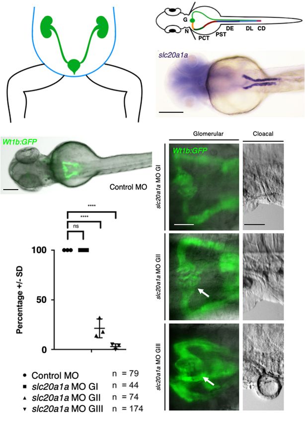
Rieke et al.                                                                                                     SLC20A1: Urinary Tract and Urorectal Development

                                            Frankfurt am Main, Germany, 40 Department of Pediatric and Adolescent Psychiatry and Psychotherapy, University Medical
                                            Centre, Johannes Gutenberg University of Mainz, Mainz, Germany, 41 Institute of Integrative Medicine, Witten/Herdecke
                                            University, Herdecke, Germany, 42 Cologne Center for Genomics, University of Cologne, Cologne, Germany, 43 Department
                                            of Clinical Chemistry and Clinical Pharmacology, University of Bonn, Bonn, Germany, 44 Royal Manchester Children’s
                                            Hospital, Manchester University NHS Foundation Trust, Manchester Academic Health Science Centre, Manchester,
                                            United Kingdom, 45 Institute for Neuroanatomy, University Hospital Bonn, University of Bonn, Bonn, Germany

                                            Previous studies in developing Xenopus and zebrafish reported that the phosphate
                                            transporter slc20a1a is expressed in pronephric kidneys. The recent identification of
                                            SLC20A1 as a monoallelic candidate gene for cloacal exstrophy further suggests its
                                            involvement in the urinary tract and urorectal development. However, little is known
                                            of the functional role of SLC20A1 in urinary tract development. Here, we investigated
                                            this using morpholino oligonucleotide knockdown of the zebrafish ortholog slc20a1a.
                                            This caused kidney cysts and malformations of the cloaca. Moreover, in morphants we
                                            demonstrated dysfunctional voiding and hindgut opening defects mimicking imperforate
                                            anus in human cloacal exstrophy. Furthermore, we performed immunohistochemistry
                                            of an unaffected 6-week-old human embryo and detected SLC20A1 in the urinary
                                            tract and the abdominal midline, structures implicated in the pathogenesis of cloacal
                                            exstrophy. Additionally, we resequenced SLC20A1 in 690 individuals with bladder
                                            exstrophy-epispadias complex (BEEC) including 84 individuals with cloacal exstrophy.
                                            We identified two additional monoallelic de novo variants. One was identified in a case-
                                            parent trio with classic bladder exstrophy, and one additional novel de novo variant
                                            was detected in an affected mother who transmitted this variant to her affected son.
                                            To study the potential cellular impact of SLC20A1 variants, we expressed them in
                                            HEK293 cells. Here, phosphate transport was not compromised, suggesting that it is
                                            not a disease mechanism. However, there was a tendency for lower levels of cleaved
                                            caspase-3, perhaps implicating apoptosis pathways in the disease. Our results suggest
                                            SLC20A1 is involved in urinary tract and urorectal development and implicate SLC20A1
                                            as a disease-gene for BEEC.
                                            Keywords: SLC20A1, urinary tract development, kidney formation, zebrafish development, cloacal malformation,
                                            functional genetics, CAKUT, bladder exstrophy-epispadias complex

INTRODUCTION                                                                          regulating endocytosis and microautophagy within yolk sac
                                                                                      visceral endoderm (Beck et al., 2009; Festing et al., 2009;
The recent identification of the phosphate transporter                                Wallingford and Giachelli, 2014).
SLC20A1 as a candidate gene for cloacal exstrophy (CE)                                   Due to their ease of molecular manipulation and real-time
(OMIM 258040) suggests its involvement also in the lower                              observation, and the high fecundity of the species, we successfully
urinary tract and urorectal development (Reutter et al.,                              used zebrafish larvae (zfl) to functionally characterize dominant
2016). While previous studies in developing Xenopus and                               variants reported in individuals with lower urinary tract
zebrafish (zf) reported the expression pattern of slc20a1a                            obstruction (Kolvenbach et al., 2019). Here, we apply a similar
in pronephric kidneys (Nichane et al., 2006; Raciti et al.,                           zf model to investigate the role of slc20a1a in the zf urinary
2008; Howe et al., 2012; Zhang et al., 2017), the possible                            tract and urorectal development, and we combine this with
role of SLC20A1 in urinary tract formation is unknown.                                human genomic, cell culture, and immunohistochemistry with
SLC20A1 encodes for the sodium-phosphate symporter                                    regard to SLC20A1. Our results suggest SLC20A1 is involved
called the solute carrier family 20 member 1 (PiT-1). The                             in human and zf urinary tract and urorectal development, and
SLC20A1 protein comprises 12 transmembrane domains                                    implicate SLC20A1 as a disease-gene for bladder exstrophy-
(TMDs) (O’Hara et al., 1990; Farrell et al., 2009). Slc20a1                           epispadias complex (BEEC). Our study also provides early
(PiT-1) knock-out mice die by embryonic day 12 (Beck                                  cell culture data to suggest that SLC20A1 variants found in
et al., 2009; Festing et al., 2009). While the exact cause                            patients affect cleaved caspase-3, consistent with a reported
of death is unknown, they have gross defects in yolk sac                              role of SLC20A1 in tumor necrosis factor-induced apoptosis
vascular development putatively correlating with Slc20a1                              (Salaün et al., 2010).

Frontiers in Cell and Developmental Biology | www.frontiersin.org                 2                                             August 2020 | Volume 8 | Article 567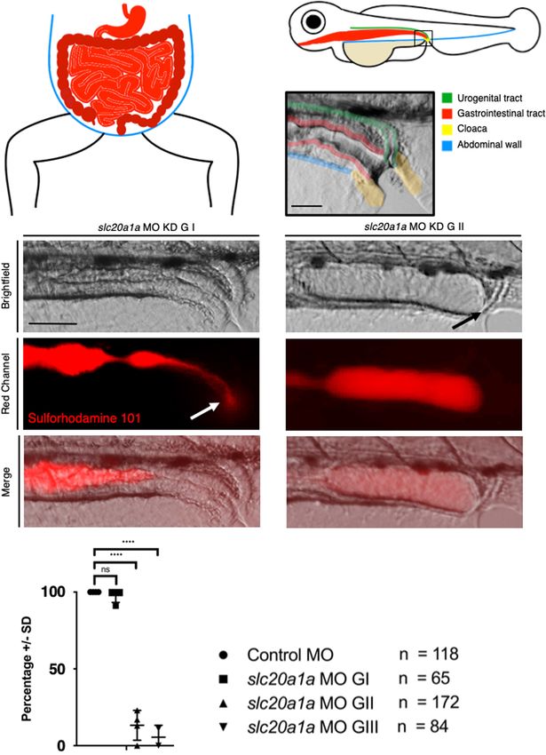
Rieke et al.                                                                                        SLC20A1: Urinary Tract and Urorectal Development

MATERIALS AND METHODS                                                       Sulforhodamine 101 (SR101) Excretion
                                                                            Assay
Zebrafish Husbandry and Embryo                                              Excretion assay was performed on days 4–5 dpf. Zfl were kept in
Preparation                                                                 the dark in 0.02 mM SR101 in Danieau 30% + 0.003% 1-phenyl-
Zf were kept according to national law and to recommendations               2-thiourea solution for 1 h. After incubation zfl were washed with
by Westerfield (Westerfield, 2000) in our fish facility. Zfl                Danieau 30% three times for 10 min before imaging.
of wild-type AB/TL and transgenic strain Tg(wt1b:eGFP)
(Perner et al., 2007) were gained by natural fish spawning                  Whole-Mount Zebrafish in situ
and raised at 28 C in Danieau (30%) medium on a 14 h                        Hybridization (WISH)
light:10 h dark cycle. All zf experiments were performed                    cDNA plasmids for the preparation of antisense and sense probes
at ≤ 5 dpf before independent feeding. To suppress                          for pax2a, evx1, slc20a1a, and slc20a1b were generated by PCR
pigmentation for later, WISH analysis or fluorescent                        from zebrafish poly-T embryonic cDNA (primer sequences are
microscopy 1-phenyl-2-thiourea (final concentration 0.003%)                 provided in Supplementary Data Sheet S15). The resulting
was added to the Danieau solution for respective zfl from                   amplified PCR products were cloned into SK(-) pBluescript .             R

1 dpf onward. Staging of zfl was performed according to                     Constructs were linearized by corresponding restriction enzymes
Kimmel et al. (1995).                                                       and DIG-labeled sense and anti-sense RNA was synthesized using
                                                                            Roche DIG RNA Labeling Kit (Cat. No. 11 175 025 910). WISH
Microinjections of Morpholino                                               was performed following modified instructions of Thisse and
Oligonucleotides and mRNA                                                   Thisse (2008).
Zebrafish embryos were collected 15 min after breeding and were
pressure injected into the yolk at the one-cell stage (up to 30–40          Immunohistochemistry in
min after fertilization) with an ATG-blocking Morpholino            R

                                                                            Whole-Mount Zfl
antisense oligonucleotide (MO) by GeneTools, LLC. Injections
                                                                            Zfl were fixed in 4% paraformaldehyde overnight at 4◦ C and
were carried out with 0.75 ng of slc20a1a MO (1.7 nL/embryo)
                                                                            washed afterward with methanol in increasing concentration
(50 CTGGAGAAAAACACTTCTGGCCTAC 30 ) and 0.75 ng of
                                                                            (25, 50, 75, 100%). Heat-induced antigen retrieval was
standard control MO (50 CCTCTTACCTCAGTTACAATTTATA
                                                                            performed in Tris-HCL (pH = 8.5) at 70◦ C for 15 min.
30 ). For pressure injection, we used the “Milli-Pulse
                                                                            For permeabilization digest with Proteinase K at room
Pressure Injector, Model MPPI-3” (Applied Scientific
                                                                            temperature was adapted to age of the zfl. They were then
Instrumentation, Inc. 29391 W. Enid Rd. Eugene, OR
                                                                            incubated in primary antibodies for 3 days at 4◦ C (1:500;
97402-9533, United States). The MPPI-3 is a self-contained
                                                                            Anti-Acetulated Tubulin: Sigma-Aldrich – T7451, mouse; Anti-
device for producing gas pressure pulses to an injection
                                                                            GFP: Invitrogen – A11122, rabbit) and secondary antibodies
needle (pulled glass capillary–GB120F-10, Science Products
                                                                            for 2 days at 4◦ C (1:1000; Alexa Fluor 546 goat anti-mouse:
GmbH). The unit offers linear control of both pressure and
                                                                            LifeTechnologies – A11030; Alexa Fluor 488 goat anti-rabbit:
pulse duration.
                                                                            LifeTechnologies – A11034).
Morpholino (MO) mRNA Rescue
Rescue experiments were performed by co-injection of MO                     High Resolution in vivo Fluorescent Zfl
together with 35 pg of human SLC20A1 polyA mRNA.                            Imaging
In vitro transcription of SLC20A1 mRNA was performed                        Embryos were pre analyzed under a Nikon AZ100 Macro-Zoom
using mMessage mMachine Kit (Ambion 1340M) and                              microscope and selected embryos were further anesthetized
Poly-A-Tailing-Kit (Ambion AM1350) on IMAGE-clone:                          with 0.016% tricaine, mounted in 2% low-melting agarose and
3918690. slc20a1a MO sequence is not homologous to                          imaged by two-photon scanning fluorescence in vivo microscopy
the hSLC20A1 mRNA (0% homology), excluding binding                          (LaVision Trim-Scope II; ImSpector and ImageJ software).
of the MO to hSLC20A1 mRNA (Supplementary Data
Sheet S16).                                                                 Statistical Analysis
                                                                            Statistical analysis was performed using GraphPad Prism version
Western Blot (WB) Analysis in slc20a1a                                      8.0.0 for Mac, GraphPad Software, San Diego, CA, United States1 .
MO Knockdown (KD) zfl                                                       Differences with a p-value of < 0.05 (∗ ) were considered as being
After grading, zfl were pooled into samples of 20–30                        statistically significant. Error bars show standard deviation (SD)
larvae of equal grades and lysed in RIPA buffer on ice                      in all experiments.
with 4% protease inhibitor using a sonicator. Proteins
were separated by SDS-PAGE, transferred on PVDF                             Human Embryo Immunohistochemistry
membranes and were probed with anti-SLC20A1 (1:1000;                        Human tissues, collected after maternal consent and ethical
Sigma-Aldrich; AV43905) at 4◦ C overnight. Enhanced                         approval (REC 08/H0906/21 + 5), were provided by the MRC
chemiluminescent (ECL) HRP substrate for low-femtogram-level
detection was used.                                                         1
                                                                                www.graphpad.com

Frontiers in Cell and Developmental Biology | www.frontiersin.org       3                                         August 2020 | Volume 8 | Article 567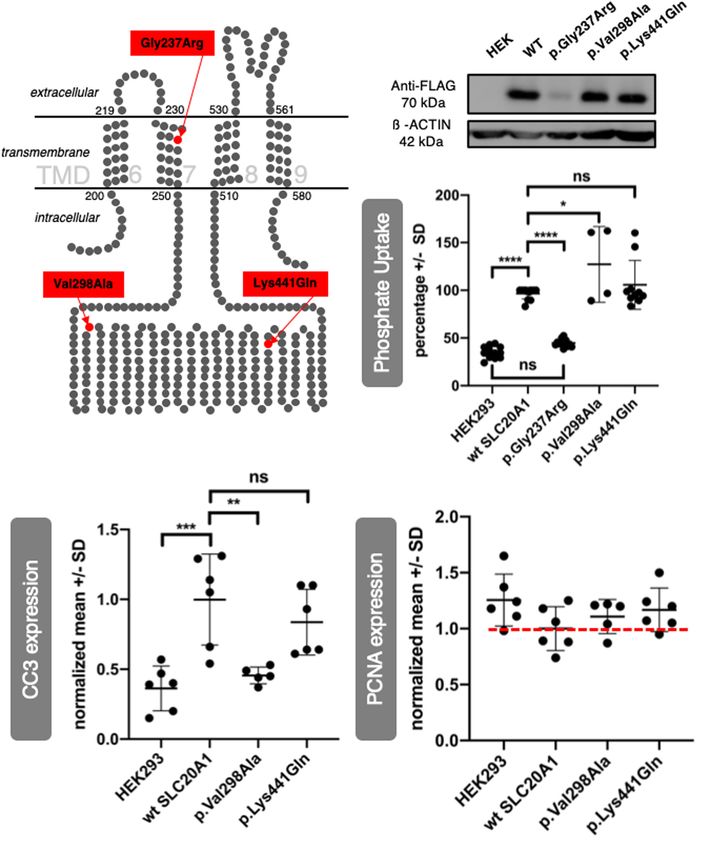
Rieke et al.                                                                                                           SLC20A1: Urinary Tract and Urorectal Development

  FIGURE 1 | slc20a1a MO KD. KD was performed by injecting 0.75 ng of ATG-blocking MO into one-cell-staged wt zf eggs. Lateral view, dorsal to top, cranial on the
  left. (A) Phenotypical grading of MO injected zfl at 2 dpf in four grades increasing in severity and lethality. Scale bar: 500 µm. See main Results text for grading
  details. In brief, GI were normal in direct inspection; GII had mild defects; GIII had moderate defects, and GIV had severe defects. G V resembles dead zfl.
  Supplementary Data Sheet S3 shows close-ups for better demonstration of hydrocephalus and eye development in all four grades presented. (B) Examples for
  cloacal abnormalities in MO KD zfl at 2 dpf. Scale bar: 100 µm. Inspection of cloacal region after previous grading revealed malformations in cloacal region in G II
  and G III sorted zfl increasing in amount and severity with grading. G I sorted zfl had normal cloacal morphology, that is, a thin and curved organ with a distal opening
  (arrow). GII and GIII zfl have abnormally shaped cloacae, with dilated and/or apparently blind-ending lumens. Further cloacal close-ups of G II and III sorted zfl are
  shown in Supplementary Data Sheet S4. (C) WB shows efficacy of MO KD in zebrafish protein lysates from 2 dpf. 70 kDa: slc20a1a, 42 kDa: ß-Actin loading
  control; WT = uninjected control, Crtl = control MO injected, G II/III/IV = slc20a1a MO KD zfl sorted by grading II–IV. Slc20a1a can be detected in uninjected wt
  control and injected control MO group. Only a weak slc20a1a signal was seen in MO KD groups G II, III, and IV, which showed phenotypical features as described in
  (A). WB shows correlation between phenotype and protein expression. Raw data of WB is shown in Supplementary Data Sheet S6. (D) Co-injection of slc20a1a
  MO with 35 µg in vitro transcribed human SLC20A1 polyA mRNA shows partial rescue of various phenotypes underlining the Morpholino’s specificity. n = 5 (here
  n = 1 represents the average score in each experimental batch). Error bars show SD. X-axis shows groups at 3 dpf that were compared: zfl showing no or only a
  very mild phenotype (G I + G II), larvae with a moderate phenotype (G III) and a last group of larvae with a severe and lethal phenotype (G IV) together with those who
  were already dead at time of comparison (G V). Y -axis shows the percentage of zfl in the corresponding groups described before. A significant difference (∗ p = 0.01)
  was seen within the first group of MO and MO-mRNA rescue group, showing a partial rescue of slc20a1a MO KD phenotype reflected in bigger group of
  phenotypically not or only mildly affected zfl. Mere overexpression of SLC20A1 wt mRNA in zfl resulted in phenotypical aberrations, which did not fit the grading
  characteristics and the phenotypes observed in slc20a1a MO KD. Further, pure SLC20A1 overexpression in zfl resulted in higher lethality compared to non-injected
  control groups (data not shown). These findings suggest that the MO rescue effect of SLC20A1 wt mRNA is weakened and disguised by the mRNA’s general
  negative overexpression effect. (E) Kaplan-Meyer curve shows significantly reduced survival (p < 0.0001) in slc20a1a MO-injected group compared to uninjected
  and control MO. Survival rates by day five post-fertilization: WT = 100%, control MO injected group = 93%, MO injected group = 35% (including all grades).
  Exclusion of embryos dying/not developing by 8 h post-fertilization (hpf) due to failed fertilization or consequences of tissue damage caused by injections with
  mechanical manipulation. Within these initial 8 hpf intervals, no difference was seen between control MO and slc20a1a MO injected groups.

Frontiers in Cell and Developmental Biology | www.frontiersin.org                    4                                                 August 2020 | Volume 8 | Article 567
Rieke et al.                                                                                          SLC20A1: Urinary Tract and Urorectal Development

and Wellcome Trust Human Developmental Biology Resource2 .              HEPES; 1 mM EDTA in distilled H2 O, pH 7.4) with 1 mM
Paraffin sections were processed for immunostaining after               dithiothreitol; 100 µg of whole cell lysates were separated on 10%
antigen retrieval, essentially as described (Kolvenbach et al.,         sodium dodecyl sulfate (SDS) gels, transferred on nitrocellulose
2019). Sections were probed with antibody to SLC20A1 (1:200;            membranes, and probed with antibodies against FLAG-tag
Proteintech 12423-1-AP).                                                (1:1,000; Sigma Aldrich; F3165) and ß-ACTIN (1:40,000; Sigma
                                                                        Aldrich; A3854). Non-transfected HEK293 cells served as
Resequencing of SLC20A1 in Individuals                                  negative control. For analysis of cleaved caspase-3 (CC3) levels
With BEEC                                                               20 µg of cell lysates were loaded and probed with anti-CC3
The resequencing study was conducted in adherence to the                (1:2,000; Cell Signaling; 9661) and ß-ACTIN (1:10,000; Sigma
Declaration of Helsinki. Informed consents were obtained from           Aldrich; A5441). Analysis of proliferation cell nuclear antigen
affected individuals or by proxies in the case of minors. The           (PCNA) levels was performed by blotting 2 µg of cell lysates
study was approved by the ethics committee of the medical               of all respective groups and probing those with anti-PCNA
faculty of the University of Bonn (No. 031/19) as well as the           (1:5,000; Abcam; ab2426) and ß-ACTIN. ImageJ was used for
respective ethic committee of the collaborating centers in              WB densitometry.
Manchester (United Kingdom), Nijmegen (AGORA data- and
biobank; Netherlands; Rooij et al., 2016), Torino (Italy), and          Phosphate Uptake Assay in Human
Stockholm (for Sweden on behalf of Lund, Göteborg, and                  Embryonic Kidney 293 (HEK293) Cells
Uppsala). For resequencing, 690 (440 male and 250 female)               Transient transfected HEK293 cells seeded on 24 well plates
BEEC individuals were included [epispadias n = 42; classic              were incubated in uptake buffer (96 mM NaCl; 2 mM KCl;
bladder exstrophy (CBE) n = 564; CE n = 84]. All three human            1.8 mM CaCl2 ; 1 mM MgCl2 ; 50 mM HEPES in distilled
SLC20A1 protein coding transcripts (ENST00000272542.7,                  H2 O) supplemented with 200 µM potassium phosphate buffer
ENST00000423633.5, ENST00000433924.5) listed in “ensembl                and 1 µCi 32 PO4 3− per ml uptake buffer for 3and 15 min
database” (September 30, 2017)3 were sequenced. PCR-                    before washing, respectively. Cell-associated radioactivity was
amplified DNA products (primer sequences are provided in                measured with a ß-counter (Tri-Carb Liquid Scintillation
                                                                                                                           R

Supplementary Data Sheet S15) were subjected to sequencing              Analyzer, Perkin Elmer). Values are given as counts per minute
using a 3130XL Genetic Analyzer (Applied Biosystems, Foster             and calculated as percentage. Non-transfected HEK293 cells
City, United States).                                                   served as background control.

Generation of Variants for in vitro                                     Computational 3D Structural Modeling
Analysis                                                                For 3D structure, modeling of human SLC20A1 variants
QuikChange Lightning Site-Directed Mutagenesis Kit (Agilent             (Uniprot ID: Q8WUM9) I-Tasser4 was used. This employs
#210518) was used to generate variants from IMAGE clone                 an integrated combinatorial approach comprising comparative
3918690 (primer sequences are provided in Supplementary Data            modeling, threading, and ab initio modeling (Roy et al., 2010)
Sheet S15).                                                             using the procedure adopted by Dakal et al. (2017). The generated
                                                                        structures were visualized in Chimera 1.13rc version.
Cloning for Cell Culture Experiments
N-terminally Flag tagged wild type (wt) and mutant SLC20A1
cDNA of human origin were cloned into pcDNA3 plasmid                    RESULTS
backbone (Clontech) (primer sequences are provided in
Supplementary Data Sheet S15).                                          Morpholino KD of slc20a1a in Embryonic
                                                                        Zebrafish
Cell Culture and Transient Transfection                                 To further functionally characterize SLC20A1 we performed
HEK293 cells were cultured in DMEM/F12 (1:1) (GIBCO) + 10%              ATG-blocking MO KD experiments in zfl. The zf has two
fetal calf serum (FCS; GIBCO) + 1% penicillin (5,000                    ortholog genes, slc20a1a and slc20a1b. Focusing on the segment
U/ml)/streptomycin (5,000 µg/ml). Cells were seeded 1:1 into 24         of chromosome 2 harboring the SLC20A1 locus in humans, none
and six well plates, followed by transient transfection with 250–       of the distinct chromosome loci of the two zf orthologs show
1,000 ng plasmid DNA per cm2 well surface using PANfect A               conserved syntenies to the human segment. NCBI Unigene’s
transfection reagent (PAN Biotech, Germany). The experiments            EST profile viewer reveals distinct expression patterns for both
were performed 48 h after transfection.                                 zf orthologs (Nichane et al., 2006): only slc20a1a appears
                                                                        to be strongly and specifically expressed in the embryonic
Western Blot Analysis in HEK293 Cells                                   kidney. While in the developing zfl no specific expression has
Transient transfected HEK293 cells were harvested and lysed             been described for slc20a1b so far (January 2020)5 , clearly
in 75 µl homogenization buffer (250 mM sucrose; 20 mM                   slc20a1a has been established as a pronephric tubular marker

2                                                                       4
    http://www.hdbr.org/                                                    http://zhanglab.ccmb.med.umich.edu/I-TASSER/
3                                                                       5
    www.ensembl.org/                                                        http://zfin.org/

Frontiers in Cell and Developmental Biology | www.frontiersin.org   5                                               August 2020 | Volume 8 | Article 567
Rieke et al. SLC20A1: Urinary Tract and Urorectal Development
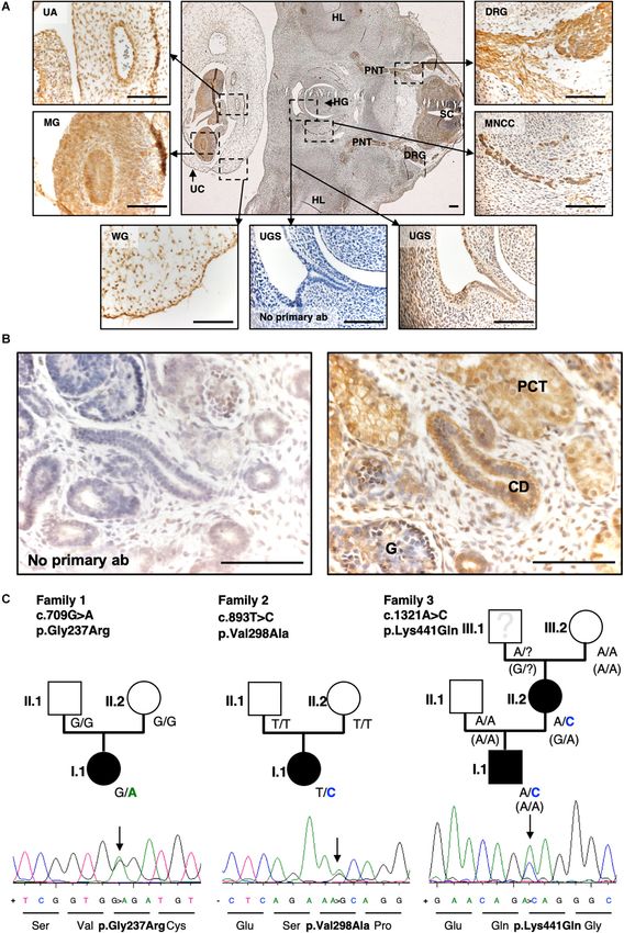
FIGURE 2 | Pronephric cysts in slc20a1a MO KD resulting from cloacal obstruction due to malformations. (A) Scheme of human abdomen, green: urogenital tract
with kidneys, ureter, bladder, urethra; blue: abdominal wall. (B) Pronephros in zfl (green: right side) as equivalent to human urinary tract. Scheme of zfl, dorsal view at
(Continued)
Frontiers in Cell and Developmental Biology | www.frontiersin.org 6 August 2020 | Volume 8 | Article 567Rieke et al. SLC20A1: Urinary Tract and Urorectal Development
FIGURE 2 | Continued
2 dpf, patterning of zebrafish pronephros is similar to human nephron segmentation. Specific segments are color coded (left side) for better identification: G,
Glomerulus; N, Neck; PCT, Proximal Convoluted Tubule; PST, Proximal Straight Tubule; DE, Distal Early; DL, Distal Late; CD, Collecting Duct. (C) Whole mount
in situ hybridization (WISH) against slc20a1a in zfl at 2 dpf, labeling proximal part of pronephros. Scale bar: 500 µm. (D) Control MO zfl in Tg(wt1b:GFP) (Perner
et al., 2007) marking proximal part of pronephros, Scale bar: 100 µm. (E) Glomerular close-ups in Tg(wt1b:GFP) zfl in dorsal view at 2 dpf (Scale bar: 50 µm). On
the left, pronephric cysts (arrows) and dilatation of proximal part of pronephros increasing in severity with grading are shown. G I (upper pictures) showing no cystic
phenotype, G II (middle pictures) showing a mild cyst formation, and G III (bottom pictures) showing severe cysts and a wide dilatation of the pronephros.
Corresponding cloacal close-ups, shown on the right in lateral view (Scale bar: 50 µm), underline correlation between malformations in urinary outflow tract and
cysts as well as pronephric dilatations in corresponding groups. (F) Graph shows percentage of zfl (Y -axis) in the following groups: Control MO, slc20a1a MO G I,
slc20a1a MO G II, slc20a1a MO G III (X-axis). Each dot stands for one individual experiment (here N = 1 represents the average score in each experimental batch).
Whereas control MO and slc20a1a MO G I larvae do not show any cystic phenotype, only an average of 21% of MO G II, and solely 3% of G III MO zfl show normal
configuration of the proximal part of pronephros. The graph shows significant differences between control MO and phenotypically normal group MO G I compared to
MO G II and G III, N = 3, ****p < 0.0001. Error bars show SD.
(Howe et al., 2012; Zhang et al., 2017). Here, WISH analysis abnormally shaped cloacae, with dilated and/or apparently blind-
confirmed slc20a1a as a pronephric marker, with expression ending lumens. Further cloacal close-ups of G II and III sorted
in the proximal (i.e., near to the glomerulus) section, while zfl are shown in Supplementary Data Sheet S4. Importantly,
WISH analysis of slc20a1b did not show any signal in tissues the finding of cloacal anomalies in larvae that had only a mild
relevant to urinary tract development (Supplementary Data whole-body phenotype suggests that the former is a “strong”
Sheets S1A–F). Hence, we studied MO KD against slc20a1a to primary effect and not simply a side effect of a more major whole-
characterize its possible developmental impact on urinary tract body malformation. For further characterization, we performed
and urorectal development. The injection procedure was uniform WISH in slc20a1a MO, control MO, and uninjected wt zfl with
in all experiments. slc20a1a MO injected larvae showed a range two different cloacal marker probes at two timepoints each.
of phenotypes on direct inspection. To facilitate analyses, we pax2a marks the distal part of the pronephric ducts up to their
graded all MO injected zfl at 2 dpf (Long-pec, according to fusion at the cloaca. For quantification of pax2a expression, we
Kimmel et al., 1995) on the basis of several phenotypical features. determined the maximal distance orthogonal to the pronephric
Representative images of the grades (G) are depicted in Figure 1. midline within the stained cloacal region (Supplementary Data
In brief: G I embryos (5% at 2 dpf) appeared normal and identical Sheets S5A–C). evx1 is a WISH marker for the cloaca. The area
to uninjected and control MO injected zfl; G II zfl (6% at 2 dpf) of staining was measured using a common threshold in all zfl
showed a mild phenotype with minimal reduction in body and (Supplementary Data Sheets S5D–F). For pax2a, a wider cloaca
head sizes, no or moderate hydrocephalus, mild pigmentations was found in slc20a1a MO KD zfl at both timepoints. Cloacal area
defects, and benign reduction of the yolk sac extension, but of expression of evx1 was significantly larger in slc20a1a MO KD
without changes in body curvature or eye abnormalities; G III zfl at both timepoints as well. These results suggest a defect in
zfl (46% at 2 dpf) showed a moderate phenotype characterized tissue development in the cloacal region of slc20a1a MO KD zfl.
by decreases in body and head sizes, overt hydrocephalus
(arrow heads, additional images in Supplementary Data Sheet Efficiency and Specificity of MO KD
S3), yolk endocytosis defects, lack of yolk sac extension along Shown by WB Analysis and mRNA
abdominal wall (arrows), eye abnormalities especially in size
(Supplementary Data Sheet S3), straight body with or without Rescue
kinking of the tail tip (no kinking shown here), pigmentation Efficacy of slc20a1a MO KD, at the protein level, was
defects, and pericardial effusion; and G IV zfl (27% at 2 dpf) had demonstrated by WB analysis at 2 dpf (Figure 1C). Slc20a1a
marked malformations of various organ systems and additional protein was detected in both control groups at about 70 kDa
defects in body curvature. Remarkably, no G IV larvae survived molecular weight. We could only detect weak slc20a1a protein
until 5 dpf and were therefore not further analyzed but scored signal in the MO KD grades (G II–IV).
as “dead” (G V) in the statistics presented. Supplementary To test the specificity of our slc20a1a MO, we co-injected
Data Sheet S3 shows close-ups for better demonstration of in vitro transcribed polyA mRNA of human wt SLC20A1. We
hydrocephalus and eye development in all four grades presented. detected a rescue effect of the human SLC20A1 mRNA in MO
zfl, as evidenced by significant increase of the proportion of
overtly normal or mildly affected zfl (GI + GII) (Figure 1D). This
Cloacal Anomalies in slc20a1a MO KD effect was even more notable given the fact that overexpression
Though cloacal anomalies were not part of the preceding of SLC20A1 wt mRNA in non-morphant zfl resulted in
grading, we frequently found malformations in the cloacal region higher lethality and phenotypical aberrations, which did not
and therefore the urinary outflow tract in mild (GII) and fit the grading characteristics and the phenotypes observed in
moderate (GIII) slc20a1a MO KD zfl (Figure 1B). These cloacal slc20a1a MO KD. Collectively, the results support the slc20a1a
malformations could themselves be graded as moderate or severe MO’s specificity.
(additional images in Supplementary Data Sheet S4). G I sorted Survival of uninjected wt, control MO, and slc20a1a MO KD
zfl had normal cloacal morphology, that is, a thin and curved zfl were monitored until 5 dpf, showing a significant decrease
organ with a distal opening (arrow). G II and G III zfl has (>50%) in survival rate in the MO KD larvae (Figure 1E).
Frontiers in Cell and Developmental Biology | www.frontiersin.org 7 August 2020 | Volume 8 | Article 567Rieke et al. SLC20A1: Urinary Tract and Urorectal Development
FIGURE 3 | Sulforhodamine 101 excretion assay shows imperforate hindgut in slc20a1a MO KD zfl. (A) Scheme of human abdomen, red = GIT with stomach,
duodenum, jejunum, ileum in light red and colon, rectum, anus in dark red; blue = abdominal wall. (B) Scheme of zfl, lateral view at 5 dpf; green = urogenital tract
(Continued)
Frontiers in Cell and Developmental Biology | www.frontiersin.org 8 August 2020 | Volume 8 | Article 567Rieke et al. SLC20A1: Urinary Tract and Urorectal Development
FIGURE 3 | Continued
(pronephros), red = GIT, blue = abdominal wall, beige = yolk sac, yellow = cloaca. (C) Cloaca in zfl at 5 dpf: fusion and opening of pronephros and GIT at cloaca
between 4 and 5 dpf. Pseudocolored for identification as above in B: green = urogenital tract (pronephros), red = GIT, yellow = cloaca. Scale bar: 50 µm.
(D) Opening of cloaca and excretion of SR101, a red fluorescent dye labeling the content of zfl intestine. Upper panel shows brightfield, middle panel red channel,
lower panel shows a merged view of both channels. On the left we show slc20a1a MO KD G I zfl at 5 dpf compared to slc20a1a MO KD G II zfl at 5 dpf on the right
side of the panel. In control MO and slc20a1a MO GI, zfl dye uptake is not disturbed; we could detect clear and bright red dye fluorescence in the gut of all animals.
Dye excretion and opening of the cloaca was not disturbed. White arrow marks dye excretion from the cloaca. In contrary, we observed cloacal opening and
excretion defects in slc20a1a MO KD G II zfl at 5 dpf mimicking an imperforate anus as shown on the right side of the panel. Black arrow marks opening defect and
therefore resulting dilatation of intestine due to bag log. No changes in peristalsis of the GIT was observed; hence, expansion of distal part of intestine as shown here
is solely caused by lack of cloacal opening. Scale bar: 50 µm. (E) Significant differences in opening of cloaca at 5 dpf in zfl between phenotypically affected and
control MO. Cloacal opening was monitored for several minutes up to 1 h. Only 13.25% of G II and 5.5% of G III zfl showed cloacal opening and therefore excretion
of SR101 from the GIT, whereas 82.5% of G II and 74% of G III zfl did not show any excretion. In the remaining 4.25% of G II and 20.5% of G III, zfl cloacal opening
could not be assessed resulting from failure of SR101 uptake in the first place or misshape and tissue malformations not allowing to assess the cloacal region in the
respective zfl. N = 4, ****p < 0.0001. Error bars show SD.
Pronephric Cysts and Dilatation of Sulforhodamine 101 Excretion Assay in
Pronephric Ducts in slc20a1a MO KD slc20a1a MO KD zfl Shows Gut Outlet
Implicate slc20a1a as Important Player in Obstruction at the Cloaca
Early Kidney Development Figure 3 depicts the gastrointestinal tract (GIT) and cloacal
In zfl, the pronephros represents the anatomical structure that opening of the hindgut. Schematic comparison of GIT of humans
corresponds to the human urinary tract. The pronephric pattern and zf is shown in Figures 3A,B (red). The close-up of the cloacal
is analogous to the mammalian nephron. The two pronephric region in zfl shows opening of pronephros and GIT at cloaca in a
ducts fuse and excrete the urine through the cloacal opening healthy zfl at 5 dpf (Figure 3C).
(Figures 2A,B). Figure 2C shows clear WISH signal for slc20a1a Fluorescent dye (Sulforhodamine 101, SR101) uptake and
in the proximal zfl pronephros at 2 dpf. slc20a1a WISH in excretion assay in zfl at 5 dpf showed normal excretion and
earlier developmental zfl stages show expression in intermediate opening of the cloaca in all control MO zfl and in an average
mesoderm, which later forms the pronephros, red blood cells, of 98% in slc20a1a MO G I zfl (Figures 3D,E). On the
and trunk endothelium (Supplementary Data Sheet S1). We contrary, excretion of SR101 was severely disturbed and absent
used Tg(wt1b:eGFP) reporter fish (Perner et al., 2007) to assess in G II (82%) and G III (74%) slc20a1a MO zfl. This assay
the impact of slc20a1a on the development of the glomeruli confirmed the high abundance of cloacal opening defects in
and the proximal region of the pronephros (Figure 2D). slc20a1a morphants resembling an imperforate anus in humans.
Whereas control MO and slc20a1a MO G I zfl did not show For clarification of the performed dye assay, representative
any phenotypical differences concerning the morphology of fluorescent videos are provided in the supporting information
the glomeruli and pronephros at 2 dpf, the majority of G (Supplementary Data Sheets S8, S9).
II (79%) and G III (97%) sorted larvae showed glomerular
cysts and a dilatation of the proximal part of the pronephric Embryonic Protein Expression of
ducts (Figures 2E,F). SLC20A1 in Human Embryonic
We sought cilia within the pronephros using
immunofluorescence staining against alpha-acetylated tubulin
Urogenital Tissue
Figure 4A shows a transverse section of a healthy (non-BEEC) 6-
and GFP in Tg(wt1b:GFP) at 2 dpf. Our two-photon microscopy
week gestation human embryo. SLC20A1 was immunodetected
(Supplementary Data Sheet S7) revealed the presence of
in several locations including the urogenital sinus and the
cilia. There were no gross structural anomalies, such as gross
urinary bladder precursor implicated in BEEC. Additionally, we
shortening or elongation, between the G II and G III MO-KD zfl.
immunodetected SLC20A1 in a 10-week-gestation metanephric
We did not, however, formally quantify cilia length nor did not
kidney, with prominent signals in the proximal tubules and
assess motility study in G II and G III MO-KD zfl. The dilatation
collecting ducts (Figure 4B).
of their pronephric ducts was confirmed.
Dilatation of the ureter and pelvis of the kidney in human can
be caused by a backlog of urine due to a functional or anatomical Resequencing of SLC20A1 Identifies Two
blockage of the urinary tract. This blockage occurs usually distal Additional Variants
to the dilated parts of the renal tract. slc20a1a MO KD zfl Resequencing of all three genomic SLC20A1 transcripts identified
show malformations in the urinary outflow tract (Figures 1B, an additional de novo variant in a case-parent trio (c.893T > C,
2E and Supplementary Data Sheet S4). Here, malformations of p.Val298Ala, ENST00000272542.7, allele frequency 0.000003979,
urinary outflow tract in morphants correlate with severe cystic Figure 4C, family 2, person I.1) as well as a novel de novo
dilatation of the pronephric kidney. This strongly supports the variant in an affected mother (Figure 4C, family 3, person II.2)
hypothesis that the cloacal malformations seen in slc20a1a MO who transmitted this variant to her affected son (c.1321A > C,
KD zfl cause a backlog of urine, which leads to cystic and dilated p.Lys441Gln, ENST00000272542.7, Figure 4C, family 3, person
pronephric kidneys. I.1) (Supplementary Data Sheet S10 Table for additional
Frontiers in Cell and Developmental Biology | www.frontiersin.org 9 August 2020 | Volume 8 | Article 567Rieke et al. SLC20A1: Urinary Tract and Urorectal Development
FIGURE 4 | Exome sequencing and targeted Resequencing in families with BEEC phenotype identified disease variants in SLC20A1. (A) IHC of a transverse section
of a 6-week-old human embryo. SLC20A1 was immunodetected (brown) in the urogenital sinus (UGS), which develops into the urinary bladder. Furthermore,
(Continued)
Frontiers in Cell and Developmental Biology | www.frontiersin.org 10 August 2020 | Volume 8 | Article 567Rieke et al. SLC20A1: Urinary Tract and Urorectal Development
FIGURE 4 | Continued
SLC20A1 was also detected in the spinal cord (SC), dorsal root ganglia (DRG), peripheric nerve trunk (PNT), hind limb (HL), hindgut (HG), migrating neural crest cells
(MNCC), umbilical cord (UC), Wharton’s jelly (WG), midgut (MG), and umbilical artery (UA). Scale bars = 100 µm. (B) Histology section of the human
10-week-gestation metanephric metanephros. Note prominent SLC20A1 immunostaining (brown) in the proximal tubule (PT) and the collecting duct (CD). The
glomerulus (G) shows a fainter signal. Scale bars: 100 µm. (C) Exome sequencing of eight CE case-parent-trios revealed de novo variant c.709G > A (p.Gly237Arg)
in SLC20A1 in family 1 (Reutter et al., 2016). Resequencing of 690 individuals with BEEC led to identification of two more variants in individuals with CBE:
c.893T > C (p.Val298Ala) in family 2 as de novo change c.1321A > C (p.Lys441Gln) with maternal inheritance (maternal phenotype: fusion defect of pelvic bone,
mild phenotype) in family 3. Pedigrees of all three families are shown with genotypes of all individuals indicated. In family 3, the maternal grandfather (Figure 1B,
family 3, person III.1) was not available for testing. For haplotype analysis of all available family members (Figure 1B, family 3, III.2, II.1, II.2, I.1), we used the
synonymous marker rs4849091 at chromosomal position chr2:113404708 A > G (p.Leu101=) of the canonical transcript ENST00000272542.7. Genotypes of
rs4849091 are shown in brackets. All variants are heterozygote changes and result in missense variants as shown in Sanger sequences including amino acid
sequences below each pedigree.
information). In this family, the maternal grandfather of the FLAG-tagged. Transfection efficiency was confirmed by WB
index person (Figure 4C, family 3, person III.1) was not available (Figure 5B). Variant c.709G > A (p.Gly237Arg) was not
for testing. Haplotype analysis of all available family members expressed efficiently in HEK293 cells as protein. A dosage effect
(Figure 1B, family 3, III.2, II.1, II.2, I.1) showed that the disease was excluded (Supplementary Data Sheet S12). Expression of
variant (c.1321A > C) must have occurred de novo in the c.893T > C (p.Val298Ala) and c.1321A > C (p.Lys441Gln)
grandmother’s derived germ cell (Figure 4C, family 3, person variants resulted in protein levels similar to that of expressed
III.2). For haplotype analysis, we used the synonymous marker wt SLC20A1 protein. Phosphate uptake in the transfected
rs4849091 at chromosomal position chr2:113404708 A > G cells was measured in a radioactive labeled phosphate assay
(p.Leu101) of the canonical transcript ENST00000272542.7. (Figure 5C). Untransfected HEK293 cells and c.709G > A
According to gnomAD this marker has a MAF of 0.4871 across (p.Gly237Arg) transfected cells displayed a basal phosphate
all ethnicities and resides in exon 8 of SLC20A1 in proximity of uptake. Cells transfected with either human SLC20A1 wt,
12 kb to variant c.1321A > C. c.893T > C (p.Val298Ala) or c.1321A > C (p.Lys441Gln) had
a threefold increased capability of phosphate uptake compared
Prediction of Variant Localizations in to untransfected control cells. We therefore conclude that
SLC20A1 SLC20A1-linked phosphate uptake is not disturbed by the two
The SLC20A1 (PiT-1) protein is a sodium-dependent inorganic de novo variants.
phosphate (Pi) symporter that contains 12 TMDs (O’Hara et al., Given that SLC20A1 protein is known to play a role in
1990; Farrell et al., 2009). Three-dimensional crystal structures of apoptosis pathways (Salaün et al., 2010; Husseini et al., 2013)
most SLC family transmembrane proteins are unknown (Dakal we used WB to assess the apoptosis marker CC3 in the
et al., 2017). Figure 5A shows TMDs 6 to 9 of a putative 2D above described transfected HEK293 cells. Overexpression of wt
structure of the SLC20A1 protein, modeled by Beck et al. (2009). SLC20A1 increased the level of CC3. In order to quantify CC3
Here, variant c.709G > A (p.Gly237Arg) of family 1 is located detection in WB, we performed densitometry (Figure 5D and
in TMD 7, and both variants, c.893T > C (p.Val298Ala), of Supplementary Data Sheet S13). In contrast to wt SLC20A1,
family 2 and variant c.1321A > C (p.Lys441Gln) of family 3 variant c.893T > C (p.Val298Ala) failed to increase CC3 levels
are located in a large intracellular loop between TMDs 7 and 8. above those in control cells. Variant c.1321A > C (p.Lys441Gln)
A further attempt of ours to predict localization of the variants increased CC3 levels but to a lesser extent compared to wt
in a 3D model of SLC20A1 protein is shown in Supplementary SLC20A1 overexpression. Variant c.709G > A (p.Gly237Arg)
Data Sheet S11. Our 3D model confirmed the localization of was not studied further due to its expression deficiency in
the two variants c.893T > C (p.Val298Ala) and c.1321A > C HEK cells previously mentioned (Figure 5B and Supplementary
(p.Lys441Gln) in a large intracellular loop between TMD 7 and Data Sheet S12). We conclude that overexpression of BEEC
8. The focus is on c.709G > A (p.Gly237Arg), though we predict variants shows differences compared to wt overexpression
a possible shift from TMD 7 (as described by Beck et al., 2009) when analyzing expression of CC3 as an apoptosis marker.
to TMD 6 in our 3D model. In transmembrane proteins, glycine Additionally, we performed WB analysis of the proliferation
resides in helices, predominantly at the helix-helix interface, marker PCNA (Figure 5E and Supplementary Data Sheet S13)
which makes it an important structural player (Li and Deber, and found no significant differences in PCNA levels between
1992; Javadpour et al., 1999). In our proposed 3D model, the experimental groups; yet, our data shows a trend: while wt
p.Gly237Arg is present at the interface of TMD 6 and TMD 1. SLC20A1 overexpression was associated with a reduced PCNA
Accordingly, p.Gly237Arg might cause instability in TMD 6. level, neither c.893T > C (p.Val298Ala) nor c.1321A > C
(p.Lys441Gln) had a similar strong effect.
Functional Characterization of SLC20A1
Variants in vitro DISCUSSION
In vitro characterization of all three SLC20A1 variants was
performed using HEK293 cells transfected with either human The results of our study suggest that SLC20A1 is not only
wt SLC20A1 or one of the three respective variants – all involved in embryonic kidney formation but also in urinary
Frontiers in Cell and Developmental Biology | www.frontiersin.org 11 August 2020 | Volume 8 | Article 567Rieke et al. SLC20A1: Urinary Tract and Urorectal Development
FIGURE 5 | SLC20A1 as transmembrane phosphate transporter and in vitro characterization of its variants found in BEEC individuals. (A) In silico 2D model of
SLC20A1, a multi-pass integral membrane protein, indicating localization of variants found in affected individuals, c.709G > A (p.Gly237Arg) in TMD 7, c.893T > C
(p.Val298Ala) and c.1321A > C (p.Lys441Gln) are located in an intracellular loop. Only four (6–9) of a total of twelve TMDs are shown in this simplified model. The
model was generated based on the data of Beck et al. (2009). (B,C) SLC20A1 mediated 32 PO4 transport in transiently transfected HEK293 cells. (B) WB analysis of
100 µg whole cell homogenates obtained from HEK293 cells transfected with 500 ng plasmid DNA per cm2 well surface. Plasmid DNA was FLAG tagged and
transfection efficiency was detected using anti-FLAG antibody (70 kDa) and anti-ß-ACTIN antibody (42 kDa), which served as loading control. As expected, no FLAG
signal could be detected in negative control (untransfected HEK293 cells, marked as HEK). Transfection worked for wt SLC20A1 overexpression as well as the
variants c.893T > C (p.Val298Ala) and c.1321A > C (p.Lys441Gln). No FLAG signal could be detected for c.709G > A (p.Gly237Arg) transfected cells. Even when
transfected with higher plasmid concentrations, p.Gly237Arg was not detectable in HEK293 cells (Supplementary Data Sheet S12). (C) Endpoint assay of
transient transfected HEK293 cells. Cells were incubated with 1 µCi 32 PO4 3 - and 200 µM K3 PO4 for 15 min. For better comparison, a highest number of counts per
minute in wt SLC20A1 overexpression group was set as 100% in each experiment (N = 6 with two datasets each), values of HEK293 and variants were calculated
correspondingly, and resulting values in percentage are shown on the y-axis (Error bars show SD). A two-way ANOVA of the grouped analysis was significant
(Continued)
Frontiers in Cell and Developmental Biology | www.frontiersin.org 12 August 2020 | Volume 8 | Article 567Rieke et al. SLC20A1: Urinary Tract and Urorectal Development
FIGURE 5 | Continued
(p < 0.0001). p.Gly237Arg did not show any differences of phosphate uptake to negative HEK293 control. This is in line with the expression deficiency of
p.Gly237Arg described before. Wt SLC20A1 overexpression showed a significant increase of phosphate uptake compared to untransfected HEK293 (Tukey’s
multiple comparison: ****p < 0.0001). Amin acid change p.Lys441Gln and p.Val298Ala overexpression resulted in an even higher phosphate uptake than wt
SLC20A1 overexpression with a significant difference between p.Val298Ala and wt SLC20A1 (Tukey’s multiple comparison: *p < 0.05). Therefore, variant
overexpression does not impair phosphate uptake capability in vitro. (D,E) Densitometric analysis of WBs (N = 6) from whole cell homogenates was obtained from
transfected HEK293 cells. WBs are provided in Supplementary Data Sheet S13. Y -axis shows normalized values against wt SLC20A1 overexpression. Error bars
show SD. (D) Expression of CC3 as apoptosis marker was measured in six WBs of corresponding independent transfection experiments. The one-way ANOVA was
significant (p = 0.0002), Tukey’s multiple comparison test was significant for HEK293 vs. WT (***p = 0.0005), and WT vs. p.Val298Ala (**p = 0.0038). Wt SLC20A1
overexpression in HEK293 cells increased apoptosis when compared to untransfected negative control (HEK). There is no induction of apoptosis inc.893T > C
(p.Val298Ala) transfected cells, comparable to untransfected negative control (HEK). c.1321A > C (p.Lys441Gln) does not result in significant reduction of CC3
expression. However, a trend of reduced CC3 expression in comparison to wt SLC20A1 overexpression can be seen. (E) Same analysis was used to study
expression of PCNA as a proliferation marker. A one-way ANOVA did not show significant results (p = 0.1903). Nevertheless, wt SLC20A1 overexpression seems to
reduce PCNA expression when compared to negative control (untransfected HEK). Variants analyzed [c.893T > C (p.Val298Ala) and c.1321A > C (p.Lys441Gln)]
tend to reduce PCNA less than WT overexpression (red dotted line for better comparison).
tract and urorectal development. Furthermore, our findings apoptosis due to KD of slc20a1a. This is in line with
suggest that monoallelic de novo variants in SLC20A1 are our overexpression studies of identified BEEC variants in
involved in BEEC formation. These conclusions are supported HEK293 cells. Overexpression of both newly identified de
by the immunodetection of SLC20A1 in the BEEC relevant novo variants in HEK293 cells resulted in lower CC3 levels
developmental organ field comprising the urogenital sinus compared to overexpression of wt hSLC20A1 (Figure 5D and
and early human embryonic kidney. In this context, CBE Supplementary Data Sheet S13) suggesting that both variants
and CE individuals present with an increased incidence interfere with apoptosis.
of kidney and upper urinary tract anomalies comprising Previous exome sequencing in case-parent-trios with CE
ureteropelvic junction obstruction, ectopic pelvic kidney, identified a novel de novo variant in SLC20A1 (Reutter et al.,
horseshoe kidney, kidney hypo- or agenesis, megaureter, ureteral 2016). Here, we identified two additional de novo variants in
ectopy, and ureterocele (Stec et al., 2012). Accordingly, we SLC20A1 in two independent BEEC families. Of the three de
observed dilatation of the proximal part of pronephric ducts novo variants identified so far, the variant with the highest
in slc20a1a MO KD zfl. From our morphological description predicted functional impact (c.709G > A, p.Gly237Arg, family
of slc20a1a MO KD, it appears that the observed cystic 1) locates in TMD 7 of SLC20A1 (Figures 5A–D model). In
dilatations and the dilatation of the pronephros are due to the case of transmembrane proteins, glycine resides in helices
urinary backlog caused by pronephric outlet obstruction predominantly at the helix-helix interface and thus plays a major
(Figures 1B, 2E and Supplementary Data Sheet S4). The structural role (Li and Deber, 1992; Javadpour et al., 1999). The
latter interpretation resembles human hydronephrosis due to amino acid change p.Gly237Arg is located in a transmembrane
vesicoureteral reflux rather than a primary architectural defect of helix, but whether it is lying at a helix-helix interface cannot be
the pronephric mesenchyme. determined using the 2D model by Beck et al. (2009). However,
In accordance with previous work, our WISH analysis in our 3D model, p.Gly237Arg lies in TMD 6 and is present at
confirms slc20a1a in zfl as specific pronephric marker in 48 the interface of TMD 6 and TMD 1 (Supplementary Data Sheet
hpf zfl (Figure 2C). Interestingly, expression of slc20a1a in zfl S11), suggesting that this variant might lead to instability in TMD
in earlier stages is not restricted to the proximal pronephric 6. Expression deficiency of c.709G > A (p.Gly237Arg) in HEK293
area but can be seen in the intermediate mesoderm as well cells points to the high impact of this variant on gene and
as in the cloacal region (Supplementary Data Sheet S1). protein function. According to the 2D and 3D model of SLC20A1
Whether slc20a1a is expressed in the cloacal tissue cannot be protein, variant c.893T > C (p.Val298Ala, family 2) and variant
confirmed be our current data. Our data suggests slc20a1a c.1321A > C (p.Lys441Gln, family 3) locate both in the large
as regulator of early urinary tract and kidney formation. intracellular loop of SLC20A1 between TMD 7 and 8 (Figure 5A
The wide range of phenotypes that affect several organ and Supplementary Data Sheet S11). Compared to variant of
systems seen in slc20a1a MO KD zfl stresses the importance c.709G > A (p.Gly237Arg), the latter two variants were predicted
of slc20a1a in early zf development. It not only alters the to have less functional impact on SLC20A1 protein function.
cloacal formation but leads to developmental defects of As outlined earlier, the BEEC incorporates a spectrum
the eye, spine, and brain. Interestingly, the description of of severity, which includes the mildest form, epispadias; the
the phenotypical features of slc20a1a MO KD zfl matches intermediate form, CBE; and the most severe form, CE, also
phenotypes seen by Nathaniel Abraham (Abraham, 2004). In called the omphalocele, exstrophy, imperforate anus, and spinal
their study, Abraham used the autophagy specific inhibitor defects (OEIS) complex (BEEC; OMIM%600057) (phenotypical
3 methyladenine (3MA) and caspase 3 inhibitor Z-DEVD- pictures and more detailed description in Supplementary Data
FMK in developing zf embryos to study inhibition of Sheet S12 (taken from Ebert et al., 2009). The BEEC is the
two apoptosis pathways. The striking similarities between most severe of all human CAKUT. Most affected individuals
those zfl treated by Abraham with 3MA and Z-DEVD- have impaired fertility despite operative reconstruction, and
FMK and our slc20a1a MO KD zfl suggest a defect in therefore the anomaly remains nearly always sporadic. Hence,
Frontiers in Cell and Developmental Biology | www.frontiersin.org 13 August 2020 | Volume 8 | Article 567Rieke et al. SLC20A1: Urinary Tract and Urorectal Development
the pathogenesis of BEEC might be explained by as yet undefined the publication of any potentially identifiable images or data
genetic de novo perturbations or environmental. included in this article. Zebrafish were kept according to national
The SR101 assay showed high abundance of opening defects law and to recommendations by Westerfield (Westerfield, 2000)
of the hindgut among slc20a1a MO KD zfl, resembling in our fish facility. Written informed consent was obtained
the imperforate anus and rectal agenesis in human CE from the individual(s), and minor(s)’ legal guardian/next
individuals (Diamond and Jeffs, 1985). The enhanced embryonic of kin.
lethality of slc20a1a morphants reflects the high mortality
of CE, which was always fatal prior to the 1960s, when
most affected individuals would die in the neonatal period AUTHOR CONTRIBUTIONS
(Lund and Hendren, 1993; Inouye et al., 2014). Therefore,
our findings of cystic dilatations of the pronephros, cloacal HR initiated the complete study. HR and BO acquired the
disorganization, and hindgut opening defects in slc20a1a respective funding and provided the laboratory resources. JR
morphants together with the observed expression of SLC20A1 and BO conceived and planned the zebrafish experiments.
in the urogenital sinus in a 6-week-old human embryo and JR together with ÖY, AJ, and MPl carried out the main
the expression of SLC20A1 in a 10-week-gestation metanephric experiments in zfl. DB and US planned and performed the
kidney suggest SLC20A1 to be involved in urinary tract and in vitro experiments in HEK293 cells. FL and AW ran the
urorectal development. IHC in human embryonic tissue. AS and TD designed the
In slc20a1a morphants, we saw several additional phenotypic 3D model of SLC20A1. JR designed the figures. BO and HR
features including growth retardation, defects of the tail supervised the work and together with JR and AW took
and body formation, hydrocephalus, and defects in yolk sac the main lead in writing the manuscript. WN, GMB, AN,
endocytosis (Figure 1A). Accordingly, human CE presents with A-KE, MPr, WR, RS, KH, F-MS, ES, TB, MLa, DKl, J-HG,
the exstrophic bladder, omphalocele, a rudimentary hindgut MA, GB, GH, GL, DKe, RC, EG, MD, WF, CM, IV, ABö,
proximal to an imperforate anus, spinal defects, and intracranial GMAB, CK, LW, PG, NZ, EJ, ABr, HT, and HR collected
anomalies comprising hydrocephalus, Chiari malformations, patients with clinical information and DNA that build the
and craniosynostosis (Diamond and Jeffs, 1985), resembling basis for the genetic analyses of this study. RZ, MLu, AH,
all affected organ systems seen in slc20a1a morphants. While SS, HT, and HR carried out the main analysis of the genetic
this phenotypic overlap of human CE phenotypes and slc20a1a data. All authors discussed the results and contributed to the
morphants might be phenotypic overlap by chance, the multitude final manuscript.
of affected overlapping organ system is suggestive of a specific
effect in slc20a1a morphants. While our G III–IV MO KD
phenotype (Figure 1A) resembles almost all if not all affected FUNDING
organ systems of the human CE phenotype, the milder
affected slc20a1a MO zfl (G II) might resemble the broad JR was supported by the BONFOR program of the
phenotypic spectrum of BEEC ranging from diastasis of the University of Bonn (Grant No. O-149.0112). HR and HT
symphysis only to epispadias, CBE, CE, and rare variants were supported by the grants RE 1723/1-1, TH 1327/1-
(Maruf et al., 2019). 1 from the German Research Foundation (Deutsche
Our present study supports zfl experiments for functional Forschungsgemeinschaft, DFG). HR and BO were further
characterization of apparent disease genes and variants in human supported by the grants RE 1723/1-3 and OD 102/1-
CAKUT phenotypes and/or congenital anorectal malformations. 3 from the DFG. AH was supported by the BONFOR
Additionally, our results suggest SLC20A1 to be involved in grant O-149.0123. We acknowledge grant support from:
urinary tract and urorectal development and implicate SLC20A1 Medical Research Council project grant MR/L002744/1
as a disease-gene for human BEEC. (AW) and Horizon 2020 Marie Skłodowska-Curie Actions
Initial Training Network (942937) RENALTRACT (AW
and FL). Histology Core Facility equipment was purchased
DATA AVAILABILITY STATEMENT with grants from the University of Manchester Strategic
Fund. This study made use of data generated by the
The raw data supporting the conclusions of this article will be
DECIPHER Consortium. A full list of centers who
made available by the authors, without undue reservation, to any
contributed to the generation of the data is available
qualified researcher.
from http://decipher.sanger.ac.uk/and via email from
decipher@sanger.ac.uk. Funding for the project to AW was
ETHICS STATEMENT provided by the Wellcome Trust. Tg(wt1b:GFP) zebrafish
were provided by Dr. Christoph Englert from the Fritz-
The study was approved by the Ethics Committee of the Medical Lipmann-Institut (FLI) für Alternsforschung in Jena, Germany.
Faculty of the University of Bonn (No. 031/19) as well as The two-photon microscope for zfl in vivo imaging was
the respective ethic committee of the collaborating centers. funded by the DFG grant INST 1172/37-1 FUGG. Zebrafish
Written informed consent to participate in this study was maintenance and work was supported by the Bonn Medical
provided by the participants’ legal guardian/next of kin, for Faculty zebrafish core facility.
Frontiers in Cell and Developmental Biology | www.frontiersin.org 14 August 2020 | Volume 8 | Article 567You can also read