STEREOTYPIC NEUTRALIZING VH ANTIBODIES AGAINST SARS-COV-2 SPIKE PROTEIN RECEPTOR BINDING DOMAIN IN COVID-19 PATIENTS AND HEALTHY INDIVIDUALS
←
→
Page content transcription
If your browser does not render page correctly, please read the page content below
RESEARCH ARTICLES

                                                                                                                       Cite as: S. I. Kim et al., Sci. Transl. Med.
                                                                                                                         10.1126/scitranslmed.abd6990 (2021).
CORONAVIRUS

Stereotypic neutralizing VH antibodies against SARS-CoV-2
spike protein receptor binding domain in COVID-19
patients and healthy individuals
Sang Il Kim1,2†, Jinsung Noh3†, Sujeong Kim1,4, Younggeun Choi1, Duck Kyun Yoo1,2,4, Yonghee Lee3, Hyunho
Lee3, Jongtak Jung5, Chang Kyung Kang5, Kyoung-Ho Song5, Pyoeng Gyun Choe5, Hong Bin Kim5, Eu Suk Kim5,
Nam-Joong Kim5, Moon-Woo Seong6, Wan Beom Park5, Myoung-don Oh5, Sunghoon Kwon3,7,8,9,10*, and Junho
Chung1,4,11*

                                                                                                                                                                             Downloaded from http://stm.sciencemag.org/ by guest on January 22, 2021
1Department of Biochemistry and Molecular Biology, Seoul National University College of Medicine, Seoul 03080, Republic of Korea. 2Ischemic/Hypoxic Disease Institute,

Seoul National University Medical Research Center, Seoul 03080, Republic of Korea. 3Department of Electrical and Computer Engineering, Seoul National University, Seoul
08826, Republic of Korea. 4Department of Biomedical Science, Seoul National University College of Medicine, Seoul 03080, Republic of Korea. 5Department of Internal
Medicine, Seoul National University College of Medicine, Seoul 03080, Republic of Korea. 6Department of Laboratory Medicine, Seoul National University College of
Medicine, Seoul 03080, Republic of Korea. 7Interdisciplinary Program in Bioengineering, Seoul National University, Seoul 08826, Republic of Korea. 8BK21+ Creative
Research Engineer Development for IT, Seoul National University, Seoul 08826, Republic of Korea. 9Biomedical Research Institute, Seoul National University Hospital, Seoul
03080, Republic of Korea. 10Institutes of Entrepreneurial BioConvergence, Seoul National University, Seoul 08826, Republic of Korea. 11Cancer Research Institute, Seoul
National University College of Medicine, Seoul 03080, Republic of Korea.
†These authors contributed equally to this work.
*To whom correspondence should be addressed. Email: skwon@snu.ac.kr (S.K.); jjhchung@snu.ac.kr (J.C.)

Stereotypic antibody clonotypes exist in healthy individuals and may provide protective immunity against
viral infections by neutralization. We observed that 13 out of 17 patients with COVID-19 had stereotypic
variable heavy chain (VH) antibody clonotypes directed against the receptor-binding domain (RBD) of
SARS-CoV-2 spike protein. These antibody clonotypes were comprised of immunoglobulin heavy variable
(IGHV)3-53 or IGHV3-66 and immunoglobulin heavy joining (IGHJ)6 genes. These clonotypes included IgM,
IgG3, IgG1, IgA1, IgG2, and IgA2 subtypes and had minimal somatic mutations, which suggested swift class
switching after SARS-CoV-2 infection. The different immunoglobulin heavy variable chains were paired with
diverse light chains resulting in binding to the RBD of SARS-CoV-2 spike protein. Human antibodies specific
for the RBD can neutralize SARS-CoV-2 by inhibiting entry into host cells. We observed that one of these
stereotypic neutralizing antibodies could inhibit viral replication in vitro using a clinical isolate of SARS-
CoV-2. We also found that these VH clonotypes existed in six out of 10 healthy individuals, with IgM isotypes
predominating. These findings suggest that stereotypic clonotypes can develop de novo from naïve B cells
and not from memory B cells established from prior exposure to similar viruses. The expeditious and
stereotypic expansion of these clonotypes may have occurred in patients infected with SARS-CoV-2
because they were already present.

INTRODUCTION                                                                            infections, which can be potentially fatal, as observed during
    Stereotypic antibodies (Abs), which share similar se-                               the development of experimental dengue virus vaccines (3–
quences across multiple individuals, can be produced in re-                             6). Several groups have identified nAbs for severe acute res-
sponse to immunological stimulation upon infection. A                                   piratory syndrome coronavirus 2 (SARS-CoV-2) (7–11), and
subset of stereotypic neutralizing Abs (nAbs) encoded by na-                            one report suggests that stereotypic nAbs using germline im-
ïve B cells that have not undergone somatic hypermutation                               munoglobulin heavy variable (IGHV)3-53 and IGHV3-66
and class switching from IgM or IgD isotypes are of great in-                           genes may exist among convalescent COVID-19 patients (7).
terest (1, 2) because their origin effectively excludes the pos-                        The structural basis of the stereotypic nAb reaction to SARS-
sibility that these nAbs evolved from pre-existing clonotypes                           CoV-2 has been clarified with the cocrystal structure of two
that are reactive to similar viruses. This phenomenon is re-                            IGHV3-53 nAbs in complex with the SARS-CoV-2 receptor-
ferred to as original antigenic sin (OAS) and has been linked                           binding domain (RBD), which defines the critical germline-
to antibody-dependent enhancement (ADE) of viral                                        encoded residues in the binding site of angiotensin-

First release: 4 January 2021                                             stm.sciencemag.org               (Page numbers not final at time of first release) 1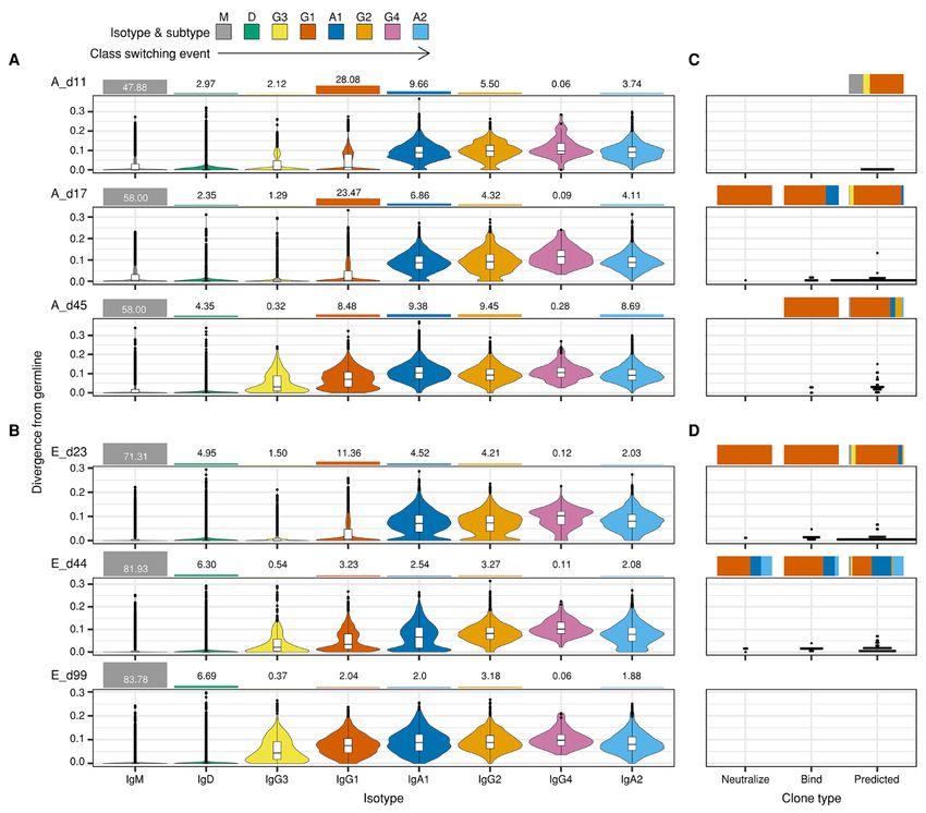
converting enzyme II (ACE2), the functional receptor of single-chain variable fragment (scFv) clones that were reac-
SARS-CoV-2 (12). The prevalence of these stereotypic nAb tive against recombinant SARS-CoV-2 RBD as measured with
clonotypes among SARS-CoV-2 patients and their character- SARS-CoV-2 RBD-specific enzyme-linked immunosorbent as-
istics, such as frequency within immunoglobulin (Ig) reper- say (ELISA) (Figure S2 and Table S2). The half-maximal bind-
toires, somatic mutations, isotypes, and chronological ing of scFv-human kappa light chain fragment (hCκ) to
changes remain to be elucidated. SARS-CoV-2 RBD occurred at concentrations ranging from
Here, we report stereotypic nAb clonotypes for SARS-CoV- 0.32 to 364 nM, which was comparable to findings in previ-
2, which were identified by mapping nAbs onto deep Ig rep- ous reports that have described human mAbs against the
ertoires that were profiled from infected patients. The stere- same protein (8, 11). We tested whether these antibody clones
otypic-naïve nAb clonotypes encoded by the IGHV3- could inhibit the binding between recombinant SARS-CoV-2
53/IGHV3-66 and immunoglobulin heavy joining (IGHJ)6 S protein and Vero E6 cells expressing the ACE2 receptor. A
genes existed in the majority of convalescent COVID-19 pa- recombinant polyhistidine (HIS)-tagged SARS-CoV-2 S pro-
tients and showed little evidence of somatic mutations along tein showed binding saturation at 200 nM when incubated
with swift isotype class switching to IgG1, IgA1, and even IgA2 with 1.5 × 10⁵ Vero E6 cells, as measured using flow cytometry
Downloaded from http://stm.sciencemag.org/ by guest on January 22, 2021
subtypes. Subsequently, we found that these same variable analysis with a fluorescein isothiocyanate (FITC)-labeled
heavy chain (VH) clonotypes pre-exist predominantly as an anti-HIS antibody. For the analysis of different scFv clones,
IgM isotype in healthy individuals, possibly encoded by naïve recombinant S protein (200 nM) was mixed with scFv-hFc fu-
B cells, which suggests that these stereotypic nAbs are not sion proteins, at a final concentration of either 200 nM
originated from clonotypes developed by previous infection (equimolar) or 600 nM (molar ratio of 1:3). Eleven clones (A-
with similar viruses. Individuals with these VH clonotypes are 1A1, A-1H4, A-1H12, A-2F1, A-2H4, A-2G3, E-3A12, E-3B1, E-
may be able to rapidly produce potent nAbs and potentially 3G9, E-3H31, and E-4D12) almost completely inhibited bind-
afford partial protection against SARS-CoV-2. ing between recombinant S protein and Vero E6 cells at 600
nM, and some showed inhibitory activity at 200 nM (Figure
RESULTS
S3). We used an in vitro viral neutralization assay in which
Isolation and characterization of human nAbs
Vero cells were infected with a clinical isolate of SARS-CoV-2
To obtain monoclonal nAbs against SARS-CoV-2, we col-
(BetaCoV/Korea/SNU01/2020) encoding D614 in the viral S
lected blood samples from 17 SARS-CoV-2-infected patients
protein at a medium tissue culture infectious dose (TCID50)
(Patients A–Q), which were used to generate human antibody
of 2,500 and in the presence of one of the 11 scFv-hCκ fusion
libraries. SARS-CoV-2 also uses spike (S) protein for receptor
proteins, at concentrations of 0.5, 5, or 50 μg/mL. Viral RNA
binding and membrane fusion in a mechanism similar to
concentrations in the culture supernatant were determined
SARS-CoV (13) and requires binding with cellular receptor
0, 24, 48, and 72 hours after infection. Nine scFv-hCκ fusion
ACE2 to gain entry into the host cell (14, 15). A previous re-
proteins exhibited complete neutralizing activity at 50 μg/mL
port suggests that a human monoclonal antibody (mAb) that
(Figure S4), and two antibodies (A-1H4 and E-3G9) showed
reacts with the RBD within the S1 region of the S protein can
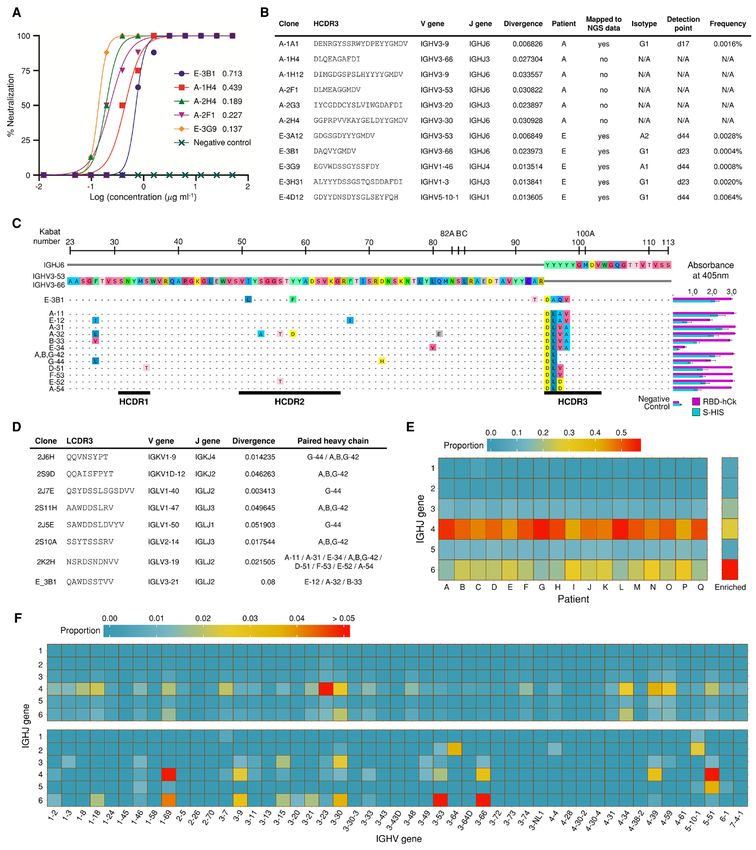
potent neutralization even at 5 μg/mL (Figure S4). Five nAbs
hinder the initial binding interaction between the virus and
with IgG2/4 isotypes (E-3B1, A-1H4, A-2H4, A-2F1, and E-
the cell, effectively neutralizing SARS-CoV-2 (11). We con-
3G9) exhibited potent neutralizing activity against authentic
firmed the reactivity of convalescent patient sera with recom-
SARS-CoV-2, with half-maximal inhibitory concentration
binant SARS-CoV-2 S and RBD proteins. Patients A and E,
(IC50) ranging from 0.137 to 0.713 μg/mL (Fig. 2A).
who presented with extensive pneumonic infiltrates, also
Identification of stereotypic clonotypes from IGH rep-
showed high plasma IgG titers against recombinant SARS-
ertoire of SARS-CoV-2-infected patients
CoV-2 nucleocapsid (N), S, S1, S2, and RBD proteins, which
We performed deep profiling of the Ig repertoire in three
was detected 11, 17, and 45 days after symptom onset in Pa-
chronological blood samples each from Patients A and E, two
tient A and 23, 44, and 99 days after symptom onset in Pa-
chronological samples each from Patients B, C, D, F, and G,
tient E (Table S1 and Fig. 1). Sera samples from Middle East
and a single time point sample from the remaining ten pa-
respiratory syndrome coronavirus (MERS-CoV) patients
tients (H-Q). We searched for nAb clonotypes that possessed
crossreacted with the SARS-CoV-2 S protein, showed a higher
identical variable (V) and joining (J) gene combinations and
titer against the S2 domain and crossreactivity to MERS-CoV
perfectly matched heavy chain complementarity-determining
S2 was detected in SARS-CoV-2 sera samples (Figs. 1 and S1),
region 3 (HCDR3) amino acid sequences among the immuno-
suggesting the potential risk for ADE. We generated four hu-
globulin heavy chain (IGH) repertoires of Patients A and E.
man Ab libraries using a phage-display system, based on the
One and five nAb clonotypes that met these criteria were
blood samples from Patient A, which were collected on days
identified in Patients A and E, respectively (Fig. 2B), and
17 and 45 (A_d17 and A_d45), and Patient E, which were col-
three nAbs (A-2F1, E-3A12, and E-3B1) were encoded by
lected on days 23 and 44 (E_d23 and E_d44). We isolated 38
First release: 4 January 2021 stm.sciencemag.org (Page numbers not final at time of first release) 2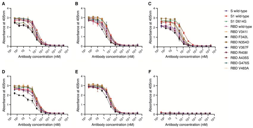
IGHV3-53/IGHV3-66 and IGHJ6 (Fig. 2B). These two VH                Stereotypic-naïve IGH clonotype against SARS-CoV-2
genes, IGHV3-53*01 and IGHV3-66*01 share an identical              pre-exist in the healthy population
amino acid sequence, except for the H12 residue (isoleucine             Among IGH clonotypes, A,B,G-42 was unique, presenting
in IGHV3-53 and valine in IGHV3-66), and only five nucleo-         little to no evidence of somatic mutations (0.6% ± 0.8%) and
tides differ between their sequences. Four clonotypes were         containing an HCDR3 (DLYYYGMDV) formed by the simple
IgG1, and two clonotypes were class-switched to IgA1 and IgA2,     joining of IGHV3-53 and IGHJ6. This naïve VH sequence ex-
when examined 44 days after symptom onset (Fig. 2B). These         isted in the IGH repertoire of five patients (Patients A, B, G,
clonotypes had a very low frequency of somatic mutations           I, and K), as IgM and IgG1, IgM and IgG1, IgG1 and IgA1, IgM,
(1.03% ± 0.51%), which was compatible with findings about          or IgG1 subtypes, respectively (Table 1). The IGH clonotypes
other nAbs in previous reports (7, 8). We evaluated all VH se-     encoded by IGHV3-53/IGHV3-66 and IGHJ6 that possessed
quences from the 17 patients and searched the clonotypes of        an HCDR3 (DLYYYGMDV) with zero to one somatic mutation
11 nAbs that were encoded by the same VJ genes and showed          could be identified within the IGH repertoire of six of 10
66.6% or higher identity in the amino acid sequence for            healthy individuals, predominantly as an IgM isotype (16),
HCDR3 (Figure S5). Clonotypes that were highly homologous          based on publicly available IGH repertoires (Table 1). The

                                                                                                                                        Downloaded from http://stm.sciencemag.org/ by guest on January 22, 2021
to the E-3B1 nAb were found among 13 out of 17 patients, with      A,B,G-42 clonotype showed light chain plasticity and could
a total of 126 clonotypes having the isotype of IgG3 (Patients     pair with five Vκ /Vλ genes to achieve RBD binding. In partic-
I, K, and P), IgG1 (Patients A, B, D-I, K, M, O, and P), IgA1      ular, the Vκ gene (2J6H) accumulated only five somatic mu-
(Patients E, G, I, and J), IgG2 (Patients I-K) and IgA2 (Patient   tations (1.4% divergence). None of the 12 clones, including
E) (Table S3). These clonotypes shared nearly identical VH se-     A,B,G-42, reacted against the recombinant RBD proteins
quences (92.45% ± 3.04% identity between amino acid se-            from either SARS-CoV or MERS-CoV (Figure S9). None of the
quences), with E-3B1 displaying an extremely low frequency         37 identified MERS-RBD-binding human mAbs (from two pa-
of somatic mutations (0.98% ± 1.48%). Among these 126              tients) were encoded by IGHV3-53/IGHV3-66 and IGHJ6 (Ta-
clonotypes, 43 unique HCDR3s were identified by amino acid         ble S4) based on analysis of a prior study (17). Therefore, the
sequence, and 12 unique HCDR3s were present in more than           presence of these stereotypic-naïve IGH clonotypes in the
one patient (Table 1).                                             healthy population, and their light chain plasticity needed to
Light chain plasticity of the stereotypic VH clonotypes            achieve SARS-CoV-2 RBD binding, may be unique to SARS-
for binding to SARS-CoV-2 RBD                                      CoV-2, which might provide a rapid and effective humoral re-
    To test the reactivity of clonotypes homologous to E-3B1       sponse to the virus among patients who express these clono-
against the SARS-CoV-2 S protein, we arbitrarily sampled 12        types. These findings suggest that a portion of the population
IGH clonotypes (Fig. 2C), containing five different HCDR3s,        possesses germline-precursor B cells, encoded by IGHV3-
from the IGH repertoires of 13 patients. The genes encoding        53/IGHV3-66 and IGHJ6, which can actively initiate virus
these IGH clonotypes were chemically synthesized and used          neutralization upon SARS-CoV-2 infection.
to construct scFv genes with the variable lambda chain (Vλ)        Distinctive V and J gene usage of the SARS-CoV-2 RBD-
gene from the E-3B1 clone. The reactivity of these scFv clones     binding antibodies
to recombinant S and RBD was tested using a phage ELISA,                To further elucidate the preferential use of IGHV3-
and three clones (E-12, A-32, and B-33) reacted against recom-     53/IGHV3-66 and IGHJ6 genes during the generation of
binant S and RBD proteins (Fig. 2C). scFv libraries were con-      SARS-CoV-2 RBD-binding antibodies, we extracted 252 pre-
structed, using the A-11, A-31, E-34, A,B,G-42, G-44, D-51, F-     dicted RBD-binding clones from our biopanning data. We
53, E-52, and A-54 genes, and the variable kappa chain             previously showed that antibody clones with binding proper-
(Vκ)/Vλ genes amplified from Patients A, E, and G, and all 12      ties can be predicted by employing next-generation sequenc-
IGH clonotypes were reactive against both recombinant S            ing (NGS) technology and analyzing the enrichment patterns
and RBD proteins when paired with eight different Vκ and Vλ        of biopanned clones (18, 19). The IGHJ4 gene was more prom-
genes (Figs. 2C and 2D). All seven light chain-profiled pa-        inent within the IGH repertoires of 17 patients, similar to
tients (A–G) possessed these Vκ/Vλ clonotypes with identical       healthy human samples (16, 20), but the predicted RBD-
VJ gene usage and perfectly matching light chain CDR3              binding clones primarily used the IGHJ6 gene (Fig. 2E). Fur-
(LCDR3) amino acid sequences (Figure S6). Immunoglobulin           thermore, the predicted RBD-binding clones showed the
lambda variable (IGLV)2-14/immunoglobulin lambda joining           dominant usage of IGHV3-53/IGHJ6 and IGHV3-66/IGHJ6
(IGLJ)3, IGLV3-19/IGLJ2, and IGLV3-21/IGLJ2 were fre-              pairs, which was not observed in the whole IGH repertoires
quently used across all seven patients (Figures S7 and S8).        of patients (Fig. 2F).
Because E-3B1 effectively inhibited the replication of SARS-       Chronological follow-up of IGH repertoire and the
CoV-2 (Fig. 2A), these 126 clonotypes are likely to neutralize     SARS-CoV-2 RBD-binding antibodies from patients
SARS-CoV-2 when paired with an optimal light chain.                     Naïve B cells typically undergo somatic hypermutations,

First release: 4 January 2021                           stm.sciencemag.org      (Page numbers not final at time of first release)   3
clonal selection, and class-switching following antigen expo-    of either IGHV3-53 or IGHV3-66 and IGHJ6 genes, using sin-
sure. We examined the chronological events that occurred in      gle B cell sequencing technology (7–11). The crystal structures
all IGH clonotypes identified in Patients A - G and those that   of two IGHV3-53 nAbs has been determined, which has
were reactive against the SARS-CoV-2 RBD. In the entire pa-      shown that two key motifs within HCDR1 and HCDR2 that
tient IGH repertoire, naïve-derived IGH clonotypes with min-     are encoded in the IGHV3-53 germline can bind to SARS-
imal somatic mutations (< 2.695% ± 0.700%) showed                CoV-2 S protein RBD (12). Therefore, the preferential use of
increased IgG3 and IgG1 subtypes, and the proportion of the      IGHV3-53/IGHV3-66 and IGHJ6 in the development of nAbs
IgG1 subtype was dramatically increased for a period (Fig. 3A    to SARS-CoV-2 appears promising, especially given evidence
and 3B and Figure S10). The naïve-derived IGH clonotypes         that IGHV3-53/IGHV3-66 and IGHJ6 are able to pair with di-
were detected as minor populations of IgA1 and IgG2 subtypes     verse light chains in order to form nAbs that can bind to the
in Patients A and E (Figs. 3A and 3B), and as an IgA2 subtype    RBD. We found eight different light chains from our experi-
in Patient E (Fig. 3B). We categorized RBD-reactive clones       ments, and other groups have identified nine different light
into three groups: 1) neutralizing antibodies (neutralize), 2)   chains (12). It is expected that the extent of light chain plas-
binding-confirmed antibodies (bind), and 3) binding-pre-         ticity is broad enough for virus-exposed individuals to suc-

                                                                                                                                      Downloaded from http://stm.sciencemag.org/ by guest on January 22, 2021
dicted antibodies (predicted). In all three groups, these IGH    cessfully evolve nAbs given that class-switched IGHV3-
clonotypes appeared and disappeared throughout the disease       53/IGHV3-66 and IGHJ6 clonotypes were present in 13 of 17
course, showed a low frequency of somatic mutations (Figs.       patients from our study.
3C and 3D), and displayed rapid class-switching, especially to       Currently, we do not know whether the stereotypic nAbs
IgG1, IgA1, and IgA2. These results suggest that RBD-reactive    are polyreactive or autoreactive. Rather, our selected stereo-
IGH clonotypes can emerge rapidly and undergo class-             typic nAbs, including A,B,G-42, do not appear to crossreact
switching to IgG1, IgA1, and IgA2, without accumulating many     with recombinant RBD proteins of either SARS-CoV or
somatic mutations. This dramatic temporal surge of naïve         MERS-CoV. Several reports have evaluated auto- or polyreac-
IGH clonotypes, with rapid class-switching, occurred across      tivity of SARS-CoV-2 nAbs with minimal somatic mutations,
the entire IGH repertoire of the patients and was not con-       and one report showed that 18 nAbs, including four antibod-
fined to those reactive to the SARS-CoV-2 RBD.                   ies encoded by IGHV3-53/IGHJ4, IGHV3-53/IGHJ6, or
Selected nAbs retained its ability to bind to most current       IGHV3-66/IGHJ4, did not show evidence of being polyreac-
SARS-CoV-2 mutants                                               tive or autoreactive (22). Another group characterized 29 Abs,
    Several mutations within the S1 domain of the SARS-CoV-      including three nAbs encoded by IGHV3-53, with limited so-
2 S protein have been identified during the 2019-20 global       matic mutations (
contradictory report suggests that early seroconversion along “predicted” clones were individually mapped for Patient E.
with high antibody titers correlate with a less severe clinical A major limitation of this study was the limited number
course (26). In addition, there was a report claiming no asso- of patients enrolled made it difficult to analyze whether these
ciation between comorbidity and antibody titer (27). stereotypic nAb clonotypes affect the clinical course of
SARS-CoV-2 can cause a severe respiratory infection, COVID-19. We found that stereotypic nAb clonotypes pre-ex-
which suggests that patients will need to produce both sys- isted in the majority of the naïve population, were prevalent
temic and mucosal nAbs, including those of the IgA isotype, among the patients who displayed rapid class-switching to
for protective immunity. Our results showed that IGHV3- IgG and IgA isotypes, and exhibited light chain plasticity
53/IGHV3-66 and IGHJ6 class-switched to IgA1 in Patients G, among the SARS-CoV-2 RBD-binding antibodies. These re-
I, and J, and class-switched to IgA1 and IgA2 in Patient E (Ta- sults suggest that stereotypic nAb clonotypes may contribute
ble S3). After 99 days from the onset of symptoms, we did not to a milder clinical course and lower mortality rate in SARS-
detect any RBD-reactive IGH clonotypes in the peripheral CoV-2 patients compared with SARS-CoV (9.5%) or MERS-
blood of Patient E; however, the antibody titer to RBD re- CoV (34.4%) patients (33), for which similar stereotypic nAb
mained high (Fig. 3D and Fig. 1). This observation is in line clonotypes have not yet been reported. Future chronological
Downloaded from http://stm.sciencemag.org/ by guest on January 22, 2021
with the findings that nAb titers remain detectable among a follow-ups of Ig repertoires in a larger population of SARS-
fraction of SARS and MERS patients 1–2 years after infection CoV-2 patients may provide insight into the effects of stereo-
(28, 29). Therefore, it can be inferred that nAb-producing typic nAb clonotypes on patient outcomes. Furthermore,
plasmablasts may be mobilized from the peripheral blood to studies assessing changes in the Ig repertoire among the na-
bone marrow niches and continue to produce nAbs in Patient ïve populations after SARS-CoV-2 vaccination may provide
E. In these niches, plasmablasts are able to differentiate into details about the contribution of these stereotypic nAb clono-
mature Ab-secreting plasma cells and can survive for decades types to protective immunity.
(30).
MATERIALS AND METHODS
In the rhesus macaque model of SARS-CoV-2 infection, vi-
Study design
ral rechallenge generated a greater nAb titer than induced by
This study was designed to investigate stereotypic nAb
primary infection and protected animals from reinfection
clonotypes of SARS-CoV-2. 26 blood samples were collected
(31). A single case of SARS-CoV-2 reinfection that has been
from 17 SARS-CoV-2-positive patients and were subjected to
reported suggests that it is possible for humans to become
NGS analysis of Ig sequences. Human antibody libraries were
infected multiple times by SARS-CoV-2 (32). It is still unclear
prepared and subjected to biopanning against recombinant
whether a SARS-CoV-2 vaccine would provide prolonged pro-
SARS-CoV-2 RBD proteins. RBD-binders were selected using
tective immunity to humans. The long-term production of
ELISA and their neutralization activity was tested using flow
nAbs, as seen in Patient E out to day 99 after symptom onset,
cytometry with ACE2-expressing Vero cells and recombinant
suggests that nAb production by plasma cells in bone marrow
SARS-CoV-2 S protein and microneutralization assay. NGS
niches may be a critical mechanism triggered by infection or
analysis of the enrichment patterns of clones through biopan-
vaccination that leads to protective immunity against SARS-
ning was performed for in silico selection of RBD-binding
CoV-2.
clones. Ig repertoire analyses were conducted to identify and
It has been reported that the frequency of RBD-reactive B
characterize nAb clonotypes, including their prevalence
cell clones is extremely low (0.07% to 0.005%) among circu-
among patients, frequency in Ig repertoires, somatic muta-
lating B cells (24). In our study, the frequency of isolated nAb
tions, isotypes, chronological changes, and existence in a na-
clonotypes in the IGH repertoire was also extremely low
ïve uninfected population.
(0.0004%-0.0064%), and Patient A had only one out of six
Clinical protocol and blood sample collection
nAbs that mapped to the IGH repertoire (Fig. 2B). As the
All patients were confirmed to be infected by SARS-CoV-
complexity of scFv phage-display libraries exceeded 3.8 × 10⁸
2 by a positive reverse transcriptase-quantitative polymerase
and 6.7 × 10⁸ colony-forming units for Patient A, diverse RBD-
chain reaction (RT-qPCR) result, and sample collection was
binding clones could be enriched by biopanning. Only 199,561
performed at Seoul National University Hospital. The study
unique IGH sequences were sampled by NGS in Patient A,
involving human sample collection was approved by the In-
whereas 515,994 IGH sequences were sampled in Patient E,
stitutional Ethics Review Board of Seoul National University
when scFv phage-display libraries were constructed. This dif-
Hospital (IRB approval number: 2004-230-1119). Three
ference in NGS throughput might explain the discrepant al-
chronological peripheral blood samples were drawn from Pa-
location of nAb clonotypes in the IGH repertoire of Patients
tients A and E, and two chronological samples were obtained
A and E. Consistent with this hypothesis, only 38.3% and
from Patients B, C, D, F, and G. A single blood sample was
22.0% of “bind” and “predicted” clones were mapped for Pa-
collected from Patients H-Q. PBMCs and plasma were iso-
tient A, respectively, whereas 77.8% and 32.1% of “bind” and
lated using Lymphoprep (Stemcell Technologies, Vancouver,
First release: 4 January 2021 stm.sciencemag.org (Page numbers not final at time of first release) 5BC, Canada), according to the manufacturer’s protocol. from phagemid DNA, using the primers listed in Table S8.
PBMCs were subjected to total RNA isolation, using the TRI SPRI-purified sequencing libraries were quantified with a
Reagent (Invitrogen, Carlsbad, CA, USA), according to the 4200 TapeStation System (Agilent Technologies), using a
manufacturer’s protocol. D1000 ScreenTape Assay, before performing sequencing on
Next-generation sequencing (NGS) an Illumina MiSeq Platform.
Genes encoding VH and part of the CH1 domain were am- NGS data processing
plified, using specific primers, as described previously (16, Pre-processing of the NGS data for the IG repertoire
34). All primers used are listed in Table S8. Briefly, total RNA The raw NGS forward (R1) and reverse (R2) reads were
was used as a template to synthesize cDNA, using the Super- merged by PEAR, v0.9.10, in default setting (36). The merged
script IV First-Strand Synthesis System (Invitrogen), with reads were q-filtered using the condition q20p95, which re-
specific primers targeting the constant region (CH1 domain) sults in 95% of the base-pairs in a read having Phread scores
of each isotype (IgM, IgD, IgG, IgA, and IgE) (34), according higher than 20. The location of the primers was recognized
to the manufacturer’s protocol. Following cDNA synthesis, 1.8 from the q-filtered reads while allowing one substitution or
volumes of SPRI beads (AmpureXP, Beckman Coulter, Brea, deletion (Table S8). Primer regions that specifically bind to
Downloaded from http://stm.sciencemag.org/ by guest on January 22, 2021
CA, USA) were used to purify cDNA, which was eluted in 40 the molecules were trimmed in the reads to eliminate the ef-
μL water. The purified cDNA (18 μL) was subjected to second- fects of primer synthesis errors. Based on the primer recog-
strand synthesis in a 25-μL reaction volume, using V gene- nition results, unique molecular identifier (UMI) sequences
specific primers (16) and KAPA Biosystems (KAPA HiFi Hot- were extracted, and the reads were clustered according to the
Start, Roche, Basel, Switzerland). The PCR conditions were as UMI sequences. To eliminate the possibility that the same
follows: 95°C for 3 min, 98°C for 1 min, 55°C for 1 min, and UMI sequences might be used for different read amplifica-
72°C for 5 min. Following the second-strand synthesis, dou- tions, the clustered reads were subclustered, according to the
ble-strand DNA (dsDNA) was purified, using SPRI beads, as similarity of the reads (Five mismatches were allowed in each
described above. VH genes were amplified using 15 μL eluted subcluster). The subclustered reads were aligned using a mul-
dsDNA and 2.5 pmol of the primers listed in Table S8, in a tiple sequence alignment tool, Clustal Omega, v1.2.4, in de-
50-μL total reaction volume (KAPA Biosystems), using the fault setting (37, 38). From the aligned reads, the frequency
following thermal cycling program: 95°C for 3 min; 17 cycles of each nucleotide was calculated, and a consensus sequence
of 98°C for 30 s, 65°C for 30 s, and 72°C for 1 min 10 s; and of each subcluster was defined using the frequency infor-
72°C for 5 min. The number of PCR cycles was increased, mation. The read count of the consensus sequence was rede-
from 17 to 19, for samples from Patients B (d10 and 19), C (d6), fined as the number of UMI subclusters that belong to the
E (d23), and G (d9 and 22). PCR products were purified using consensus sequences.
SPRI beads and eluted in 30 μL water. Genes encoding Vκ and Sequence annotation, functionality filtering, and
Vλ were amplified using specific primers, as described previ- throughput adjustment
ously (20, 35). Briefly, total RNA was used as a template to Sequence annotation consisted of two parts: isotype an-
synthesize cDNA, using the Superscript IV First-Strand Syn- notation and VDJ annotation. For annotation, the consensus
thesis System (Invitrogen), with specific primers targeting sequence was divided into two sections, a VDJ region and a
the constant region, which are listed in Table S8, according constant region, in a location-based manner. For isotype an-
to the manufacturer’s protocol. Following cDNA synthesis, notation, the extracted constant region was aligned with the
SPRI beads were used to purify cDNA, which was eluted in IMGT (International Immunogenetics Information System)
40 μL water. Purified cDNA (18 μL) was used for the first am- constant gene database (39). Based on the alignment results,
plification, in a 25-μL reaction volume, using VJ gene-specific the isotypes of the consensus sequences were annotated.
primers, which are listed in Table S8, and KAPA Biosystems. Then, the VDJ regions of the consensus sequences were an-
The PCR conditions were as follows: 95°C for 3 min, 4 cycles notated using IgBLAST, v1.8.0 (40). Among the annotation
of 98°C for 1 min, 55°C for 1 min, and 72°C for 1 min; and results, V/D/J genes (V/J genes for VL), CDR1/2/3 sequences,
72°C for 10 min. Subsequently, DNA was purified using SPRI and the number of mutations from the corresponding V
beads, and the Vκ and Vλ genes were amplified using 15 μL genes were extracted for further analysis. Divergence values
eluted dsDNA and 2.5 pmol of the primers listed in Table S8, were defined as the number of mutations identified in the
in a 50-μL total reaction volume (KAPA Biosystems). The PCR aligned V gene, divided by the aligned length. The non-func-
conditions were as follows: 95°C for 3 min; 17 cycles of 98°C tional consensus reads were defined using the following cri-
for 30 s, 65°C for 30 s, and 72°C for 1 min 10 s; and 72°C for teria and filtered-out: 1. sequence length shorter than 250 bp;
5 min. PCR products were purified using SPRI beads, as de- 2. existence of stop-codon or frame-shift in the full amino
scribed above. For the amplification of VH from each round acid sequence; 3. annotation failure in one or more of the
of biopanning (rounds 0–4), gene fragments were amplified CDR1/2/3 regions; and 4. isotype annotation failure. The
First release: 4 January 2021 stm.sciencemag.org (Page numbers not final at time of first release) 6functional consensus reads were random-sampled, to adjust PC2 was primarily composed of the frequency in round 0
the throughput of the VH data (Table S5). Throughput adjust- (Figure S13). Thus, we defined PC1-major clones as the pre-
ment was not conducted for VL data (Table S6). dicted clones, by setting constant threshold values on the PC1
Preprocessing of the biopanning NGS data value and the ratio between PC1 and PC2 (Table S7). 94.74%
Pre-processing of the biopanning NGS data was per- of the RBD-binding clones were mapped to the predicted
formed as previously reported, except for the application of clones (Figure S13).
the q-filtering condition q20p95 instead of q20p100 (41). Construction of a human scFv phage-display library and
Overlapping IGH repertoire construction VL shuffled libraries
To investigate the shared IGH sequences among the pa- For the VH gene, the cDNA prepared for the NGS analysis
tients, we defined the overlapping IGH repertoire of the pa- was used. For the VΚ and Vλ genes, total RNA was used to
tients. Histograms for the nearest-neighbor distances of the synthesize cDNA, using the Superscript IV First-Strand Syn-
HCDR3 amino acid sequences were calculated for the reper- thesis System (Invitrogen), with oligo(dT) primers, according
toire data. A hierarchical, distance-based analysis, which was to the manufacturer’s instructions. Then, the genes encoding
reported previously (42), was applied to the HCDR3 amino VΚ/Vλ and VH were amplified, from the oligo(dT)-synthesized
Downloaded from http://stm.sciencemag.org/ by guest on January 22, 2021
acid sequences, to cluster functionally similar IGH sequences. cDNA and the cDNA prepared for NGS analysis, respectively,
The IGH sequences for all repertoire data could be approxi- using the primers listed in Table S8 and KAPA Biosystems.
mated into a bimodal distribution, allowing the functionally The PCR conditions were as follows: preliminary denatura-
similar IGH sequences to be extracted by capturing the first tion at 95°C for 3 min; 4 cycles of 98°C for 1 min, 55°C for 1
peak of the distribution (Figure S11). Threshold values for min, and 72°C for 1 min; and 72°C for 10 min. Subsequently,
each data set were defined as the nearest-neighbor distance DNA was purified using SPRI beads, as described above. The
value of those points with a minimum frequency between the purified DNA was amplified using the primers listed in Table
two peaks of the distribution. Then, the minimum value S8 and KAPA Biosystems. The PCR conditions were as fol-
among all threshold values, 0.113871, was used to construct lows: preliminary denaturation, at 95°C for 3 min; 25 cycles
the overlapping IGH repertoire, which means that 11.3871% of 98°C for 30 s, 58°C for 30 s, and 72°C for 90 s; and 72°C
of mismatches in the HCDR3 amino acid sequence were al- for 10 min. Then, the VH and VΚ/Vλ fragments were subjected
lowed in the overlapping IGH repertoire construction. To to electrophoresis, on a 1% agarose gel, and purified, using a
construct the overlapping IGH repertoire, the repertoire data QIAquick Gel Extraction Kit (Qiagen Inc., Valencia, CA, USA),
sets of all patients were merged into one data set. The IGH according to the manufacturer’s instructions. The purified VH
sequences in the merged data set were then clustered, using and VΚ/Vλ fragments were mixed, at equal ratios at 50 ng, and
the following conditions: 1. the same V and J gene usage; and subjected to overlap extension, to generate scFv genes, using
2. mismatch smaller than 11.3871% among the HCDR3 amino the primers listed in Table S8 and KAPA Biosystems. The PCR
acid sequences. Subsequently, clusters containing IGH se- conditions were as follows: preliminary denaturation, at
quences from more than one patient were included in the 94°C for 5 min; 25 cycles of 98°C for 15 s, 56°C for 15 s, and
overlapping IGH repertoire data set. 72°C for 2 min; and 72°C for 10 min. The amplified scFv frag-
Extraction of binding-predicted clones ment was purified and cloned into a phagemid vector, as de-
From each round of biopanning (rounds 0, 2, 3, and 4), scribed previously (43).
the VH genes were amplified and subjected to NGS analysis, For the construction of VΚ/Vλ shuffled libraries, gBlocks
using the MiSeq platform, as described previously (19). Bind- Gene Fragments (Integrated DNA Technologies, Coralville,
ing-predicted clones from biopanning were selected by ana- IA, USA), encoding A-11, E-12, A-31, A-32, B-33, E-34, A,B,G-
lyzing the enrichment or diminishment patterns of clones in 42, G-44, D-51, F-53, E-52, and A-54, were synthesized. Syn-
the NGS data from four libraries, A_d17, A_d45, E_d23, and thesized VH and the VΚ/Vλ genes from Patients A, E, and G
E_d44, at each round of biopanning. The enrichment of were used to synthesize the scFv libraries using PCR, as de-
clones primarily occurred during the second round of biopan- scribed previously (43). Then, the amplified scFv fragments
ning based on the input/output virus titer values for each were purified and cloned into the phagemid vector, as de-
round of biopanning and the frequencies of the clones in the scribed above.
NGS data (Figure S12). The frequency information in the NGS Biopanning
data sets for biopanning rounds 0, 2, 3, and 4 was subject to Phage display of the human scFv libraries exceeded com-
principal component analysis (PCA) for dimension reduction. plexity of 3.8 × 10⁸, 6.7 × 10⁸, 2.0 × 10⁸, and 7.2 × 10⁸ colony-
Accordingly, principal component (PC)1 and PC2, which rep- forming units for A_d17, A_d45, E_d23, and E_d44, respec-
resented clone enrichment and clone depletion, respectively, tively. These libraries were subjected to four rounds of bi-
were extracted. In the biopanning data, PC1 was primarily opanning against the recombinant SARS-CoV-2 RBD protein
composed of the frequencies in rounds 2, 3, and 4, whereas (Sino Biological Inc., Beijing, China), fused to mFc or hCκ, as
First release: 4 January 2021 stm.sciencemag.org (Page numbers not final at time of first release) 7described previously (44). Briefly, 3 μg of the recombinant Inc.), S2 (Sino Biological Inc.), NP (Sino Biological Inc.), RBD,
SARS-CoV-2 RBD protein was conjugated to 1.0 × 10⁷ mag- RBD mutants, SARS-CoV RBD (Sino Biological Inc.), MERS-
netic beads (Dynabeads M-270 epoxy, Invitrogen) and incu- CoV S (Sino Biological Inc.), RBD (Sino Biological Inc.), S2
bated with the scFv phage-display libraries (approximately (Sino Biological Inc.) proteins were added to microtiter plates
1012 phages), for 2 hours at 37°C. During the first round of (Costar), in coating buffer (0.1 M sodium bicarbonate, pH
biopanning, the beads were washed once with 500 μL of 8.6). After incubation at 4°C, overnight and blocking with 3%
0.05% (v/v) Tween-20 (Sigma-Aldrich, St. Louis, MO, USA) in bovine serum albumin (BSA) in PBS, for 1 hour at 37°C, seri-
phosphate-buffered saline (PBST). For the other rounds of bi- ally diluted plasma (5-fold, 6 dilutions, starting from 1:100)
opanning, 1.5 μg of recombinant SARS-CoV-2 RBD protein or scFv-hFc (5-fold, 12 dilutions, starting from 1,000 or 500
was conjugated to 5.0 × 10⁶ magnetic beads, and the number nM) in blocking buffer was added to individual wells and in-
of washes was increased to three. After each round of biopan- cubated for 1, h at 37°C. Then, plates were washed three times
ning, the bound phages were eluted and rescued, as described with 0.05% PBST. Horseradish peroxidase (HRP)-conjugated
previously (44). rabbit anti-human IgG antibody (Invitrogen) or anti-human
Phage ELISA Ig kappa light chain antibody (Millipore, Temecula, CA,
Downloaded from http://stm.sciencemag.org/ by guest on January 22, 2021
To select SARS-CoV-2 S reactive clones, phage ELISA was USA), in blocking buffer (1:5,000), was added to wells and in-
performed, using recombinant S and RBD protein-coated mi- cubated for 1 hour at 37°C. After washing three times with
crotiter plates, as described previously (45). Reactive scFv PBST, 2,2′-azino-bis-3-ethylbenzothiazoline-6-sulfonic
clones were subjected to Sanger sequencing (Cosmogenetech, (ThermoFisher Scientific Inc., Waltham, MA, USA) or
Seoul, Republic of Korea), to determine their nucleotide se- 3,3′,5,5′-Tetramethylbenzidine liquid substrate system
quences. (ThermoFisher Scientific Inc.) was added to the wells. Ab-
Expression of recombinant proteins sorbance was measured at 405 nm or 650 nm, respectively,
A human, codon-optimized, SARS-CoV-2 RBD using a microplate spectrophotometer (Multiskan GO;
(YP_009724390.1, amino acids 306–543) gene was synthe- Thermo Scientific).
sized (Integrated DNA Technologies). Using a synthesized Flow cytometry
wild-type RBD gene as a template, RBD mutants (V341I, The recombinant SARS-CoV-2 S protein (200 nM), fused
F342L, N354D, N354D/D364Y, V367F, R408I, A435S, W436R, with a HIS-tag at the C terminus (Sino Biological Inc.), was
G476S, and V483A) were generated through two-step PCR, incubated with scFv-hFc fusion proteins at a final concentra-
using the primers listed in Table S8. The genes encoding wild- tion of either 200 nM (equimolar) or 600 nM (molar ratio of
type or mutant SARS-CoV-2 RBD were cloned into a modified 1:3), in 50 μL of 1% (w/v) BSA in PBS, containing 0.02% (w/v)
mammalian expression vector, containing the hCκ gene (44), sodium azide (FACS buffer), at 37°C for 1 hour. Irrelevant
and transfected into Expi293F (Invitrogen) cells. The fusion scFv-hFc or scFv-hCκ fusion proteins were used as negative
proteins were purified by affinity chromatography, using controls. Vero E6 cells (ACE2+) were seeded into v-bottom 96-
KappaSelect Columns (GE Healthcare, Chicago, IL, USA), as well plates (Corning, Corning, NY, USA), at a density of 1.5 ×
described previously (46). Due to low expression yields, two 105 cells per well. Then, the mixture was added to each well
RBD mutants (N354D/D364Y, W436R) were excluded from and incubated, at 37°C for 1 hour. After washing three times
further studies. with FACS buffer, FITC-labeled rabbit anti-HIS Ab (Abcam,
The genes encoding the selected scFv clones were cloned Cambridge, UK) was incubated, at 37°C for 1 hour. Then, the
into a modified mammalian expression vector, containing the cells were washed three times with FACS buffer, resuspended
hIgG1 Fc regions (hFc) or hCκ at the C terminus (44, 47), be- in 150 μL of PBS, and subjected to analysis by flow cytometry,
fore being transfected and purified by affinity chromatog- using a FACS Canto II instrument (BD Bioscience, San Jose,
raphy, as described above. CA, USA). For each sample, 10,000 cells were analyzed.
Genes encoding VH and VL were amplified, cloned into a Microneutralization assay
mammalian expression vector containing the CH1 and hinge The virus (BetaCoV/Korea/SNU01/2020, accession num-
regions of human IgG2 fused to the CH2 and CH3 regions of ber MT039890) was isolated at the Seoul National University
human IgG4 (48, 49), and transfected into Expi293F cells Hospital and propagated in Vero cells (ATCC CCL-81), using
(Invitrogen) as described previously (50). Then, IgG2/4 was Dulbecco’s Modified Eagle’s Medium (DMEM, Welgene,
purified by affinity chromatography using MabSelect col- Gyeongsan, Republic of Korea) supplemented with 2% fetal
umns with the AKTA Pure chromatography system (GE bovine serum (Gibco) (51). The cells were grown in T-25
Healthcare) following the manufacturer’s protocol. flasks, (ThermoFisher Scientific Inc.), inoculated with SARS-
ELISA CoV-2, and incubated at 37°C, in a 5% CO2 environment. 3
100 ng of each recombinant SARS-CoV-2 S (Sino Biologi- days after inoculation, virus was harvested from culture su-
cal Inc.), S1 (Sino Biological Inc.), S1 D614G (Sino Biological pernatants and stored at -80°C. The virus titer was
First release: 4 January 2021 stm.sciencemag.org (Page numbers not final at time of first release) 8determined via a TCID50 assay (52). Fig. S8. VJ gene usage among the IG lambda light chain repertoire of patients.
Vero cells were seeded in T-25 flasks and grown for 24 Fig. S9. Reactivity of phage-displayed scFv clones in phage ELISA.
Fig. S10. Deep profiling of the IGH repertoire of Patients B, C, D, F, and G.
hours, at 37°C, in a 5% CO2 environment, to ensure 80% con- Fig. S11. The nearest-neighbor distance histogram for HCDR3 amino acid sequences
fluency on the day of inoculation. The recombinant scFv-hCκ in the IGH repertoires of patients.
fusion proteins (0.5, 5, or 50 μg/mL) were mixed with 2,500 Fig. S12. Frequency scatter plots for NGS data from four libraries after each round of
biopanning.
TCID50 of SARS-CoV-2, and the mixture was incubated for 2
Fig. S13. The results of principal component analysis (PCA) applied to the NGS data of
hours, at 37°C. The mixture (1 mL) was added to Vero cells four libraries after each round of biopanning.
and incubated for 1 hour, at 37°C, in a 5% CO2 environment. Table S1. Demographic and clinical characteristics.
After incubation for 1 hour, 6 mL of complete media was Table S2. SARS-CoV-2 RBD-reactive scFv clones.
Table S3. Class-switched IGH clonotypes homologous to E-3B1.
added to the flasks and incubated, at 37°C, in a 5% CO2 envi-
Table S4. Human mAbs reactive against MERS-CoV RBD.
ronment. After 0, 24, 48, and 72 hours of infection, the culture Table S5. Statistics for the pre-processing of the IGH NGS data.
supernatant was collected, to measure the virus titers. Viral Table S6. Statistics for the pre-processing of the IGκ and IGλ NGS data.
RNA was extracted using the MagNA Pure 96 DNA and Viral Table S7. The RBD-binding prediction clones.
NA small volume kit (Roche, Germany), according to the Table S8. Primers used in the study.
Downloaded from http://stm.sciencemag.org/ by guest on January 22, 2021
Data file S1. Individual level data.
manufacturer’s instructions. Viral RNA was detected using
the PowerChek 2019-nCoV Real-time PCR Kit (Kogene Bio-
REFERENCES AND NOTES
tech, Seoul, Republic of Korea), for the amplification of the E 1. J. G. Jardine, T. Ota, D. Sok, M. Pauthner, D. W. Kulp, O. Kalyuzhniy, P. D. Skog, T. C.
gene and quantified according to a standard curve, which was Thinnes, D. Bhullar, B. Briney, S. Menis, M. Jones, M. Kubitz, S. Spencer, Y. Adachi,
constructed using in vitro transcribed RNA provided by the D. R. Burton, W. R. Schief, D. Nemazee, HIV-1 VACCINES. Priming a broadly
European Virus Archive (https://www.european-virus- neutralizing antibody response to HIV-1 using a germline-targeting immunogen.
Science 349, 156–161 (2015). doi:10.1126/science.aac5894 Medline
archive.com). An alternative neutralization assay was per- 2. J. R. Willis, J. A. Finn, B. Briney, G. Sapparapu, V. Singh, H. King, C. C. LaBranche, D.
formed as described previously (53). Briefly, Vero cells were C. Montefiori, J. Meiler, J. E. Crowe Jr., Long antibody HCDR3s from HIV-naïve
seeded in 96-well flat-bottom tissue culture microtiter plates donors presented on a PG9 neutralizing antibody background mediate HIV
in DMEM medium were grown for 24 hours at 37°C in a 5% neutralization. Proc. Natl. Acad. Sci. U.S.A. 113, 4446–4451 (2016).
doi:10.1073/pnas.1518405113 Medline
CO2 environment. 50 μl of two-fold serially diluted IgG2/4 3. N. Eroshenko, T. Gill, M. K. Keaveney, G. M. Church, J. M. Trevejo, H. Rajaniemi,
were mixed with an equal volume of SARS-CoV-2 containing Implications of antibody-dependent enhancement of infection for SARS-CoV-2
100 TCID50 and the IgG2/4-virus mixture was incubated at countermeasures. Nat. Biotechnol. 38, 789–791 (2020). doi:10.1038/s41587-
37 °C for 1 hour. The mixture was then transferred into a 96- 020-0577-1 Medline
4. A. Iwasaki, Y. Yang, The potential danger of suboptimal antibody responses in
well microtiter plate containing Vero cells with 8 repeats and COVID-19. Nat. Rev. Immunol. 20, 339–341 (2020). doi:10.1038/s41577-020-
incubated for 5 days at 37°C in a 5% CO2 environment. Cells 0321-6 Medline
infected with 100 TCID50 of SARS-CoV-2, isotype IgG2/4 con- 5. M. K. Smatti, A. A. Al Thani, H. M. Yassine, Viral-Induced Enhanced Disease Illness.
trol, or without the virus, were applied as positive, negative, Front. Microbiol. 9, 2991 (2018). doi:10.3389/fmicb.2018.02991 Medline
6. A. Zhang, H. D. Stacey, C. E. Mullarkey, M. S. Miller, Original Antigenic Sin: How First
and uninfected controls, respectively. The cytopathic effect Exposure Shapes Lifelong Anti-Influenza Virus Immune Responses. J. Immunol.
(CPE) in each well was observed 5 days post-infection. The 202, 335–340 (2019). doi:10.4049/jimmunol.1801149 Medline
IC50 was calculated using GraphPad Prism 8 (GraphPad Soft- 7. Y. Cao, B. Su, X. Guo, W. Sun, Y. Deng, L. Bao, Q. Zhu, X. Zhang, Y. Zheng, C. Geng,
ware, San Diego, CA, USA). All experiments using authentic X. Chai, R. He, X. Li, Q. Lv, H. Zhu, W. Deng, Y. Xu, Y. Wang, L. Qiao, Y. Tan, L. Song,
G. Wang, X. Du, N. Gao, J. Liu, J. Xiao, X. D. Su, Z. Du, Y. Feng, C. Qin, C. Qin, R. Jin,
SARS-CoV-2 were conducted in Biosafety Level 3 laboratory. X. S. Xie, Potent neutralizing antibodies against SARS-CoV-2 identified by high-
Statistical analyses throughput single-cell sequencing of convalescent patients’ B cells. Cell 182, 73–
Data are represented as mean ± standard deviation. Sta- 84.e16 (2020). doi:10.1016/j.cell.2020.05.025 Medline
tistical analyses were performed using R software v.3.4.3. For 8. B. Ju, Q. Zhang, J. Ge, R. Wang, J. Sun, X. Ge, J. Yu, S. Shan, B. Zhou, S. Song, X.
Tang, J. Yu, J. Lan, J. Yuan, H. Wang, J. Zhao, S. Zhang, Y. Wang, X. Shi, L. Liu, J.
the flow cytometry analysis using ACE2-expressing cells and Zhao, X. Wang, Z. Zhang, L. Zhang, Human neutralizing antibodies elicited by
recombinant SARS-CoV-2 S protein, results were analyzed by SARS-CoV-2 infection. Nature 584, 115–119 (2020). doi:10.1038/s41586-020-
independent t-tests. 2380-z Medline
9. T. F. Rogers, F. Zhao, D. Huang, N. Beutler, A. Burns, W. T. He, O. Limbo, C. Smith,
SUPPLEMENTARY MATERIALS G. Song, J. Woehl, L. Yang, R. K. Abbott, S. Callaghan, E. Garcia, J. Hurtado, M.
stm.sciencemag.org/cgi/content/full/scitranslmed.abd6990/DC1 Parren, L. Peng, S. Ramirez, J. Ricketts, M. J. Ricciardi, S. A. Rawlings, N. C. Wu,
Fig. S1. Titrations of serum IgG by ELISAs specific to MERS-CoV. M. Yuan, D. M. Smith, D. Nemazee, J. R. Teijaro, J. E. Voss, I. A. Wilson, R. Andrabi,
Fig. S2. Reactivity of anti-SARS-CoV-2 scFv antibodies against recombinant SARS- B. Briney, E. Landais, D. Sok, J. G. Jardine, D. R. Burton, Isolation of potent SARS-
CoV-2 RBD CoV-2 neutralizing antibodies and protection from disease in a small animal
Fig. S3. Inhibition of recombinant SARS-CoV-2 S glycoprotein binding to ACE2- model. Science 369, 956–963 (2020). doi:10.1126/science.abc7520 Medline
expressing cells. 10. R. Shi, C. Shan, X. Duan, Z. Chen, P. Liu, J. Song, T. Song, X. Bi, C. Han, L. Wu, G.
Fig. S4. In vitro neutralization of SARS-COV-2. Gao, X. Hu, Y. Zhang, Z. Tong, W. Huang, W. J. Liu, G. Wu, B. Zhang, L. Wang, J. Qi,
Fig. S5. Mapping of 11 nAbs to the overlapping IGH repertoire. H. Feng, F. S. Wang, Q. Wang, G. F. Gao, Z. Yuan, J. Yan, A human neutralizing
Fig. S6. Existence of VL that can be paired with the stereotypic VH. antibody targets the receptor-binding site of SARS-CoV-2. Nature 584, 120–124
Fig. S7. VJ gene usage among the IG kappa light chain repertoire of patients. (2020). doi:10.1038/s41586-020-2381-y Medline
First release: 4 January 2021 stm.sciencemag.org (Page numbers not final at time of first release) 911. Y. Wu, F. Wang, C. Shen, W. Peng, D. Li, C. Zhao, Z. Li, S. Li, Y. Bi, Y. Yang, Y. Gong, A. Hurley, H. H. Hoffmann, K. G. Millard, R. G. Kost, M. Cipolla, K. Gordon, F.
H. Xiao, Z. Fan, S. Tan, G. Wu, W. Tan, X. Lu, C. Fan, Q. Wang, Y. Liu, C. Zhang, J. Bianchini, S. T. Chen, V. Ramos, R. Patel, J. Dizon, I. Shimeliovich, P. Mendoza, H.
Qi, G. F. Gao, F. Gao, L. Liu, A noncompeting pair of human neutralizing antibodies Hartweger, L. Nogueira, M. Pack, J. Horowitz, F. Schmidt, Y. Weisblum, E.
block COVID-19 virus binding to its receptor ACE2. Science 368, 1274–1278 Michailidis, A. W. Ashbrook, E. Waltari, J. E. Pak, K. E. Huey-Tubman, N. Koranda,
(2020). doi:10.1126/science.abc2241 Medline P. R. Hoffman, A. P. West Jr., C. M. Rice, T. Hatziioannou, P. J. Bjorkman, P. D.
12. M. Yuan, H. Liu, N. C. Wu, C. D. Lee, X. Zhu, F. Zhao, D. Huang, W. Yu, Y. Hua, H. Bieniasz, M. Caskey, M. C. Nussenzweig, Convergent antibody responses to
Tien, T. F. Rogers, E. Landais, D. Sok, J. G. Jardine, D. R. Burton, I. A. Wilson, SARS-CoV-2 in convalescent individuals. Nature 584, 437–442 (2020).
Structural basis of a shared antibody response to SARS-CoV-2. Science 369, doi:10.1038/s41586-020-2456-9 Medline
1119–1123 (2020). doi:10.1126/science.abd2321 Medline 25. J. Zhao, Q. Yuan, H. Wang, W. Liu, X. Liao, Y. Su, X. Wang, J. Yuan, T. Li, J. Li, S.
13. D. Wrapp, N. Wang, K. S. Corbett, J. A. Goldsmith, C. L. Hsieh, O. Abiona, B. S. Qian, C. Hong, F. Wang, Y. Liu, Z. Wang, Q. He, Z. Li, B. He, T. Zhang, Y. Fu, S. Ge,
Graham, J. S. McLellan, Cryo-EM structure of the 2019-nCoV spike in the L. Liu, J. Zhang, N. Xia, Z. Zhang, Antibody responses to SARS-CoV-2 in patients
prefusion conformation. Science 367, 1260–1263 (2020). with novel coronavirus disease 2019. Clin. Infect. Dis. 71, 2027–2034 (2020).
doi:10.1126/science.abb2507 Medline doi:10.1093/cid/ciaa344 Medline
14. Q. Wang, Y. Zhang, L. Wu, S. Niu, C. Song, Z. Zhang, G. Lu, C. Qiao, Y. Hu, K. Y. Yuen, 26. W. H. Kong, R. Zhao, J. B. Zhou, F. Wang, D. G. Kong, J. B. Sun, Q. F. Ruan, M. Q.
Q. Wang, H. Zhou, J. Yan, J. Qi, Structural and Functional Basis of SARS-CoV-2 Liu, Serologic Response to SARS-CoV-2 in COVID-19 Patients with Different
Entry by Using Human ACE2. Cell 181, 894–904.e9 (2020). Severity. Virol. Sin. (2020). doi:10.1007/s12250-020-00270-x Medline
doi:10.1016/j.cell.2020.03.045 Medline 27. K. K. To, O. T. Tsang, W. S. Leung, A. R. Tam, T. C. Wu, D. C. Lung, C. C. Yip, J. P.
Downloaded from http://stm.sciencemag.org/ by guest on January 22, 2021
15. P. Zhou, X. L. Yang, X. G. Wang, B. Hu, L. Zhang, W. Zhang, H. R. Si, Y. Zhu, B. Li, C. Cai, J. M. Chan, T. S. Chik, D. P. Lau, C. Y. Choi, L. L. Chen, W. M. Chan, K. H. Chan,
L. Huang, H. D. Chen, J. Chen, Y. Luo, H. Guo, R. D. Jiang, M. Q. Liu, Y. Chen, X. R. J. D. Ip, A. C. Ng, R. W. Poon, C. T. Luo, V. C. Cheng, J. F. Chan, I. F. Hung, Z. Chen,
Shen, X. Wang, X. S. Zheng, K. Zhao, Q. J. Chen, F. Deng, L. L. Liu, B. Yan, F. X. H. Chen, K. Y. Yuen, Temporal profiles of viral load in posterior oropharyngeal
Zhan, Y. Y. Wang, G. F. Xiao, Z. L. Shi, A pneumonia outbreak associated with a saliva samples and serum antibody responses during infection by SARS-CoV-2:
new coronavirus of probable bat origin. Nature 579, 270–273 (2020). An observational cohort study. Lancet Infect. Dis. 20, 565–574 (2020).
doi:10.1038/s41586-020-2012-7 Medline doi:10.1016/S1473-3099(20)30196-1 Medline
16. B. Briney, A. Inderbitzin, C. Joyce, D. R. Burton, Commonality despite exceptional 28. P. G. Choe, R. A. P. M. Perera, W. B. Park, K. H. Song, J. H. Bang, E. S. Kim, H. B.
diversity in the baseline human antibody repertoire. Nature 566, 393–397 (2019). Kim, L. W. R. Ko, S. W. Park, N. J. Kim, E. H. Y. Lau, L. L. M. Poon, M. Peiris, M. D.
doi:10.1038/s41586-019-0879-y Medline Oh, MERS-CoV Antibody Responses 1 Year after Symptom Onset, South Korea,
17. S. I. Kim, S. Kim, J. Kim, S. Y. Chang, J. M. Shim, J. Jin, C. Lim, S. Baek, J. Y. Min, W. 2015. Emerg. Infect. Dis. 23, 1079–1084 (2017). doi:10.3201/eid2307.170310
B. Park, M. D. Oh, S. Kim, J. Chung, Generation of a Nebulizable CDR-Modified Medline
MERS-CoV Neutralizing Human Antibody. Int. J. Mol. Sci. 20, 5073 (2019). 29. L. P. Wu, N. C. Wang, Y. H. Chang, X. Y. Tian, D. Y. Na, L. Y. Zhang, L. Zheng, T. Lan,
doi:10.3390/ijms20205073 Medline L. F. Wang, G. D. Liang, Duration of antibody responses after severe acute
18. W. Yang, A. Yoon, S. Lee, S. Kim, J. Han, J. Chung, Next-generation sequencing respiratory syndrome. Emerg. Infect. Dis. 13, 1562–1564 (2007).
enables the discovery of more diverse positive clones from a phage-displayed doi:10.3201/eid1310.070576 Medline
antibody library. Exp. Mol. Med. 49, e308 (2017). doi:10.1038/emm.2017.22 30. D. Tarlinton, A. Radbruch, F. Hiepe, T. Dörner, Plasma cell differentiation and
Medline survival. Curr. Opin. Immunol. 20, 162–169 (2008).
19. D. K. Yoo, S. R. Lee, Y. Jung, H. Han, H. K. Lee, J. Han, S. Kim, J. Chae, T. Ryu, J. doi:10.1016/j.coi.2008.03.016 Medline
Chung, Machine Learning-Guided Prediction of Antigen-Reactive In Silico 31. W. Deng, L. Bao, J. Liu, C. Xiao, J. Liu, J. Xue, Q. Lv, F. Qi, H. Gao, P. Yu, Y. Xu, Y. Qu,
Clonotypes Based on Changes in Clonal Abundance through Bio-Panning. F. Li, Z. Xiang, H. Yu, S. Gong, M. Liu, G. Wang, S. Wang, Z. Song, Y. Liu, W. Zhao,
Biomolecules 10, 421 (2020). doi:10.3390/biom10030421 Medline Y. Han, L. Zhao, X. Liu, Q. Wei, C. Qin, Primary exposure to SARS-CoV-2 protects
20. C. Soto, R. G. Bombardi, A. Branchizio, N. Kose, P. Matta, A. M. Sevy, R. S. against reinfection in rhesus macaques. Science 369, 818–823 (2020).
Sinkovits, P. Gilchuk, J. A. Finn, J. E. Crowe Jr., High frequency of shared doi:10.1126/science.abc5343 Medline
clonotypes in human B cell receptor repertoires. Nature 566, 398–402 (2019). 32. R. L. Tillett, J. R. Sevinsky, P. D. Hartley, H. Kerwin, N. Crawford, A. Gorzalski, C.
doi:10.1038/s41586-019-0934-8 Medline Laverdure, S. C. Verma, C. C. Rossetto, D. Jackson, M. J. Farrell, S. V. Hooser, M.
21. J. Ou, Z. Zhou, R. Dai, J. Zhang, W. Lan, S. Zhao, J. Wu, D. Seto, L. Cui, G. Zhang, Q. Pandori, Genomic evidence for reinfection with SARS-CoV-2: A case study. Lancet
Zhang, Emergence of RBD mutations in circulating SARS-CoV-2 strains enhancing Infect. Dis. (2020). Medline
the structural stability and human ACE2 receptor affinity of the spike protein. 33. V. J. Munster, M. Koopmans, N. van Doremalen, D. van Riel, E. de Wit, A Novel
bioRxiv, 2020.2003.2015.991844 (2020). Coronavirus Emerging in China - Key Questions for Impact Assessment. N. Engl.
22. J. Kreye, S. M. Reincke, H.-C. Kornau, E. Sánchez-Sendin, V. Max Corman, H. Liu, J. Med. 382, 692–694 (2020). doi:10.1056/NEJMp2000929 Medline
M. Yuan, N. C. Wu, X. Zhu, C. D. Lee, J. Trimpert, M. Höltje, K. Dietert, L. Stöffler, 34. F. Horns, C. Vollmers, D. Croote, S. F. Mackey, G. E. Swan, C. L. Dekker, M. M. Davis,
N. von Wardenburg, S. van Hoof, M. A. Homeyer, J. Hoffmann, A. Abdelgawad, A. S. R. Quake, Lineage tracing of human B cells reveals the in vivo landscape of
D. Gruber, L. D. Bertzbach, D. Vladimirova, L. Y. Li, P. C. Barthel, K. Skriner, A. C. human antibody class switching. eLife 5, e16578 (2016). doi:10.7554/eLife.16578
Hocke, S. Hippenstiel, M. Witzenrath, N. Suttorp, F. Kurth, C. Franke, M. Endres, Medline
D. Schmitz, L. M. Jeworowski, A. Richter, M. L. Schmidt, T. Schwarz, M. A. Müller, 35. H. Wardemann, S. Yurasov, A. Schaefer, J. W. Young, E. Meffre, M. C. Nussenzweig,
C. Drosten, D. Wendisch, L. E. Sander, N. Osterrieder, I. A. Wilson, H. Prüss, A Predominant autoantibody production by early human B cell precursors. Science
SARS-CoV-2 neutralizing antibody protects from lung pathology in a COVID-19 301, 1374–1377 (2003). doi:10.1126/science.1086907 Medline
hamster model. bioRxiv 2020.2008.2015.252320 (2020). 36. J. Zhang, K. Kobert, T. Flouri, A. Stamatakis, PEAR: A fast and accurate Illumina
doi:10.2139/ssrn.3680870 Medline Paired-End reAd mergeR. Bioinformatics 30, 614–620 (2014).
23. C. Kreer, M. Zehner, T. Weber, M. S. Ercanoglu, L. Gieselmann, C. Rohde, S. Halwe, doi:10.1093/bioinformatics/btt593 Medline
M. Korenkov, P. Schommers, K. Vanshylla, V. Di Cristanziano, H. Janicki, R. 37. F. Sievers, D. G. Higgins, Clustal Omega for making accurate alignments of many
Brinker, A. Ashurov, V. Krähling, A. Kupke, H. Cohen-Dvashi, M. Koch, J. M. Eckert, protein sequences. Protein Sci. 27, 135–145 (2018). doi:10.1002/pro.3290
S. Lederer, N. Pfeifer, T. Wolf, M. J. G. T. Vehreschild, C. Wendtner, R. Diskin, H. Medline
Gruell, S. Becker, F. Klein, Longitudinal Isolation of Potent Near-Germline SARS- 38. F. Sievers, A. Wilm, D. Dineen, T. J. Gibson, K. Karplus, W. Li, R. Lopez, H.
CoV-2-Neutralizing Antibodies from COVID-19 Patients. Cell 182, 843–854.e12 McWilliam, M. Remmert, J. Söding, J. D. Thompson, D. G. Higgins, Fast, scalable
(2020). doi:10.1016/j.cell.2020.06.044 Medline generation of high-quality protein multiple sequence alignments using Clustal
24. D. F. Robbiani, C. Gaebler, F. Muecksch, J. C. C. Lorenzi, Z. Wang, A. Cho, M. Omega. Mol. Syst. Biol. 7, 539 (2011). doi:10.1038/msb.2011.75 Medline
Agudelo, C. O. Barnes, A. Gazumyan, S. Finkin, T. Hägglöf, T. Y. Oliveira, C. Viant, 39. M. P. Lefranc, IMGT databases, web resources and tools for immunoglobulin and
First release: 4 January 2021 stm.sciencemag.org (Page numbers not final at time of first release) 10You can also read