Brown versus White Adipose Tissue: A Mini-Review - Karger ...
←
→
Page content transcription
If your browser does not render page correctly, please read the page content below
Clinical Section / Mini-Review

                                                    Gerontology 2012;58:15–23                                             Received: April 1, 2010
                                                                                                                          Accepted: September 20, 2010
                                                    DOI: 10.1159/000321319
                                                                                                                          Published online: December 7, 2010

Brown versus White Adipose Tissue:
A Mini-Review
Christoph H. Saely a–c Kathrin Geiger b, c Heinz Drexel a–d
a
 Department of Medicine and Cardiology, Academic Teaching Hospital Feldkirch, and b Vorarlberg Institute
for Vascular Investigation and Treatment (VIVIT), Feldkirch, Austria; c Private University of the Principality of
Liechtenstein, Triesen, Liechtenstein; d Drexel University College of Medicine, Philadelphia, Pa., USA

Key Words                                                                  do not allow definite conclusions to be drawn concerning a
Brown adipose tissue ⴢ White adipose tissue ⴢ Brown fat ⴢ                  causal relationship between loss of BAT and increasing body
White fat ⴢ Aging                                                          weight with advancing age or obesity-related metabolic
                                                                           disorders of older age, stimulation of BAT appears to be an
                                                                           attractive novel candidate target for the treatment of age-
Abstract                                                                   related obesity.                  Copyright © 2010 S. Karger AG, Basel
Background: Brown adipose tissue (BAT) is abundant in
small mammals and in newborns and helps them to survive
cold temperatures. In adults, it had long been considered to
be absent or at least of no relevance. Recent investigations,                  Introduction
however, have fuelled interest in adult BAT. Objective: We
aimed at (1) summarizing structural and physiological char-                   Two types of adipose tissue can be distinguished,
acteristics of BAT versus white adipose tissue (WAT); (2) dis-             which have essentially antagonistic functions: white adi-
cussing the development of the two adipose tissue types; (3)               pose tissue (WAT) stores excess energy as triglycerides
reviewing the data available from human studies on BAT, and                and brown adipose tissue (BAT) is specialized in the dis-
(4) discussing the impact of aging. Methods: We summarize                  sipation of energy through the production of heat.
recent descriptions of BAT and WAT based on the original                      BAT is abundant in small mammals and in newborns
literature and reviews in the field, with emphasis on human                and helps them to survive cold temperatures. In adults, it
BAT. Results: WAT and BAT have essentially antagonistic                    had long been considered to be absent or at least of no
functions: WAT stores excess energy as triglycerides and BAT               relevance. Recent investigations, however, have shown
is specialized in the dissipation of energy through the pro-               that adults also have metabolically active BAT and that
duction of heat. Considerable amounts of BAT are present in                BAT may play an important role in energy homeostasis
a substantial proportion of adult humans and relatively high               in adults.
quantities of BAT are associated with lower body weight.                      From a clinical point of view, BAT is thus of great in-
With increasing age, BAT decreases and body weight increas-                terest as a potential target to treat obesity and associated
es. Conclusions: Although the available cross-sectional data               metabolic disorders. This work first summarizes struc-

                          © 2010 S. Karger AG, Basel                       Christoph H. Saely, MD
                          0304–324X/12/0581–0015$38.00/0                   Vorarlberg Institute for Vascular Investigation and Treatment (VIVIT)
Fax +41 61 306 12 34                                                       Carinagasse 47
E-Mail karger@karger.ch   Accessible online at:                            AT–6807 Feldkirch (Austria)
www.karger.com            www.karger.com/ger                               Tel. +43 5522 303 2670, Fax +43 5522 303 7533, E-Mail vivit @ lkhf.at
Table 1. Characteristics of white and brown fat
White fat Brown fat
Function Energy storage Heat production
Morphology Single lipid droplet Multiple small vacuolae
Variable amount of mitochondria Abundant mitochondria
Characteristic proteins Leptin UCP1
Development From Myf5-negative progenitor cells From Myf5-positive progenitor cells (but there are
also Myf5-negative brown fat cells which are derived
from other lineages)
Human data Large amounts are associated with increased risk Large amounts are associated with decreased risk of
of obesity-related disorders obesity-related disorders
Impact of aging Increases with age relative to total body weight Decreases with age
tural and physiological characteristics of BAT versus through muscle shivering. In adult humans, brown fat
WAT and discusses the development of the two adipose has been found to be distributed throughout the cervical,
tissue types. It then reviews the data available from hu- supraclavicular, axillary, paravertebral, mediastinal, and
man studies on BAT and discusses the impact of aging. upper abdominal regions [3]. This distribution appears to
serve as a warming mechanism for the blood supply to
vital organs.
Structure and Physiology Mitochondria in eukaryotic cells store energy as a pro-
ton gradient across the inner mitochondrial membrane.
Characteristics of white and brown fat are summa- This energy is used to synthesize adenosine trisphosphate
rized in table 1. White adipocytes are spherical cells (ATP) when the protons flow down their concentration
whose variable size mainly depends on the size of the sin- gradient across the membrane through the enzyme ATP
gle lipid droplet stored in them. This lipid droplet consists synthase. When protons run back along the gradient
of triglycerides and accounts for more than 90% of the without producing ATP, the stored energy is dissipated as
cell volume. Mitochondria in white adipocytes are thin, heat. This can occur via a short-circuit route for the pro-
elongated, and variable in amount. tons, the uncoupling protein 1 (UCP1) in the inner mito-
Brown adipocytes in contrast contain triglycerides as chondrial membrane [4], which immunohistochemically
multiple small vacuoles; they are typically polygonal with is the defining protein marker of BAT [2].
a variable diameter. The most characteristic organelles of Most other differentially expressed genes show only
BAT cells are the mitochondria. They are large, spherical, relative differences between brown and white adipocytes.
packed with laminar cristae and usually numerous. Be- These include type 2 iodothyronine deiodinase; the
cause of its greater oxygen demand brown fat also con- transmembrane glycoprotein Elovl3; the fatty-acid-acti-
tains more capillaries than white fat. Also, nerve supply vated transcription factor peroxisome-proliferator-acti-
is denser in BAT than in WAT. The brown color of BAT vated receptor-␣ (PPAR␣); the nuclear coactivator PGC-
is attributable to its high mitochondrial density and high 1␣, and factors involved in mitochondrial biogenesis and
vascularization [1]. function. All of these genes are preferentially expressed
Whereas WAT stores excess energy as triglycerides, in BAT, whereas leptin, the nuclear corepressor RIP140,
the function of BAT is to dissipate energy through the and matrix protein fibrillin-1 are more highly expressed
production of heat. This is referred to as nonshivering in WAT than in BAT. Among the developmental genes,
thermogenesis [2]. Therefore, brown fat is abundant in homeobox genes HoxA1 and HoxC4 are preferentially
small mammals or newborns who are predisposed to expressed in human fetal BAT, whereas HoxA4 and
temperature loss by a small body volume to body surface HoxC8 are more abundant in human WAT [4].
ratio and for whom it is difficult to maintain an adequate Cold exposure and feeding increase BAT activity and
core body temperature through isolation by WAT or UCP1 expression via norepinephrine released from the
16 Gerontology 2012;58:15–23 Saely /Geiger /Drexelsympathetic nervous system. Concordantly, other -ad-
Color version available online
renergic agonists as well as cAMP analogs increase UCP1
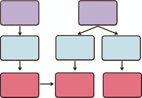
White Myf5+
expression. Further stimuli that can induce UCP1 expres- progenitor progenitor
sion include thyroid hormone, insulin, thiazolidinediones, cells cells
and retinoic acid. Glucocorticoids inhibit UCP1 gene ex-
pression in response to adrenergic stimulation [4]. Further,
rat experiments suggest that nicotine exerts its energy ex- White Brown
Myoblasts
penditure-increasing effect by activating BAT function [5]. adipoblasts adipoblasts
Development
White ? Brown
Myocytes
adipocytes adipocytes
Adipocytes (as well as myocytes) arise from the meso-
derm [6]. During fetal development, BAT emerges earlier
than WAT, which begins to develop in midgestation. It is
at its maximal size relative to body weight at birth. WAT
Fig. 1. Development of white and brown fat. Brown fat shares
depots in humans typically increase gradually through- common progenitor cells with muscle cells. It is uncertain wheth-
out life [4]. er white fat cells transdifferentiate into brown fat cells.
Previously, a common origin of white and brown adi-
pocytes was assumed. However, more recent evidence
does not support this concept. Rather, brown fat cells and
muscle cells both seem to be derived from the same stem tion, including induction of PRDM16 and UCP1 gene ex-
cells (fig. 1). pression [12]. Synthetic chemicals or endogenous factors
Indeed, apart from their lack of contractility, brown that activate PRDM16 function or mimic its action in
fat cells have much in common with muscle cells: muscle brown adipocyte development are of potential interest as
like brown fat is specialized for lipid catabolism rather future antiobesity drugs.
than storage, is innervated by the sympathetic nervous Importantly, however, there are two types of brown fat
system, contains abundant mitochondria, and facilitates cells [11]: besides the Myf5-positive cells described above,
adaptive thermogenesis [7]. Already in 2006, Atit et al. [8] which are located at classic locations for brown fat cells,
clearly showed that BAT and muscle (and dermis) had the there are brown fat cells interspersed in WAT which are
same developmental origin. Timmons et al. [9] observed negative for the Myf5 marker and thus are derived from
that both muscle cells and brown fat cells, in contrast to another lineage: they probably stem from the activation
white adipose fat cells, carry the marker myogenic factor of dormant precursor cells. Transdifferentiation from
5 (Myf5), which suggests a bipotent precursor cell for white to brown fat cells has been discussed as one possi-
muscle and brown adipocytes (but does not rule out the bility as well [1]. An interesting recent study demonstrates
possibility that these cells are derived from separate lin- that inhibition of peripheral cannabinoid type 1 receptor
eages of Myf5-positive cells). These findings were con- (CB1R) action in adipocytes directly promotes transdif-
firmed and extended by Seale et al. [10, 11]. These authors ferentiation of white adipocytes into a mitochondrion-
showed that the Zn-finger transcriptional regulator rich, thermogenic brown fat phenotype [13]. Enhanced
PRD1-BF1-RIZ1 homologous domain containing 16 thermogenesis, therefore, may represent a peripheral
(PRDM16) controls a bidirectional cell fate switch be- mechanism contributing to the weight loss and improved
tween skeletal myoblasts and brown fat cells. Loss of glucose homeostasis induced by the CB1R blocking agent
PRDM16 from brown fat precursors causes a loss of rimonabant.
brown fat characteristics and promotes muscle differen-
tiation. Conversely, ectopic expression of PRDM16 in
myoblasts induces their differentiation into brown fat Human Data
cells. PRDM16 stimulates brown adipogenesis by binding
to PPAR-␥ and activating its transcriptional function High-quality data on BAT in humans remain scarce.
[11]. Also, bone morphogenetic protein 7 (BMP7) was In human fetuses and newborns, BAT is found in axillary,
shown to specifically direct brown adipocyte differentia- cervical, perirenal, and periadrenal regions [4] but de-
Brown versus White Adipose Tissue: Gerontology 2012;58:15–23 17
A Mini-Reviewcreases shortly after birth and has been considered to be An important limitation of these studies is that no his-
irrelevant in adults until recently. Several investigations, tological verification of BAT was performed. In 2007, Ne-
however, now have conclusively demonstrated the pres- dergaard et al. [29] reviewed those observations from
ence and, more importantly, the functional relevance of PET-CT scans in a physiological perspective. Overall, the
BAT in adults. data extracted from the literature suggested that human
Already in 1981, an anatomical study described BAT depots of putative brown fat are somewhat differently lo-
in the neck region of outdoor workers but not in indoor cated from those in rodents, the main depots being found
workers [14]. The authors suggested a role of BAT in tem- in the supraclavicular and the neck regions with some ad-
perature regulation. Another morphological study [15] ditional paravertebral, mediastinal, para-aortic, and su-
found that there is a large amount of a tissue around the prarenal localizations. From the reviewed data the overall
adrenal glands and kidneys in patients with pheochro- conclusion was reached that BAT activity in man is in-
mocytoma that fulfills the ultrastructural and biochemi- duced by acute cold and is stimulated by the sympathetic
cal criteria for BAT. This pointed at the possibility of BAT nervous system. The prevalence of active BAT in the pop-
stimulation through adrenergic mechanisms. ulation was estimated to be in the range of some tens of
Brown adipocytes are metabolically highly active and percent.
can take up high amounts of glucose. Tissues with high Scientific interest in BAT was recently fuelled by the
rates of glucose incorporation can be identified with pos- publication of investigations in the New England Journal
itron emission tomography (PET) using radioactively la- of Medicine [24, 30, 31], which not only conclusively dem-
beled fluorodeoxyglucose (FDG). This technique is used onstrated the presence of BAT in adult humans but also
clinically to detect metabolically active tumor sites. For a its dependence on environmental temperature and its as-
long time, false-positive results had been obtained in the sociation with body weight.
parasternal or supraclavicular regions. By simultaneous Using PET, Virtanen et al. [31] found that cold-in-
examinations with FDG-PET and X-ray computed to- duced glucose uptake was increased by a factor of 15 in
mography (CT) these sites could be identified as adipose paracervical and supraclavicular adipose tissue in 5
tissue. This PET-CT technique has disclosed large healthy subjects. The authors obtained biopsy specimens
amounts of metabolically active BAT in adult humans. of this tissue from the first 3 consecutive subjects and
A series of investigators have retrospectively exam- documented messenger RNA (mRNA) and protein levels
ined scans from clinical monitoring studies of cancer pa- of the brown-adipocyte marker UCP1. Together with
tients. As these studies were performed under environ- morphologic assessment, which showed numerous mul-
mental conditions that could affect the outcome in an tilocular, intracellular lipid droplets, and with the results
uncontrolled way, the quantitative data reported are not of biochemical analysis, these findings documented the
meaningful and will not be discussed further here [16– presence of substantial amounts of metabolically active
26]. BAT in healthy adult humans.
In a study by Garcia et al. [27], two teenage female pa- van Marten Lichtenbelt et al. [30] studied 24 healthy
tients with abnormal FDG uptake patterns on PET scans men of whom 14 were overweight or obese under thermo-
performed with oral benzodiazepine administration un- neutral conditions and during mild cold exposure. BAT
derwent repeat imaging in temperature-controlled envi- activity was determined with the use of integrated FDG
ronment settings. The authors observed a resolution of PET-CT. Body composition and energy expenditure were
supraclavicular FDG uptake with temperature control in measured with the use of dual-energy X-ray absorptiom-
these two patients in whom benzodiazepines had no ef- etry and indirect calorimetry. BAT activity was observed
fect. in 23 of the 24 subjects (96%) during cold exposure but
Another study [28] described bilateral FDG tracer ac- not under thermoneutral conditions. This activity was
cumulation in the shoulder and lower neck of a patient significantly lower in the overweight or obese subjects
with extra-adrenal pheochromocytoma and high plasma than in the lean subjects. Body mass index (BMI) and
norepinephrine levels. The authors suggested that exces- percentage of body fat were both significantly negatively
sive sympathetic stimulation by high circulating cate- correlated with BAT, whereas the resting metabolic rate
cholamine concentrations may have augmented the met- was significantly positively correlated. The authors con-
abolic activity and tracer uptake in BAT and that radio- cluded that the percentage of young men with BAT was
nuclide imaging can noninvasively visualize human BAT high, but that its activity was reduced in overweight or
in terms of metabolic and functional activity. obese men.
18 Gerontology 2012;58:15–23 Saely /Geiger /DrexelIn response to these articles, several interesting obser- be considered that epicardial fat functions like brown fat
vations were published as letters to the editor. One impor- to protect the myocardium and coronary vessels from hy-
tant comment hypothesized that the prevalence of BAT pothermia. This process could be blunted in the elderly.
may have been underestimated because no repeat mea- Zingaretti et al. [36] analyzed samples of adipose tis-
surements were performed. Lee et al. [32] analyzed 4,834 sue from the neck of 35 patients undergoing surgery for
consecutive PET-CT scans of 2,934 patients. BAT was de- thyroid diseases. In 1/3 of the subjects (the younger and
tected in 250 patients, yielding a prevalence of 8.5%. leaner), distinct islands composed of UCP1-immunore-
Among 747 patients who underwent scanning more than active brown adipocytes could clearly be discerned. The
once, 145 patients had at least one positive scan, yielding brown adipose islands were richly sympathetically inner-
a much higher prevalence of almost 20%. vated (indicating acute central control), whereas adjacent
Timmons and Pedersen [33], using gene profiling, white adipose areas were not. Cells with features of brown
found that mRNA of the BAT marker UCP1 was posi- adipocyte precursors were found in pericapillary areas.
tively associated with BMI in human subcutaneous adi- A common shortcoming of the data discussed is the
pose tissue from 33 subjects. These associations with BMI cross-sectional approach which does not allow assump-
were in the opposite direction as those previously ob- tion of a causal relationship between low amounts of ac-
served and were markedly influenced by the presence of tive BAT and overweight or obesity. Alternatively, over-
diabetes. The authors pointed out that disseminated weight patients and in particular diabetic patients may
brown adipocytes within the large subcutaneous adipose be unable to sustain BAT stores. Lean adults may further
tissue mass may cumulatively represent substantial require increased BAT metabolism for nonshivering
brown adipocyte activity that may not be detected with thermogenesis to maintain body temperature. Whether
the use of PET-CT or crude biochemical studies. A limi- thinner patients have more BAT to meet these thermo-
tation of this observation, however, is that mRNA expres- genic needs or are thin because they have more active
sion cannot be translated into the amount of BAT activ- BAT at this stage is a classical hen-and-egg question
ity. Further studies would be needed to expand observa- which requires further study.
tions on UCP1 mRNA into hypotheses about functional
BAT activity in subcutaneous fat.
Saito et al. [34] also examined the prevalence of meta- The Impact of Aging
bolically active BAT in healthy adult humans and the ef-
fects of cold exposure and adiposity. FDG uptake into Mass, distribution and function of white fat undergo
adipose tissue was measured by PET-CT in 56 healthy dramatic changes throughout life. Fat mass reaches a
volunteers (31 male and 25 female subjects) aged 23–65 peak by middle or early old age, followed by a substantial
years. When exposed to cold for 2 h, 17 of 32 subjects aged decline in advanced old age. However, the observed de-
23–35 years and 2 of 24 subjects aged 38–65 years showed crease in total body fat with old age does not coincide
a substantial FDG uptake into adipose tissue of the supra- with a decline in percent body fat, which typically re-
clavicular and paraspinal regions, whereas they showed mains constant or increases. The age-associated decline
no detectable uptake when kept warm. Histological ex- in the sizes of adipose depots is accompanied by the ac-
aminations confirmed the presence of brown adipocytes cumulation of fat outside adipose tissue and loss of lean
in these regions. The cold-activated FDG uptake was in- body mass, particularly of muscle. Ectopic fat accumula-
creased in winter compared with summer and was in- tion occurs in bone marrow, muscle, liver, and at other
versely related to BMI and total and visceral fat areas es- sites, potentially contributing to age-dependent dysfunc-
timated from CT images at the umbilical level. These tion of these tissues [37].
findings indicate a high prevalence of metabolically ac- Further, different fat depots undergo age-related
tive BAT in adult humans and suggest a role in the control changes at different rates. Retro-orbital and peripheral
of body temperature and adiposity. The authors state that subcutaneous fat, for example, tend to be lost first, where-
if BAT were fully activated, it would burn an amount of as visceral fat is rather preserved. The earlier loss of sub-
energy equivalent to approximately 4.1 kg of adipose tis- cutaneous fat and consequent relative increase in intra-
sue over a year. abdominal fat with aging could predispose to metabolic
In another recent work, Sacks et al. [35] found that dysfunction and related health complications [37]. This
UCP1 is expressed at high levels in epicardial fat as com- indicates that depot-specific changes in fat tissue func-
pared to other fat depots. The possibility should therefore tion with aging may contribute to development of age-
Brown versus White Adipose Tissue: Gerontology 2012;58:15–23 19
A Mini-Reviewrelated metabolic disorders. On a cellular level, the capac- lying these beneficial effects of caloric restriction are not
ity of preadipocytes to become fully functional mature completely understood. However, it has been demon-
adipocytes declines with age [37]. Age-dependent lipo- strated that caloric restriction reduces the generation of
toxicity is related to a decreased capacity of adipose tissue free radicals by mitochondria [47] and prevents the age-
to store free fatty acids. The resulting lipotoxic environ- associated decline in mitochondrial function [48].
ment is detrimental to nonadipose tissues. This may con- Valle et al. [49] investigated the effect of long-term ca-
tribute to systemic lipotoxicity, and the increased preva- loric restriction on BAT mitochondrial function and bio-
lence of metabolic syndrome in older populations. genesis. In their study, caloric restriction induced resis-
The functionality of BAT is impaired, too, with in- tance to the loss of total and mitochondrial protein, COX
creasing age. The ability to regulate body temperature di- activity and uncoupling capacity with advancing age.
minishes with age both in humans and rodents [38, 39]. These results demonstrated that caloric restriction pre-
This decline in thermoregulation often results in a de- vents the age-associated decline of mitochondrial func-
crease in cold tolerance and in impaired body weight con- tion in BAT.
trol, promoting an age-related increase in body weight. In Far from being an inert reservoir for triglycerides, fat
rodent models, the age-associated decline in thermoreg- tissue is a highly active endocrine organ producing a wide
ulation has been related to BAT atrophy [40] which paral- array of peptide hormones, referred to as adipokines [50].
lels a loss of UCP1 activity [41]. This age-related decline The pathophysiological role of some of these peptide hor-
in BAT functionality has been shown to be influenced by mones has been extensively studied in recent years.
gender: females show a less severe loss of thermogenic ca- For example, adiponectin, the most abundant peptide
pacity with advancing age than males [42]. Pathophysi- secreted by adipocytes, enhances insulin sensitivity [50].
ologically, impairment of BAT function with increasing Low serum levels of adiponectin are associated with the
age appears to be mediated, at least in part, by disturbed metabolic syndrome and coronary atherosclerosis [51].
adrenergic signaling [43, 44]. Studies on aging humans revealed that enhanced adipo-
Ueno et al. [45] investigated the effect of aging on BAT nectin values are a distinctive feature of centenarians
activity in lean and obese (ob/ob) mice. Unlike old lean [52]. In humans, there is a significant positive relation-
animals, old obese animals exhibited decreased expres- ship between plasma adiponectin and age, even after ad-
sion of mRNA for UCP1. These findings suggest a marked justment for visceral adiposity [53].
decrease in BAT thermogenic capacity and activity in old Leptin, another important adipocyte-derived peptide
obese mice. Age and obesity thus seem to interact regard- hormone plays an important role in the regulation of food
ing their association with low amounts of BAT. intake and thermogenesis [50]. Insufficient action of
The interaction between body weight and age as con- leptin causes a reduction in metabolic rate in ob/ob mice
cerns their association with low amounts of BAT, which [54]. Leptin resistance has been implicated in the patho-
was observed in mice, may be present also in humans: in genesis of obesity-related complications involving abnor-
the above study by Saito et al. [34], the prevalence of cold- malities of lipid metabolism that resemble those of old
activated BAT activity decreased in elderly subjects: in a age. To determine whether development of leptin resis-
total of 24 elderly subjects, BAT was detected in only 2 tance in advancing age might account for such abnor-
males who were rather lean. In the above-mentioned malities, an interesting study [55] compared the effects of
study by Zingaretti et al. [36], the prevalence of brown fat hyperleptinemia induced by adenovirus gene transfer in
in the cervical adipose tissue was also positively corre- 2-month-old and 18-month-old lean wild-type Zucker
lated with young age and leanness. The authors pointed diabetic fatty rats. The leptin-induced decline in food in-
out that because of the covariation of these two factors, it take, body weight, and body fat in old rats was only 25,
was not possible to ascribe the high prevalence to young 50, and 16%, respectively, of that of young rats. Whereas
age or leanness separately. in young rats, plasma free fatty acids exhibited a 44% and
Mitochondria are the predominant cell organelles in triacylglycerol a 94% decrease, neither changed in the
brown adipocytes, as has been discussed above. Mito- older rats. In hyperleptinemic young rats, adipocyte ex-
chondria also play a crucial role in the aging process since pression of preadipocyte factor 1 increased dramatically
they are the main source of free radicals and, at the same and leptin mRNA virtually disappeared; there was in-
time, the most immediate target of oxidative damage. In creased expression of acyl CoA oxidase, carnitine palmi-
many species, caloric restriction can increase lifespan toyl transferase 1, and their transcription factor PPAR␣,
and delay the rate of aging [46]. The mechanisms under- accounting for the reduction in body fat. These hyperlep-
20 Gerontology 2012;58:15–23 Saely /Geiger /Drexeltinemia-induced changes were profoundly reduced in the ylases. SIR2 has been shown to extend lifespan in yeast,
old rats. On a high-fat diet, old rats consumed 28% more worms and flies [60]. The activation of sirtuin 1 (SIRT1),
calories than the young and gained 1.5 times as much fat, the mammalian ortholog of SIR2, resulted in enhanced
despite greater endogenous hyperleptinemia. These data lipid oxidation in BAT, skeletal muscle and liver cells and
illustrate that the actions of leptin decline with age and protected mice against diet-induced insulin resistance
that this could account for the associated abnormalities and obesity [61]. Further, SIRT1 regulates acetylation and
in lipid metabolism in the elderly. The role of obesity with transcriptional activity of FOXO, a member of the fork-
regard to the development of leptin resistance during ag- head transcription factors, in response to oxidative stress
ing is not definitely clear. Whereas some data point to a [62]. Deacetylation of FOXO3a results in the activation of
causal role of obesity in the development of leptin resis- genes involved in cell cycle arrest and resistance to oxida-
tance [56] other data suggest that obesity develops as a tive stress, away from its transcriptional targets involved
consequence of leptin resistance [57]. in apoptosis towards longevity [63]. Interestingly, resve-
Of note, leptin also facilitates long-term potentiation ratrol, which is present in grapes and red wine, is a potent
and synaptic plasticity in the hippocampus, promotes - activator of SIRT1 and inhibits preadipocyte prolifera-
amyloid clearance, and improves memory function in tion and adipogenic differentiation in a SIRT1-dependent
animal models of aging and Alzheimer’s disease; circu- manner [64].
lating leptin was recently shown to be associated with a
reduced incidence of dementia and Alzheimer’s disease
and with brain volume in asymptomatic older adults [58]. Conclusion
Although the discovery of longevity genes supports
the concept that life span is genetically determined, adi- Considerable amounts of BAT are present in a sub-
pose tissue seems to be a pivotal organ in the aging pro- stantial proportion of adult humans and relatively high
cess and in the determination of life span, as has been quantities of brown fat are associated with lower body
reviewed by Bluher [59]: leanness and caloric restriction weight. With increasing age, BAT decreases and body
increase longevity in various organisms, including mam- weight increases. Although the available cross-sectional
mals. Increased longevity in mice with a fat-specific dis- data do not allow definite conclusions to be drawn con-
ruption of the insulin receptor gene (FIRKO) suggests cerning a causal relationship between loss of BAT and
that reduced adiposity leads to an extended life span even increasing body weight with advancing age or with the
in the presence of normal or increased food intake. Re- obesity-related metabolic disorders of older age, stimula-
duced fat mass also has an impact on longevity in a num- tion of BAT appears an attractive novel candidate target
ber of other model organisms. In Drosophila, a specific for the treatment of age-related obesity.
reduction in body fat through overexpression of the fork-
head type transcription factor (dFOXO) extends life span.
Acknowledgements
One possible gene mediating the life-extending effect
of caloric restriction is silent information regulator 2 This work was supported by the Austrian Science Fund (FWF,
(SIR2), a member of the family of sirtuin histone deacet- Project 21057).
References 1 Cinti S: Transdifferentiation properties of 4 Gesta S, Tseng YH, Kahn CR: Developmen-
adipocytes in the adipose organ. Am J Physi- tal origin of fat: tracking obesity to its source.
ol Endocrinol Metab 2009. Epub ahead of Cell 2007;131:242–256.
print. 5 Mano-Otagiri A, Iwasaki-Sekino A, Ohata
2 Mattson MP: Perspective: does brown fat H, Arai K, Shibasaki T: Nicotine suppresses
protect against diseases of aging? Ageing Res energy storage through activation of sympa-
Rev 2010;9:69–76. thetic outflow to brown adipose tissue via
3 Wehrli NE, Bural G, Houseni M, Alkhawal- corticotropin-releasing factor type 1 recep-
deh K, Alavi A, Torigian DA: Determination tor. Neurosci Lett 2009;455:26–29.
of age-related changes in structure and func- 6 Enerback S: The origins of brown adipose
tion of skin, adipose tissue, and skeletal mus- tissue. N Engl J Med 2009;360:2021–2023.
cle with computed tomography, magnetic 7 Farmer SR: Brown fat and skeletal muscle:
resonance imaging, and positron emission unlikely cousins? Cell 2008;134:726–727.
tomography. Semin Nucl Med 2007; 37: 195–
205.
Brown versus White Adipose Tissue: Gerontology 2012;58:15–23 21
A Mini-Review8 Atit R, Sgaier SK, Mohamed OA, Taketo 20 Hany TF, Gharehpapagh E, Kamel EM, Buck 33 Timmons JA, Pedersen BK: The importance MM, Dufort D, Joyner AL, Niswander L, A, Himms-Hagen J, von Schulthess GK: of brown adipose tissue. N Engl J Med 2009; Conlon RA: -Catenin activation is neces- Brown adipose tissue: a factor to consider in 361:415–416. sary and sufficient to specify the dorsal der- symmetrical tracer uptake in the neck and 34 Saito M, Okamatsu-Ogura Y, Matsushita M, mal fate in the mouse. Dev Biol 2006; 296: upper chest region. Eur J Nucl Med Mol Im- Watanabe K, Yoneshiro T, Nio-Kobayashi J, 164–176. aging 2002;29:1393–1398. Iwanaga T, Miyagawa M, Kameya T, Nakada 9 Timmons JA, Wennmalm K, Larsson O, 21 Sturkenboom MG, Franssen EJ, Berkhof J, K, Kawai Y, Tsujisaki M: High incidence of Walden TB, Lassmann T, Petrovic N, Ham- Hoekstra OS: Physiological uptake of [18F] metabolically active brown adipose tissue in ilton DL, Gimeno RE, Wahlestedt C, Baar K, fluorodeoxyglucose in the neck and upper healthy adult humans: effects of cold expo- Nedergaard J, Cannon B: Myogenic gene ex- chest region: are there predictive character- sure and adiposity. Diabetes 2009; 58: 1526– pression signature establishes that brown istics? Nucl Med Commun 2004; 25: 1109– 1531. and white adipocytes originate from distinct 1111. 35 Sacks HS, Fain JN, Holman B, Cheema P, cell lineages. Proc Natl Acad Sci USA 2007; 22 Dobert N, Menzel C, Hamscho N, Worde- Chary A, Parks F, Karas J, Optican R, Ba- 104:4401–4406. hoff W, Kranert WT, Grunwald F: Atypical houth SW, Garrett E, Wolf RY, Carter RA, 10 Seale P, Kajimura S, Yang W, Chin S, Rohas thoracic and supraclavicular FDG-uptake in Robbins T, Wolford D, Samaha J: Uncou- LM, Uldry M, Tavernier G, Langin D, Spie- patients with Hodgkin’s and non-Hodgkin’s pling protein-1 and related messenger ribo- gelman BM: Transcriptional control of lymphoma. Q J Nucl Med Mol Imaging 2004; nucleic acids in human epicardial and other brown fat determination by PRDM16. Cell 48:33–38. adipose tissues: epicardial fat functioning as Metab 2007;6:38–54. 23 Rousseau C, Bourbouloux E, Campion L, brown fat. J Clin Endocrinol Metab 2009;94: 11 Seale P, Bjork B, Yang W, Kajimura S, Chin Fleury N, Bridji B, Chatal JF, Resche I, Cam- 3611–3615. S, Kuang S, Scime A, Devarakonda S, Conroe pone M: Brown fat in breast cancer patients: 36 Zingaretti MC, Crosta F, Vitali A, Guerrieri HM, Erdjument-Bromage H, Tempst P, Rud- analysis of serial 18F-FDG PET/CT scans. M, Frontini A, Cannon B, Nedergaard J, Cin- nicki MA, Beier DR, Spiegelman BM: Eur J Nucl Med Mol Imaging 2006; 33: 785– ti S: The presence of UCP1 demonstrates that PRDM16 controls a brown fat/skeletal mus- 791. metabolically active adipose tissue in the cle switch. Nature 2008;454:961–967. 24 Cypess AM, Lehman S, Williams G, Tal I, neck of adult humans truly represents brown 12 Tseng YH, Kokkotou E, Schulz TJ, Huang Rodman D, Goldfine AB, Kuo FC, Palmer adipose tissue. FASEB J 2009;23:3113–3120. TL, Winnay JN, Taniguchi CM, Tran TT, Su- EL, Tseng YH, Doria A, Kolodny GM, Kahn 37 Cartwright MJ, Tchkonia T, Kirkland JL: zuki R, Espinoza DO, Yamamoto Y, Ahrens CR: Identification and importance of brown Aging in adipocytes: potential impact of in- MJ, Dudley AT, Norris AW, Kulkarni RN, adipose tissue in adult humans. N Engl J Med herent, depot-specific mechanisms. Exp Kahn CR: New role of bone morphogenetic 2009;360:1509–1517. Gerontol 2007;42:463–471. protein 7 in brown adipogenesis and energy 25 Stefan N, Pfannenberg C, Haring HU: The 38 McDonald RB, Horwitz BA, Hamilton JS, expenditure. Nature 2008;454:1000–1004. importance of brown adipose tissue. N Engl Stern JS: Cold- and norepinephrine-induced 13 Perwitz N, Wenzel J, Wagner I, Buning J, J Med 2009;361:416–417. thermogenesis in younger and older Fischer Drenckhan M, Zarse K, Ristow M, Lilienthal 26 Au-Yong IT, Thorn N, Ganatra R, Perkins 344 rats. Am J Physiol 1988;254:R457–R462. W, Lehnert H, Klein J: Cannabinoid type 1 AC, Symonds ME: Brown adipose tissue and 39 Norman DC, Grahn D, Yoshikawa TT: Fever receptor blockade induces transdifferentia- seasonal variation in humans. Diabetes and aging. J Am Geriatr Soc 1985; 33: 859– tion towards a brown fat phenotype in white 2009;58:2583–2587. 863. adipocytes. Diabetes Obes Metab 2010; 12: 27 Garcia CA, Van ND, Majd M, Atkins F, Acio 40 Scarpace PJ, Matheny M, Borst SE: Thermo- 158–166. E, Sheikh A, Butler C: Benzodiazepine-resis- genesis and mitochondrial GDP binding 14 Huttunen P, Hirvonen J, Kinnula V: The oc- tant ‘brown fat’ pattern in positron emission with age in response to the novel agonist currence of brown adipose tissue in outdoor tomography: two case reports of resolution CGP-12177A. Am J Physiol 1992; 262:E185– workers. Eur J Appl Physiol Occup Physiol with temperature control. Mol Imaging Biol E190. 1981;46:339–345. 2004;6:368–372. 41 Cannon B, Nedergaard J: Brown adipose tis- 15 Ricquier D, Nechad M, Mory G: Ultrastruc- 28 Fukuchi K, Tatsumi M, Ishida Y, Oku N, sue: function and physiological significance. tural and biochemical characterization of Hatazawa J, Wahl RL: Radionuclide imaging Physiol Rev 2004;84:277–359. human brown adipose tissue in pheochro- metabolic activity of brown adipose tissue in 42 McDonald RB, Day C, Carlson K, Stern JS, mocytoma. J Clin Endocrinol Metab 1982; a patient with pheochromocytoma. Exp Clin Horwitz BA: Effect of age and gender on 54:803–807. Endocrinol Diabetes 2004; 112:601–603. thermoregulation. Am J Physiol 1989; 16 Cohade C, Mourtzikos KA, Wahl RL: ‘USA- 29 Nedergaard J, Bengtsson T, Cannon B: Unex- 257:R700–R704. Fat’: prevalence is related to ambient outdoor pected evidence for active brown adipose tis- 43 Langin D, Tavernier G, Lafontan M: Regula- temperature-evaluation with 18F-FDG PET/ sue in adult humans. Am J Physiol Endocri- tion of 3-adrenoceptor expression in white CT. J Nucl Med 2003;44:1267–1270. nol Metab 2007;293:E444–E452. fat cells. Fundam Clin Pharmacol 1995;9:97– 17 Cohade C, Osman M, Pannu HK, Wahl RL: 30 van Marken Lichtenbelt WD, Vanhommerig 106. Uptake in supraclavicular area fat (‘USA- JW, Smulders NM, Drossaerts JM, Kemerink 44 Scarpace PJ, Dove J, Matheny M: Effects of fat’): description on 18F-FDG PET/CT. J Nucl GJ, Bouvy ND, Schrauwen P, Teule GJ: Cold- age on -adrenergic subtype activation of Med 2003;44:170–176. activated brown adipose tissue in healthy adenylyl cyclase in brown adipose tissue. 18 Yeung HW, Grewal RK, Gonen M, Schoder men. N Engl J Med 2009;360:1500–1508. Proc Soc Exp Biol Med 1996; 213:262–267. H, Larson SM: Patterns of 18F-FDG uptake in 31 Virtanen KA, Lidell ME, Orava J, Heglind 45 Ueno N, Oh-ishi S, Segawa M, Nishida M, adipose tissue and muscle: a potential source M, Westergren R, Niemi T, Taittonen M, Fukuwatari Y, Kizaki T, Ookawara T, Ohno of false-positives for PET. J Nucl Med 2003; Laine J, Savisto NJ, Enerback S, Nuutila P: H: Effect of age on brown adipose tissue ac- 44:1789–1796. Functional brown adipose tissue in healthy tivity in the obese (ob/ob) mouse. Mech Age- 19 Truong MT, Erasmus JJ, Munden RF, Ma- adults. N Engl J Med 2009;360:1518–1525. ing Dev 1998;100:67–76. rom EM, Sabloff BS, Gladish GW, Podoloff 32 Lee P, Ho KK, Fulham MJ: The importance 46 Weindruch R, Sohal RS: Seminars in medi- DA, Macapinlac HA: Focal FDG uptake in of brown adipose tissue. N Engl J Med 2009; cine of the Beth Israel Deaconess Medical mediastinal brown fat mimicking malignan- 361:418–420. Center. Caloric intake and aging. N Engl J cy: a potential pitfall resolved on PET/CT. Med 1997;337:986–994. AJR Am J Roentgenol 2004;183:1127–1132. 22 Gerontology 2012;58:15–23 Saely /Geiger /Drexel
47 Colom B, Oliver J, Roca P, Garcia-Palmer FJ:       53 Koh SJ, Hyun YJ, Choi SY, Chae JS, Kim JY,        60 Guarente L, Picard F: Calorie restriction –
   Caloric restriction and gender modulate car-          Park S, Ahn CM, Jang Y, Lee JH: Influence of         the SIR2 connection. Cell 2005; 120: 473–
   diac muscle mitochondrial H2O2 production             age and visceral fat area on plasma adiponec-        482.
   and oxidative damage. Cardiovasc Res 2007;            tin concentrations in women with normal           61 Feige JN, Lagouge M, Canto C, Strehle A,
   74:456–465.                                           glucose tolerance. Clin Chim Acta 2008;389:          Houten SM, Milne JC, Lambert PD, Mataki
48 Hepple RT, Baker DJ, McConkey M, Muryn-               45–50.                                               C, Elliott PJ, Auwerx J: Specific SIRT1 activa-
   ka T, Norris R: Caloric restriction protects       54 Breslow MJ, Min-Lee K, Brown DR, Chacko              tion mimics low energy levels and protects
   mitochondrial function with aging in skele-           VP, Palmer D, Berkowitz DE: Effect of leptin         against diet-induced metabolic disorders by
   tal and cardiac muscles. Rejuvenation Res             deficiency on metabolic rate in ob/ob mice.          enhancing fat oxidation. Cell Metab 2008;8:
   2006;9:219–222.                                       Am J Physiol 1999;276:E443–E449.                     347–358.
49 Valle A, Guevara R, Garcia-Palmer FJ, Roca         55 Wang ZW, Pan WT, Lee Y, Kakuma T, Zhou            62 Yang Y, Hou H, Haller EM, Nicosia SV, Bai
   P, Oliver J: Caloric restriction retards the          YT, Unger RH: The role of leptin resistance          W: Suppression of FOXO1 activity by FHL2
   age-related decline in mitochondrial func-            in the lipid abnormalities of aging. FASEB J         through SIRT1-mediated deacetylation.
   tion of brown adipose tissue. Rejuvenation            2001;15:108–114.                                     EMBO J 2005;24:1021–1032.
   Res 2008;11:597–604.                               56 Fernandez-Galaz C, Fernandez-Agullo T,            63 Brunet A, Sweeney LB, Sturgill JF, Chua KF,
50 Galic S, Oakhill JS, Steinberg GR: Adipose            Perez C, Peralta S, Arribas C, Andres A,             Greer PL, Lin Y, Tran H, Ross SE, Mosto-
   tissue as an endocrine organ. Mol Cell Endo-          Carrascosa JM, Ros M: Long-term food re-             slavsky R, Cohen HY, Hu LS, Cheng HL, Je-
   crinol 2010;316:129–139.                              striction prevents ageing-associated central         drychowski MP, Gygi SP, Sinclair DA, Alt
51 Saely CH, Risch L, Hoefle G, Rein P,                  leptin resistance in Wistar rats. Diabetologia       FW, Greenberg ME: Stress-dependent regu-
   Muendlein A, Marte T, Aczel S, Langer P,              2002;45:997–1003.                                    lation of FOXO transcription factors by the
   Drexel H: Low serum adiponectin is inde-           57 Gabriely I, Ma XH, Yang XM, Rossetti L,              SIRT1 deacetylase. Science 2004; 303: 2011–
   pendently associated with both the metabol-           Barzilai N: Leptin resistance during aging is        2015.
   ic syndrome and angiographically deter-               independent of fat mass. Diabetes 2002; 51:       64 Fischer-Posovszky P, Kukulus V, Tews D,
   mined coronary atherosclerosis. Clin Chim             1016–1021.                                           Unterkircher T, Debatin KM, Fulda S,
   Acta 2007;383:97–102.                              58 Lieb W, Beiser AS, Vasan RS, Tan ZS, Au R,           Wabitsch M: Resveratrol regulates human
52 Bik W, Baranowska B: Adiponectin – a pre-             Harris TB, Roubenoff R, Auerbach S, DeCar-           adipocyte number and function in a Sirt1-
   dictor of higher mortality in cardiovascular          li C, Wolf PA, Seshadri S: Association of plas-      dependent manner. Am J Clin Nutr 2010;92:
   disease or a factor contributing to longer life?      ma leptin levels with incident Alzheimer dis-        5–15.
   Neuro Endocrinol Lett 2009;30:180–184.                ease and MRI measures of brain aging.
                                                         JAMA 2009;302:2565–2572.
                                                      59 Bluher M: Fat tissue and long life. Obes Facts
                                                         2008;1:176–182.

Brown versus White Adipose Tissue:                                              Gerontology 2012;58:15–23                                                 23
A Mini-Review
You can also read