Heterogeneity in patterns of helminth infections across populations of mountain gorillas (Gorilla beringei beringei)
←
→
Page content transcription
If your browser does not render page correctly, please read the page content below
www.nature.com/scientificreports

                OPEN              Heterogeneity in patterns
                                  of helminth infections
                                  across populations of mountain
                                  gorillas (Gorilla beringei beringei)
                                  Klara J. Petrželková1,2,3*, Carine Uwamahoro4, Barbora Pafčo1, Barbora Červená1,5,
                                  Peter Samaš1, Antoine Mudakikwa6, Richard Muvunyi6, Prosper Uwingeli6, Kirsten Gilardi7,
                                  Julius Nziza7, Jean Bosco Noheri7, Winnie Eckardt4, Felix Ndagijimana4, Benard Ssebide7,
                                  Ricky Okwirokello7, Fred Nizeyimana7, Eddy Kambale Syaluha7, Gaspard Nzayisenga7,
                                  Luis Flores Girón8,9, Méthode Bahizi7, Adrien Emile Ntwari7, Jean‑Paul Lukusa7,
                                  Jean Claude Tumushime7, Damien Mangura7, Jeff Mapilanga10, Arthur Kalonji11,
                                  Robert Aruho12, Anna Stryková5, Zuzana Tehlarová1, Rita Cameira1,5, Linda Lowenstine13,
                                  Jan Šlapeta14, Dušan Romportl15, Nicola Ferrari16,17, Michael Cranfield7 & David Modrý2,5,18

                                  Conservation efforts have led to the recovery of the endangered mountain gorilla populations. Due
                                  to their limited potential for spatial expansion, population densities increased, which may alter the
                                  epidemiology of infectious diseases. Recently, clinical gastrointestinal illnesses linked to helminth
                                  infections have been recorded in both gorilla populations. To understand drivers and patterns of
                                  helminth infections we quantified strongylid and tapeworm infections across both Virunga Massif
                                  and Bwindi populations using fecal egg counts. We assessed the impact of age, sex, group size,
                                  season and spatial differences used as a proxy, which reflects observed variation in the occurrence
                                  of gastrointestinal problems, vegetation types, gorilla subpopulation growth and associated social
                                  structure on helminth infections. We revealed striking geographic differences in strongylid infections
                                  with higher egg counts mostly in areas with high occurrences of gastrointestinal disease. Increased
                                  helminth egg counts were also associated with decreasing group size in some areas. Observed spatial
                                  differences may reflect mutual effects of variations in subpopulation growth rates, gorilla social
                                  structure, and vegetation associated with altitude across mountain gorilla habitat. Helminth infection
                                  intensities in Virunga gorillas were lowest in the youngest and the oldest animals. Elucidating parasite
                                  infection patterns of endangered species with low genetic diversity is crucial for their conservation
                                  management.

                                  1
                                   Institute of Vertebrate Biology, The Czech Academy of Sciences, Brno, Czech Republic. 2Institute of Parasitology,
                                  Biology Centre, The Czech Academy of Sciences, Ceske Budejovice, Czech Republic. 3Liberec Zoo, Liberec, Czech
                                  Republic. 4Dian Fossey Gorilla Fund, Musanze, Rwanda. 5Department of Pathology and Parasitology, Faculty of
                                  Veterinary Medicine, University of Veterinary Sciences Brno, Brno, Czech Republic. 6Rwanda Development Board,
                                  Kigali, Rwanda. 7Gorilla Doctors (MGVP, Inc.), Davis, CA, USA. 8Centre de Recherche en Sciences Naturelles
                                  de Lwiro, Lwiro, Democratic Republic of Congo. 9Lwiro Primate Rehabilitation Center, Democratic Republic of
                                  Congo and Ivan Carter Wildlife Conservation Alliance, Orlando, FL, USA. 10Institut Congolais pour la Conservation
                                  de la Nature, Kinshasa, Democratic Republic of Congo. 11Institut Congolais pour la Conservation de la Nature,
                                  Parc National de Kahuzi Biega, Bukavu, Democratic Republic of Congo. 12Uganda Wildlife Authority, Kampala,
                                  Uganda. 13School of Veterinary Medicine, University of California Davis, Davis, CA, USA. 14Sydney School of
                                  Veterinary Science, Faculty of Science, University of Sydney, Sydney, Australia. 15Department of Physical
                                  Geography and Geoecology, Faculty of Science, Charles University, Prague, Czech Republic. 16Department of
                                  Veterinary Medicine, Università degli Studi di Milano, Milan, Italy. 17Research Center Epidemiology and Molecular
                                  Surveillance of Infections ‘‘EpiSoMI’’, Università degli Studi di Milano, Milan, Italy. 18Department of Botany and
                                  Zoology, Faculty of Science, Masaryk University, Brno, Czech Republic. *email: petrzelkova@ivb.cz

Scientific Reports |   (2021) 11:10869                | https://doi.org/10.1038/s41598-021-89283-4                                                1

                                                                                                                                             Vol.:(0123456789)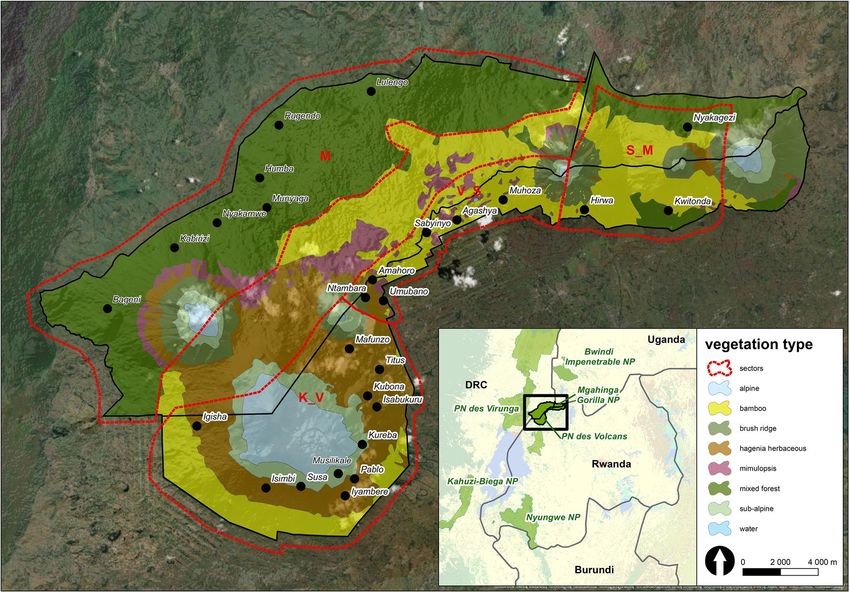
www.nature.com/scientificreports/
Endangered mountain gorillas (Gorilla beringei beringei) live in two populations and entirely within protected
areas: one ranging within the Virunga Massif (VM) spanning the borders of Rwanda, Uganda and the Democratic
Republic of the Congo (DRC), and the second ranging in the Bwindi-Sarambwe ecosystem in Uganda and DRC.
The VM population declined dramatically in 1 970s1, but the numbers stabilized in the 1980s, and since then
the population has been steadily increasing with annual growth rates around 3–4% between 2003 and 20162,3.
However, this increase has been almost entirely attributed to the growth rate in habituated g roups2–4 due to the
“extreme conservation” measures these groups benefit from, such as daily group monitoring and protection, vet-
erinary interventions and anti-poaching p atrols5. Moreover, the population increase was not observed uniformly
across VM, which may be due to varying ecological conditions that are linked to different habitat types3,4,6,7. We
have less information about the history of the Bwindi-Sarambwe population dynamics. Robbins et al.8 previously
indicated that this second mountain gorilla population showed little or no growth before 2009 in comparison to
VM population, but subsequent censuses in 2011 and 2018 detected growth of the population9,10.
Although both mountain gorilla populations are growing, their potential spatial expansion is extremely
limited due to high human densities adjacent to protected areas inhabited by mountain gorillas. Consequently,
increasing numbers of gorillas in both populations are likely to result in rising individual and group densities
at least in some areas. The VM population has already exceeded the previously predicted carrying capacity of
the environment11, as defined by the maximum number of animals an area will support based on r esources9.
In addition, between Mt. Karisimbi and Mt. Visoke (aka Bisoke) in the VM, where some of the highest growth
rates of gorilla numbers occurred3,4, the mountain gorillas experienced major changes in the social structure
leading locally to a threefold increase in group densities. Namely, three stable multi‐male groups, which have
been steadily growing from the mid‐1990s until 2006, underwent a series of group fissions and formation of
smaller groups including multiple one-male g roups12,13. As a result, there has been an increase in home range
overlap, a reduction in home range areas used exclusively by a single group and annual intergroup encounter rates
tripled in this area13. In addition, small groups, which are often one-male groups, may experience higher stress
levels than large groups, which are often multi-male groups, because they have higher home range overlaps with
neighboring groups, inter-group encounter rates, and are more vulnerable to infanticide by external m ales14–17.
All those observed changes in population dynamics, social structure, and habitat use may be altering stress levels
and cause changes in pathogen epidemiology with subsequent health problems12,18,19.
Parasites influence the dynamics of free-living animal populations20,21 through their impact on host survival22
or reproduction4. The severity of parasite-induced diseases often depends on the intensity of infection (number of
parasite individuals in the host animal)23. Identification of factors affecting parasite infections and their intensities
as well as resulting patterns of parasite infections across host populations is essential for optimizing effectiveness
of parasite control strategies and management of wildlife, particularly for species of conservation concerns24.
For soil-transmitted parasites, host population density and habitat use play a central role in the host-parasite
interaction by increasing the probability that parasite infectious stages will be ingested by a host25. Data from
6670 individual hosts representing 19 mammalian species showed a strong positive correlation between the host’s
population density and average gastrointestinal (GIT) strongylid nematode infection i ntensity26. In non-human
primates this phenomenon has been demonstrated in fragmented populations, where host densities may increase
due to restricted available habitat (e.g.27,28). In addition, host habitat characteristics can alter the risk of helminth
infections29. For example, plant species composition, the leaf form of p lants30, temperature, and h umidity31,32
29
are known to influence helminth larval abundance, development and their migratory behavior . Moreover, it is
generally accepted that parasite abundance decreases with elevation33, but it is not always true across all parasite
taxa. For example, the survival of Cooperia and Ostertagia strongylids in livestock is optimized for cooler weather
due to larval inhibition and thus the infection risk increases at higher e levations34. Seasonal differences in hel-
minths (especially strongylid nematodes) are commonly observed in non-human primates (e.g.35–38). However,
some studies reported highest infection intensities in the wet, while others in the dry season. The impact of
host factors, such as sex and age, on parasite infections has also been well documented in non-human primates
(e.g.38,39). It is widely accepted that males of vertebrate hosts tend to exhibit higher rates of parasitism and disease
than females40–42, with several main factors claimed to cause sex differences in parasitism: body size dimorphism
with possibly larger nutritional requirements in the larger sex, social relationship, diet, habitat and h ormones43.
Three theoretical models are generally used to describe the host age–infection relationship in parasites; Type I
(linear) and Type II (asymptotic) relationships are thought to occur in the absence of an effective host immune
response and instead depend on immigration-death processes linked to parasite transmission and longevity44.
Type III (convex) associations have been suggested to indicate adaptive immunity to parasites45 and have been
reported in non-human primates36.
Gastrointestinal helminthiases are typically asymptomatic in wild non-human primates, but host factors like
immune status, or extrinsic factors, such as habitat loss, fragmentation and other anthropogenic pressures, can
alter the transmission dynamics of helminths and increase host susceptibility, which may exacerbate negative
effects of infections on h ealth46,47. However, clinical diseases caused by parasites have been observed in moun-
tain gorillas; post-mortem examinations of 60 gorillas from 1985 to 2007 (Mountain Gorilla Veterinary Project
[MGVP], publication in review) revealed histopathologic evidence for enteritis in more than 50% of cases,
gastritis in more than 35% of cases, and colitis in more than 25% of cases. More recently (2013–2017), several
cases of severe gastritis have been diagnosed in VM in Rwanda, particularly in adult gorillas ranging between Mt.
Karisimbi and Mt. Visoke, with most cases involving young silverbacks (MGVP, data on file). Trichostrongylid
nematodes have been observed in the stomach and small intestines in these cases and the histopathology of
observed stomach lesions are typical of those caused by nematodes in a range of domestic and wild mammals,
including non-human primates48–50. In Bwindi Impenetrable National Park (BINP), the Ugandan part of the
Bwindi-Sarambwe ecosystem, similar alarming observations indicating change in parasitic infections have been
recorded (MGVP, data on file). Many gorillas have exhibited weight loss, declining body condition and poor
Scientific Reports | (2021) 11:10869 | https://doi.org/10.1038/s41598-021-89283-4 2
Vol:.(1234567890)www.nature.com/scientificreports/
Figure 1. Division of the Virunga Massif into four areas according to volcano positions (M: Mikeno, Virunga
National Park; K_V: Karisimbi–Visoke, Volcanoes National Park; V_S: Visoke–Sabyinyo, Volcanoes National
Park; S_M: Sabyinyo–Muhavura, Mgahinga Gorilla National Park, Volcanoes National Park) with vegetation
zones (based on Robbins et al.5) and distribution of the studied groups. Map was created using ArcGIS Desktop
10.6 (ESRI 2019. ArcGIS Desktop: Release 10.6. Redlands, CA: Environmental Systems Research Institute; esri.
com). Vegetation data were adopted according to Robbins et al.5; boundaries of protected areas were derived
from ProtectedPlanet.net database.
haircoats in recent years, and in severe cases (n > 10) anthelmintic treatment were administered to sick goril-
las in BINP resulting in marked clinical improvement. Anoplocephalid tapeworms have also been commonly
observed during necropsies in gorillas from both VM and BINP, and intensities of infection have been high in
some animals (e.g., > 100 tapeworms in one animal) (MGVP, data on file).
Although parasites of mountain gorillas have been studied for almost 100 y ears51, surveys of the spatio-
temporal pattern of parasite infections over the entire geographic distribution of mountain gorillas are lacking
(for basic studies covering gorilla subpopulations see e.g. 38,52–54). We conducted an extensive species-wide survey
of helminth infections across the VM and BINP populations and across different seasons to uncover the driv-
ers and patterns of helminth infections and provide a comprehensive foundation for future assessments of the
impact of these parasites on gorilla population dynamics. We utilized samples collected from night nests across
both populations and samples from individually-identified gorillas from five social groups in VM in Rwanda that
experienced differing levels of strongylid infections based on a small-scale pilot study using the same methods
applied in this larger-scale study. We divided the VM into four areas according to volcano positions (modification
of Weber and V edder1, Fig. 1). This geographic division allowed for a comparison of areas with differences in the
occurrence of parasite-related GIT disease, as well as differences in vegetation types, and in historical subpopu-
lation growth and associated current social structure across mountain gorilla distribution r ange55,56. The areas
were namely: Karisimbi–Visoke (K_V), Visoke–Sabyinyo (V_S), Sabyinyo–Muhavura (aka Muhabura) (S_M),
Mikeno (M). The entirety of BINP was considered as one area. We then investigated the impact of area, sampling
period (season), group size and host factors (sex, age) on helminth infections.
In the samples collected from the night nests, we predict that the helminth infections (quantified as eggs per
gram in fecal samples; hereafter EPG or egg counts) are higher in the areas with a high occurrence of GIT prob-
lems (K_V) than in areas where less or no GIT problems were reported (M, S_M). We predicted that the helminth
infections are higher in silverback gorillas in comparison to other age/sex classes and higher in smaller gorilla
groups than in larger groups as smaller groups may experience more stress. We further predicted that these effects
of age/sex class and group size on the helminth infections are stronger in the areas with higher occurrence of GIT
problems and historically higher subpopulation growth. In individually sampled gorillas we predicted that the
helminth infections are higher in males than in females, change with age following the convex pattern, and differ
between groups as our small-scale pilot project involving five groups suggested. We predicted that age and sex
effects are more pronounced in groups with higher helminth egg counts and that convex pattern of egg counts
Scientific Reports | (2021) 11:10869 | https://doi.org/10.1038/s41598-021-89283-4 3
Vol.:(0123456789)www.nature.com/scientificreports/
Park Area (Weber & Vedder, 1983)1 Sampling period (2018) Season Sample type
Karisimbi–Visoke (K_V) January/February Dry Nest
VoNP, Rwanda Visoke–Sabyinyo (V_S) May/June Boundary wet/dry Individual
Sabyinyo–Muhavura (S_M) September/October Wet Nest + individual
January/February Dry Nest
ViNP, DRC Mikeno (M)
September/October Wet Nest
MGNP, Uganda Sabyinyo–Muhavura (S_M) September/October Wet Nest
BINP, Uganda BINP September/October Wet Nest
Table 1. Sampling design by area (with a national park and country), sampling period and its corresponding
season and sample type (for more detailed information about sampled groups, see the Supplementary
Table S1). VoNP Volcanoes National Park in Rwanda, ViNP Virunga National Park in Democratic Republic of
the Congo, MGNP Mgahinga Gorilla National Park in Uganda, BINP Bwindi Impenetrable National Park in
Uganda.
is more pronounced in males than females. In both night nest and individual samples we expect differences in
helminth infections between sampling periods, which correspond with different seasons and predicted higher
egg counts in the wet season38.
Results
We used three different datasets to test our hypotheses aiming to explain drivers and patterns of helminth
infections across five subpopulations (K_V, V_S, S_M, M, BINP) of the mountain gorillas (Fig. 1, Table 1 and
Table S1, for details see “Material and methods”). Dataset 1 included nest samples collected across the whole
VM and BINP during a single period (n = 392) and was used to investigate the effects of area, age/sex class and
group size. Dataset 2 was composed of nest samples but restricted to VM covering two periods (January/Febru-
ary and September/October 2018) (n = 491) and used to examine the effect of area, age/sex class, group size and
sampling period (season). Dataset 3 with samples from individually identified gorillas from selected groups in
VM collected across two periods (May/June 2018 and September/October 2018) (n = 450) was used to investigate
the effects of group identity, sex, exact a ge2 and sampling period (season).
We observed helminth eggs in the majority of examined gorilla fecal samples (Fig. 2): in dataset 1, the preva-
lence of the strongylids was 97% (median EPG = 480; range 0–10,050), while the prevalence of the tapeworms
was 88% (median EPG = 188; range 0–5775). For the information on strongylid and tapeworm infections in
datasets 2 and 3 see Table S1.
Strongylid infections. In dataset 1, we detected an interactive effect of area and age/sex class (Wald’s Chi-
square = 26.7, df = 12, p = 0.008; Table 2) with no clear pattern in M and S_M areas, while in other areas the
infants had lower EPG values than non-infant classes (Fig. 3A). M and S_M areas had similar EPG values (Wald’s
Chi-square = 1.7, df = 1, p = 0.19) and both had lower EPG values than the other areas (Wald’s Chi-square test,
all p < 0.001; Fig. 3A, Table S1). Area K_V had lower EPG values than V_S and BINP areas (Wald’s Chi-square
test, both p < 0.01), while the V_S and BINP had similar infection rates (Wald’s Chi-square = 0.1, df = 1, p = 0.92).
Infants had lower EPG values than any other age/sex classes (Wald’s Chi-square test, all p < 0.01), while no dif-
ferences were observed among other sex/age classes (Fig. 3A, Table S1).
In dataset 2, we found an interactive effect of area and group size (Wald’s Chi-square = 20.0, df = 3, p < 0.001;
Table 3). The EPG values decreased with increasing group size in the S_M area (estimate = − 0.14 ± 0.03; z = − 5.0,
p < 0.001) but there was no relationship in other areas (Fig. 4A). All the areas differed from each other (Wald’s
Chi-square test, all p < 0.04; Fig. 4A,B, Table 3). EPG values were lower in infants compared to other age/sex
classes (Wald’s Chi-square test, all p < 0.002), while no differences were observed among other age/sex classes
(Fig. 4B, Table S1).
In dataset 3, EPG values differed among groups (Wald’s Chi-square = 323.9, df = 4, p < 0.001; Table 4) with
Kwitonda group (from S_M sector) showing lower EPG values compared to any other group (Wald’s Chi-
square test, all p < 0.001), while the other groups had similar values (Fig. 5A, Table S1). There was a quadratic
age effect (Wald’s Chi-square = 6.8, df = 2, p = 0.03) on strongylid EPG values with a peak occurring between
10 and 30 years (Fig. 5A). Gorillas showed higher EPG values in September/October (wet season) (estimated
marginal mean ± SE = 645 ± 197 EPG) compared to May/June (boundary wet/dry season) (429 ± 130; Wald’s
Chi-square = 41.3, df = 1, p < 0.001; Fig. 5B).
Tapeworm infections. In dataset 1, there was interactive effect of area with both group size (Wald’s Chi-
square = 10.8, df = 4, p = 0.03) and age/sex class (Wald’s Chi-square = 32.1, df = 12, p = 0.001) on tapeworm EPG
values (Table 2, Fig. 3B,C). Specifically, the EPG values decreased with increasing group size in K_V (esti-
mate = − 0.11 ± 0.03; z = − 4.3, p < 0.001) and M area (estimate = − 0.08 ± 0.03; z = − 2.3, p = 0.02; Fig. 3B) but no
relationship was found in other areas. Differences in EPG values between areas were detected only between
K_V and BINP (Wald’s Chi-square = 10.0, df = 1, p = 0.002), with higher values in BINP. There was a significant
interaction of area and age/sex on EPG values, where K_V area showed a non-significant tendency to have lower
EPG values in infants than in other age/sex classes while other areas had rather similar EPG values across age/
sex classes (Fig. 3C).
Scientific Reports | (2021) 11:10869 | https://doi.org/10.1038/s41598-021-89283-4 4
Vol:.(1234567890)www.nature.com/scientificreports/
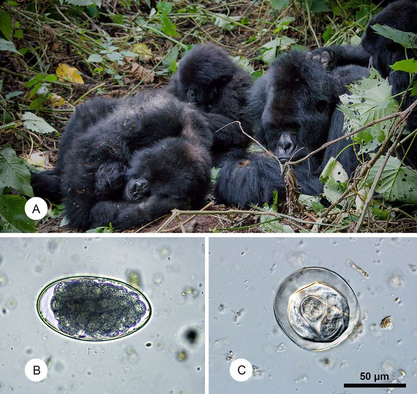
Figure 2. Examined host and their parasites quantified in the study: (A) resting group of mountain gorillas in
Virunga Massif (Virunga National Park, Democratic Republic of the Congo); (B) egg of strongylid nematode;
(C) egg of anoplocephalid tapeworm; both microphotographs in the same scale.
Strongylids Tapeworms
Effect d.f. Chi_square P value Chi_square P value
Area 4 264.6 < 0.001 19.4 < 0.001
Age/sex 3 33.9 < 0.001 8.4 0.04
Group size 1 0.01 0.91 14.4 < 0.001
Area: age/sex 12 26.7 0.008 32.1 0.001
Area: group size 4 2.7 0.61 10.8 0.03
Table 2. Factors influencing helminth infections in Virunga Massif and Bwindi Impenetrable National Park
(dataset 1: nest samples from all national parks).
In dataset 2, we found an interactive effect of area and group size (Wald’s Chi-square = 14.0, df = 3, p = 0.003;
Table 3, Fig. 4C). The EPG values decreased with increasing group size in the K_V area (estimate = − 0.12 ± 0.02;
z = − 5.2, p < 0.001) but no changes in EPG values with group size were observed in other areas (Fig. 4C). EPG
values in K_V area differed significantly from S_M (Wald’s Chi-square = 5.1, df = 1, p = 0.02) and V_S areas
(Wald’s Chi-square = 6.5, df = 1, p = 0.01; Fig. 4C, Table S1). Similar to strongylid EPG values, infants had lower
EPG values for tapeworms compared to any other age/sex classes (Wald’s Chi-square test, all p < 0.01), while the
egg counts between other classes were similar (Fig. 4D, Table S1).
In dataset 3, there was an interactive effect of quadratic age and group identity on EPG values (Wald’s Chi-
square = 40.9, df = 8, p < 0.001; Table 4). In particular, EPG values peaked between the age of 10 and 20 years in
groups Titus, Hirwa and Kureba with the highest peak found in Titus group (Fig. 5C, Tables 4 and S1). Kwitonda
group had the lowest maximum EPG value with the latest peak (age = 26 years). Individuals had higher EPG
Scientific Reports | (2021) 11:10869 | https://doi.org/10.1038/s41598-021-89283-4 5
Vol.:(0123456789)www.nature.com/scientificreports/
A B
C
Figure 3. Models’ predicted mean values with 95% confidence intervals for the factors affecting helminth
infections in mountain gorillas in Virunga Massif and Bwindi Impenetrable National Park (dataset 1). (A)
strongylid eggs per gram (EPG)—sampling area and age/sex class effects, (B) tapeworm EPG—sampling area
and group size effects, (C) tapeworm EPG—sampling area and age/sex class effects. juv/sub: juvenile/subadult
class; M: Mikeno (Virunga National Park); K_V: Karisimbi–Visoke (Volcanoes National Park), V_S: Visoke–
Sabyinyo (Volcanoes National Park), S_M: Sabyinyo–Muhavura (Mgahinga Gorilla National Park, Volcanoes
National Park).
Strongylids Tapeworms
Effect d.f. Chi_square P value Chi_square P value
Area 3 376.3 < 0.001 8.6 0.03
Age/sex 3 41.6 < 0.001 21.6 < 0.001
Group size 1 6.1 0.01 15.2 < 0.001
Season 1 0.04 0.85 0.07 0.79
Area: age/sex 9 15.0 0.09 15.2 0.09
Area: group size 3 20.0 < 0.001 14.0 0.003
Table 3. Factors influencing helminth infections in Virunga Massif (dataset 2: nest samples from Volcanos
and Virunga National Parks from wet and dry season).
values in September/October (wet season) (estimated marginal mean ± SE = 625 ± 97 EPG) compared to May/
June (boundary wet/dry season) (361 ± 97 EPG; Wald’s Chi-square = 37.6, df = 1, p < 0.001; Fig. 5D).
Discussion
The majority of mountain gorilla fecal samples examined were positive for both strongylid nematodes and anop-
locephalid tapeworms, which is in accordance with previous studies of mountain gorilla gastrointestinal (GIT)
parasites38,52,53,57. The overall highest strongylid egg counts were recorded in the gorilla groups in the BINP, where
individual cases of weight loss and poor body condition have been documented, followed by the groups from the
Visoke–Sabyinyo area (V_S) of the VM. Gorilla groups ranging in the Karisimbi–Visoke area (K_V), where the
highest number of severe gastritis in the VM have been documented, had notably high strongylid EPG values,
especially when compared with the groups ranging in the Mikeno (M in in DRC) and the Sabyinyo–Muhavura
(S_M) area of the VM, which exhibited substantially lower egg counts and no fatal GIT diseases in recent years
(MGVP, unpublished data). Strongylid egg counts increased with decreasing group size in the S_M area (without
Scientific Reports | (2021) 11:10869 | https://doi.org/10.1038/s41598-021-89283-4 6
Vol:.(1234567890)www.nature.com/scientificreports/
A B
C D
Figure 4. Models’ predicted mean values with 95% confidence intervals for the factors affecting helminth
infections in mountain gorillas in Virunga Massif in two periods (dataset 2). (A) strongylid egg per gram
(EPG)—sampling area and group size effects, (B) strongylid EPG—sampling area and age/sex class effects,
(C) tapeworm EPG—sampling area and group size effects, (D) tapeworm EPG—sampling area and age/sex
class effects. juv/sub: juvenile/subadult class; M: Mikeno (Virunga National Park); K_V: Karisimbi–Visoke
(Volcanoes National Park), V_S: Visoke–Sabyiniyo (Volcanoes National Park), S_M: Sabyinyo–Muhavura
(Volcanoes National Park).
Strongylids Tapeworms
Effect d.f. Chi_square P value Chi_square P value
Age2 2 6.8 0.03 9.2 0.01
Group ID 4 323.9 < 0.001 16.8 0.002
Season 1 41.3 < 0.001 29.6 < 0.001
Sex 1 1.8 0.18 0.8 0.38
Age2: group ID 8 11.7 0.17 40.9 < 0.001
Age2: sex 2 2.4 0.31 3.1 0.21
Group ID: sex 4 0.4 0.98 7.5 0.11
Table 4. Factors influencing helminth infections in the individual samples from five selected groups from
Volcanos National Park collected in wet and dry season.
GIT problems) and we did not observe higher helminth infections in silverbacks compared to other age/sex
classes, which does not support our predictions. On the other hand, according to our prediction tapeworm infec-
tion intensities increased with decreasing group size in an area with high occurrence of GIT problems (K_V) but
also in an area with no GIT problem (M). The observed differences in helminth infection rates among sampling
areas thus only partially reflect occurrence of detected GIT problems in mountain gorillas.
Mutual effects of different population growth of VM mountain gorilla subpopulations and the BINP popu-
lation, differences in the social structure particularly in VM areas, as well as differences in habitat characteris-
tics (vegetation types interlinked with altitude) across the distribution range of mountain gorillas may explain
observed differences in strongylid infections among areas. Unfortunately, information about current gorilla
densities across the sampled areas are lacking, but different population dynamics have been observed in the
VM areas with low strongylid egg counts (see M and S_M in Fig. 4A) in comparison to the other VM areas. For
Scientific Reports | (2021) 11:10869 | https://doi.org/10.1038/s41598-021-89283-4 7
Vol.:(0123456789)www.nature.com/scientificreports/
A B
C D
Figure 5. Models’ predicted mean values with 95% confidence intervals for the factors affecting helminth
infections in individually sampled mountain gorillas in Virunga Massif (dataset 3). (A) strongylid eggs per
gram (EPG)—group identity and age effects, (B) strongylid EPG—sampling period (season) and group identity
effects, (C) tapeworm EPG—group identity and age effects, (D) tapeworm EPG—sampling period (season) and
group identity effects.
example, the VM population decline between 1960 and 1973 was driven by a severe decrease in the M (Mikeno)
subpopulation in DRC (67%), accounting for 85% of the total VM decline, while in the other VM areas the
population decline was much lower1. Since the 1980s the number of gorillas in M either declined a gain7 or did
not significantly increase4,6. Considerably less gorillas have been found historically in S_M than in other areas
of the VM and the size of this subpopulation has been stable for y ears1,3,4,6,7. In contrast, in the central areas of
VM where strongylid egg counts were high (e.g. in K_V and V_S), the increase in gorilla numbers and groups
since the 1980s has been the most s ignificant1,3,4,7,11.
Our results thus indirectly suggest that high growth rates of gorilla subpopulations in specific VM areas in the
last 40 years can be linked to high strongylid infection intensities found in these areas today. Existing data from
BINP does not allow comparisons of population changes between VM and BINP, but although differences in
strongylid egg counts among groups in BINP were observed (see Supplementary Table S1), we did not detect any
groups with apparent low EPG, as was observed in some areas of VM (M and S_M areas). However, groups from
the northern BINP area, which is characterized by lower gorilla and group densities as well as medium-altitude
moist evergreen f orest9, were not included in this study and thus should be investigated in future sampling efforts.
Comparisons of our results with previous studies conducted in BINP are difficult due to significant differences
in employed quantification methods. However, egg counts reported in previous studies were also high, exceed-
ing 1000 and reaching values up to 11,000 EPG even in formalin-fixed fecal samples, where lower egg counts
are always observed due to fixation38,53,54. Comparison of strongylid egg counts between both mountain gorilla
populations and conclusions on observed high strongylid egg counts in BINP should be done with caution, as
BINP is characterized by different vegetation linked to different environmental factors, presence of different
sympatric non-human primates9 and thus the structure of the strongylid communities could differ between both
gorilla populations. The very high egg counts in BINP groups and observed clinical cases of chronic weight loss
and declining body condition, which may be attributable at least in part to helminth infection intensities warrant
further and more detailed research of helminth infections across BINP including precise strongylid identifica-
tion. In general, further studies with detailed information about current gorilla and group densities across the
whole VM and BINP are urgently needed to fully understand the ability of helminths, in particular strongylids,
to regulate the growth of mountain gorilla populations.
Scientific Reports | (2021) 11:10869 | https://doi.org/10.1038/s41598-021-89283-4 8
Vol:.(1234567890)www.nature.com/scientificreports/
Observed relatively high strongylid EPG values and the increase of tapeworm EPG values with decreas-
ing group size in K_V may be associated with the disintegration of a few larger multi-male groups after 2006,
which led to high group densities, a higher proportion of one‐male groups and groups below average size in this
area12,13. One-male groups, which are often smaller in size, are more vulnerable to attacks from external males,
which can cause lethal wounds in adult males and infants16,17. Therefore, higher home range overlaps, the higher
rates of interaction between groups in K_V, which can be extremely stressful events18, may have compromised
the immune system of the gorillas, in particular the small and one-male groups, leading to an increased disease
susceptibility, probable higher parasite infection intensities, and subsequently to impairing h ealth58. Gorilla social
structure has not been studied in detail in other areas, however census results suggest a gradual increase in the
number of gorilla groups in V_S (with the highest strongylid egg counts) area since 2 0003,4,6,7, which may have
resulted in similar conditions as documented in K _V13 leading to high strongylid EPG values.
Marked differences in the vegetation cover (changing with elevation) that dominate gorilla habitat in our
studied VM areas may also in part explain observed strongylid EPG differences among these areas. The gorilla
groups with lower strongylid EPG values in the M and the S_M area predominantly utilize lower altitude forest
(mixed forest) habitat, while the groups with higher strongylid EPG values in K_V and V_M exclusively range
in vegetation zones at higher altitudes, including the bamboo zones, Hagenia – Hypericum, and s ubalpine5,59.
Moreover, the recent increase in group densities in K_V12 also caused more groups in this area to routinely range
at extremely high elevations above 3300 m (Dian Fossey Gorilla Fund—DFGF, unpublished data). In addition,
the strongylid infections could be affected by gorilla diet, particularly by the consumption of medicinal p lants60,
which may vary in their availability across the VM vegetation zones. Very little is known about self-medication
or even on diet composition in less studied VM areas (M, S_M). Comprehensive studies on the survival of stron-
gylid larvae in different vegetation/elevation zones in VM are needed to better understand the impact of factors
interlinked to vegetation cover on infection intensities. We cannot rule out that VM gorillas living in the lower
altitude mixed forest with higher tree densities and more food in trees11 are more arboreal than those ranging in
the other higher vegetation zones with lower tree densities, which may reduce their contact with infective larvae
and result in lower strongylid intensities.
In our study, the strongylid infection intensities were lower only in infants compared to other broad age/sex
classes3. Silverbacks, which have been diagnosed more frequently with fatal gastritis, did not exhibit the high-
est strongylid infections as predicted. The dataset composed of samples from individually-identified gorillas
revealed no differences in helminth infections between males and females and that helminth infections in VM
gorillas exhibited a convex (Type III) age-parasite intensity profile21, with the peak of infections around the age
of 10–20 years for tapeworms and 10–30 years for strongylids. The youngest and the oldest animals had the
lowest egg counts. There are several mechanisms that might account for convex age-intensity curves, includ-
ing parasite-induced host mortality, acquired immunity, age-related changes in susceptibility to infection and
age-dependent changes in exposure to parasites39. If acquired immunity influences helminth infections, than a
negative correlation between infection level peak of infection and the age at which the peak occurs is expected—a
phenomenon known as the ‘peak shift’ ( see23,61). However, we observed different trends in the relationship of
age and helminth egg counts and we suggest that other factors may be the drivers of the age-related helminth
infection dynamics in VM gorillas. Namely, parasite-induced host mortality may be responsible for the convex
(Type III) age-helminth intensity profile in mountain gorillas, which should be a focus for further study. Studies
have shown that host-acquired immunity seems to be less effective at controlling strongylid infections62,63, while
in equids age-related exposure to tapeworm infection may be an important determinant of acquired immunity
and up-to-date results do not preclude the development of acquired i mmunity64.
Seasonal effects on both helminth infections were observed only when comparing helminth egg counts from
May/June to those from September/October in individually sampled gorillas. We found that both strongylid and
tapeworm EPG values were higher in September/October (wet season), which is in accordance with many studies
in non-human primates as strongylid larvae have better conditions for survival and development during the wet
season (e.g.35,38). In anaplocephalid tapeworms, epidemiological studies demonstrated significant seasonality
with high infections linked to months with high humidity and moderate t emperatures65.
Extreme conservation efforts have led to the recovery of the mountain gorilla, but our study points to new
challenges emerging as possible “side effects” of this remarkable conservation success. Unraveling the patterns of
parasite infections in a population, evaluating host exposure to infective parasite stages and susceptibility to infec-
tion and its consequences on host health is important for the conservation and survival of endangered animal
species21,66. Small, isolated populations with low genetic diversity such as the mountain g orillas67 are particularly
sensitive to pathogens as heterozygosity levels are linked directly to reduced population fitness via inbreeding
depression68. The growing gorilla population has limited spatial expansion opportunities and those areas will be
reaching their carrying capacity. Our study highlights that any results originating from the groups in K_V, which
have been intensively studied since 1967, and from which most of our existing knowledge of the Virunga gorilla
population stems, may not be applicable to the whole VM population and thus research efforts should be further
expanded to groups across the VM including those ranging in the Democratic Republic of the Congo (M area).
It is important to mention that strongylid nematodes in large terrestrial herbivores, such as great apes, occur
in complex communities and identification of species within those communities using traditional coproscopic
or even conventional molecular methods is not possible. Therefore, a strain-level identification of strongylids
infecting gorillas using a metabarcoding approach is urgently w arranted69, as the gorillas from the areas with
increased incidence of GIT disease may host different communities of strongylids in comparison to other areas.
Scientific Reports | (2021) 11:10869 | https://doi.org/10.1038/s41598-021-89283-4 9
Vol.:(0123456789)www.nature.com/scientificreports/
Material and methods
Study sites. The study was carried out across the entire range of the mountain gorilla including: (1) the
Virunga Massif (VM, 451 km2), which spans three national parks—Volcanoes National Park (VoNP) in north-
western Rwanda, the Mikeno sector of the Virunga National Park (ViNP) in the North Kivu Province, eastern
DRC and Mgahinga Gorilla National Park (MGNP) in southwestern Uganda; and (2) Bwindi Impenetrable
National Park (BINP, 331 km2) located in southwestern Uganda. Sarambwe Reserve, a small protected area
in DRC that is contiguous with BINP, was excluded for logistical reasons (Fig. 1). There are two rainy seasons
(March–May and September–November) and two dry seasons (December–February and June–August) in the
study region. VM is characterized by an altitude ranging from 2000 to 4500 m 70, with different vegetation types
mainly determined by elevation, including the alpine zone (above 3600 m), sub-alpine zone (3200–3600 m),
Hagenia–Hypericum zone (2800–3200 m), lower altitude forest (mixed forest) (1600–2500 m), bamboo zone
(2500–2800 m), disturbed woodland (2300–2800 m), open grassland and swamp (occurring in various alti-
tudes)55 (Fig. 1). The most abundant vegetation type in ViNP is the mixed forest, while in VoNP it is bamboo,
followed by Hagenia—Hypericum woodland55. Historically, most of the mixed forest in VoNP was destroyed by
human activities, especially in the 1950s and 1960s, which pushed the gorilla population on the Rwandan side to
higher elevations, where temperatures can drop to 0 °C. A single small section of lower altitude forest remains
between Mt. Gahinga and Sabyinyo (the S_M). MGNP is dominated by mixed forest and b amboo55 (Fig. 1). The
vegetation in BINP is classified as medium‐altitude moist evergreen forest and high‐altitude submontane forest.
The altitude in BINP ranges from 1160 to 2607 m56.
Sample collection. In 2018 fresh fecal samples were collected from habituated mountain gorillas across
the VM and BINP (without northern part). Study groups in Bwindi predominantly ranged in the high‐altitude
submontane forest. In the morning, feces were collected from gorilla nests of the previous night, put into plas-
tic bags, labeled with age/sex class (silverback > 12 years, adult > 8 years, subadult/juvenile > 3.5–8 years, infant
0–3.5 years) based on the estimation of the size of fecal lobes3,71. Groups from ViNP and VoNP (VM) were
sampled twice, once in the dry season (January/February) and once in the wet season (September/October), to
assess the impact of season (sampling period) on helminth infection intensities. To investigate the impact of age
and sex as host factors more accurately than estimating age/sex classes from nest samples, individual samples
were also collected during focal follows in five habituated gorilla groups in VoNP in VM in May/June and in
September/October (Table 1).
The groups were assigned to particular areas in VM (see above for division) based on 2018 GPS points
recorded daily by staff of MGVP, Rwanda Development Board (RDB) and DFGF in Rwanda and by ViNP and
MGVP in DRC (Table 1; Table S1; Fig. 5). Data on group size (number of individuals), exact age, and sex of
individually sampled gorillas were obtained from DFGF, MGVP, VoNP/RDB, ViNP, MGNP, BINP and Uganda
Wildlife Authority databases. Fecal samples were transported to the MGVP laboratories in Musanze (Rwanda),
Kisoro (Uganda) and Goma (DRC), stored in a refrigerator, and examined within a maximum of 48 h from
estimated defecation.
Parasite quantification. The number of parasite eggs shed in feces (egg counts or eggs per gram; EPG)
can be used as a proxy for nematode infection intensities in both domestic and wild animals, including non-
human primates (e.g.72,73). Although the numbers may be influenced by individual and temporal changes in
egg shedding74,75, the relationship between egg counts and the number of adult worms in the host has been
repeatedly confirmed25,26. In anoplolocephalid tapeworms this relationship can be more complicated; termi-
nated gravid proglottids detach from an adult and can either disintegrate along their route leading to the release
orses76,77, we also implemented egg
of eggs in feces, or proglottids are shed intact. However, based on studies in h
counts as a proxy for the quantification of tapeworm infections in gorillas.
From the inside of each fecal bolus we subsampled 3 g of feces, homogenized it in water, strained the sus-
pended feces through a sieve into a 50 ml tube and centrifuged the mixture at 2500 rpm for 5 min. After
decanting the supernatant, we quantified the strongylid and tapeworm eggs in the remaining sediment using
the Mini-FLOTAC d evice78. Specifically, we resuspended the sediment in a saturated salt solution (NaCl, spe-
cific gravity = 1.2) to a total volume of 45 ml and filled both chambers of the Mini-FLOTAC using disposable
Pasteur pipettes. After 10 min, the Mini-FLOTAC was read under a light microscope using 10× magnification.
We counted all tapeworm and strongylid eggs and calculated eggs per gram of feces (EPG) using the following
formula: EPG = total egg count in two chambers × 7.5.
Statistical analyses. We investigated factors influencing helminth infection intensities using univari-
ate generalized linear mixed models (GLMMs) with strongylid and tapeworm fecal EPGs as response variables.
With respect to research aims, we analyzed the EPGs in three different datasets: (1) dataset 1 (n = 392); (2) data-
set 2 (n = 491); and (3) dataset 3 (n = 450) (Table S1).
In datasets 1 and 2, sampling area (categorical), group size (continuous; number of individuals) and age/sex
class (categorical; "infants", "juvenile/subadults", "adults", "silverbacks") were considered as explanatory vari-
ables. In dataset 2, period (binary; season: "January/February" and "September/October") was included as an
additional explanatory variable. We also included the interaction of sampling area with age/sex class or group
size, respectively in both dataset 1 and 2. We could not rule out repeated sampling of an individual when using
nest samples because a gorilla may build and use more than one nest per n ight79. Therefore, we included age/sex
class nested within group identity as random effect in all models using datasets 1 and 2 to minimize the risk of
Type I error. Therefore, the probability of considering the same sample as independent was lower80. In dataset
3, period (season), group identity (categorical), sex (binary) and age (continuous; in years) were considered
Scientific Reports | (2021) 11:10869 | https://doi.org/10.1038/s41598-021-89283-4 10
Vol:.(1234567890)www.nature.com/scientificreports/
as explanatory variables. To include possible quadratic effects of age on helminth egg counts, its second-order
polynomial effect was included. We further included interactions of group identity with polynomial age or sex,
and the interaction of polynomial age with sex. GLMMs applied on the dataset 3 included individual identity as
a random factor to account for the non-independence of repeated samples collected from the same individual.
We applied a negative binomial distribution to all models to account for the aggregated distribution of para-
sites within-host populations. Models were fitted with the function glmer.nb of the package “lme4” (version
1.1-2381) in R 3.5.082. We validated each model fit using the variance inflation factor to assess multicollinearity,
visual inspection of the model residuals, homoscedasticity of each variable residuals, random effect quantiles
Q–Q plots and found them satisfactory. We presented and interpreted results from the full model including all
the variables and their interactions or polynomials for which we made predictions in the introduction. This
model approach allowed us to test our hypotheses and reduce Type I error and problems of biased effect s izes83.
However, main effects are generally not biologically interpretable if involved in interactions or polynomial
terms84. Thus, to enable interpretation of main effects in the presence of interactions or polynomials, we refitted
each model with manually re-coded categorical variables (i.e. constructing dummy variables) and then centered
(subtracted the sample mean from all the variable values) all the independent variables entering the full model
(e.g.85,86). Significance tests for fixed effects were performed using Type II Wald’s Chi-square tests under which
we compared a focal dummy variable against remaining dummy variables. This was accomplished by setting
the focal dummy variables as the reference (e.g. “infant” and “M” dummy variables were assigned with value
0) in the model structure. To compare/test all the relevant combinations, we re-fitted the model with a slightly
modified model structure where we only changed the reference dummy variables. Albeit the full models allowed
us to address predictions, we also wanted to identify biologically most relevant factors by finding model best
fitting the d ata87. Thus, we further performed a complementary analysis to the best models using model selec-
tion procedure according to the Akaike information criterion corrected for small sample sizes ( AICc)88 with R
package MuMIn89. Best models and their outputs were presented in the Supplementary material.
Ethics approval. This research involved only non-invasive work with wild non-human primates. All work
was done in accordance with guidelines of the national authorities.
Data availability
Data are available in the Figshare repository https://figshare.com/s/61461cfcb8d9657c3bcb.
Received: 7 October 2020; Accepted: 20 April 2021
References
1. Weber, A. W. & Vedder, A. Population dynamics of the Virunga gorillas: 1959–1978. Biol. Conserv. 26, 341–366 (1983).
2. Granjon, A.-C. et al. Estimating abundance and growth rates in a wild mountain gorilla population. Anim. Conserv. 23, 455–465
(2020).
3. Gray, M. et al. Virunga Massif Mountain Gorilla Census—2010 Summary Report (IGCP & Partners, 2010).
4. Gray, M. et al. Genetic census reveals increased but uneven growth of a critically endangered mountain gorilla population. Biol.
Conserv. 158, 230–238 (2013).
5. Robbins, M. M. et al. Extreme conservation leads to recovery of the Virunga mountain gorillas. PLoS One 6, e19788 (2011).
6. Hickey, J. R., Granjon, A.-C. & Vigilant, L. Virunga 2015–2016 Surveys: Monitoring Mountain Gorillas, Other Select Mammals, and
Illegal Activities (IGCP & Partners, 2019).
7. Kalpers, J. et al. Gorillas in the crossfire: Population dynamics of the Virunga mountain gorillas over the past three decades. Oryx
37, 326–337 (2003).
8. Robbins, M. M., Gray, M., Kagoda, E. & Robbins, A. M. Population dynamics of the Bwindi mountain gorillas. Biol. Conserv. 142,
2886–2895 (2009).
9. Hickey, J. R., Uzabaho, E. & Akantorana, M. Bwindi-Sarambwe EM 2018 Surveys: Monitoring Mountain Gorillas, Other Select
Mammals, and Human Activities 40 (GVTC, IGCP & Partners, 2019).
10. Roy, J. et al. Challenges in the use of genetic mark-recapture to estimate the population size of Bwindi mountain gorillas (Gorilla
beringei beringei). Biol. Conserv. 180, 249–261 (2014).
11. McNeilage, A. J. Mountain Gorillas in the Virunga Volcanoes: Ecology and Carrying Capacity (University of Bristol, 1995).
12. Caillaud, D., Ndagijimana, F., Giarrusso, A. J., Vecellio, V. & Stoinski, T. S. Mountain gorilla ranging patterns: Influence of group
size and group dynamics. Am. J. Primatol. 76, 730–746 (2014).
13. Caillaud, D. et al. Violent encounters between social units hinder the growth of a high-density mountain gorilla population. Sci.
Adv. 6, eaba0724 (2020).
14. Watts, D. P. Causes and consequences of variation in male mountain gorilla life histories and group membership. In Primate Males
(ed. Kappeler, P. M.) 169–179 (Cambridge University Press, 2000).
15. Robbins, M. M., Robbins, A. M., Gerald-Steklis, N. & Steklis, H. D. Socioecological influences on the reproductive success of
female mountain gorillas (Gorilla beringei beringei). Behav. Ecol. Sociobiol. 61, 919–931 (2007).
16. Robbins, A. M. et al. Impact of male Infanticide on the social structure of mountain gorillas. PLoS One 8, e78256 (2013).
17. Grueter, C. C. et al. Quadratic relationships between group size and foraging efficiency in a herbivorous primate. Sci. Rep. 8, 16718
(2018).
18. Eckardt, W., Stoinski, T. S., Rosenbaum, S. & Santymire, R. Social and ecological factors alter stress physiology of Virunga mountain
gorillas (Gorilla beringei beringei). Ecol. Evol. 9, 5248–5259 (2019).
19. Harcourt, A. H., Parks, S. A. & Woodroffe, R. Human density as an influence on species/area relationships: Double jeopardy for
small African reserves?. Biodivers. Conserv. 10, 1011–1026 (2001).
20. Citterio, C. V. et al. Abomasal nematode community in an alpine chamois (Rupicapra r. rupicapra) population before and after a
die-off. J. Parasitol. 92, 918–927 (2006).
21. Hudson, P. J. Macroparasites: Observed patterns. Ecol. Infect. Dis. Nat. Popul. 20, 144–176 (1995).
22. Albon, S. D. et al. The role of parasites in the dynamics of a reindeer population. Proc. R. Soc. Lond. B Biol. Sci. 269, 1625–1632
(2002).
Scientific Reports | (2021) 11:10869 | https://doi.org/10.1038/s41598-021-89283-4 11
Vol.:(0123456789)www.nature.com/scientificreports/
23. Anderson, R. M. & May, R. M. Age-related changes in the rate of disease transmission: Implications for the design of vaccination
programmes. Epidemiol. Infect. 94, 365–436 (1985).
24. Lloyd-Smith, J. O., Schreiber, S. J., Kopp, P. E. & Getz, W. M. Superspreading and the effect of individual variation on disease
emergence. Nature 438, 355–359 (2005).
25. Anderson, R. M. & May, R. M. Regulation and stability of host-parasite population interactions: I. Regulatory processes. J. Anim.
Ecol. 47, 219–247 (1978).
26. Arneberg, P., Skorping, A., Grenfell, B. & Read, A. F. Host densities as determinants of abundance in parasite communities. Proc.
R. Soc. Lond. B Biol. Sci. 265, 1283–1289 (1998).
27. Gillespie, T. R. & Chapman, C. A. Forest fragmentation, the decline of an endangered primate, and changes in host–parasite
interactions relative to an unfragmented forest. Am. J. Primatol. 70, 222–230 (2008).
28. Mbora, D. N. M. & McPeek, M. A. Host density and human activities mediate increased parasite prevalence and richness in primates
threatened by habitat loss and fragmentation. J. Anim. Ecol. 78, 210–218 (2009).
29. dos Santos, C. N. et al. Seasonal dynamics of cyathostomin (Nematoda–Cyathostominae) infective larvae in Brachiaria humidicola
grass in tropical southeast Brazil. Vet. Parasitol. 180, 274–278 (2011).
30. Silangwa, S. M. & Todd, A. C. Vertical migration of trichostrongylid larvae on grasses. J. Parasitol. 50, 278–285 (1964).
31. Callinan, A. P. L. & Westcott, J. M. Vertical distribution of trichostrongylid larvae on herbage and in soil. Int. J. Parasitol. 16,
241–244 (1986).
32. Crofton, H. D. The ecology of immature phases of trichostrongyle nematodes: II. The effect of climatic factors on the availability
of the infective larvae of Trichostrongylus retortaeformis to the host. Parasitology 39, 26–38 (1948).
33. Zanet, S. et al. Higher risk of gastrointestinal parasite infection at lower elevation suggests possible constraints in the distributional
niche of Alpine marmots. PLoS One 12, e0182477 (2017).
34. Derek Scasta, J. Livestock parasite management on high-elevation rangelands: Ecological interactions of climate, habitat, and
wildlife. J. Integr. Pest Manag. 6, 20 (2015).
35. Huffman, M. A., Gotoh, S., Turner, L. A., Hamai, M. & Yoshida, K. Seasonal trends in intestinal nematode infection and medicinal
plant use among chimpanzees in the Mahale Mountains, Tanzania. Primates 38, 111–125 (1997).
36. MacIntosh, A. J. J., Hernandez, A. D. & Huffman, M. A. Host age, sex, and reproductive seasonality affect nematode parasitism in
wild Japanese macaques. Primates 51, 353–364 (2010).
37. Pafčo, B. et al. Do habituation, host traits and seasonality have an impact on protist and helminth infections of wild western lowland
gorillas?. Parasitol. Res. 116, 3401–3410 (2017).
38. Rothman, J. M., Pell, A. N. & Bowman, D. D. Host-parasiteecology of the helminths in mountain gorillas. J. Parasitol. 94, 834–840
(2008).
39. Müller-Graf, C. D. M., Collins, D. A. & Woolhouse, M. E. J. Intestinal parasite burden in five troops of olive baboons (Papio cyno-
cephalus anubis) in Gombe Stream National Park, Tanzania. Parasitology 112, 489–497 (1996).
40. Alexander, J. & Stimson, W. H. Sex hormones and the course of parasitic infection. Parasitol. Today 4, 189–193 (1988).
41. Bundy, D. A. P. Gender-dependent patterns of infections and disease. Parasitol. Today 4, 186–189 (1988).
42. Zuk, M. Reproductive strategies and disease susceptibility: An evolutionary viewpoint. Parasitol. Today 6, 231–233 (1990).
43. Nunn, C. & Altizer, S. Infectious Diseases in Primates: Behavior (Ecology and Evolution. Oxford University Press, Oxford, 2006).
44. Wilson, K. et al. Heterogeneities in macroparasite infections: Patterns and processes. In The Ecology of Wildlife Diseases 6–44
(2002).
45. Cattadori, I. M., Boag, B., Bjørnstad, O. N., Cornell, S. J. & Hudson, P. J. Peak shift and epidemiology in a seasonal host–nematode
system. Proc. R. Soc. B Biol. Sci. 272, 1163–1169 (2005).
46. Terio, K. A. et al. Oesophagostomiasis in non-human primates of Gombe National Park, Tanzania. Am. J. Primatol. 80, e22572
(2018).
47. Gillespie, T. R., Nunn, C. L. & Leendertz, F. H. Integrative approaches to the study of primate infectious disease: Implications for
biodiversity conservation and global health. Am. J. Phys. Anthropol. 137, 53–69 (2008).
48. Collett, M. G. et al. Gastric Ollulanus tricuspis infection identified in captive cheetahs (Acinonyx jubatus) with chronic vomiting:
Case report. J. S. Afr. Vet. Assoc. 71, 251–255 (2000).
49. Dennis, M. M., Bennett, N. & Ehrhart, E. J. Gastric adenocarcinoma and chronic gastritis in two related Persian cats. Vet. Pathol.
43, 358–362 (2006).
50. Smetana, H. F. & Orihel, T. C. Gastric papillomata in Macaca speciosa induced by Nochtia nochti (Nematoda: Trichostrongyloidea).
J. Parasitol. 55, 349–351 (1969).
51. Nybelin, O. Anoplocephala gorillae n. sp. Ark Zool. 19, 1–3 (1924).
52. Sleeman, J. M., Meader, L. L., Mudakikwa, A. B., Foster, J. W. & Patton, S. Gastrointestinal parasites of mountain gorillas (Gorilla
gorilla beringei) in the Parc National des Volcans, Rwanda. J. Zool. Wildl. Med. 31, 322–328 (2000).
53. Ashford, R. W., Lawson, H., Butynski, T. M. & Reid, G. D. F. Patterns of intestinal parasitism in the mountain gorilla Gorilla gorilla
in the Bwindi-Impenetrable Forest, Uganda. J. Zool. 239, 507–514 (1996).
54. Kalema-Zikusoka, G., Rothman, J. M. & Fox, M. T. Intestinal parasites and bacteria of mountain gorillas (Gorilla beringei beringei)
in Bwindi Impenetrable National Park, Uganda. Primates 46, 59–63 (2005).
55. Owiunji, I, et al. The biodiversity of the Virunga Volcanoes. https://programs.wcs.org/portals/49/media/file/volcanoes_biodiv_
survey.pdf (2005).
56. Langdale-Brown, I., Osmaston, H. & Wilson, J. G. The Vegetation of Uganda and Its Bearing on Land-Use (Governmentt of Uganda,
1964).
57. Ashford, R. W., Reid, G. D. F. & Butynski, T. M. The intestinal faunas of man and mountain gorillas in a shared habitat. Ann. Trop.
Med. Parasitol. 84, 337–340 (1990).
58. Shutt, K. et al. Effects of habituation, research and ecotourism on faecal glucocorticoid metabolites in wild western lowland gorillas:
Implications for conservation management. Biol. Conserv. 172, 72–79 (2014).
59. Kayiranga, A. et al. Analysis of climate and topography impacts on the spatial distribution of vegetation in the Virunga Volcanoes
Massif of East-Central Africa. Geosciences 7, 17 (2017).
60. Cousins, D. & Huffman, M. A. Medicinal properties in the diet of gorillas: An ethno-phramacological evaluation. Afr. Stud. Monogr.
23, 65–89 (2002).
61. Woolhouse, M. E. J. Patterns in parasite epidemiology: The peak shift. Parasitol. Today 14, 428–434 (1998).
62. Hayes, K. S., Bancroft, A. J. & Grencis, R. K. Immune-mediated regulation of chronic intestinal nematode infection. Immunol. Rev.
201, 75–88 (2004).
63. Maizels, R. M. et al. Helminth parasites—masters of regulation. Immunol. Rev. 201, 89–116 (2004).
64. Proudman, C. J., Holmes, M. A., Sheoran, A. S., Edwards, S. E. R. & Trees, A. J. Immunoepidemiology of the equine tapeworm
Anoplocephala perfoliata: Age-intensity profile and age-dependency of antibody subtype responses. Parasitology 114, 89–94 (1997).
65. Gergócs, V., Garamvölgyi, Á., Homoródi, R. & Hufnagel, L. Seasonal change of oribatid mite communities (Acari, Oribatida) in
three different types of microhabitats in an oak forest. Appl. Ecol. Environ. Res. 9, 181–195 (2011).
66. Dobson, A. & Foufopoulos, J. Emerging infectious pathogens of wildlife. Philos. Trans. R. Soc. Lond. B. Biol. Sci. 356, 1001–1012
(2001).
Scientific Reports | (2021) 11:10869 | https://doi.org/10.1038/s41598-021-89283-4 12
Vol:.(1234567890)You can also read