Dengue Fever and Severe Dengue in Barbados, 2008-2016 - MDPI
←
→
Page content transcription
If your browser does not render page correctly, please read the page content below
Tropical Medicine and
            Infectious Disease

Article
Dengue Fever and Severe Dengue in
Barbados, 2008–2016
Kirk Osmond Douglas 1, * , Sudip Kumar Dutta 2 , Byron Martina 2,3 , Fatih Anfasa 3,4 ,
T. Alafia Samuels 5 and Marquita Gittens-St. Hilaire 1,6
 1    Faculty of Medical Sciences, University of the West Indies, Cave Hill, St. Michael BB11000, Barbados;
      marquita.gittens@cavehill.uwi.edu
 2    Artemis One Health Research Foundation, Molengraaffsingel 10, 2629 JD Delft, The Netherlands;
      s.dutta@artemisonehealth.com (S.K.D.); b.martina@artemisonehealth.com (B.M.)
 3    Department of Viroscience, Postgraduate School Molecular Medicine, Erasmus University Medical Center,
      Wytemaweg 80, 3015 CN Rotterdam, The Netherlands; fatih.anfasa@gmail.com
 4    Department of Internal Medicine, Faculty of Medicine, Universitas Indonesia, Diponegoro 71,
      Central Jakarta 10430, Indonesia
 5    Epidemiology Research Unit, Caribbean Institute for Health Research (CAIHR), University of the West
      Indies, Mona, Kingston 7, Jamaica; alafia.samuels@cavehill.uwi.edu
 6    Best–dos Santos Public Health Laboratory, Enmore Complex, Martindales Road,
      St. Michael BB11155, Barbados
 *    Correspondence: kirk.douglas.7911@gmail.com; Tel.: +246-834-3963

 Received: 31 March 2020; Accepted: 29 April 2020; Published: 2 May 2020

 Abstract: Analysis of the temporal, seasonal and demographic distribution of dengue virus (DENV)
 infections in Barbados was conducted using national surveillance data from a total of 3994 confirmed
 dengue cases. Diagnosis was confirmed either by DENV–specific real time reverse transcriptase
 polymerase chain reaction (rRT–PCR), or non–structural protein 1 (NS1) antigen or enzyme linked
 immunosorbent assay (ELISA) tests; a case fatality rate of 0.4% (10/3994) was observed. The dengue
 fever (DF) prevalence varied from 27.5 to 453.9 cases per 100,000 population among febrile patients
 who sought medical attention annually. DF cases occurred throughout the year with low level of
 transmission observed during the dry season (December to June), then increased transmission during
 rainy season (July to November) peaking in October. Three major dengue epidemics occurred in
 Barbados during 2010, 2013 and possibly 2016 with an emerging three–year interval. DF prevalence
 among febrile patients who sought medical attention overall was highest among the 10–19 years
 old age group. The highest DF hospitalisation prevalence was observed in 2013. Multiple serotypes
 circulated during the study period and Dengue virus serotype 2 (DENV–2) was the most prevalent
 serotype during 2010, whilst DENV–1 was the most prevalent serotype in 2013. Two DENV–1
 strains from the 2013 DENV epidemic were genetically more closely related to South East Asian
 strains, than Caribbean or South American strains, and represent the first ever sequencing of DENV
 strains in Barbados. However, the small sample size (n = 2) limits any meaningful conclusions.
 DF prevalence was not significantly different between females and males. Public health planning
 should consider DENV inter–epidemic periodicity, the current COVID–19 pandemic and similar
 clinical symptomology between DF and COVID–19. The implementation of routine sequencing of
 DENV strains to obtain critical data can aid in battling DENV epidemics in Barbados.

 Keywords: dengue; severe dengue; genetic sequencing; DENV; epidemiology; arbovirus; public
 health; epidemics; Barbados; Caribbean

Trop. Med. Infect. Dis. 2020, 5, 68; doi:10.3390/tropicalmed5020068         www.mdpi.com/journal/tropicalmed
Trop. Med. Infect. Dis. 2020, 5, 68                                                                2 of 20

1. Introduction
     Dengue fever (DF) is the most prevalent human arboviral disease in tropical and subtropical
regions worldwide. Dengue virus (DENV) is a global health threat that is primarily acquired through
the bites of infected mosquitoes and responsible for over 100 million infections and 20,000 deaths
annually [1,2].
     Current dengue epidemiology research aids in vaccine planning initiatives as it describes the
transmission dynamics within a population and the possible risk factors for DENV infections. In the
Latin America and Caribbean, such epidemiological data is varied in approach which can lead to
inconsistencies on the impact of age on disease burden and severity [3]. Dengue has been endemic in
Barbados for over 30 years with the circulation of all four DENV serotypes [4–12]. Barbados has two
annual seasons namely a dry and wet season. The dry season occurs from December–May and the
wet/rainy season from June to November. DENV infection is transmitted year–round in Barbados and
dengue epidemics have occurred in 1995, 1997, 2001, and 2007 [6,7,9]. Notably, dengue haemorrhagic
fever (DHF) in Barbados and the Caribbean does not appear to be as prevalent as in other Asian
countries where dengue is more severe. Current systematic population data on dengue epidemiology
in Barbados within the last 10 years has been absent. A recent dengue epidemiology study involved
children up to 16 years old (2000–2009) but lacked detailed data on the entire population [8].
     We present epidemiological data across Barbados from a centralised laboratory where all suspected
and confirmed dengue cases in the island are tested. We also performed virus sequencing on the
envelope (E) gene of two DENV strains that were obtained from two real–time reverse transcriptase
polymerase chain reaction– (rRT–PCR) positive out of 33 patient sera.

2. Materials and Methods

2.1. Ethical Approval
      The study (IRB No. 181110–A) was approved by Institutional Review Board (IRB) on Ethics in
Research on Human Subjects at The University of the West Indies (UWI), Cave Hill, St. Michael,
Barbados combined with the Ministry of Health on 11 July 2013 and the Ethics Committee at the Queen
Elizabeth Hospital (QEH), Martindale’s Road, St. Michael, Barbados on 19 August 2013 prior to the
start of data collection and analyses.

2.2. Sample Collection
      All suspected febrile patients who seek medical attention on the island are referred to the central
public health laboratory based on the similarity of clinical symptoms of DENV, chikungunya virus
(CHIKV), Zika virus (ZIKV), Leptospira and hantavirus infections including fever, malaise, myalgia,
arthralgia, rash, retro–orbital pain, abdominal pain, nausea and vomiting. Random sampling of
patients from this database then permits a good representation of the entire population in Barbados
with febrile illness who sought medical attention. Dengue is a reportable disease in Barbados using a
functional passive surveillance system. The amalgamated Barbados public health laboratory is the
sole laboratory where all suspected dengue/leptospirosis febrile patients are tested for confirmation of
DENV, hantavirus, CHIKV, ZIKV and Leptospira infections.
      All dengue cases, including probable and confirmed cases were diagnosed per the case definitions
issued by Barbados Ministry of Health. A DF and DHF case were defined as per the 1997 WHO
dengue guidelines for 2008 and a DF and severe dengue (SD) case were defined as per the 2009 WHO
dengue guidelines for 2009–2016. Dengue cases were registered using surveillance forms issued by
the Barbados Ministry of Health. These data were from suspected febrile patients tested for several
infections including DENV, Leptospira, CHIKV, ZIKV and hantavirus. A dengue epidemic was defined
by a significant difference in DF prevalence between the preceding year and the epidemic year where
the DF prevalence is significantly higher in the epidemic year compared to the preceding year.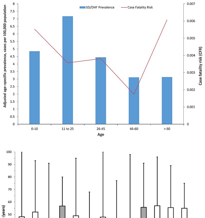
Trop. Med. Infect. Dis. 2020, 5, 68                                                                   3 of 20

      Using a centralised patient database at the laboratory, a list of 3994 laboratory confirmed dengue
cases were identified by DENV–specific rRT–PCR, immunoglobulin M (IgM) and immunoglobulin G
(IgG) enzyme linked immunosorbent assay (ELISA) and non-specific protein 1 (NS1) antigen testing
in Barbados during 2008 to 2016. This list of patients was analysed and pertinent epidemiological
characteristics of infection including age, new dengue case, gender and geographical location during
this period were reported. The list of patients was exported from Microsoft Access as a Microsoft
Excel file for analysis. The patients were first grouped by year of dengue infection for the period of
study 2008–2016 (Figure 1). Using Microsoft Excel, each of the groups were sorted by age and the
list of total patients was reduced to 3894 out of the original 3994 patients (97.5%) as some of patients’
ages were not recorded. Each year group was sorted by age and the tally of each age group recorded.
Age standardisation was done using World Health Organization (WHO) standard [13].

                Figure 1. Sampling and analysis of dengue fever (DF) patients, Barbados, 2008–2016.

     For analysis by gender, all 3994 laboratory confirmed dengue patients were used (100%) as all
patients had their gender recorded. Each group was sorted by gender and the tally of patients for each
gender was recorded. For analysis by geographic location, only 80.4% (3340/3994) were used as a parish
address was missing for 19.6% of dengue cases on the list generated. From these, 3340 dengue patients
each group was sorted by parish and the tally of patients for each parish was recorded. For analysis of
new dengue cases, 3095 laboratory confirmed dengue patients were used (77.5%) with both IgM and
IgG results. These new cases were sorted by age and gender and the tallies of patients were recorded.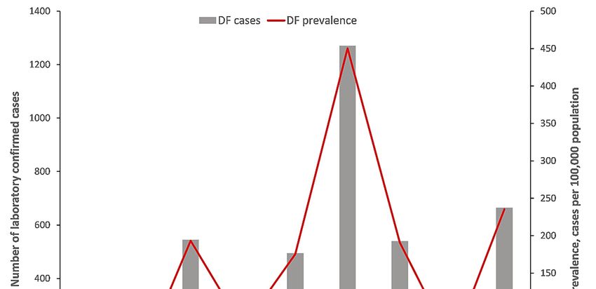
Trop. Med. Infect. Dis. 2020, 5, 68                                                                4 of 20

2.3. Dengue Diagnosis
     Laboratory confirmation of dengue cases were conducted via either detection of DENV–specific
IgM and IgG in patients’ serum with dengue IgM and IgG antibody–capture ELISA kits (samples
within 5–15 days of illness), or NS1 antigen or rRT–PCR (samples within the first five days of illness).
IgG and IgM antibody–capture ELISAs were performed per manufacturer’s instructions (Focus
Diagnostics, Cypress, CA90630 USA) [14,15]. Platelia™ Dengue NS1 Ag–ELISA (Biorad Laboratories,
Marnes–La–Coquette, France) was used for the NS1 antigen detection [16]. The patient sera were tested
using rRT–PCR to serotype the infecting DENV strain(s) as per the public health laboratory’s dengue
RT–PCR serotyping procedure [17]. For each testing assay, the relevant kit and in–house positive and
negative controls were used for quality control. A primary/new dengue case was defined by either the
presence of DENV–specific viral RNA (vRNA), NS1 antigen or DENV–specific IgM along with the
absence of DENV–specific IgG.

2.4. Virus Sequencing—Next Generation Sequencing (NGS)
       Primers were designed for the E gene that allowed five fragments of sizes between up to 500
nucleotides, with about 50 nucleotides of overlap to be generated. Real time reverse transcriptase
polymerase chain reaction (rRT-PCR) was conducted using random primers (Invitrogen) and Superscript
III (Invitrogen), and deoxyribonucleotide (DNA) amplification was performed using the specific primers
and PFU polymerase (Invitrogen). Fragments were gel–purified using QIAquick gel extraction kit
(Qiagen, Venlo, The Netherlands) and were organized in libraries of equal concentrations. Libraries
were created for each virus without DNA fragmentation (GS FLX Titanium Rapid Library Preparation,
Roche), emPCR (Amplification Method Lib–L) and GS junior sequencing runs were performed
according to instructions of the manufacturer (Roche). Amplicons were sequenced in a blinded fashion
using 454 technology. Reads from the GS–FLX sequencing data were sorted by bar code and aligned to
reference sequences using CLC Genomics software 4.6.1. Using the alignment, a single nucleotide
polymorphism (SNP) table was made with a minimum coverage of 10 reads and a minimum variant
frequency of 1.0%. Raw nucleotide sequences were filtered, aligned, trimmed and translated using
pre–specified criteria applied uniformly so that all the protein E sequences used in the analyses spanned
the exo–domain and the transmembrane region.

2.5. Phylogenetic Analysis
     The phylogenetic analysis was performed using MEGA version 6, considering sequences dengue
virus envelope (DENV–E) gene obtained in the present study (viz. DS18 and DS29) with global
reference sequences of different dengue serotype (DENV–1, –2, –3 and –4) and DENV–1 sequences
from different geographic regions, available in the GenBank database. The nucleotide sequences were
aligned using MUSCLE v.3.6. and the phylogenetic tree was generated considering complete envelop
gene (E) of DENV, using the maximal likelihood method (ML) based on Tamura–Nei (TN93) + gamma
model. Reliability of the analysis was evaluated by a bootstrap test with 1000 replications.

2.6. Data Analysis
     Data analysis was done using Microsoft Excel (Microsoft, Redmond, WA, USA) and SPSS 16.0
software (SPSS Inc., Chicago, IL, USA) for statistical analysis. Confidence intervals (95%) were
calculated for prevalence, case fatality risk/rate (CFR) and hospitalisation prevalence using Barbados
2010 census data and Barbados population data from 2014. CFR is the number of persons who died ÷
number of persons infected. A probability (p) of ≤ 0.05 was considered statistically significant.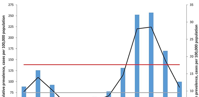
Trop. Med. Infect. Dis. 2020, 5, 68                                                                                     5 of 20

3. Results

3.1. Laboratory Testing and Clinical Symptoms
      The largest number of diagnostic tests were performed in 2013 (Table 1). The most common
clinical symptoms observed among DENV–infected patients in Barbados were fever, headache, joint
pain, retro–orbital pain, muscle pain and gastrointestinal symptoms (Table 2). More severe symptoms
such as bleeding, jaundice, thrombocytopenia and hepatomegaly (usually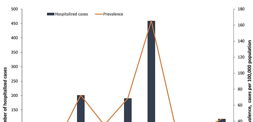
Trop. Med. Infect. Dis. 2020, 5, 68                                                                      6 of 20

194.7 (95% CI, 178.3–211.1) cases per 100,000 population, respectively), 2012 vs. 2013, (176.7 (95% CI,
161.0–192.4) and 453.9 (95% CI, 428.8–478.9) cases per 100,000 population, respectively) and 2015 vs.
2016 (53.3 (95% CI, 44.7–61.9) and 237.2 (95% CI, 219.1–255.3) cases per 100,000 population, respectively)
(Figure 2A). Thus, major dengue epidemics occurred in 2010, 2013 and 2016; the 2013 epidemic started
in late 2012, peaked in 2013 and spilled over into 2014 (Figure 2A; data not shown). The highest DF
prevalence was observed during 2013 and was significantly higher than all the years in the study
period (Figure 2A).
      The highest age adjusted DF prevalence was observed in patients 10–19 years of age with 404.7
(95% CI, 378.2–431.4) cases per 100,000 population, which was significantly higher than all other age
groups, and the next highest was the 20–29 age group 264.3 (95% CI, 243.4–285.4) cases per 100,000
population (Figure 2B).
      The mean monthly DF prevalence in Barbados differed significantly from the rainy season (June
to November), with 17.6 (95% CI, 16.0–19.3) cases per 100,000 population, to the dry season (December
to May), with 9.5 (95% CI, 7.9–11.1) cases per 100,000 population (Figure 2C). During the study period,
the cumulative DF prevalence increased from June, peaking in October, with 257.8 (95% CI, 238.2–275.9)
cases
   Trop.per 100,000
         Med.           population,
              Infect. Dis. 2020, 5, x FORthen
                                          PEERdecreasing
                                               REVIEW    to lower levels in December (Figure 2C). Consistently
                                                                                                         7 of 25
low cumulative DF prevalence occurred from January to May (Figure 2C).

     A

                                                Figure 2. Cont.
Trop.
Trop.    Med.
      Med.    Infect.
           Infect.    Dis.
                   Dis.    2020,
                        2020, 5, 5,
                                 68x FOR PEER REVIEW                                                    77 of
                                                                                                           of 25
                                                                                                              20

   B

  C

     Figure 2. (A) Number of dengue fever (DF) cases and prevalence among patients seeking medical
        Figure 2. (A) Number of dengue fever (DF) cases and prevalence among patients seeking medical attention
     attention in Barbados, 2008–2016; (B) DF prevalence and age groups among patients seeking medical
        in Barbados, 2008–2016; (B) DF prevalence and age groups among patients seeking medical attention in
     attention in Barbados, 2008–2016; (C) monthly cumulative DF, mean DF, mean dry season and mean
        Barbados, 2008–2016; (C) monthly cumulative DF, mean DF, mean dry season and mean wet season
     wet season prevalence among patients seeking medical attention in Barbados, 2008–2016.
        prevalence among patients seeking medical attention in Barbados, 2008–2016.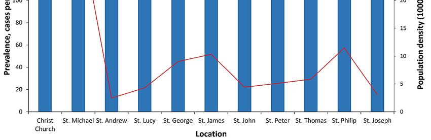
Trop. Med. Infect. Dis. 2020, 5, 68                                                                    8 of 20

              All four DENV serotypes were observed during the study period 2008–2016, with DENV–2 being
       the predominant DENV serotype observed in 2010 and 2011, whilst DENV–1 became the predominant
       DENV serotype in 2012 and 2013 (Table 1). DENV serotyping was absent for 2008 and 2014–2016
       (Table 1). The predominant serotypes during 2010 and 2013 epidemics were DENV–2 and DENV–1,
       respectively, and may explain the fewer SD cases observed in 2013 than in 2010 (Table 1). DENV–3
       serotype was the most dominant serotype in 2007; therefore, is it not unusual that the DENV epidemics
       observed in 2010 and 2013 were caused predominantly by DENV–2 and DENV–1, respectively.
              During the 2010 and 2013 dengue epidemics, the geographic distribution of dengue infections
       in Barbados was different. No significant difference was observed in crude DF prevalence among
       different parishes (Figure 3A). No significant difference was observed for the mean crude DF prevalence
       between males and females: A total of 153.6 (95% CI, 146.3–164.4) vs. 164.7 (95% CI, 158.2–168.5) cases
       per 100,000 population respectively; a ratio of 1:1.
              A total of 576 primary dengue cases were observed in the period 2008–2016. The highest primary
       dengue prevalence were observed in 2013, with 104.0 (95% CI, 92.0–116.0) cases per 100,000 population,
       and 2012, with 34.2 (95% CI, 27.3–41.1) cases per 100,000 population, which were significantly higher
Trop. Med.
       thanInfect. Dis. years
               other    2020, 5, (Figure
                                 x FOR PEER REVIEW
                                         3B).                                                      7 of 25 (14.4%),
                                              Primary DENV infection accounted for 576 confirmed patients
       and at least 2382 (59.6%) secondary DENV infections occurred during 2008 to 2016.

           A

                                                     Figure 3. Cont.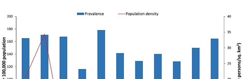
Trop. Med. Infect. Dis. 2020, 5, x FOR PEER REVIEW                                                    7 of 25
Trop. Med. Infect. Dis. 2020, 5, 68                                                                      9 of 20

     B

    C

      Figure 3. (A) Dengue fever (DF) prevalence by geographic location among patients seeking medical
      attention in Barbados, 2008–2016; (B) primary (IgM positive only) and secondary (IgM and IgG
      positive) DF prevalence for patients seeking medical attention in Barbados, 2008–2016; (C) number of
      hospitalized DF cases and hospitalized DF prevalence among patients seeking medical attention in
      Barbados, 2008–2016.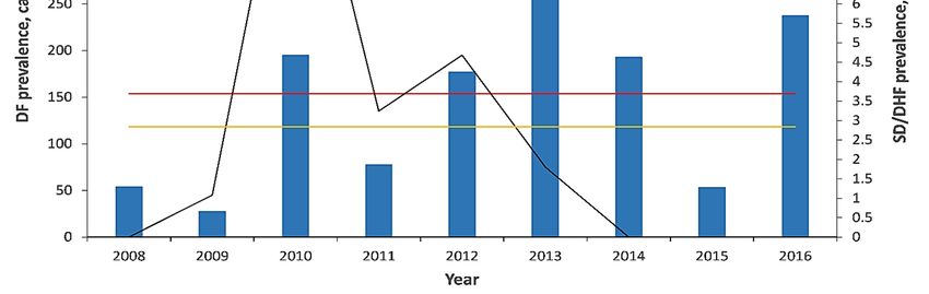
Trop. Med. Infect. Dis. 2020, 5, 68                                                                            10 of 20

3.3. Severe Dengue in Barbados
     DENV infection in 1356 out of 3994 (34.0%) patients resulted in hospitalizations, and the crude
prevalence of hospitalized DF patients ranged from 14.2 (95% CI, 8.2–19.8) cases per 100,000 population
to 165.2 (95% CI, 150.1–180.3) cases per 100,000 population between 2008 and 2016 (Figure 3C). During
2013, the crude prevalence of hospitalized DF patients, with 165.2 (95% CI, 150.1–180.3) cases per
100,000 population, was significantly higher than every year during the study and was followed by
the crude hospitalized dengue prevalence in 2010, with 72.4 (95% CI, 62.3–82.4) cases per 100,000
population (Figure 3C). SD/DHF prevalence varied from 0 to 11.9 (95% CI, 7.8–15.9) cases per 100,000
population (Figure 4A). The mean SD/DHF prevalence observed during the study period was 2.5
(95% CI, 1.2–3.8) cases per 100,000 population. A significantly higher SD/DHF prevalence was observed
during 2010, 11.9 (95% CI, 7.8–15.9) cases per 100,000 population than in every other year in the study,
even though a higher DENV prevalence was observed in 2013 than in 2010 (Figures 2A and 4A).
The 2010 dengue epidemic was more severe than 2013 epidemic as the SD/DHF prevalence in 2010,
with 11.9 (95% CI, 7.8–15.9) cases per 100,000 population, was significantly higher than the SD/DHF
prevalence in 2013, with 1.8 (95% CI, 0.2–3.4) cases per 100,000 population (Figure 4A).
     The CFR among SD/DHF cases varied by age and ranged from 0.0017 to 0.0061, and was highest
among the elderly, persons 60 years or older and the young (0.075 per 100 DF cases), even though the
age adjusted prevalence was higher in the 11–25 years old age group (Figure 4B). An overall CFR of
(10/3994) 0.4% was observed over the study period.
     Median
    Trop.       age Dis.
          Med. Infect. of SD/DHF      patients
                          2020, 5, x FOR        varied during 2008–2016, with the highest median age observed
                                         PEER REVIEW                                                           7 of 25
in 2010 (Figure 4C). Most SD/DHF cases in 2012 were young (30 years old; Figure 4C). The age distribution of the DF patients ranged
          Figure 3. (A) Dengue fever (DF) prevalence by geographic location among patients seeking medical
from one to 100 years (median 29.6 years) and the median age varied over time (Figure 4C). The median
          attention in Barbados, 2008–2016; (B) primary (IgM positive only) and secondary (IgM and IgG positive) DF
age of SD/DHF       cases was 40 years old (interquartile age range of 23–56 years old) and the median age
          prevalence for patients seeking medical attention in Barbados, 2008–2016; (C) number of hospitalized DF
varied between
          cases and2010    to 2013DF
                       hospitalized    (Figure  4C).among
                                         prevalence  SD cases  were
                                                          patients    observed
                                                                   seeking medicalinattention
                                                                                     2009–2013     and an2008–2016.
                                                                                              in Barbados, overall mean
age of 35.8 years old was observed over the study period (Figure 4C).

       A

                                                   Figure 4. Cont.
Trop. Med. Infect.
Trop. Med. Dis.Dis.
Infect. 2020, 5, 68
2020, 5, x FOR PEER REVIEW 7 of1125of 20
B
C
Figure
Figure 4. (A)
4. (A) Denguefever
Dengue fever(DF),
(DF), mean
meanDF,DF,severe
severedengue (SD)/dengue
dengue (SD)/denguehaemorrhagic fever (DHF)
haemorrhagic fever and mean
(DHF)
and SD/DHF
mean SD/DHFprevalence among patients
prevalence seeking seeking
among patients medical attention in Barbados,
medical attention 2008–2016;2008–2016;
in Barbados, (B) SD/DHF
prevalence
(B) SD/DHF and case fatality
prevalence risk fatality
and case (CFR) byrisk
age (CFR)
groupsby
among patients among
age groups seeking medical
patientsattention
seekinginmedical
Barbados,
2008–2016; (C) DF and SD/DHF age distribution among patients seeking medical attention
attention in Barbados, 2008–2016; (C) DF and SD/DHF age distribution among patients seeking medical in Barbados,
attention in Barbados, 2008–2016. Boxes encompass 25th and 75th percentiles. Black lines within the
boxes represent medians. Error bars represent the minimum and maximum ranges.
3.4. DENV Sequencing in Dengue Patient Sera
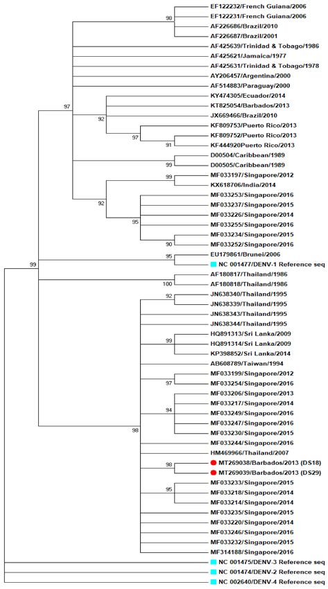
Two out of thirty–three (2/33) DENV specific rRT–PCR positive sera samples yielded E gene
sequences using NGS methods (Figure 5). The viral loads on the remaining 31 samples were too low toTrop. Med. Infect. Dis. 2020, 5, 68 12 of 20
permit sequencing using NGS. Two samples were positive in the rRT–PCR, patients DS18 and DS29,
with cycle threshold (Ct) values of, respectively, 28.5 and 30. Both sequences, GenBank accession
numbers MT269038 (from DS18) and MT269039 (from DS29), grouped closer to DENV–1 strains of
South–East (SE) Asian origin than to DENV–1 strains from Caribbean or South American origin;
however, the small sample size (n = 2) limits the drawing of any meaningful conclusions (Figure 5).
Figure 5. Phylogenetic analysis of dengue virus type 1 (DENV–1), based on complete envelop gene (E).
The tree was constructed using maximum likelihood (ML) method, with 1000 boot strap resampling.
The boot strap values were mention adjacent to the branch. Patient sample DS18 (MT269038) and DS29
(MT269039) were marked with red dots, different serotypes of dengue NCBI reference strains were
marked with cyan dots.Trop. Med. Infect. Dis. 2020, 5, 68 13 of 20
4. Discussion
Epidemiological data are important in vaccine planning and are critical in measuring vaccine
efficacy and disease burden [18]. The mean dengue prevalence observed in this study was comparable
with previous studies in Barbados (163.0 vs. 162.5 cases per 100,000 population) and more than
double that of other countries within the Caribbean and Latin America (163.0 vs. 72.1 cases per
100,000; P < 0.01) [19]. This prevalence remains one of the highest in Caribbean along with Trinidad,
Martinique, Guadeloupe, French Guiana and Puerto Rico [19]. However other Caribbean territories
have significantly lower prevalence, as low as 140 times lower [19]. These disparities in DF prevalence
may be due to differences in the extensiveness of the DENV laboratory diagnostic testing and laboratory
surveillance systems in each English–speaking Caribbean country. The more extensive the reporting,
the more accurate the prevalence. Other factors that contribute are reduced public health awareness,
population density, infecting DENV strain, urban planning activities, topography, water storage
practices, change of lifestyle, apathy and heightened medical seeking behaviour [20–23]. Variation in
the number of laboratory tests conducted can also influence this was well as more tests conducted will
results in more cases detected. A cross–sectional serological survey within the community may be
more representative and accurate as it would be a random sampling of the population without the bias
of patients seeking medical attention which can result in higher prevalence.
A period between dengue epidemics is often observed though the periodic length may vary but
usually is 3 to 5 years in length [12,24–26]. A cyclic pattern of dengue epidemics every 3 years has been
observed in Barbados since 2007 including 2007, 2010, 2013 and possibly 2016 as evidenced by peak
prevalence in the period of study [9]. It is noted that ZIKV was first detected in Barbados during 2016
and the introduction of ZIKV likely increased the number of persons with DF–like symptoms having
flavivirus IgM positive serological results. Only ZIKV molecular testing can conclusively determine
the nature of the clinical infection(s). If interepidemic period of 3–5 years is consistent the next DENV
epidemic in Barbados is likely due in 2020 or 2021 and the possibility of the concurrent occurrence
of severe acute respiratory syndrome coronavirus 2 (SARS CoV–2) pandemic and a DENV epidemic
exists. Reports of suspected DENV infections later being confirmed as SARS–CoV–2 infections
and the similarity with clinical symptoms could lead to complication of SARS–CoV–2 pandemic
response [27–30].
Median age is a predictor of the change in force of infection and mean force of infection [18].
Force of infection is defined as the rate at which susceptible individuals acquire an infectious disease
and it is dependent on several factors including disease prevalence, rate of contact of individuals
(communicable diseases) and infectiousness of individuals [18]. As the median age increases, the force
of infection decreases. The age groups most affected by dengue and DHF/SD can differ in different
geographical locations including SE Asia, Europe and the Americas based partly on differences in the
relative force of infection [31–33]. Dengue endemic areas have a higher force of infection due to the
greater exposure risk to DENV infection by vectors and the higher number of new DENV infections.
The high DF prevalence observed in patients 10–19 years is consistent with prevalence data obtained
from other DF endemic locations including Laos [34].
Previous studies have illustrated that gentrification, a shift of the age distribution of human
population toward older ages, increases the risk of severe dengue prevalence [35,36]. In endemic
countries, such as Barbados, the greater proportion of DENV infections were secondary occurring in
personsTrop. Med. Infect. Dis. 2020, 5, 68 14 of 20
4.1. Seasonal Factors
Climatic factors can enhance vector population growth and increase the risk of DENV vectors
biting susceptible humans [39–41]. Dengue transmission is often seasonal and driven by many climatic
factors including temperature, short and long term rainfall and relative humidity influencing mosquito
vector growth and density [42–45]. The significant difference between DF prevalence during the rainy
and dry seasons in Barbados highlights rainfall as a key factor in DENV transmission as it permits
the proliferation of dengue vectors and increased risk of bites by DENV infected vectors as observed
previously [46–49].
Primarily a higher DF prevalence is observed in urban areas versus rural areas as the population
density is higher in urban areas resulting in more susceptible human hosts [50] however in Barbados,
no significant difference in DF prevalence was observed among parishes during the study period.
Population density alone does not influence dengue transmission as other factors such as degree of
vegetation cover and availability of water supply could also influence [51–54].
4.2. Dengue and Gender
Sex bias does occur in infectious disease epidemiology including dengue infections [55]. Among
patients tested positive for DENV infection, males were as likely as females to be infected with DENV
during the study period in Barbados. Higher prevalence of DENV infection were observed in males
from studies in Asia while reports from the Americas showed either females are more often infected or
the proportion is nearly equal [55–59]. In Asia this disparity was linked to more males in the labour
force and working outdoors [60]. Unlike in Singapore where there are more working males than
females, the skilled labour force in Barbados has a similar number of males and females, 30,454 males
vs. 29,872 females, (2010 Barbados census data) [60].
4.3. Dengue Disease Severity
In the Americas, the severe forms of dengue are not as common as in Southeast (SE) Asia as DHF
or DSS attack rates are 18–fold greater in SE Asia than in the Americas [61]. In comparison the mean
SD/DHF prevalence in Barbados is lower than Indonesia (30–85 cases per 100,000 population) and
several other to SE Asian countries [62,63]. Mean SD/DHF prevalence in this study was comparatively
higher to previous studies in Barbados, and other Caribbean countries excluding Trinidad, Turks
and Caicos, Suriname, Bermuda, Montserrat and Cayman Islands [19]. One possible explanation
is the improvement of DENV surveillance in Barbados. However, there could be other possibilities
such as viral strain differences, public health awareness, basis of surveillance (i.e., population based),
population age structure, and effectiveness of vector control programs. An example is the use of only
rapid test kits with low sensitivity to diagnose dengue. Moreover, only samples of severe cases that are
send to CARPHA for more sensitive diagnostic testing.
Among SD/DHF cases in Barbados, the highest prevalence prevalence was observed among
persons 11–25 years of age which is slightly higher than other countries including Brazil, Sri Lanka and
Thailand where SD/DHF is mostly observed in persons aged 15 or younger [64,65]. Younger members
of the population are more susceptible to dengue infection as they lack substantial immunity however,
but this immunity increases with age as observed in the decline in DF prevalence as age increased.
Even though dengue prevalence has increased in Barbados as in other areas [66–68], attenuation
of the severity has occurred, resulting in a shift in the age distribution of DHF toward older age
groups [66,69,70].
The CFR in Barbados is relatively lower than dengue CFRs in most dengue endemic countries
except for Singapore during 2004 and Vietnam during 2010 [62,63]. This lower CFR maybe due
to several factors including easier access to health care, effective mosquito control, water storage
practices, host population genetics, vector biology, geophysical factors and other socioeconomic
differences [49,51,64]. Fatal dengue cases did occur during the study period reviewed but these wereTrop. Med. Infect. Dis. 2020, 5, 68 15 of 20
likely underestimated as the reliance on passive case reporting and/or review of death certificates can
fail to identify fatal cases due to misdiagnosis [71–76]. No gender bias exists in Barbados for DHF/SD
as in other countries since males (n = 35) were as likely as females (n = 38) to be infected with DENV
and develop DHF or SD during the study period [55].
All 4 DENV serotypes were in circulation during 2010 epidemic and this likely increased the risk
of developing SD/DHF [12,77,78]. The infecting DENV strain influences the severity of dengue disease
and the infecting strains in 2010 likely differed from those in 2013 [79]. Sequence data from DENVs in
circulation especially during epidemics can provide useful information regarding strain ‘fitness’ and
the ability to cause severe disease thus should be included as part of routine dengue surveillance and
public health plans [80–82].
4.4. Sequencing of DENV Envelope (E) Gene Region
DENV–1 serotype has been present in the Caribbean since 1970s, being first reported in Jamaica
and was reported as the first DENV serotype to enter Brazil from the Caribbean [83,84]. One study
revealed that DENV–1 was first present in Grenada in the Lesser Antilles, then rapidly spread to South
and Central America and then to the Greater Antilles [83,84]. One phylogenetic study of DENV–1
envelope genes revealed the role of East Asian countries as possible sources of dengue epidemics
and the close relatedness of at least one Caribbean DENV–1 virus to East Asian DENV–1 viruses [85].
The E gene sequence of two RT–PCR positive patient sera samples yielded E gene sequences closer in
relation to DENV–1 isolates of Asian origin, Thailand and Singapore than those from the Caribbean
and South America. Sequencing of DENVs from 2010 outbreak would have been of great interest to
observe their genetic relationship with other sequenced DENVs. However, no deeper insights into
DENVs strain can be since the sample number of sequenced strains (n = 2) is too small. More sequence
data from different time points would be important to perform a thorough analysis.
4.5. Study Limitations
Our study has several limitations including selection bias due to the selection and testing of patients
based on clinical suspicion and is biased due to surveillance, incomplete referral and dengue diagnostic
testing, the potential to miss mild cases and non–symptomatic dengue cases and persistence of
DENV–specific IgM [86]. There are however several strengths of this study due to the key observations
obtained including (a) first sequencing of DENVs from Barbados which bear closer relation to Asian
strains than Caribbean or South American ones, (b) a DENV epidemic periodicity of 3–5 years and the
possible complications for public health officials with current coronavirus disease 2019 (COVID–19)
pandemic planning, (c) lack of gender bias of DENV infection and d) generally a low DENV CFR.
4.6. Future Research
Future research efforts should include analysis of sociodemographic and socioeconomic
determinants of DENV infection, DENV and arboviral research in mosquito vectors to identify
DENV strains in circulation, DENV, ZIKV and CHIKV diagnostic testing of donated blood for
blood banking, DENV seroprevalence studies for any future DENV vaccination efforts and genetic
characterisation of DENV strains to assess the evolution of DENV in Barbados.
Author Contributions: Conceptualization, K.O.D., T.A.S. and M.G.-S.H.; methodology, K.O.D., T.A.S., B.M., F.A.,
S.K.D., and M.G.-S.H.; software, K.O.D. and S.K.D; validation, K.O.D., B.M., F.A., M.G.-S.H. and S.K.D.; formal
analysis, K.O.D., T.A.S., B.M., F.A., S.K.D., and M.G.-S.H.; investigation, K.O.D., T.A.S., B.M., F.A., S.K.D., and
M.G.-S.H.; resources, K.O.D., B.M., and M.G.-S.H.; data curation, K.O.D. and S.K.D.; writing—original draft
preparation, K.O.D.; writing—review and editing, K.O.D., T.A.S., B.M., F.A., S.K.D., and M.G.-S.H.; visualization,
K.O.D. and S.K.D.; supervision, B.M., and M.G.-S.H.; project administration, B.M., T.A.S., and M.G.-S.H.;
funding acquisition, K.O.D., B.M., and M.G.-S.H. All authors have read and agreed to the published version of
the manuscript.Trop. Med. Infect. Dis. 2020, 5, 68 16 of 20
Funding: This study was funded by the University of the West Indies (UWI), Cave Hill, St. Michael, Barbados and
Artemis One Health Institute, Netherlands. Fatih Anfasa was supported with a PhD grant from the Directorate of
Higher Education of the Ministry of Research and Education, Republic of Indonesia.
Acknowledgments: The authors would like to thank the entire staff at the Best–dos Santos Public Health
Laboratory for laboratory testing and provision of the raw data, and the staff of the Clinical Chemistry Department
and the Medical Records Department of the Queen Elizabeth Hospital (QEH), Barbados and the staff at Artemis
One Health in the Netherlands for sequencing of DENV strains in human sera.
Conflicts of Interest: The authors declare no conflict of interest.
References
1. Dick, O.B.; San Martín, J.L.; Montoya, R.H.; del Diego, J.; Zambrano, B.; Dayan, G.H. The history of dengue
outbreaks in the Americas. Am. J. Trop. Med. Hyg. 2012, 87, 584–593. [CrossRef]
2. Murray, N.E.A.; Quam, M.B.; Wilder-Smith, A. Epidemiology of dengue: Past, present and future prospects.
Clin. Epidemiol. 2013, 5, 299–309. [CrossRef]
3. Torres, J.R.; Orduna, T.A.; Piña-Pozas, M.; Vázquez-Vega, D.; Sarti, E. Epidemiological characteristics of
dengue disease in Latin America and in the Caribbean: A systematic review of the literature. J. Trop. Med.
2017, 2017, 8045435. [CrossRef]
4. Bellon, M.M.; MacLean, J.D. A point source dengue outbreak in Canadian tourists in Barbados. Can. Commun.
Dis. Rep. 1998, 24, 161–164. [PubMed]
5. Branch, S.L.; Levett, P.N. Evaluation of four methods for detection of immunoglobulin M antibodies to
dengue virus. Clin. Diagn. Lab. Immunol. 1999, 6, 555–557. [CrossRef]
6. Levett, P.N.; Branch, S.L.; Edwards, C.N. Detection of dengue infection in patients investigated for leptospirosis
in Barbados. Am. J. Trop. Med. Hyg. 2000, 62, 112–114. [CrossRef]
7. Gittens-St Hilaire, M.; Clarke-Greenidge, N. An analysis of the subtypes of dengue fever infections in
Barbados 2003–2007 by reverse transcriptase polymerase chain reaction. Virol. J. 2008, 5, 152. [CrossRef]
8. Kumar, A.; Gittens-St Hilaire, M.; Nielsen, A.L. Epidemiological trends and clinical manifestations of Dengue
among children in one of the English-speaking Caribbean countries. Trans. R. Soc. Trop. Med. Hyg. 2013, 107,
254–260. [CrossRef]
9. Kumar, A.; Gittens-St Hilaire, M.; Clarke-Greenidge, N.; Nielsen, A.L. Epidemiological Trend and Clinical
Observations among Children and Adults with Dengue in Barbados. West Indian Med. J. 2015, 64, 37–42.
[CrossRef]
10. Kumar, A.; Nielsen, A.L. Trends in the patterns of IgM and IgG antibodies in febrile persons with suspected
dengue in Barbados, an English-speaking Caribbean country, 2006–2013. J. Infect. Public Health 2015, 8,
583–592. [CrossRef]
11. Kumar, A.; Gittens-St Hilair, M.; Jason, V.; Ugwuagu, C.; Krishnamurthy, K. The clinical characteristics
and outcome of children hospitalized with dengue in Barbados, an English Caribbean country. J. Infect.
Dev. Ctries. 2015, 9, 394–401. [CrossRef]
12. Chadee, D.; Mahabir, R.S.; Sutherland, J.M. Dengue Fever Epidemiology and Control in the Caribbean:
A tatus Report (2012). Carib. Med. J. 2012, 74, 17–21.
13. Ahmad, O.; Boschi-Pinto, C.; Lopez, A.; Murray, C.; Lozano, R.; Inoue, M. Age Standardization of Rates: A New
WHO Standard; World Health Organization: Geneva, Switzerland, 2001.
14. Groen, J.; Koraka, P.; Velzing, J.; Copra, C.; Osterhaus, A.D. Evaluation of six immunoassays for detection of
dengue virus-specific immunoglobulin M and G antibodies. Clin. Diagn. Lab. Immunol. 2000, 7, 867–871.
[CrossRef]
15. Koraka, P.; Zeller, H.; Niedrig, M.; Osterhaus, A.D.; Groen, J. Reactivity of serum samples from patients with
a flavivirus infection measured by immunofluorescence assay and ELISA. Microbes Infect. 2002, 4, 1209–1215.
[CrossRef]
16. Kumarasamy, V.; Wahab, A.A.; Chua, S.; Hassan, Z.; Mohamad, M.; Chua, K. Evaluation of a commercial
dengue NS1 antigen-capture ELISA for laboratory diagnosis of acute dengue virus infection. J. Virol. Methods
2007, 140, 75–79. [CrossRef]
17. Johnson, B.W.; Russell, B.J.; Lanciotti, R.S. Serotype-specific detection of dengue viruses in a fourplex
real-time reverse transcriptase PCR assay. J. Clin. Microbiol. 2005, 43, 4977–4983. [CrossRef]
18. Lahariya, C. Vaccine epidemiology: A review. J. Fam. Med. Prim. Care 2016, 5, 7. [CrossRef]Trop. Med. Infect. Dis. 2020, 5, 68 17 of 20
19. Cafferata, M.L.; Bardach, A.; Rey-Ares, L.; Alcaraz, A.; Cormick, G.; Gibbons, L.; Romano, M.; Cesaroni, S.;
Ruvinsky, S. Dengue epidemiology and burden of disease in Latin America and the Caribbean: A systematic
review of the literature and meta-analysis. Value Health Reg. Issues 2013, 2, 347–356. [CrossRef]
20. Gubler, D.J. Aedes aegypti and Aedes aegypti-borne disease control in the 1990s: Top down or bottom up.
Am. J. Trop. Med. Hyg. 1989, 40, 571–578. [CrossRef]
21. Hemme, R.R.; Tank, J.L.; Chadee, D.D.; Severson, D.W. Environmental conditions in water storage drums
and influences on Aedes aegypti in Trinidad, West Indies. Acta Trop. 2009, 112, 59–66. [CrossRef]
22. Getachew, D.; Tekie, H.; Gebre-Michael, T.; Balkew, M.; Mesfin, A. Breeding sites of Aedes aegypti: Potential
dengue vectors in Dire Dawa, East Ethiopia. Interdiscip. Perspect. Infect. Dis. 2015, 2015, 706276. [CrossRef]
[PubMed]
23. Gubler, D.J. Dengue, urbanization and globalization: The unholy trinity of the 21st century. Trop. Med. Health
2011, 39 (Suppl. 4), S3–S11. [CrossRef]
24. Shepard, D.S.; Coudeville, L.; Halasa, Y.A.; Zambrano, B.; Dayan, G.H. Economic impact of dengue illness in
the Americas. Am. J. Trop. Med. Hyg. 2011, 84, 200–207. [CrossRef]
25. Ferreira, G.L. Global dengue epidemiology trends. Rev. Inst. Med. Trop. Sao Paulo 2012, 54 (Suppl. 18), S5–S6.
[CrossRef]
26. World Health Organization. Guidelines for Diagnosis, Treatment, Prevention and Control; World Health
Organization: Geneva, Switzerland, 2009.
27. Lorenz, C.; Azevedo, T.S.; Chiaravalloti-Neto, F. COVID-19 and dengue fever: A dangerous combination for
the health system in Brazil. Travel Med. Infect. Dis. 2020. [CrossRef]
28. Joob, B.; Wiwanitkit, V. COVID-19 can present with a rash and be mistaken for Dengue. J. Am. Acad. Dermatol.
2020, 82, e177. [CrossRef]
29. Yan, G.; Lee, C.K.; Lam, L.T.; Yan, B.; Chua, Y.X.; Lim, A.Y.; Phang, K.F.; Kew, G.S.; Teng, H.; Ngai, C.H.
Covert COVID-19 and false-positive dengue serology in Singapore. Lancet Infect. Dis. 2020. [CrossRef]
30. Saavedra-Velasco, M.; Chiara-Chilet, C.; Pichardo-Rodriguez, R.; Grandez-Urbina, A.; Inga-Berrospi, F.
Coinfection between dengue and covid-19: Need for approach in endemic zones. Revista de la Facultad de
Ciencias Medicas (Cordoba, Argentina) 2020, 77, 52–54. [CrossRef] [PubMed]
31. Castanha, P.; Cordeiro, M.; Martelli, C.; Souza, W.; Marques, E.; Braga, C. Force of infection of dengue
serotypes in a population-based study in the northeast of Brazil. Epidemiol. Infect. 2013, 141, 1080–1088.
[CrossRef]
32. Rodríguez-Barraquer, I.; Buathong, R.; Iamsirithaworn, S.; Nisalak, A.; Lessler, J.; Jarman, R.G.; Gibbons, R.V.;
Cummings, D.A. Revisiting Rayong: Shifting seroprofiles of dengue in Thailand and their implications for
transmission and control. Am. J. Epidemiol. 2014, 179, 353–360. [CrossRef]
33. Tam, C.C.; Tissera, H.; de Silva, A.M.; De Silva, A.D.; Margolis, H.S.; Amarasinge, A. Estimates of dengue
force of infection in children in Colombo, Sri Lanka. PLoS Negl. Trop. Dis. 2013, 7, e2259. [CrossRef]
[PubMed]
34. Khampapongpane, B.; Lewis, H.C.; Ketmayoon, P.; Phonekeo, D.; Somoulay, V.; Khamsing, A.; Phengxay, M.;
Sisouk, T.; Vongphrachanh, P.; Bryant, J.E. National dengue surveillance in the Lao People’s Democratic
Republic, 2006–2012: Epidemiological and laboratory findings. West. Pac. Surveill. Response J. WPSAR 2014,
5, 7.
35. Cummings, D.A.; Iamsirithaworn, S.; Lessler, J.T.; McDermott, A.; Prasanthong, R.; Nisalak, A.; Jarman, R.G.;
Burke, D.S.; Gibbons, R.V. The impact of the demographic transition on dengue in Thailand: Insights from a
statistical analysis and mathematical modeling. PLoS Med. 2009, 6, e1000139. [CrossRef]
36. Thai, K.T.; Nishiura, H.; Hoang, P.L.; Tran, N.T.T.; Phan, G.T.; Le, H.Q.; Tran, B.Q.; Van Nguyen, N.;
de Vries, P.J. Age-specificity of clinical dengue during primary and secondary infections. PLoS Negl. Trop.
Dis. 2011, 5, e1180. [CrossRef] [PubMed]
37. Prince, H.E.; Yeh, C.; Lapé-Nixon, M. Primary and probable secondary dengue virus (DV) infection rates in
relation to age among DV IgM-positive patients residing in the United States mainland versus the Caribbean
islands. Clin. Vaccine Immunol. 2012, 19, 105–108. [CrossRef] [PubMed]
38. Mizumoto, K.; Ejima, K.; Yamamoto, T.; Nishiura, H. On the risk of severe dengue during secondary infection:
A systematic review coupled with mathematical modeling. J. Vector Borne Dis. 2014, 51, 153. [PubMed]
39. Morales, I.; Salje, H.; Saha, S.; Gurley, E.S. Seasonal Distribution and Climatic Correlates of Dengue Disease
in Dhaka, Bangladesh. Am. J. Trop. Med. Hyg. 2016, 94, 1359–1361. [CrossRef]Trop. Med. Infect. Dis. 2020, 5, 68 18 of 20
40. Choi, Y.; Tang, C.S.; McIver, L.; Hashizume, M.; Chan, V.; Abeyasinghe, R.R.; Iddings, S.; Huy, R. Effects of
weather factors on dengue fever incidence and implications for interventions in Cambodia. BMC Public
Health 2016, 16, 241. [CrossRef]
41. Hii, Y.L.; Zaki, R.A.; Aghamohammadi, N.; Rocklöv, J. Research on Climate and Dengue in Malaysia:
A Systematic Review. Curr. Environ. Health Rep. 2016, 3, 81–90. [CrossRef]
42. Bennett, S.N.; Drummond, A.J.; Kapan, D.D.; Suchard, M.A.; Munoz-Jordan, J.L.; Pybus, O.G.; Holmes, E.C.;
Gubler, D.J. Epidemic dynamics revealed in dengue evolution. Mol. Biol. Evol. 2010, 27, 811–818. [CrossRef]
43. Johansson, M.A.; Cummings, D.A.; Glass, G.E. Multiyear climate variability and dengue—El Nino southern
oscillation, weather, and dengue incidence in Puerto Rico, Mexico, and Thailand: A longitudinal data
analysis. PLoS Med. 2009, 6, e1000168. [CrossRef] [PubMed]
44. Morrison, A.C.; Getis, A.; Santiago, M.; Rigau-Perez, J.G.; Reiter, P. Exploratory space-time analysis of
reported dengue cases during an outbreak in Florida, Puerto Rico, 1991–1992. Am. J. Trop. Med. Hyg. 1998,
58, 287–298. [CrossRef] [PubMed]
45. Chadee, D.; Shivnauth, B.; Rawlins, S.; Chen, A. Climate, mosquito indices and the epidemiology of dengue
fever in Trinidad (2002–2004). Ann. Trop. Med. Parasitol. 2007, 101, 69–77. [CrossRef]
46. Depradine, C.; Lovell, E. Climatological variables and the incidence of Dengue fever in Barbados. Int. J.
Environ. Health Res. 2004, 14, 429–441. [CrossRef] [PubMed]
47. Nagao, Y.; Thavara, U.; Chitnumsup, P.; Tawatsin, A.; Chansang, C.; Campbell-Lendrum, D. Climatic and
social risk factors for Aedes infestation in rural Thailand. Trop. Med. Int. Health 2003, 8, 650–659. [CrossRef]
[PubMed]
48. Arcari, P.; Tapper, N.; Pfueller, S. Regional variability in relationships between climate and dengue/DHF in
Indonesia. Singap. J. Trop. Geogr. 2007, 28, 251–272. [CrossRef]
49. Banu, S.; Hu, W.; Hurst, C.; Tong, S. Dengue transmission in the Asia-Pacific region: Impact of climate change
and socio-environmental factors. Trop. Med. Int. Health 2011, 16, 598–607. [CrossRef]
50. Araujo, R.V.; Albertini, M.R.; Costa-da-Silva, A.L.; Suesdek, L.; Franceschi, N.C.S.; Bastos, N.M.; Katz, G.;
Cardoso, V.A.; Castro, B.C.; Capurro, M.L. São Paulo urban heat islands have a higher incidence of dengue
than other urban areas. Braz. J. Infect. Dis. 2015, 19, 146–155. [CrossRef]
51. Schmidt, W.-P.; Suzuki, M.; Thiem, V.D.; White, R.G.; Tsuzuki, A.; Yoshida, L.-M.; Yanai, H.; Haque, U.;
Anh, D.D.; Ariyoshi, K. Population density, water supply, and the risk of dengue fever in Vietnam: Cohort
study and spatial analysis. PLoS Med. 2011, 8, e1001082. [CrossRef]
52. Nonomura, A.; Kitahara, M.; Masuda, T. Impact of land use and land cover changes on the ambient
temperature in a middle scale city, Takamatsu, in Southwest Japan. J. Environ. Manag. 2009, 90, 3297–3304.
[CrossRef]
53. Vanwambeke, S.O.; Bennett, S.N.; Kapan, D.D. Spatially disaggregated disease transmission risk: Land cover,
land use and risk of dengue transmission on the island of Oahu. Trop. Med. Int. Health 2011, 16, 174–185.
[CrossRef] [PubMed]
54. Zellweger, R.M.; Cano, J.; Mangeas, M.; Taglioni, F.; Mercier, A.; Despinoy, M.; Menkès, C.E.;
Dupont-Rouzeyrol, M.; Nikolay, B.; Teurlai, M. Socioeconomic and environmental determinants of dengue
transmission in an urban setting: An ecological study in Nouméa, New Caledonia. PLoS Negl. Trop. Dis.
2017, 11, e0005471. [CrossRef] [PubMed]
55. Anker, M.; Arima, Y. Male-female differences in the number of reported incident dengue fever cases in six
Asian countries. West. Pac. Surveill. Response J. WPSAR 2011, 2, 17–23. [CrossRef] [PubMed]
56. Yew, Y.W.; Ye, T.; Ang, L.W.; Ng, L.C.; Yap, G.; James, L.; Chew, S.K.; Goh, K.T. Seroepidemiology of dengue
virus infection among adults in Singapore. Ann. Acad. Med. Singap. 2009, 38, 667–675. [PubMed]
57. Kaplan, J.E.; Eliason, D.A.; Moore, M.; Sather, G.E.; Schonberger, L.B.; Cabrera-Coello, L.; Fernandez de
Castro, J. Epidemiologic investigations of dengue infection in Mexico, 1980. Am. J. Epidemiol. 1983, 117,
335–343. [CrossRef]
58. Lin, C.C.; Huang, Y.H.; Shu, P.Y.; Wu, H.S.; Lin, Y.S.; Yeh, T.M.; Liu, H.S.; Liu, C.C.; Lei, H.Y. Characteristic of
dengue disease in Taiwan: 2002–2007. Am. J. Trop. Med. Hyg. 2010, 82, 731–739. [CrossRef]
59. Da Rosa, A.T.; Vasconcelos, P.; Da Rosa, E.T.; Rodrigues, S.G.; Mondet, B.; Cruz, A.; Sousa, M.R.; Da Rosa, J.T.
Dengue epidemic in Belém, Pará, Brazil, 1996–1997. Emerg. Infect. Dis. 2000, 6, 298. [CrossRef] [PubMed]
60. Ooi, E.E. Changing pattern of dengue transmission in Singapore. Dengue Bull. 2001, 25, 40–44.Trop. Med. Infect. Dis. 2020, 5, 68 19 of 20
61. Halstead, S.B. Dengue in the Americas and Southeast Asia: Do they differ? Revista Panamericana de
Salud Publica 2006, 20, 407–415. [CrossRef]
62. Case Fatality Rates of Dengue Fever and Dengue Haemorrhagic Fever (DF/DHF) in the Western Pacific Region,
2000–2010; World Health Organization: Geneva, Switzerland, 2011.
63. Bravo, L.; Roque, V.G.; Brett, J.; Dizon, R.; L’Azou, M. Epidemiology of dengue disease in the Philippines
(2000–2011): A systematic literature review. PLoS Negl. Trop. Dis. 2014, 8, e3027. [CrossRef]
64. Karyanti, M.R.; Uiterwaal, C.S.; Kusriastuti, R.; Hadinegoro, S.R.; Rovers, M.M.; Heesterbeek, H.; Hoes, A.W.;
Bruijning-Verhagen, P. The changing incidence of dengue haemorrhagic fever in Indonesia: A 45-year
registry-based analysis. BMC Infect. Dis. 2014, 14, 412. [CrossRef] [PubMed]
65. Teixeira, M.G.; Costa, M.C.; Coelho, G.; Barreto, M.L. Recent shift in age pattern of dengue hemorrhagic
fever, Brazil. Emerg. Infect. Dis. 2008, 14, 1663. [CrossRef] [PubMed]
66. Huy, R.; Buchy, P.; Conan, A.; Ngan, C.; Ong, S.; Ali, R.; Duong, V.; Yit, S.; Ung, S.; Te, V. National dengue
surveillance in Cambodia 1980–2008: Epidemiological and virological trends and the impact of vector control.
Bull. World Health Organ. 2010, 88, 650–657. [CrossRef] [PubMed]
67. Ooi, E.-E.; Goh, K.-T.; Gubler, D.J. Dengue prevention and 35 years of vector control in Singapore. Emerg. Infect.
Dis. 2006, 12, 887. [CrossRef] [PubMed]
68. Dengue and Dengue Haemorrhagic Fever Fact Sheet No. 117; World Health Organization: Geneva,
Switzerland, March 2009. Available online: http://www.who.int/mediacentre/factsheets/fs117/en/ (accessed
on 6 April 2011).
69. Kularatne, S.; Seneviratne, S.; Malavige, G.; Fernando, S.; Velathanthiri, V.; Ranatunga, P.; Wijewickrama, E.;
Gurugama, P.; Karunatilaka, D.; Aaskov, J. Synopsis of findings from recent studies on dengue in Sri Lanka.
Dengue Bull. 2006, 30, 80.
70. Setiatia, T.E.; Wagenaarb, J.F.; de Kruifb, M.D.; Mairuhub, A.T.; van Gorpb, E.C.; Soemantria, A. Changing
Epidemiology of Dengue Haemorrhagic Fever in Indonesia. Dengue Bull. 2006, 30, 1.
71. Sharp, T.M.; Tomashek, K.M.; Read, J.S.; Margolis, H.S.; Waterman, S.H. A New Look at an Old Disease:
Recent Insights into the Global Epidemiology of Dengue. Curr. Epidemiol. Rep. 2017, 4, 11–21. [CrossRef]
72. Stanaway, J.D.; Shepard, D.S.; Undurraga, E.A.; Halasa, Y.A.; Coffeng, L.E.; Brady, O.J.; Hay, S.I.; Bedi, N.;
Bensenor, I.M.; Castañeda-Orjuela, C.A. The global burden of dengue: An analysis from the Global Burden
of Disease Study 2013. Lancet Infect. Dis. 2016, 16, 712–723. [CrossRef]
73. Tomashek, K.M.; Gregory, C.J.; Sánchez, A.R.; Bartek, M.A.; Rivera, E.J.G.; Hunsperger, E.; Muñoz-Jordán, J.L.;
Sun, W. Dengue deaths in Puerto Rico: Lessons learned from the 2007 epidemic. PLoS Negl. Trop. Dis. 2012,
6, e1614. [CrossRef]
74. Leo, Y.-S.; Thein, T.L.; Fisher, D.A.; Low, J.G.; Oh, H.M.; Narayanan, R.L.; Gan, V.C.; Lee, V.J.; Lye, D.C.
Confirmed adult dengue deaths in Singapore: 5-year multi-center retrospective study. BMC Infect. Dis. 2011,
11, 123. [CrossRef]
75. Tomashek, K.M.; Rivera, A.; Torres-Velasquez, B.; Hunsperger, E.A.; Munoz-Jordan, J.L.; Sharp, T.M.;
Rivera, I.; Sanabria, D.; Blau, D.M.; Galloway, R. Enhanced Surveillance for Fatal Dengue-Like Acute Febrile
Illness in Puerto Rico, 2010–2012. PLoS Negl. Trop. Dis. 2016, 10, e0005025. [CrossRef] [PubMed]
76. De Góes Cavalcanti, L.P.; de Melo Braga, D.N.; da Silva, L.M.A.; Aguiar, M.G.; Castiglioni, M.; Silva-Junior, J.U.;
de Carvalho Araújo, F.M.; da Costa Pereira, R.A.; Malta, D.L.; de Lima Pompeu, M.M. Postmortem diagnosis
of dengue as an epidemiological surveillance tool. Am. J. Trop. Med. Hyg. 2016, 94, 187–192. [CrossRef]
[PubMed]
77. Legrand, L.F.; Hotta, H.; Hotta, S.; Homma, M. Antibody-mediated enhancement of infection by dengue
virus of the P815 murine mastocytoma cell line. Biken J. 1986, 29, 51–55. [PubMed]
78. Guzman, M.G.; Vazquez, S. The complexity of antibody-dependent enhancement of dengue virus infection.
Viruses 2010, 2, 2649–2662. [CrossRef]
79. Rodriguez-Roche, R.; Sanchez, L.; Burgher, Y.; Rosario, D.; Alvarez, M.; Kouri, G.; Halstead, S.B.; Gould, E.A.;
Guzman, M.G. Virus role during intraepidemic increase in dengue disease severity. Vector Borne Zoonotic Dis.
2011, 11, 675–681. [CrossRef]
80. Tajima, S.; Nakayama, E.; Kotaki, A.; Moi, M.L.; Ikeda, M.; Yagasaki, K.; Saito, Y.; Shibasaki, K.-I.; Saijo, M.;
Takasaki, T. Whole genome sequencing–based molecular epidemiologic analysis of autochthonous dengue
virus type 1 strains circulating in Japan in 2014. Jpn. J. Infect. Dis. 2016. [CrossRef]Trop. Med. Infect. Dis. 2020, 5, 68 20 of 20
81. Cruz, C.D.; Torre, A.; Troncos, G.; Lambrechts, L.; Leguia, M. Targeted full-genome amplification and
sequencing of dengue virus types 1–4 from South America. J. Virol. Methods 2016, 235, 158–167. [CrossRef]
82. Rodriguez-Roche, R.; Blanc, H.; Bordería, A.V.; Díaz, G.; Henningsson, R.; Gonzalez, D.; Santana, E.;
Alvarez, M.; Castro, O.; Fontes, M. Increasing clinical severity during a dengue virus type 3 Cuban epidemic:
Deep sequencing of evolving viral populations. J. Virol. 2016, 90, 4320–4333. [CrossRef]
83. Dos Passos Cunha, M.; Guimarães, V.N.; Souza, M.; de Paula Cardoso, D.D.D.; de Almeida, T.N.V.;
de Oliveira, T.S.; Fiaccadori, F.S. Phylodynamics of DENV-1 reveals the spatiotemporal co-circulation of two
distinct lineages in 2013 and multiple introductions of dengue virus in Goiás, Brazil. Infect. Genet. Evol. 2016,
43, 130–134. [CrossRef]
84. Allicock, O.M.; Lemey, P.; Tatem, A.J.; Pybus, O.G.; Bennett, S.N.; Mueller, B.A.; Suchard, M.A.; Foster, J.E.;
Rambaut, A.; Carrington, C.V. Phylogeography and population dynamics of dengue viruses in the Americas.
Mol. Biol. Evol. 2012, 29, 1533–1543. [CrossRef]
85. Chen, R.; Vasilakis, N. Dengue—Quo tu et quo vadis? Viruses 2011, 3, 1562–1608. [CrossRef] [PubMed]
86. Chien, Y.-W.; Liu, Z.-H.; Tseng, F.-C.; Ho, T.-C.; Guo, H.-R.; Ko, N.-Y.; Ko, W.-C.; Perng, G.C. Prolonged
persistence of IgM against dengue virus detected by commonly used commercial assays. BMC Infect. Dis.
2018, 18, 156. [CrossRef] [PubMed]
© 2020 by the authors. Licensee MDPI, Basel, Switzerland. This article is an open access
article distributed under the terms and conditions of the Creative Commons Attribution
(CC BY) license (http://creativecommons.org/licenses/by/4.0/).You can also read