REVIEW Protein tyrosine phosphatases and signalling
←
→
Page content transcription
If your browser does not render page correctly, please read the page content below
19

REVIEW
Protein tyrosine phosphatases and signalling
Andrew W Stoker
Institute of Child Health, University College London, 30 Guilford Street, London WC1N 1EH, UK
(Requests for offprints should be addressed to A W Stoker; Email: astoker@ich.ucl.ac.uk)

Abstract
A cornerstone of many cell-signalling events rests on                             field, this review will present our understanding of PTP
reversible phosphorylation of tyrosine residues on proteins.                      action in selected areas and will present current knowledge
The reversibility relies on the co-ordinated actions of                           surrounding the regulatory mechanisms that control PTP
protein tyrosine kinases and protein tyrosine phosphatases                        enzymes themselves. It will be seen that PTPs control
(PTPs), both of which exist as large protein families. This                       diverse processes such as focal adhesion dynamics, cell–cell
review focuses on the rapidly evolving field of the PTPs.                         adhesion and insulin signalling, and their own actions are
We now know that rather than simply scavenging                                    in turn regulated by dimerisation, phosphorylation and
phosphotyrosine, the PTPs specifically regulate a wide                            reversible oxidation.
range of signalling pathways. To illustrate this and to high-                     Journal of Endocrinology (2005) 185, 19–33
light current areas of agreement and contention in the

Introduction                                                                      dual specificity PTP (dsPTPs) and the low Mr PTPs
                                                                                  (Alonso et al. 2004). Only RPTPs and nrPTPs will be
The development and physiology of multicellular organ-                            discussed here and readers are encouraged to refer to
isms rely heavily on dynamic interactions between hun-                            additional, recent reviews on the enzyme family (Fauman
dreds of cell types in the body. Cell–cell communication                          & Saper 1996, Ramponi & Stefani 1997, Beltran & Bixby
through biochemical signalling underpins this multicellu-                         2003, Johnson & Van Vactor 2003, Paul & Lombroso
larity and we are learning an ever-increasing amount about                        2003). RPTPs and nrPTPs fall into several subtypes based
the molecules and processes involved. One cornerstone of                          on their non-catalytic domain structures (examples in Fig.
intracellular signalling rests on the ability of proteins to be                   1) (Paul & Lombroso 2003). RPTPs are predominantly
reversibly phosphorylated by protein kinases and protein                          found in the plasma membrane, whereas nrPTPs are
phosphatases. Such phosphorylation alters target proteins                         localised to a variety of intracellular compartments, includ-
by inducing conformational changes, creating docking                              ing the cytosol, plasma membrane and endoplasmic
sites for other proteins and causing intracellular relocation.                    reticulum. The catalytic targets of most PTPs were largely
Phosphotyrosine, which makes up around 0·1% of the                                unclear until recently, as were the extracellular ligands of
total cellular phosphoamino acid content, plays a dispro-                         most RPTPs. Nevertheless, this has gradually been chang-
portionate part in cell signalling. Protein tyrosine phos-                        ing and recent years have seen important advances in our
phorylation is regulated by protein tyrosine kinases (PTKs)                       knowledge surrounding PTP regulation and signalling.
and protein tyrosine phosphatases (PTPs) and this review
will concentrate on the latter. The function of PTPs is not
simply to scavenge phosphotyrosine and to ‘reset the                              Roles in signalling
clock’. Instead, the past decade has uncovered a wide
range of signalling pathways that are regulated by PTPs as                        To illustrate our current state of understanding, as well as
will be illustrated and discussed below.                                          its deficiencies, this review will examine three topical areas
                                                                                  of PTP function, (a) cell–substrate adhesion, (b) cell–cell
                                                                                  adhesion and (c) insulin signalling. Interested readers are
The PTP family                                                                    encouraged to read other excellent reviews that cover
                                                                                  additional areas of PTP signalling as well as their cellular
PTPs fall into four classes: the classical receptor PTPs                          and developmental functions (Espanel et al. 2001, Stoker
(RPTPs), the classical non-receptor PTP (nrPTPs), the                             2001, Beltran & Bixby 2003, Johnson & Van Vactor 2003,

Journal of Endocrinology (2005) 185, 19–33                                                                                       DOI: 10.1677/joe.1.06069
0022–0795/05/0185–019  2005 Society for Endocrinology Printed in Great Britain                     Online version via http://www.endocrinology-journals.org

                                                                                                             Downloaded from Bioscientifica.com at 08/01/2021 06:28:44AM
                                                                                                                                                           via free access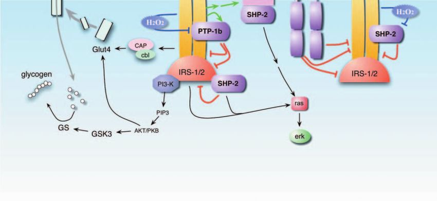
20   A W STOKER    ·    Protein tyrosine phosphatases and signalling

                       Figure 1 Schematic of the PTP family. The structures of RPTPs discussed in this review are shown: LAR,
                       PTP, PTP (type IIa); PTP, PTP, PTP// (type IIb); PTPϕ, DEP-1 (type III); PTP, PTP´ (type IV);
                       PTP / , PTP (type V). All but class III have duplicated catalytic domains D1 and D2. Each subclass has
                       unique extracellular domain structures depicted by coloured symbols as indicated. The cytoplasmic PTPs
                       only have single PTP catalytic domains, and their additional domain structures are discussed elsewhere. For
                       further references, see ‘The PTP family’ in the text. IG, immunoglobulin-like domain; FNIII, fibronectin type
                       III domain; MAM, meprin/A5-protein/PTPmu.

     Paul & Lombroso 2003, Ensslen-Craig & Brady-Kalnay                         ible (Schwarzbauer 1997) and this is the job of the PTPs.
     2004).                                                                     Several of these enzymes have now been implicated in the
                                                                                regulation of cell adhesion and FA dynamics (Angers-
                                                                                Loustau et al. 1999a, Beltran & Bixby 2003, Larsen et al.
     Substrate adhesion and motility                                            2003) (Fig. 2A).
     Interactions between cells and the extracellular matrix are
     essential for the maintenance of cell differentiation, sur-                 PTP-PEST PTP-PEST is a widely expressed nrPTP
     vival and motility. These interactions are governed largely                with interaction domains rich in proline, glutamic acid,
     by integrins, heterodimeric transmembrane protein com-                     serine and threonine (Garton & Tonks 1994). PTP-PEST
     plexes that can bind multiple extracellular molecules and                  binds directly to p130cas (Garton et al. 1997), Grb2
     can nucleate adhesive structures known as focal adhesions                  (Charest et al. 1997), Csk (Davidson et al. 1997), Shc
     (FAs) (Schwartz 2001, Zamir & Geiger 2001, Miranti &                       (Davidson & Veillette 2001) and paxillin (Shen et al.
     Brugge 2002, Parsons 2003). Integrin binding leads to                      1998). The roles of these interactions are largely unclear,
     autophosphorylation of focal adhesion kinase (FAK), acti-                  but the dephosphorylation of p130cas may be central to
     vation of the pp60src PTK and then a cascade of down-                      PTP-PEST’s actions. Overexpression of PTP-PEST in
     stream tyrosine phosphorylations (Fig. 2A) (Schaller et al.                fibroblasts induces p130cas hypophosphorylation, hinder-
     1994). For a cell to de-attach and migrate, the phos-                      ing the translocation of p130cas to membrane ruffles,
     phorylation of focal adhesion components must be revers-                   and reducing cell migration (Garton & Tonks 1999).
     Journal of Endocrinology (2005) 185, 19–33                                                                            www.endocrinology-journals.org
                                                                                                       Downloaded from Bioscientifica.com at 08/01/2021 06:28:44AM
                                                                                                                                                     via free access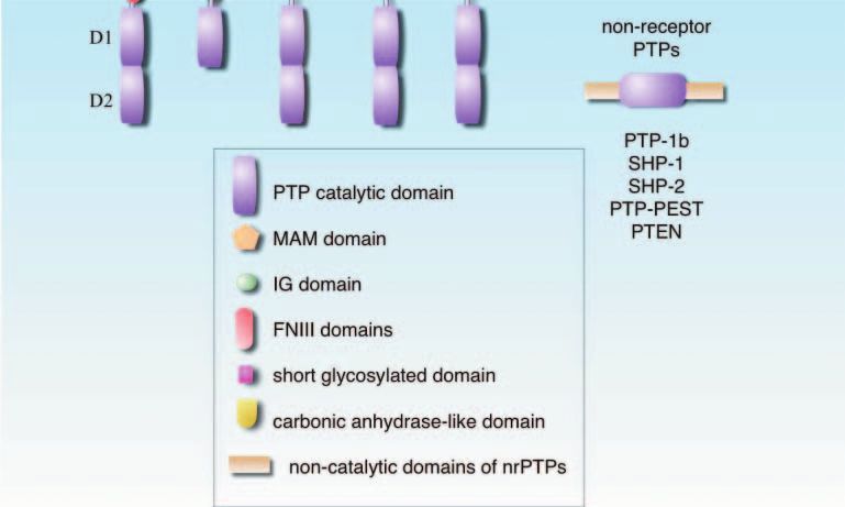
Protein tyrosine phosphatases and signalling ·              A W STOKER         21

PTP-PEST-deficient fibroblasts also exhibit increased              membrane-bound SHPS-1 (SHP substrate 1), which in
numbers of focal adhesions, increased phosphorylation of           turn recruits SHP-2. SHP-2 may then further activate
p130cas, paxillin and FAK, and defects in cell motility            pp60src, possibly via inhibition of Csk (Zhang et al. 2004),
(Angers-Loustau et al. 1999b). PTP-PEST therefore ap-              leading to increased FAK phosphorylation (Fig. 2A). To
pears to regulate FA turnover, such that either over- or           support this, SHP-2-deficient fibroblasts exhibit FAK and
underexpression of PTP-PEST can cause an imbalance                 pp60src hypophosphorylation (Oh et al. 1999). Other
resulting in impaired motility.                                    studies, however, show that SHP-2 either triggers FAK
                                                                   hypophosphorylation or has little effect (Tsuda et al. 1998,
PTP1B Evidence for the involvement of PTP1B in                     Yu et al. 1998, Manes et al. 1999, Inagaki et al. 2000).
integrin signalling, FA formation and cell motility provides       SHP-2 is also implicated in the repulsive guidance of cell
a complex, contradictory picture. Knock-down of PTP1B              migration triggered by Eph receptor tyrosine kinases
expression using antisense oligonucleotides (Hassid et al.         (RTKs), which bind to FAK constitutively. This repulsion
1999) or overexpression of wild-type PTP1B (Liu et al.             is regulated in part by preventing integrin-mediated
1998) both suggest correlations between PTP1B activity             adhesion (Miao et al. 2000, Poliakov et al. 2004). Upon
and decreased adhesion, motility and FA formation.                 EphA2 activation by ephrins, SHP-2 binds to EphA2,
PTP1B may negatively regulate adhesion-based signalling            leading to a loss of FAK and paxillin phosphorylation;
by directly dephosphorylating p130cas (Fig. 2A). However,          EphA2–FAK complexes also break down. Here again,
other studies using dominant-negative PTP1B (Arregui               SHP-2 may be a negative regulator of FAK phosphoryl-
et al. 1998) and PTP1B-deficient fibroblasts (Cheng et al.         ation and integrin signalling. Therefore, although the
2001) both support the opposite view that PTP1B                    precise mode of action of SHP-2 remains to be clarified,
activates pp60src (or fyn) directly or indirectly, thereby         and indeed it may vary between cell types, there is no
stimulating FAK and p130cas phosphorylation and FA                 doubt that SHP-2 can influence FAK activity, cell
formation. Until we can clarify whether, in different cell          adhesion and motility.
types and under different conditions, PTP1B does indeed
have divergent effects, the contradictions in these data will       PTEN PTEN is a tumour suppressor and an unusual
remain.                                                            enzyme in being able to dephosphorylate both the lipid
                                                                   phosphatidylinositol 3,4,5-triphosphate and phospho-
RPTP pp60src-like PTKs heavily influence FA dynam-                tyrosine (Yamada & Araki 2001). PTEN is implicated in
ics (Parsons 2003). pp60src is regulated by a carboxy-             many cellular processes, including cell adhesion, migra-
terminal, inhibitory tyrosine (Schwartzberg 1998), the             tion, invasion and anoikis (Yamada & Araki 2001), the
phosphorylation of which is reciprocally controlled by the         latter being apoptosis of cells after loss of matrix contact
Csk PTK and PTPs such as RPTP (Harder et al. 1998).               (Grossmann 2002). Overexpression of PTEN in cells
PTP overexpression in A431 cells causes increased                 causes hypophosphorylation of FAK and p130cas and
pp60src activity and pp60src/FAK association (Harder et al.        reduces cell motility (Tamura et al. 1998, 1999a,c). Fur-
1998). The opposite seems to occur in RPTP-deficient              thermore, dominant-negative PTEN causes FAK hyper-
cells (Su et al. 1999, Zeng et al. 2003). By acting as a           phosporylation and can co-purify FAK (Tamura et al.
pp60src activator, RPTP is thus uniquely situated to act          1998, 1999c). PTEN also targets the adaptor Shc, which
upstream of FAK in integrin signalling. PTP can also              binds to integrin complexes and in turn affects Erk MAP
target p130cas as a direct substrate in cultured cells (Buist et   kinase signalling (Gu et al. 1998). There are still doubts
al. 2000). One puzzle with RPTP is how, or indeed if,             over PTEN’s level of influence over FAK in vivo, how-
it is regulated by external cues, upstream of any effects on        ever, since PTEN / cells show no alteration in FAK
FAs. This must await the identification of an extracellular        phosphorylation even though motility is increased (Sun
ligand.                                                            et al. 1999, Liliental et al. 2000). Perhaps PTP-PEST and
                                                                   SHP-2 share the majority of control over FAK and p130cas
SHP-2 SHP-2 is an ubiquitous enzyme implicated in                  phosphorylation, while PTEN has a minor, or a very
both growth factor receptor signalling and integrin signal-        context-specific role. PTEN also appears to promote cell
ling (Feng 1999, Neel et al. 2003). The mechanism of               death through anoikis, by hydrolysing phosphatidylinositol
SHP-2 action remains controversial, though, and the                3,4,5-triphosphate (PIP3) and inactivating the AKT/PKB
critical downstream targets are largely unclear (Neel et al.       pathway. By also dampening FAK and phosphatidylinosi-
2003). SHP-2 is an nrPTP with two integral SH2 domains             tol 3-kinase (PI3)-kinase activities, PTEN may therefore
that bind to phosphotyrosine residues in receptors and             accelerate this apoptosis pathway (Tamura et al. 1999b,
docking proteins (see also ‘Regulation of PTP action’              Yamada & Araki 2001).
below) (Neel et al. 2003). Several studies point to SHP-2
being either a positive or a negative regulator of cell            LAR LAR is an adhesion molecule-like RPTP that
adhesion and motility. One model suggests that integrin            localises to FAs (Streuli et al. 1988, Serra Pages et al. 1995).
engagement causes pp60src-dependent phosphorylation of             LAR binds to liprins, which are enriched at these sites of
www.endocrinology-journals.org                                                                 Journal of Endocrinology (2005) 185, 19–33
                                                                                           Downloaded from Bioscientifica.com at 08/01/2021 06:28:44AM
                                                                                                                                         via free access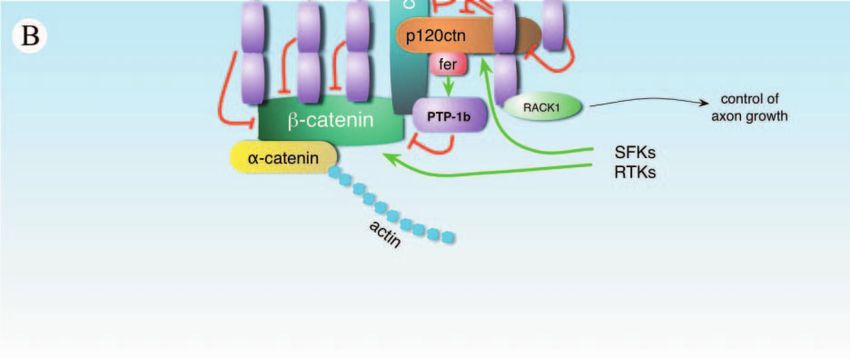
22   A W STOKER    ·   Protein tyrosine phosphatases and signalling

     Journal of Endocrinology (2005) 185, 19–33                                           www.endocrinology-journals.org
                                                                      Downloaded from Bioscientifica.com at 08/01/2021 06:28:44AM
                                                                                                                    via free access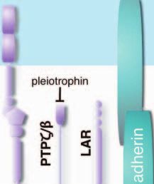
Protein tyrosine phosphatases and signalling ·               A W STOKER         23

adhesion (Serra-Pages et al. 1998), and to Trio, a large                adhesion molecules (Daniel & Reynolds 1997). Their
Rac/Rho GTPase that influences the actin cytoskeleton                   regulated adhesive properties are critical for many mor-
(Debant et al. 1996). An important role for LAR in FAs                  phogenic events including epithelial to mesenchymal
may revolve around p130cas. Overexpression of LAR in                    transitions (Nakagawa & Takeichi 1998, Lilien et al.
U2OS cells causes apoptosis and p130cas degradation                     2002), while defects in cadherin function are also linked to
(Weng et al. 1998, 1999). Anoikis can be triggered by                   cancer progression (Hirohashi 1998). Understanding
proteolytic fragments of p130cas (O’Neill et al. 2000, Wei              how cadherin-based adhesion is regulated is therefore
et al. 2004) and proteolysis of p130cas can be initiated by             of great importance in the fields of development and
hypophosphorylation, induced either by tyrosine kinase                  disease.
inhibition (Wei et al. 2002) or, in all likelihood, by                      Cadherins are linked to the actin cytoskeleton by
increased PTP activity. Indeed, LAR specifically targets                -catenin, -catenin and p120ctn (Daniel & Reynolds
p130cas for dephosphorylation, which may in turn lead                   1997) (Fig. 2B). The adhesive functions of cadherins
directly to its degradation (Weng et al. 1999). Re-                     are particularly sensitive to tyrosine phosphorylation of
introduction of excess p130cas into LAR-expressing cells                  -catenin. Phosphorylation of -catenin causes its dis-
can partially rescue apoptosis. Interestingly, LAR-induced              sociation from cadherin and loss of cell–cell adhesion
apoptosis is relatively direct and does not require prior loss          complexes (Balsamo et al. 1996, 1998, Roura et al. 1999).
of substrate adhesion (Weng et al. 1998). One isoform of                p120ctn may stabilise cadherins at the plasma membrane
LAR is known to bind to laminin (O’Grady et al. 1998)                   (Peifer & Yap 2003, Kowalczyk & Reynolds 2004) and its
and this PTP may therefore respond directly to the ECM                  tyrosine phosphorylation causes loss of cadherin function
at FAs. The effect of laminin binding on the biochemical                 (Ozawa & Ohkubo 2001). Since tyrosine phosphorylation
activity of LAR remains unclear.                                        is central to cadherin/catenin function, it is not surprising
                                                                        that PTPs are again found in the arena (Beltran & Bixby
PTPw PTPϕ is a type III RPTP, also known as GLEPP1                      2003) (Fig. 2B). PTPµ interacts with cadherin and may be
(Pixley et al. 1995). Studies in macrophages show that                  able to dephosphorylate it directly (Brady Kalnay et al.
paxillin is a PTPϕ substrate, and PTPϕ co-localises with                1995, Brady-Kalnay et al. 1998), although this remains a
paxillin in membrane ruffles. Overexpression of PTPϕ                      little controversial (Zondag et al. 1996). PTP1B binds
in BAC1.2F5 macrophages causes enhanced motility,                       directly to cadherin, but in this case its target is -catenin
possibly by reducing the quantity of phosphorylated                     (Fig. 2B). A dominant-negative form of PTP1B causes
paxillin available for focal adhesions and increasing FA                hyperphosphorylation of -catenin and loss of cadherin
turnover (Pixley et al. 2001).                                          function, possibly by displacing the wild-type enzyme
   While we probably know most of the central PTP                       (Balsamo et al. 1996, 1998, Pathre et al. 2001). PTP1B
players in integrin and FA signalling, there remain many                itself is activated by tyrosine phosphorylation on tyrosine
interesting questions. For example, which PTPs are the                  residue 152, catalysed by the PTK Fer (Xu et al. 2004); Fer
most influential for focal adhesion dynamics and do                     binds p120ctn, thereby holding it near to PTP1B. The
different PTPs act in strict concert or are their actions                p120ctn protein can be dephosphorylated by PTPµ and this
more cell type specific? Put another way, do the conflict-              has been proposed to be a route by which this RPTP
ing experimental data with individual PTPs reflect                      controls N-cadherin function in axons (Zondag et al.
genuine, cell-specific differences in enzyme action, or                  2000). DEP-1, a type III RPTP, and the nrPTP SHP-1
confusion arising from the wide range of under- and                     can also target p120ctn as a potential substrate (Keilhack
overexpression systems and cell types used? Either way, it              et al. 2000, Holsinger et al. 2002).
is clear that a range of cytoplasmic and receptor-type                        -Catenin interacts directly with several RPTPs,
PTPs are involved at several key levels in integrin                     including LAR (Kypta et al. 1996), PTP / (Meng et al.
signalling.                                                             2000), PTP (Fuchs et al. 1996) and PTP (Cheng et al.
                                                                        1997). All of these PTPs (except PTP) can dephos-
                                                                        phorylate -catenin and have been proposed to regulate
Cell–cell adhesion                                                      cadherin/ -catenin interactions. It is not clear, though,
Cell–cell adhesion within epithelia is largely controlled by            how most of these biochemical events are actually regu-
the cadherin family of homophilic, calcium-dependent                    lated in the cell. In the case of PTP / , its ligand

Figure 2 PTP involvement with integrin and cadherin signalling. This schematic shows some of the documented binding partners and
substrates of PTPs, at (A) sites of focal adhesions and (B) sites of cadherin binding. Dephosphorylation steps are depicted by red lines and
tyrosine kinase reactions are shown by green arrows. Black lines indicate pathways that are stimulated. Grey arrows indicate the binding of
extracellular ligands where known. All PTPs are shown in purple. In (B), cadherin and PTP// each have homophilic binding partners,
binding in trans from another cell. Note that all of the above interactions have been documented in at least one study, but their relative
physiological importance is not yet known in every case. Some of these interactions may be competitive or cell specific and may not
occur concurrently. See text for further details. SFKs, src family kinases; ECM, extracellular matrix.

www.endocrinology-journals.org                                                                        Journal of Endocrinology (2005) 185, 19–33
                                                                                                  Downloaded from Bioscientifica.com at 08/01/2021 06:28:44AM
                                                                                                                                                via free access
24 A W STOKER · Protein tyrosine phosphatases and signalling
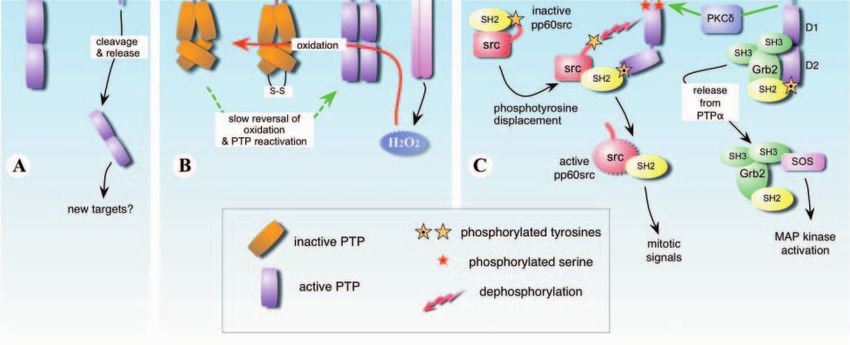
Figure 3 PTPs and insulin signalling. This diagram depicts the documented interactions between PTPs and components of the insulin
signalling pathway. On the left of the diagram are some of the major pathways stimulated by the insulin receptor (IR) tyrosine kinase,
leading to activation of glycogen synthase (GS) and movement of the glucose transporter Glut4 to the plasma membrane. PTP enzymes
are shown in purple and dephosphorylation events are shown by red lines. Phosphorylation by tyrosine kinases is shown by green
arrows; black lines indicate further pathways that are stimulated. Inactivation of PTPs by peroxide, which is produced following IR
stimulation, is shown in blue. Note that all of the above interactions have been documented in at least one study, but their relative
physiological importance is not yet known in every case. Some of these interactions may be competitive or cell specific and may not
occur concurrently. See text for further details.
pleiotrophin can inactivate the RPTP and this results in or any other PTPs are developmentally controlled in order
increased -catenin phosphorylation (Meng et al. 2000). to influence events such as epithelial–mesenchymal
Although it is evident that PTPs can strongly influence the transitions.
dynamics of cell–cell adhesion, we have much yet to learn.
For example, if cadherin/ –catenin interactions are regu-
lated by the PTPs describe above, then why do we see so
Insulin signalling
little perturbation of epithelial tissues in animals lacking
their genes? The lack of such phenotypes may indicate a One field in which PTP action has been well studied is
high level of biochemical redundancy in the control of that of insulin signalling (Cheng et al. 2002a, Asante-
-catenin phosphorylation. Whether several PTPs must Appiah & Kennedy 2003) (Fig. 3). Given the increasing
actively co-operate to regulate -catenin phosphorylation, concern surrounding diabetes and obesity, particularly
or whether there are key PTPs in specific cell types, with insulin-resistant forms, it is not surprising that the
remains to be determined. molecular regulation of the insulin receptor (IR; an RTK),
In cultured tumour cells, overexpression of LAR causes has come under scrutiny. There is particularly keen
reduced -catenin phosphorylation, stabilisation of adhe- interest in understanding how PTPs might impinge on the
rens junctions and loss of tumour formation, indicating signalling of the IR and whether PTPs are relevant
that this RPTP could have a powerful controlling therapeutic targets. Here, a number of PTPs have been
effect over epithelial integrity in vivo (Müller et al. 1999). implicated in recent years (Fig. 3), although their status as
In this context, it will be most interesting to see if LAR IR regulators is still controversial in most cases.
Journal of Endocrinology (2005) 185, 19–33 www.endocrinology-journals.org
Downloaded from Bioscientifica.com at 08/01/2021 06:28:44AM
via free accessProtein tyrosine phosphatases and signalling · A W STOKER 25
LAR LAR expression is increased in fat tissue of clinically 32D cells and these IRS-1 forms are even hyperphos-
obese humans (Ahmad et al. 1995) and increased phorylated (Myers et al. 1998). SHP-2 also binds to IRS-3
co-immunoprecipitation of LAR with the IR occurs (Ross et al. 1998) and IRS-4 (Escribano et al. 2003), so
after insulin treatment (Ahmad & Goldstein 1997). perhaps there is further degeneracy in the system. Another
Furthermore, overexpression of LAR suppresses insulin pathway through which SHP-2 may act in concert with
action (Li et al. 1996, Zhang et al. 1996) and antisense the IR involves SHPS-1 (see also Fig. 2) (Takada et al.
knock-down of LAR enhances and prolongs insulin sig- 1998). SHPS-1 is phosphorylated in response to IR
nalling in McA-RH7777 hepatoma cells (Kulas et al. stimulation, and recruits SHP-2. Overexpression of wild-
1995, 1996, Mooney et al. 1997). These data are consistent type SHPS-1 in CHO cells enhances MAP kinase
with LAR being a direct, negative regulator of the IR. activation downstream of insulin stimulation, whereas
Engineered overexpression of LAR in mouse skeletal mutated forms of SHPS-1 that cannot recruit SHP-2 show
muscle suppresses IR signalling, causing insulin resistance no enhancement (Takada et al. 1998). How SHPS-1/
possibly through deactivation of the insulin receptor sub- SHP-2 interactions stimulate MAP kinase in this system
strate IRS-2 (Zabolotny et al. 2001). In LAR-deficient remains to be determined.
mice, although there is increased insulin sensitivity at the Transgenic analyses of SHP-2 function have also
resting state, there are also unexpected defects in glucose proved equivocal. Homozygous null mice do not survive,
homeostasis after insulin treatment that are more consistent whereas heterozygotes appear normal with respect to
with insulin resistance (Ren et al. 1998). The case for LAR glucose homeostasis, and IR and IRS-1 phosphorylation
being a significant regulator of the IR therefore remains (Arrandale et al. 1996). Nevertheless, ubiquitous expres-
unresolved. sion of a dominant-negative form of SHP-2 causes signifi-
cant insulin resistance and reduced IRS-1 phosphorylation
PTP and PTP´ Overexpression of PTP in BHK-IR (Maegawa et al. 1999). This latter study again supports
cells causes dephosphorylation of the IR and suppresses the idea that SHP-2 plays a positive role in insulin
IR-mediated cell rounding (Lammers et al. 1997). The signalling.
highly related PTP´ enzyme has also been implicated as a
potential IR phosphatase in one overexpression study PTP1B PTP1B has drawn much recent attention as being
in BHK cells (Andersen et al. 2001). Furthermore, in a negative regulator of insulin action (Cheng et al. 2002a,
adipocytes, rat PTP can decrease the transport of the Asante-Appiah & Kennedy 2003). PTP1B associates with
insulin-responsive glucose transporter Glut4 after insulin the IR after insulin treatment and several critical tyrosine
stimulation (Cong et al. 1999). However, a case against residues in PTP1B become phosphorylated (Seely et al.
PTP being a physiological regulator of the IR comes 1996, Bandyopadhyay et al. 1997). Importantly, the IR is
from studies of 3T3-L1 cells, where antisense PTP a substrate for PTP1B (Bandyopadhyay et al. 1997, Dadke
does not affect insulin-induced MAP kinase activation or et al. 2000, Salmeen et al. 2000) and there is evidence that
DNA synthesis (Arnott et al. 1999), and from mice IRS-1 is also (Goldstein et al. 2000). Homozygous loss of
deficient for PTP, where there are no defects in glucose PTP1B has no gross effect on mouse development or
homeostasis yet reported (Ponniah et al. 1999, Su et al. viability. However, the mice have defects in glucose and
1999). insulin tolerance, they also remain sensitive to insulin
(when on high fat diets) and they show increases in IR
SHP-2 SHP-2 binds to the IR and to IRS-1 in yeast phosphorylation after insulin treatment (Elchebly et al.
two-hybrid assays (Rocchi et al. 1996) and in transfected 1999, Klaman et al. 2000). However, most of these effects
cells (Kharitonenkov et al. 1995), and IRS-1 is also likely only occur in muscle and liver, suggesting that further
to be a direct substrate (Hayashi et al. 2004). In another PTPs might control insulin signalling in fat cells.
study, wild-type SHP-2 did not interact with the IR in PTP1B is widely expressed and localises predominantly
yeast two-hybrids, whereas a phosphatase-dead mutant in the endoplasmic reticulum (Frangioni et al. 1992),
did (via tyrosine 1146 on the IR). This suggests that the although it does have a cleavable carboxy-terminal domain
IR is also a direct substrate (Rocchi et al. 1996). However, that allows release into the cytosol (Frangioni et al. 1993).
a further study suggests that IR is not a substrate, but It is not exactly clear, therefore, how PTP1B is getting
instead indicates that SHP-2 may bind to the IR in order access to the IR. Like some other RPTKs, the IR may
to recruit IRS-1 (Kharitonenkov et al. 1995). become endocytosed and thereby gain access to endoplas-
Several studies conclude that SHP-2 does affect the mic reticulum-associated PTP1B (Cheng et al. 2002a, Haj
insulin signalling process itself, but again there is a lack of et al. 2002). Lastly, it is worth noting that PTP1B has also
consensus. Interference with SHP-2 activity can block been implicated in controlling obesity, through the regu-
insulin-stimulated mitosis in 3T3 cells (Milarski & Saltiel lation of leptin action (Cheng A. et al. 2002b, Zabolotny
1994) and block downstream activation of Ras (Noguchi et al. 2002). Interested readers are encouraged to read
et al. 1994). In contrast, mutated forms of IRS-1 that do further details reviewed by Asante-Appiah & Kennedy
not bind SHP-2 can facilitate effective insulin signalling in (2003).
www.endocrinology-journals.org Journal of Endocrinology (2005) 185, 19–33
Downloaded from Bioscientifica.com at 08/01/2021 06:28:44AM
via free access26 A W STOKER · Protein tyrosine phosphatases and signalling
Other PTPs The PTPs that act upon the IR may not be released from PTP and becomes free to associate, via its
limited to those above. A study employing substrate- SH3 domain, with SOS, leading to downstream signalling
trapping PTPs suggested that PTP , Sap-1 and TC-PTP (Fig. 4) (Den Hertog & Hunter 1996).
could bind specifically to phosphorylated IR ‘baits’
(Walchli et al. 2000). However, further tests showed that
Cleavage and differential localisation
a wider range of wild-type PTPs could dephosphorylate an
IR-derived target peptide (containing the principal PTP Many proteins are subject to post-translational proteolytic
target tyrosines; PTPGMTRDIYETDYYRKGGKG). It cleavage and PTPs are no exception. A number of RPTPs
is not clear, therefore, how these in vitro assays will relate are cleaved by subtilisin-like proteases (Streuli et al. 1992,
to the substrate specificity or accessibility of these PTPs Jiang et al. 1993, SerraPages et al. 1994, Gebbink et al.
towards the IR in vivo. 1995, Campan et al. 1996, Aicher et al. 1997) and this can
It is clear that PTPs significantly influence the level of ultimately result in ectodomain ‘shedding’, at least in cell
insulin signalling and these enzymes do represent poten- culture. A role for shedding in vivo is currently conjectural,
tial, pharmacological targets in insulin-resistant diabetes. but it may be a mechanism to either terminate RPTP
This field remains controversial, however, with strong signalling, facilitate internalisation of catalytic domains, or
evidence pointing to PTP1B being the major player, but to release ectodomains to compete for ligands. Intracellular
with several other PTPs hovering in the wings. The field cleavage also occurs. PTP´, PTP and PTP1B can be
currently suffers from numerous apparently contradictory cleaved in a calpain-dependent manner, releasing their
findings and the loss-of-function mouse strains have added catalytic domains into the cytoplasm (Frangioni et al. 1993,
to the uncertainty by sometimes failing to corroborate cell Gil-Henn et al. 2001). This may prevent access of the
culture data. Tissue-specific action of certain PTPs, or the PTPs to membrane-associated substrates, or provide access
concerted actions of multiple PTPs, may both therefore to novel substrates.
serve to control IR phosphorylation levels in vivo. Devel-
oping any diabetes therapies based on PTP inhibition
Ligands for RPTPs
therefore remains a worthy goal, and a considerable
challenge. Upon the discovery of receptor-like RPTPs with their
highly conserved ectodomains, there was great interest in
their potential to be regulated by extracellular ligands.
Regulation of PTP action Since that time there have been great difficulties in
identifying the ligands for many RPTPs and, even more
It is evident from the discussion above that molecular so, understanding their effects on RPTP activity. A
control of the PTPs themselves is still relatively poorly number of RPTPs have homophilic binding properties,
understood. Recent research, however, is beginning to including the type IIb enzymes, as well as PTP and
provide much-needed insight and some key findings are isoforms of LAR (Yang et al. 2003, for reviews see Beltran
discussed below. & Bixby 2003, Johnson & van Vactor 2003). Of those
RPTPs with heterophilic binding properties, PTP /
binds pleiotrophin and adhesion molecules (Beltran &
Phosphorylation and SH2 domains Bixby 2003), PTP binds to heparan sulphate proteo-
PTP enzymes can be phosphorylated on tyrosines, pro- glycans (Aricescu et al. 2002) and LAR binds the laminin/
viding binding sites for SH2-containing proteins. SH2 nidogen complex (O’Grady et al. 1998). Of these RPTP
domains are protein adaptor modules with specific affinity interactions, only the interaction between pleiotrophin
for phosphorylated tyrosine residues on proteins (Pawson and PTP / has so far been shown to have a clear effect
et al. 2001, Pawson 2004). These domains act as docking on enzyme activity: pleiotrophin binding inhibits PTP /
devices, facilitating the formation of supramolecular sig- and causes an increase in -catenin phosphorylation
nalling complexes (Pawson et al. 2001, Neel et al. 2003, (Meng et al. 2000).
Pawson 2004). One RPTP that attracts such SH2 domains Several major areas must therefore be clarified with
is PTP. The tyrosine residue 789 in D2 of PTP is respect to RPTP ligands. Why has it been so hard to
constitutively phosphorylated and associates with the SH2 identify heterotypic ligands for many RPTPs? Does this
domain of Grb2 (Fig. 4) (Den Hertog & Hunter 1996, Su mean that some RPTPs do not have ligands? For example,
et al. 1996). Upon PKC phosphorylation of PTP, Grb2 work with CD45 suggests that the activity of this enzyme
is exchanged for pp60src, whose own SH2 domain now can be modulated through changes in protein isoform
associates with tyr789 (Zheng et al. 2002, Brandt et al. expression and the consequent changes in ectodomain
2003). Through a ‘phosphotyrosine displacement’ mech- glycosylation and dimerisation (Xu & Weiss 2002).
anism, the inhibitory C-terminal tyr527 of pp60src is thus Understanding whether the trans interactions of RPTP
released and is then dephosphorylated by PTP, activating ectodomains generally activate or inactivate the catalytic
pp60src (Zheng et al. 2000, 2002). Concurrently, Grb2 is activity of RPTPs also remains a challenge for the field.
Journal of Endocrinology (2005) 185, 19–33 www.endocrinology-journals.org
Downloaded from Bioscientifica.com at 08/01/2021 06:28:44AM
via free accessProtein tyrosine phosphatases and signalling · A W STOKER 27
Figure 4 The regulation of PTP activity. This diagram shows three different ways in which RPTPs are regulated (A) by cleavage, (B) by
oxidation and dimerisation and (C) by phosphorylation. The precise role of these events is still uncertain for many RPTPs. (A) Cleavage
and extracellular shedding of ectodomains has been reported for several RPTPs (shown here for LAR). (B) Some RPTPs, including PTP
and CD45 are present as dimers in the membrane. Inactivation of dimers can occur through oxidation of D1/D2 catalytic sites and
conformational changes. Oxidation is caused by peroxide that is released following growth factor receptor activation; this oxidation is
reversible in the cell. (C) Phosphorylation of PTP by PKC causes detachment of Grb2 followed by attachment of pp60src, effectively
through SH2 domain exchange. Binding of the SH2 domain of pp60src to PTP exposes phosphotyrosine 527 in pp60src (yellow star),
which is then dephosphorylated by PTP.
There remains the possibility that RPTPs require cis rather activation (Jiang et al. 1999). It was also found that an
than trans interactions to promote intramembrane com- inhibitory peptide ‘wedge’ caused reciprocal blockade of
partmentalisation into, for example, lipid rafts. Finally, cis the catalytic sites of the dimerised D1 domains, revealing
interactions between ectodomains themselves may not a potential inactivation mechanism (Majeti et al. 1998,
necessarily require a ligand, but could still be essential in Wallace et al. 1998). However, although ‘wedge’ inhibi-
enforcing dimerisation and regulating enzyme activity. tion may occur with PTP and CD45, it appears less
feasible with LAR and PTPµ (Hoffmann et al. 1997, Nam
et al. 1999), and therefore may not be a universal inhibi-
PTP dimerisation
tory mechanism. It is still likely, however, that RPTPs can
A well-known prerequisite for the activation of receptor have a dimerised, rotational state that engenders catalytic
PTKs is their dimerisation and autophosphorylation inactivity with or without involvement of a wedge. One
(Heldin 1995). In 1998, It was found that the catalytic further, outstanding question is whether the dimerisation
domains of PTP formed dimer-like structures when process itself is generally constitutive, or whether it
crystallised (Majeti et al. 1998, Wallace et al. 1998), and it requires the presence of a ligand.
was subsequently shown that the full-length protein
dimerises in live cells (Jiang et al. 1999, 2000). Dimerisa-
PTP oxidation
tion in cis has also been demonstrated for CD45 (Takeda
et al. 1992) and it could well be a common phenomenon The catalytic site cysteines of PTPs have an ionisation
for RPTPs in general. Suggestions as to how dimers might constant (PKa) of between 5 and 6. They are therefore
regulate RPTP signalling have been given. As with usually deprotonated and susceptible to oxidative attack,
RPTKs (Heldin 1995), the rotational orientation of dimer- which inactivates the catalytic site at least temporarily (Fig.
ised receptor PTP has been shown to be critical for its full 4) (Xu et al. 2002). Oxidation of PTPs could be an elegant
www.endocrinology-journals.org Journal of Endocrinology (2005) 185, 19–33
Downloaded from Bioscientifica.com at 08/01/2021 06:28:44AM
via free access28 A W STOKER · Protein tyrosine phosphatases and signalling
mechanism by which RPTKs briefly impede local PTP specificity in so many signalling pathways. As our knowl-
activity and thereby prolong PTK signals (Xu et al. 2002). edge thus increases, the ultimate goal of applying it to a
Indeed, the activation of growth factor receptors does therapeutic setting, tackling human diseases with under-
produce a local burst of peroxide capable of oxidising PTPs lying dysregulation of phosphotyrosine signalling, will
(Bae et al. 1997, Lee et al. 1998, Meng et al. 2002, 2004). come closer to fruition.
In the case of the IR, PTP1B may inhibit insulin
signalling, but the IR itself may in turn inhibit (albeit
temporarily) PTP1B through local oxidant production Acknowledgements
(Mahadev et al. 2001). For most RPTPs, the obvious
candidate for oxidative attack is the catalytically active D1 I would like to thank Radu Aricescu for helpful comments
domain. However, studies of PTP show that D2 is the on the manuscript. All figures were designed using
initial, selective target, and its oxidation causes conforma- Omnigraffle Pro software. The author declares that there
tional shifts in the catalytic domains that result in pro- is no conflict of interest that would prejudice the impar-
longed enzyme inactivation (Blanchetot et al. 2002, Pers- tiality of this scientific work.
son et al. 2004) (Fig. 4). Finally, a recent study suggests
that PTP can briefly form disulphide-linked dimers
when treated with peroxide and that the D2 active site References
cysteines are again critical for this event (van der Wijk Ahmad F & Goldstein BJ 1997 Functional association between the
et al. 2004). The relevance of this event to other native insulin receptor and the transmembrane protein-tyrosine
RPTPs remains to be addressed. phosphatase LAR in intact cells. Journal of Biological Chemistry 272
There is therefore justifiable excitement that a universal 448–457.
regulatory mechanism may now have been uncovered for Ahmad F, Considine RV & Goldstein BJ 1995 Increased abundance
of the receptor-type protein-tyrosine phosphatase LAR accounts for
PTPs, although many key questions remain. For example, the elevated insulin receptor dephosphorylating activity in adipose
how precisely do PTKs stimulate peroxide production and tissue of obese human subjects. Journal of Clinical Investigation 95
how localised is it in the cell? In addition, the percentage 2806–2812.
of a given PTP that is inactivated varies widely between Aicher B, Lerch MM, Muller T, Schilling J & Ullrich A 1997
Cellular redistribution of protein tyrosine phosphatases LAR and
studies. Although much of this may arise from the varied PTPsigma by inducible proteolytic processing. Journal of Cell Biology
experimental protocols, we have yet to define the neces- 138 681–696.
sary levels of PTP inactivation that would be required for Alonso A, Sasin J, Bottini N, Friedberg I, Osterman A, Godzik A,
the sustained activation of RPTK signalling. This is not a Hunter T, Dixon J & Mustelin T 2004 Protein tyrosine
trivial undertaking, but it has far-reaching consequences phosphatases in the human genome. Cell 117 699–711.
Andersen JN, Elson A, Lammers R, Romer J, Clausen JT, Moller KB
for our understanding of PTP regulation. & Moller NP 2001 Comparative study of protein tyrosine
phosphatase-epsilon isoforms: membrane localization confers
specificity in cellular signalling. Biochemical Journal 354 581–590.
Conclusions and future prospects Angers-Loustau A, Cote JF & Tremblay ML 1999a Roles of protein
tyrosine phosphatases in cell migration and adhesion. Biochemistry
and Cell Biology 77 493–505.
The road to discovery with the PTP family has been long Angers-Loustau A, Cote JF, Charest A, Dowbenko D, Spencer S,
and arduous, but its box of secrets is finally being prised Lasky LA & Tremblay ML 1999b Protein tyrosine
open, revealing true surprises. We see that PTPs are phosphatase-PEST regulates focal adhesion disassembly, migration,
closely involved in many levels of cell biological control. and cytokinesis in fibroblasts. Journal of Cell Biology 144 1019–1031.
Aricescu AR, McKinnell IW, Halfter W & Stoker AW 2002 Heparan
Furthermore, the enzymes can be controlled and modu- sulfate proteoglycans are ligands for receptor protein tyrosine
lated in diverse and novel ways, including through oxi- phosphatase sigma. Molecular and Cellular Biology 22 1881–1892.
dation, dimerisation, cleavage, differential localisation, use Arnott CH, Sale EM, Miller J & Sale GJ 1999 Use of an antisense
of differential mRNA splicing, or combinations of these. strategy to dissect the signaling role of protein-tyrosine phosphatase
alpha. Journal of Biological Chemistry 274 26105–26112.
There also appears to be cellular and developmental Arrandale JM, Gore Willse A, Rocks S, Ren JM, Zhu J, Davis A,
tolerance to their individual loss of function in many Livingston JN & Rabin DU 1996 Insulin signaling in mice
cases, suggesting that highly conserved redundancy or expressing reduced levels of Syp. Journal of Biological Chemistry 271
degeneracy is in place in order to maintain control of PTK 21353–21358.
signalling pathways. The discovery of oxidative regulation Arregui CO, Balsamo J & Lilien J 1998 Impaired integrin-mediated
adhesion and signaling in fibroblasts expressing a dominant-negative
in particular has profound implications and may trigger mutant PTP1B. Journal of Cell Biology 143 861–873.
novel pharmacological approaches for PTP inhibition or Asante-Appiah E & Kennedy BP 2003 Protein tyrosine phosphatases:
activation. Other exciting challenges include understand- the quest for negative regulators of insulin action. American Journal of
ing the structure and function of RPTP ectodomains, Physiology–Endocrinology and Metabolism 284 E663–E670.
Bae YS, Kang SW, Seo MS, Baines IC, Tekle E, Chock PB & Rhee
defining more precisely the substrate specificities of SG 1997 Epidermal growth factor (EGF)-induced generation of
PTPs, and finally discovering how these extremely active hydrogen peroxide. Role in EGF receptor-mediated tyrosine
enzymes are harnessed by cells to act with apparent phosphorylation. Journal of Biological Chemistry 272 217–221.
Journal of Endocrinology (2005) 185, 19–33 www.endocrinology-journals.org
Downloaded from Bioscientifica.com at 08/01/2021 06:28:44AM
via free accessProtein tyrosine phosphatases and signalling · A W STOKER 29
Balsamo J, Leung T, Ernst H, Zanin MK, Hoffman S & Lilien J 1996 Davidson D & Veillette A 2001 PTP-PEST, a scaffold protein tyrosine
Regulated binding of PTP1B-like phosphatase to N-cadherin: phosphatase, negatively regulates lymphocyte activation by targeting
control of cadherin-mediated adhesion by dephosphorylation of a unique set of substrates. EMBO Journal 20 3414–3426.
beta-catenin. Journal of Cell Biology 134 801–813. Davidson D, Cloutier JF, Gregorieff A & Veillette A 1997 Inhibitory
Balsamo J, Arregui C, Leung T & Lilien J 1998 The nonreceptor tyrosine protein kinase p50 csk is associated with protein-tyrosine
protein tyrosine phosphatase PTP1B binds to the cytoplasmic phosphatase PTP-PEST in hemopoietic and non-hemopoietic cells.
domain of N-cadherin and regulates the cadherin-actin linkage. Journal of Biological Chemistry 272 23455–23462.
Journal of Cell Biology 143 523–532. Debant A, Serra Pages C, Seipel K, O’Brien S, Tang M, Park SH &
Bandyopadhyay D, Kusari A, Kenner KA, Liu F, Chernoff J, Streuli M 1996 The multidomain protein Trio binds the LAR
Gustafson TA & Kusari J 1997 Protein-tyrosine phosphatase 1B transmembrane tyrosine phosphatase, contains a protein kinase
complexes with the insulin receptor in vivo and is tyrosine- domain, and has separate rac-specific and rho-specific guanine
phosphorylated in the presence of insulin. Journal of Biological nucleotide exchange factor domains. PNAS 93 5466–5471.
Chemistry 272 1639–1645. Den Hertog J & Hunter T 1996 Tight association of GRB2 with
Beltran PJ & Bixby JL 2003 Receptor protein tyrosine phosphatases as receptor protein-tyrosine phosphatase alpha is mediated by the
mediators of cellular adhesion. Frontiers in Bioscience 8 D87–D99. SH2 and C-terminal SH3 domains. EMBO Journal 15
Blanchetot C, Tertoolen LG & den Hertog J 2002 Regulation of 3016–3027.
receptor protein-tyrosine phosphatase alpha by oxidative stress. Elchebly M, Payette P, Michaliszyn E, Cromlish W, Collins S, Loy
EMBO Journal 21 493–503. AL, Normandin D, Cheng A, Himms-Hagen J, Chan CC et al.
Brady Kalnay SM, Rimm DL & Tonks NK 1995 Receptor protein 1999 Increased insulin sensitivity and obesity resistance in mice
tyrosine phosphatase PTPmu associates with cadherins and catenins lacking the protein tyrosine phosphatase-1B gene. Science 283
in vivo. Journal of Cell Biology 130 977–986. 1544–1548.
Brady-Kalnay SM, Mourton T, Nixon JP, Pietz GE, Kinch M, Chen Ensslen-Craig SE & Brady-Kalnay SM 2004 Receptor protein tyrosine
H, Brackenbury R, Rimm DL, Del Vecchio RL & Tonks NK phosphatases regulate neural development and axon guidance.
1998 Dynamic interaction of PTPmu with multiple cadherins in Developmental Biology 275 12–22.
vivo. Journal of Cell Biology 141 287–296. Escribano O, Fernandez-Moreno MD, Zueco JA, Menor C, Fueyo J,
Brandt DT, Goerke A, Heuer M, Gimona M, Leitges M, Kremmer Ropero RM, Diaz-Laviada I, Roman ID & Guijarro LG 2003
E, Lammers R, Haller H & Mischak H 2003 Protein kinase C Insulin receptor substrate-4 signaling in quiescent rat hepatocytes
delta induces Src kinase activity via activation of the protein and in regenerating rat liver. Hepatology 37 1461–1469.
tyrosine phsophatase PTPalpha. Journal of Biological Chemistry 278 Espanel X, Walchli S, Gobert RP, El Alama M, Curchod ML,
34073–34078. Gullu-Isler N & van Huijsduijnen RH 2001 Pulling stringsH
Buist A, Blanchetot C, Tertoolen LG & den Hertog J 2000 below the surface: hormone receptor signaling through inhibition of
Identification of p130 Cas as an in vivo substrate of receptor protein tyrosine phosphatases. Endocrine 15 19–28.
protein-tyrosine phosphatase alpha. Journal of Biological Chemistry Fauman EB & Saper MA 1996 Structure and function of the protein
275 20754–20761. tyrosine phosphatases. Trends in Biochemical Science 21 413–417.
Campan M, Yoshizumi M, Seidah NG, Lee ME, Bianchi C & Haber
Feng GS 1999 Shp-2 tyrosine phosphatase: signaling one cell or many.
E 1996 Increased proteolytic processing of protein tyrosine
Experimental Cell Research 253 47–54.
phosphatase mu in confluent vascular endothelial cells: the role of
PC5, a member of the subtilisin family. Biochemistry 35 3797–3802. Frangioni JV, Beahm PH, Shifrin V, Jost CA & Neel BG 1992 The
nontransmembrane tyrosine phosphatase PTP-1B localizes to the
Charest A, Wagner J, Kwan M & Tremblay ML 1997 Coupling of
the murine protein tyrosine phosphatase PEST to the epidermal endoplasmic reticulum via its 35 amino acid C-terminal sequence.
growth factor (EGF) receptor through a Src homology 3 (SH3) Cell 68 545–560.
domain-mediated association with Grb2. Oncogene 14 1643–1651. Frangioni JV, Oda A, Smith M, Salzman EW & Neel BG
Cheng A, Bal GS, Kennedy BP & Tremblay ML 2001 Attenuation of 1993 Calpain-catalyzed cleavage and subcellular relocation of
adhesion-dependent signaling and cell spreading in transformed protein phosphotyrosine phosphatase 1B (PTP-1B) in human
fibroblasts lacking protein tyrosine phosphatase-1B. Journal of platelets. EMBO Journal 12 4843–4856.
Biological Chemistry 276 25848–25855. Fuchs M, Mueller T, Lerch MM & Ullrich A 1996 Association of
Cheng A, Dube N, Gu F & Tremblay ML 2002a Coordinated action human protein-tyrosine phosphatase kappa with members of the
of protein tyrosine phosphatases in insulin signal transduction. armadillo family. Journal of Biological Chemistry 271 16712–16719.
European Journal of Biochemistry 269 1050–1059. Garton AJ & Tonks NK 1994 PTP-PEST: a protein tyrosine
Cheng A, Uetani N, Simoncic PD, Chaubey VP, Lee-Loy A, phosphatase regulated by serine phosphorylation. EMBO Journal 13
McGlade CJ, Kennedy BP & Tremblay ML 2002b Attenuation of 3763–3771.
leptin action and regulation of obesity by protein tyrosine Garton AJ & Tonks NK 1999 Regulation of fibroblast motility by the
phosphatase 1B. Developmental Cell 2 497–503. protein tyrosine phosphatase PTP-PEST. Journal of Biological
Cheng J, Wu K, Armanini M, O’Rourke N, Dowbenko D & Lasky Chemistry 274 3811–3818.
LA 1997 A novel protein-tyrosine phosphatase related to the Garton AJ, Burnham MR, Bouton AH & Tonks NK 1997 Association
homotypically adhering kappa and mu receptors. Journal of Biological of PTP-PEST with the SH3 domain of p130 cas; a novel
Chemistry 272 7264–7277. mechanism of protein tyrosine phosphatase substrate recognition.
Cong LN, Chen H, Li Y, Lin CH, Sap J & Quon MJ 1999 Oncogene 15 877–885.
Overexpression of protein tyrosine phosphatase-alpha (PTP-alpha) Gebbink MF, Zondag GC, Koningstein GM, Feiken E, Wubbolts
but not PTP-kappa inhibits translocation of GLUT4 in rat adipose RW & Moolenaar WH 1995 Cell surface expression of receptor
cells. Biochemical and Biophysical Research Communications 255 protein tyrosine phosphatase RPTP mu is regulated by cell–cell
200–207. contact. Journal of Cell Biology 131 251–260.
Dadke S, Kusari J & Chernoff J 2000 Down-regulation of insulin Gil-Henn H, Volohonsky G & Elson A 2001 Regulation of
signaling by protein-tyrosine phosphatase 1B is mediated by an protein-tyrosine phosphatases alpha and epsilon by calpain-mediated
N-terminal binding region. Journal of Biological Chemistry 275 proteolytic cleavage. Journal of Biological Chemistry 276
23642–23647. 31772–31779.
Daniel JM & Reynolds AB 1997 Tyrosine phosphorylation and Goldstein BJ, Bittner-Kowalczyk A, White MF & Harbeck M 2000
cadherin/catenin function. Bioessays 19 883–891. Tyrosine dephosphorylation and deactivation of insulin receptor
www.endocrinology-journals.org Journal of Endocrinology (2005) 185, 19–33
Downloaded from Bioscientifica.com at 08/01/2021 06:28:44AM
via free access30 A W STOKER · Protein tyrosine phosphatases and signalling
substrate-1 by protein-tyrosine phosphatase 1B. Possible facilitation energy expenditure, decreased adiposity, and tissue-specific insulin
by the formation of a ternary complex with the Grb2 adaptor sensitivity in protein-tyrosine phosphatase 1B-deficient mice.
protein. Journal of Biological Chemistry 275 4283–4289. Molecular and Cellular Biology 20 5479–5489.
Grossmann J 2002 Molecular mechanisms of ‘detachment-induced Kowalczyk AP & Reynolds AB 2004 Protecting your tail: regulation
apoptosis–Anoikis’. Apoptosis 7 247–260. of cadherin degradation by p120-catenin. Current Opinion in Cell
Gu J, Tamura M & Yamada KM 1998 Tumor suppressor PTEN Biology 16 522–527.
inhibits integrin- and growth factor-mediated mitogen-activated Kulas DT, Zhang WR, Goldstein BJ, Furlanetto RW & Mooney RA
protein (MAP) kinase signaling pathways. Journal of Cell Biology 143 1995 Insulin receptor signaling is augmented by antisense inhibition
1375–1383. of the protein tyrosine phosphatase LAR. Journal of Biological
Haj FG, Verveer PJ, Squire A, Neel BG & Bastiaens PI 2002 Imaging Chemistry 270 2435–2438.
sites of receptor dephosphorylation by PTP1B on the surface of the Kulas DT, Goldstein BJ & Mooney RA 1996 The transmembrane
endoplasmic reticulum. Science 295 1708–1711. protein-tyrosine phosphatase LAR modulates signaling by multiple
Harder KW, Moller NP, Peacock JW & Jirik FR 1998 receptor tyrosine kinases. Journal of Biological Chemistry 271
Protein-tyrosine phosphatase alpha regulates Src family kinases and 748–754.
alters cell-substratum adhesion. Journal of Biological Chemistry 273 Kypta RM, Su H & Reichardt LF 1996 Association between a
31890–31900. transmembrane protein tyrosine phosphatase and the
Hassid A, Huang S & Yao J 1999 Role of PTP-1B in aortic smooth cadherin–catenin complex. Journal of Cell Biology 134 1519–1529.
muscle cell motility and tyrosine phosphorylation of focal adhesion Lammers R, Moller NP & Ullrich A 1997 The transmembrane
proteins. American Journal of Physiology–Heart and Circulatory protein tyrosine phosphatase alpha dephosphorylates the insulin
Physiology 277 H192–H198. receptor in intact cells. FEBS Letters 404 37–40.
Hayashi K, Shibata K, Morita T, Iwasaki K, Watanabe M & Sobue K Larsen M, Tremblay ML & Yamada KM 2003 Phosphatases in
2004 Insulin receptor substrate-1/SHP-2 interaction, a cell-matrix adhesion and migration. Nature Reviews Molecular Cell
phenotype-dependent switching machinery of IGF-I signaling in Biology 4 700–711.
vascular smooth muscle cells. Journal of Biological Chemistry 279 Lee SR, Kwon KS, Kim SR & Rhee SG 1998 Reversible inactivation
40807–40818. of protein-tyrosine phosphatase 1B in A431 cells stimulated with
Heldin CH 1995 Dimerization of cell surface receptors in signal epidermal growth factor. Journal of Biological Chemistry 273
transduction. Cell 80 213–223. 15366–15372.
Hirohashi S 1998 Inactivation of the E-cadherin-mediated cell Li PM, Zhang WR & Goldstein BJ 1996 Suppression of insulin
adhesion system in human cancers. American Journal of Pathology 153 receptor activation by overexpression of the protein-tyrosine
phosphatase LAR in hepatoma cells. Cell Signal 8 467–473.
333–339.
Lilien J, Balsamo J, Arregui C & Xu G 2002 Turn-off, drop-out:
Hoffmann KM, Tonks NK & Barford D 1997 The crystal structure of
functional state switching of cadherins. Developmental Dynamics 224
domain 1 of receptor protein-tyrosine phosphatase mu. Journal of
18–29.
Biological Chemistry 272 27505–27508.
Liliental J, Moon SY, Lesche R, Mamillapalli R, Li D, Zheng Y, Sun
Holsinger LJ, Ward K, Duffield B, Zachwieja J & Jallal B 2002 The H & Wu H 2000 Genetic deletion of the Pten tumor suppressor
transmembrane receptor protein tyrosine phosphatase DEP1 gene promotes cell motility by activation of Rac1 and Cdc42
interacts with p120(ctn). Oncogene 21 7067–7076. GTPases. Current Biology 10 401–404.
Inagaki K, Noguchi T, Matozaki T, Horikawa T, Fukunaga K, Tsuda Liu F, Sells MA & Chernoff J 1998 Protein tyrosine phosphatase 1B
M, Ichihashi M & Kasuga M 2000 Roles for the protein tyrosine negatively regulates integrin signaling. Current Biology 8 173–176.
phosphatase SHP-2 in cytoskeletal organization, cell adhesion and Maegawa H, Hasegawa M, Sugai S, Obata T, Ugi S, Morino K,
cell migration revealed by overexpression of a dominant negative Egawa K, Fujita T, Sakamoto T, Nishio Y et al. 1999 Expression of
mutant. Oncogene 19 75–84. a dominant negative SHP-2 in transgenic mice induces insulin
Jiang G, den Hertog J, Su J, Noel J, Sap J & Hunter T 1999 resistance. Journal of Biological Chemistry 274 30236–30243.
Dimerization inhibits the activity of receptor-like protein-tyrosine Mahadev K, Zilbering A, Zhu L & Goldstein BJ 2001
phosphatase-alpha. Nature 401 606–610. Insulin-stimulated hydrogen peroxide reversibly inhibits
Jiang G, den Hertog J & Hunter T 2000 Receptor-like protein protein-tyrosine phosphatase 1b in vivo and enhances the early
tyrosine phosphatase alpha homodimerizes on the cell surface. insulin action cascade. Journal of Biological Chemistry 276
Molecular and Cellular Biology 20 5917–5929. 21938–21942.
Jiang YP, Wang H, D’Eustachio P, Musacchio JM, Schlessinger J & Majeti R, Bilwes AM, Noel JP, Hunter T & Weiss A 1998
Sap J 1993 Cloning and characterization of R-PTP-kappa, Dimerization-induced inhibition of receptor protein tyrosine
a new member of the receptor protein tyrosine phosphatase phosphatase function through an inhibitory wedge. Science 279
family with a proteolytically cleaved cellular adhesion 88–91.
molecule-like extracellular region. Molecular and Cellular Biology 13 Manes S, Mira E, Gomez-Mouton C, Zhao ZJ, Lacalle RA &
2942–2951. Martinez AC 1999 Concerted activity of tyrosine phosphatase
Johnson KG & Van Vactor D 2003 Receptor protein tyrosine SHP-2 and focal adhesion kinase in regulation of cell motility.
phosphatases in nervous system development. Physiological Reviews Molecular and Cellular Biology 19 3125–3135.
83 1–24. Meng K, Rodriguez-Pena A, Dimitrov T, Chen W, Yamin M, Noda
Keilhack H, Hellman U, van Hengel J, van Roy F, M & Deuel TF 2000 Pleiotrophin signals increased tyrosine
Godovac-Zimmermann J & Bohmer FD 2000 The protein-tyrosine phosphorylation of beta-catenin through inactivation of the intrinsic
phosphatase SHP-1 binds to and dephosphorylates p120 catenin. catalytic activity of the receptor-type protein tyrosine phosphatase
Journal of Biological Chemistry 275 26376–26384. beta/zeta. PNAS 97 2603–2608.
Kharitonenkov A, Schnekenburger J, Chen Z, Knyazev P, Ali S, Meng TC, Fukada T & Tonks NK 2002 Reversible oxidation and
Zwick E, White M & Ullrich A 1995 Adapter function of inactivation of protein tyrosine phosphatases in vivo. Molecular Cell 9
protein-tyrosine phosphatase 1D in insulin receptor/insulin receptor 387–399.
substrate-1 interaction. Journal of Biological Chemistry 270 Meng TC, Buckley DA, Galic S, Tiganis T & Tonks NK 2004
29189–29193. Regulation of insulin signaling through reversible oxidation of the
Klaman LD, Boss O, Peroni OD, Kim JK, Martino JL, Zabolotny JM, protein tyrosine phosphatases TC45 and PTP1B. Journal of Biological
Moghal N, Lubkin M, Kim YB, Sharpe AH et al. 2000 Increased Chemistry 279 37716–37725.
Journal of Endocrinology (2005) 185, 19–33 www.endocrinology-journals.org
Downloaded from Bioscientifica.com at 08/01/2021 06:28:44AM
via free accessYou can also read