Pegasus, a small extracellular peptide enhancing short-range diffusion of Wingless
←
→
Page content transcription
If your browser does not render page correctly, please read the page content below
ARTICLE
                  https://doi.org/10.1038/s41467-021-25785-z                 OPEN

                  Pegasus, a small extracellular peptide enhancing
                  short-range diffusion of Wingless
                  Emile G. Magny1, Ana Isabel Platero1, Sarah A. Bishop1,2, Jose I. Pueyo                                 2,   Daniel Aguilar-Hidalgo           3,4   &
                  Juan Pablo Couso 1 ✉
1234567890():,;

                  Small Open Reading Frames (smORFs) coding for peptides of less than 100 amino-acids are
                  an enigmatic and pervasive gene class, found in the tens of thousands in metazoan genomes.
                  Here we reveal a short 80 amino-acid peptide (Pegasus) which enhances Wingless/Wnt1
                  protein short-range diffusion and signalling. During Drosophila wing development, Wingless
                  has sequential functions, including late induction of proneural gene expression and wing
                  margin development. Pegasus mutants produce wing margin defects and proneural expres-
                  sion loss similar to those of Wingless. Pegasus is secreted, and co-localizes and co-
                  immunoprecipitates with Wingless, suggesting their physical interaction. Finally, measure-
                  ments of fixed and in-vivo Wingless gradients support that Pegasus increases Wingless
                  diffusion in order to enhance its signalling. Our results unveil a new element in Wingless
                  signalling and clarify the patterning role of Wingless diffusion, while corroborating the link
                  between small open reading frame peptides, and regulation of known proteins with
                  membrane-related functions.

                  1 Centro Andaluz de Biologia del Desarrollo, CSIC-Universidad Pablo de Olavide, Sevilla, Spain. 2 Brighton and Sussex Medical School, University of Sussex,

                  Brighton, UK. 3 School of Biomedical Engineering, University of British Columbia, Vancouver, BC, Canada. 4 Michael Smith Laboratories, University of British
                  Columbia, Vancouver, BC, Canada. ✉email: jpcou@upo.es

                  NATURE COMMUNICATIONS | (2021)12:5660 | https://doi.org/10.1038/s41467-021-25785-z | www.nature.com/naturecommunications                                  1
ARTICLE NATURE COMMUNICATIONS | https://doi.org/10.1038/s41467-021-25785-z
S
mall Open Reading Frames (smORFs) coding for peptides observed no effect within small peg- mutant clones, but in larger
of less than 100 amino-acids are emerging as a fundamental clones sens is lost in cells located more than 3–4 cell diameters
and pervasive gene class, mostly uncharacterised but away from the nearest Peg-producing cells. Inside these large
numbering tens of thousands in metazoan genomes1–3. A rich mutant clones we also observe a reduced Wg distribution, while
source of smORFs are so-called long-non-coding RNAs, which in the smaller clones with normal sens expression the spread of
can be translated4–6 producing peptides with important Wg appears normal (Fig. 2f, g, S2). Distalless (Dll) expression
functions7–10. Another group of smORFs (called short coding appeared unchanged within large peg clones (Fig. S2) suggesting
sequences or sCDSs1) are often annotated as coding genes of that peg has no effect on genes activated by lower levels of Wg
mostly unknown function, but produce peptides about signalling earlier in development (see also Fig. S8).
80aa long with biochemical properties and subcellular We generated transgenic flies expressing a UAS-PegGFP fusion
localisation typical of secreted and membrane-associated (methods). PegGFP rescues the phenotype of null peg without
peptides1,4. Characterised examples include antimicrobial pep- producing artefacts (Fig. 2e), indicating that PegGFP is a bona-
tides, organelle components1,3, and cell signals11. Here we expand fide replica of native Peg. We expressed PegGFP across the
the functional relevance of sCDSs in particular, and smORFs in developing wing margin using ptcGal4, and detected it outside the
general, by characterising a secreted peptide with a developmental expression domain, showing that PegGFP was secreted (Fig. 3a,
function in the Drosophila wing. S3). Importantly, when co-expressed, secreted PegGFP and Wg
Fly wing development is a well-studied developmental system co-localised (Fig. S3). To exclude possible artefacts of co-
where several important cell signals have been characterized. expressing peg and wg in the same cells, we generated a peg-
Amongst these is the secreted cell-signalling protein DWnt1/ Gal4 line by CRISPR-mediated homology-directed genome edit-
Wingless (Wg), which has sequential expression patterns and ing (methods) to express pegGFP in the native peg pattern
functions in wing development12,13, culminating in the patterning (Fig. 3b). pegGal4-driven PegGFP rescues the chemosensory
of the presumptive wing margin. Reducing Wg protein secretion bristle phenotype (Fig. 2e), accumulates in the extracellular space
or transport late in development produces effects ranging from (Fig. 3c–d), and co-localises with endogenous Wg along the
total abolition of the wing margin to partial loss of bristles and apico-basal axis, including in cells where neither peg nor wg are
reduction of proneural gene expression13,14. Here, we show that expressed (Fig. 3e, f). Furthermore, Wg was retained by PegGFP
the Drosophila short coding sequence CG17278 is translated into in co-immunoprecipitation experiments using protein extracts
a secreted peptide that interacts physically with, and increases the from either pegGal4 UAS-PegGFP wing discs (Fig. 3g), or from
short-range diffusion of Wg. This enhancement is essential to whole larvae expressing PegGFP ubiquitously (Fig. S3). This
establish full proneural gene expression, and hence, the full wing shows that Peg can interact physically with Wg in vivo, whether
margin pattern. We named this gene pegasus (peg) after the directly or as part of a protein complex.
mythical winged horse able to carry Greek heroes over long Having established that Peg is secreted, co-localizes and
distances. interacts physically with Wg, we analysed further the effect of Peg
on the distribution and function of Wg. In the absence of Peg, Wg
showed a narrower distribution compared to wildtype (Fig. 4a,b,
Results and S4), consistent with the previously observed narrower sens-
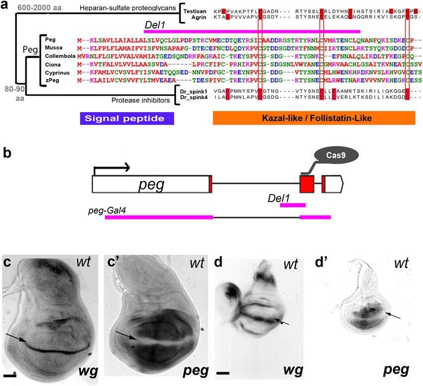
peg encodes an 80aa peptide with a signal peptide (suggesting expressing domain, loss of bristles in peg- mutants, and the lower
secretion or membrane localisation) and a Kazal2/Follistatin-like Wg spread in large peg- clones (Fig. 2). Reciprocally, when
domain (FS-like) (Fig. 1a). FS-like domains are present in Agrin PegGFP was overexpressed, we observed extended Wg diffusion
and other heparin-sulphate proteoglycans (HSPGs). HSPGs are and sens expression (Fig. 4c–f, S2). Peg had similar effects on
secreted and bind to the extracellular matrix, facilitating the WgGFP transport and signalling when UAS-WgGFP was either
anchoring or diffusion of other proteins across it; thus, HSPGs co-expressed with UAS-Peg, or expressed in peg- mutants
influence Wnt and Hh signalling in vertebrates and flies15. peg (Fig. 4g–i). Next, we corroborated that the positive effects of Peg
showed wide conservation in insects, and likely conservation in on Wg transport and signalling are linked, mapping to the Wg
uncharacterised vertebrate genes similar to SPINK extracellular protein. Firstly, we observed neither reduction nor increase of wg
protease inhibitors (Fig. 1 and S1, Table S1)16. However, the transcription upon loss or over-expression of Peg, as observed by
pattern of conserved cysteines (red) in Peg peptides differs from in-situ hybridisation and by RT-qPCR in imaginal discs (Fig. S4).
both SPINK protease inhibitors and HSPGs, highlighting a new Secondly, the correlated expansion of the Wg secreted domain
protein family (Fig. 1 and S1, Tables S1 and S2). peg was strongly and of its target sens were linked by a similar expansion of
expressed in the developing fly wing but was excluded from the Armadillo (Arm) nuclear localisation, a known marker of Wg
presumptive wing margin, a transcription pattern complementary signalling (Fig. 4j–o, S4). Thirdly, abolition of Wg transport
to that of wingless (Fig. 1c-d). was epistatic over Peg function: we observed that WgNRT flies,
We generated peg- null mutants by CRISPR-Cas9, producing which only express a membrane-tethered version of Wg17,
small deletions within the peg ORF (Fig. 1b). These mutants show showed phenotypes of bristle and sens expression loss similar to
high pupal lethality (62%) and a significant reduction in the peg- and wg- mutants; phenotypes that over-expression of Peg was
number of chemosensory bristles at the wing margin, a char- not able to rescue, and loss of function of peg did not enhance
acteristic phenotype of wg loss of function13 (Fig. 2a, b, e, (Figs. 5a–f, 2e).
Table S1). The chemosensory precursors are determined during Overall our results show that diffusible Peg: (a) interacts with
late larval development from cells within two proneural bands and enhances Wg spread, and (b) promotes Wg signalling acting
about 3–4 cells wide, induced by Wg signalling from a stripe of upstream or on the Wg protein itself. The simplest hypothesis is
Wg-expressing cells along the presumptive wing margin (Fig. 2c). that these effects are causally linked, such that the Peg peptide
In peg- null mutants, the proneural marker senseless (sens) facilitates Wg protein transport, resulting in expanded Wg sig-
revealed a significant reduction in proneural band width, con- nalling and expression of target genes. We corroborated this
sistent with the reduction in chemosensory bristles (Fig. 2d, S2). hypothesis by direct observation of Wg transport dynamics
These phenotypes suggest a positive role for Peg in Wg signalling. in vivo, performing Fluorescence Recovery after Photobleaching
We looked at the expression of Sens in peg- clones, to assess if Peg (FRAP). We observed a recovery of 23% of WgGFP fluorescence
acted non-autonomously, like secreted proteins. Indeed, we by 5 min in wt discs, whereas peg- discs reached only 11% by that
2 NATURE COMMUNICATIONS | (2021)12:5660 | https://doi.org/10.1038/s41467-021-25785-z | www.nature.com/naturecommunications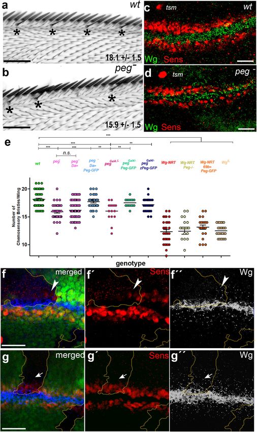
NATURE COMMUNICATIONS | https://doi.org/10.1038/s41467-021-25785-z                                                                               ARTICLE

Fig. 1 peg a conserved smORF is expressed in wing imaginal discs in a pattern complementary to wg. a Phylogenetic tree and alignments of different
proteins with FS-like domains, including Peg, the large secreted proteoglycans Agrin and Testican, and the family of small secreted Serine Protease
Inhibitors of the Kazal type (Spinks). Danio rerio (Dr_) spinks are presented here. Size range in aa is indicated for each group of genes. Note the similarity
between Peg and putative homologues in another dipteran (Musca), a primitive arthropod (Collembola), a basal chordate (Ciona, a tunicate), and the fishes
Cyprinus (Carp) and Danio rerio (zebrafish, zPeg). The pattern of conserved cysteines (red) in each family of peptides is indicated in red. The signal peptide
and FS-like domains are indicated in blue and orange, respectively. This protein structure with a signal peptide followed by a single protein domain is similar
to other secreted or membrane-associated small ORF peptides, such as antimicrobial peptides40, organelle components39,41, and cell signals11,42. The
pegDel1 allele (pink) removes a large portion of aa from the Peg sequence, including most of the Kazal domain and part of the signal peptide sequence. b peg
genomic locus indicating the site targeted for CRISPR/Cas9 mutagenesis, within the peg ORF (red), and the genomic sequences deleted in the different
alleles used in this study (pink). c, d In-situ hybridization for wg and peg in late (c, c´) and mid (d, d´) third instar wing imaginal discs. peg is expressed in a
pattern complementary to wg (arrows, see also Butler et al.43). Scale bars: c, d: 50 µm.

time (Fig. 5g–i, Movies S1 and S2). Since Peg has a predicted                      peg had no effect on Wg mRNA levels (Fig. S4), we assessed Wg
domain similar to both protease inhibitors and HSPGs15,16                          protein levels using quantitative Westerns and observed no sig-
(Fig. 1a), we asked whether Peg had an effect on Wg stability/                     nificant changes in the developing wings of peg- mutants, nor in
degradation, or on its extracellular movement. We formalized the                   those over-expressing Peg (Fig. S6). Altogether our imaging,
effective transport dynamics of Wg using the reaction diffusion                    biochemical and mathematical data support a role for the Peg
model of Kicheva et al.18 (methods; Fig. S4). We obtained a wild-                  peptide as a secreted facilitator of Wg diffusion.
type decay length for Wg (λwt = 6.14 ± 1.31 µm) similar to                            Finally, we carried out genetic interactions to explore a possible
Kicheva´s (5.8 ± 2.04 μm), but a significant reduction in peg-                      functional relationship between Peg and HSPGs, despite their
mutants (λpeg = 3.19 ± 0.57 µm), together with a significant                        different sequences and molecular sizes (Fig. 1). HSPGs have
reduction in the Wg diffusion coefficients between peg- and wt:                     complex functions, and it has been shown that they can modulate
Dwt = 0.51 ± 0.11 µm2/s and Dpeg = 0.15 ± 0.06 µm2/s. Further, we                  Wg diffusion, but also that their overriding activity in this context
found no significant difference in the effective Wg degradation                     is to bind and inactivate extracellular Wg protein in cooperation
rates between wt and peg- (Kwt = 0.014 ± 0.006/s, Kpeg = 0.015 ±                   with Notum19. Thus, the interaction between HSPGs and Notum
 0.008/s). These results imply that Wg diffuses at a slower rate in                defines the pool of active extracellular Wg, and we would expect
peg- mutants, without changes to Wg degradation. We corrobo-                       that Peg then interacts with this active Wg pool. We observe that
rated this analysis experimentally: having established above that                  the loss of wing margin bristles generated by over-expression of

NATURE COMMUNICATIONS | (2021)12:5660 | https://doi.org/10.1038/s41467-021-25785-z | www.nature.com/naturecommunications                                          3
ARTICLE                                                                 NATURE COMMUNICATIONS | https://doi.org/10.1038/s41467-021-25785-z

Sugarless, an enzyme involved in HSPG synthesis20, is not res-               requires Peg to reach its targets (see Fig. S7 for details). Further,
cued by over-expression of Peg (Fig. S7). This is as expected: since         the peg- chemosensory bristle phenotype is unaffected by a
Peg does not generate more active Wg, extra Peg cannot rescue                reduction of the main Wg-related HSPG, Dally-like (dlp) and,
the reduction of active Wg caused by the sugarless-mediated                  consistently with this lack of genetic interaction, we observed no
excess of HSPGs. However, the excess of bristles created by                  physical interaction between PegGFP and Dlp by co-
reduction of Notum (which produces an excess of active Wg19)                 immunoprecipitation (Fig. S7). Altogether our observations sug-
was corrected by reduction of Peg, because this excess of Wg still           gest that Peg acts on Wg separately from HSPGs.

4                NATURE COMMUNICATIONS | (2021)12:5660 | https://doi.org/10.1038/s41467-021-25785-z | www.nature.com/naturecommunications
NATURE COMMUNICATIONS | https://doi.org/10.1038/s41467-021-25785-z ARTICLE
Fig. 2 peg has wg-like phenotypes. a, b peg- wing margins (b) have fewer chemosensory bristles (*) compared to wild-type (a), average number±SD is
indicated, quantified in (e), c, d. peg- wing discs (d) have reduced sens expression compared to wt (c; quantified in Fig. S2a-b, f). Note that the twin
campaniform sensilla (tsm) precursor located some 3-4 cell diameters away from the sens dorsal stripe remains unaffected, providing a limit for Wg
function (see also references13,44, and Fig. S1). e Quantification of chemosensory bristles in different genetic backgrounds representing mean ± SEM. peg
mutants, either pegDel1 /pegDel1(peg-) or pegGal4/pegDel1 (pegGal4/-) show a significant reduction compared to wild-type. This peg− phenotype is rescued by
ubiquitous expression of PegGFP with daughterless (da)-Gal4 (da > PegGFP: w; da-Gal4, pegDel1 / UAS-PegGFP, pegDel1), or with pegGal4 (Peg > PegGFP: w;
PegGal4 / UAS-PegGFP, pegDel1), which reconstitutes the endogenous peg expression in the wing imaginal disc. da-Gal4 controls (w; da-Gal4, pegDel1 /
pegDel1) fail to rescue the number of chemosensory bristles in peg mutants. This peg− phenotype is also rescued by expressing the zebrafish zPeg putative
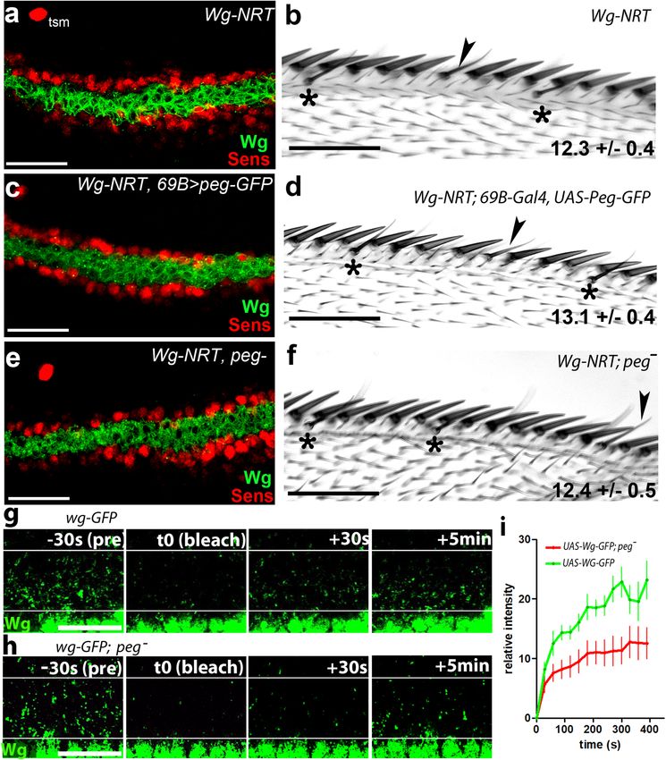
homologue with the pegGal4 driver (Peg > zPeg-GFP). Flies homozygous for a membrane bound version of Wg (WgNRT) show a similar, albeit stronger,
phenotype, which is not rescued by over-expression of PegGFP (WgNRT; 69B > PegGFP), nor worsened by its removal (WgNRT; peg Del1). Wings of the
wgIL temperature-sensitive allele flies (at 17oC) show a similar reduction of chemosensory bristles as Wg-NRT-expressing wings. (***: p < 0.001,
**:p < 0.01) assessed by one-tailed t-test, See Table S1 for statistical analyses. Source data are provided as a Source Data file f. Large peg- clones (lacking
GFP, >5 cells in width, quantified in Fig. S2) show a reduction in sens (f,f´) and in secreted Wg (white dots) (f,f´´). Wg in expressing cells (white contours)
remains normal. peg- cells neighbouring wild-type cells show near-normal expression of sens and extracellular Wg (Arrowheads). g Small clones (up to 6
cell diameters wide) show no reduction in either sens (g, g´) nor Wg distribution (g, g´´) (arrows). Scale bars: a, b: 50 µm; c, d, f, g: 20 µm.
Although the regulation of Wnt gradients has already a general Wnt signals is a complex and highly modulated process, different
developmental and clinical relevance21,22, the relevance of Peg from the elegant classical models of freely diffusing morphogens,
might be more direct, since we observed conservation of its and involves different extracellular proteins in different devel-
sequence in chordates and vertebrates (Fig. 1a, S1). Remarkably, opmental and molecular contexts19,33.
we corroborated the functional conservation between Drosophila Our results also corroborate the growing importance of
Peg and these putative homologues in two ways. Firstly, we res- smORF-encoded peptides as cellular and developmental reg-
cued the peg mutant phenotype by expressing the zebrafish ulators. Given other characterised examples1,11, their average
homologue (zPeg) in the peg pattern (Fig. 2e). Second, we observe sizes and aa composition, and their numbers1,2, it is likely that
that zPegGFP expressed in this manner co-localized with endo- more smORF peptides physically regulating well-known canoni-
genous fly Wg (Fig. S6) in the same way as found for fly PegGFP cal proteins with membrane-related and signalling functions will
(Fig. 3). be characterised.
Discussion
Wg was characterized as a ligand diffusing over several cells23,24, Methods
Drosophila lines. Fly stocks and crosses were cultured at 25 °C, unless otherwise
but the viability of WgNRT flies suggested that diffusion of Wg stated. For time-specific activation of gene expression with the Gal80ts system, the
may not be actually required during development17. However, cultures were carried out at 18 °C and then shifted to 29 °C prior to dissection after
although WgNRT appears to reconstitute most wg functions, the indicated times. The following lines were obtained from the Drosophila
different studies have shown that Wg signalling at a distance is Bloomington stock centre at Indiana University (https://bdsc.indiana.edu/): Or-R
(used as wt), w;;69BGal4 (#1774), w;;UASdsRed (#6282), w;enGal4,UAS-dsRed /CyO
required in specific developmental contexts25,26. Here we (#30557), w hsFlp;; FRT 82B tubGFP (#5188), da-Gal4 (#55851), w;UAS-mCD8-
demonstrate that short-range diffusion of Wg is required for the FRP(#86558) and w; NotumI6/ TM3 (#4117). The UAS-sgl stock (M{UAS-
appropriate patterning of the wing margin and its associated sgl.ORF.3xHA}ZH-86Fb, #F003098) was obtained from flyORF (https://flyorf.ch/).
proneural field, as initially reported13. As repeatedly shown, Wg The w;ptcGal4-tubGal80ts;MKRS/TM6b was a gift from James Castelli Gair-
Hombria, the w; UAS-wgGFP and w; wgNRT w; dlpMH20/TM6b, and UAS dlp-HA
has sequential short range signalling functions in Drosophila wing stocks were gifts from Jean Paul Vincent. rn-Gal413 was previously published in St
development, (instead of a single, long-range morphogenetic Pierre et al.34. wgIL homozygous flies raised at 17 °C were obtained as in
function), a crucial fact that reconciles results from several Couso et al.13.
groups, including those involving WgNRT13,14,27,28 (Fig. S8). Early
in wing development (aprox. 48-72 h. after egg laying, AEL) Wg
expression endows a small groups of cells with wing fate, but by Transgenic constructs. To generate the pegGFP construct, a fragment containing
the CG17278 5′ UTR and CDS sequences (devoid of stop codon) was amplified
the end of development two days later this instruction is inherited from a whole Drosophila embryo cDNA library and cloned into a pENTR/D-
and indirectly felt across hundreds of descendant cells; hence, Wg TOPO gateway vector (Invitrogen) (see Table S4 for primer sequences), this vector
perturbations at an early stage can produce either loss of the was then recombined with the pPWG vector (UAS-insert-C terminal GFP),
entire wing, or its duplication, without relying on long-range obtained from the Drosophila Genomics Resource Centre at Indiana University
(https://dgrc.bio.indiana.edu/), to obtain UAS-pegGFP.
diffusion12,27,29,30. Shortly afterwards (aprox. 72-96 h AEL), The zPegGFP construct was generated as follows: after identifying zPeg
transitory expression of Wg across the entire developing wing is (zebrafish EST sich211-195b11.3) as a putative vertebrate peg homologue, we
required for wing growth12,17,30–32, but this function does not obtained a full length zebrafish embryo cDNA library (segmentation 14-19 somites
strictly require Wg diffusion17, since Wg is then transcribed in to pharyngula prim-5 stages, when zPeg is highly expressed according to Zfin) from
nearly all cells of the developing wing. Finally, during late the Juan Ramon Morales lab at the CABD in Seville, and used it to amplify the
sich211-195b11.3 cDNA by PCR using the zPeg Dr FW and zPeg Dr Rv 1 primers
development (96–120 h AEL), Wg is expressed in a stripe along (see Table S4 for primer sequences).
the dorsal-ventral boundary of the wing, where its function is to We then carried out a strategy to replace the peg CDS by the zPeg CDS in the
pattern the wing margin13,14. Thus, the late patterning of the pegGFP construct, using the NEBuilder HiFi DNA Assembly kit (NEB), which is
wing margin offers an immediate and direct read-out of the based on the assembly of PCR amplicons with overlapping ends. We amplified and
assembled the fragments corresponding to zPeg CDS, flanked, upstream, by the peg
functional range of Wg signalling. There, complementary 5′UTR, and downstream by GFP from the original pegGFP vector, using the
expression of Peg enhances Wg diffusion to reach cells located following primers, obtained with the NEBuilder web-tool: 5′UTR_fwd, 5′UTR_rev,
3-4 cell diameters from the Wg source, which otherwise would be GFP_fwd, GFP_rev, in both cases using pegGFP as template. The Danio CDS
deprived of short-range Wg signal. This enhancement is essential fragment was amplified with the following primers: Danio CDS_fw and Danio
CDS_rev using the zPeg cDNA amplicon as template. See Table S4 for primer
to ensure the development of all the neural precursors and sen- sequences.
sory organs of the wing margin. This role of the Peg small The resulting amplicons were then assembled with a XhoI-linearised pPWG
secreted peptide joins the literature showing that transport of vector, and sequenced.
NATURE COMMUNICATIONS | (2021)12:5660 | https://doi.org/10.1038/s41467-021-25785-z | www.nature.com/naturecommunications 5ARTICLE NATURE COMMUNICATIONS | https://doi.org/10.1038/s41467-021-25785-z Fig. 3 Peg is secreted and interacts with Wg. a PegGFP (green dots) is found outside and beyond the cells expressing it (labelled with dsRed) after 12 h of induction with ptcGal4-UASGal80ts (methods). b peg-Gal4 (methods, Fig. S9) reconstitutes the peg gene expression pattern in the wing pouch (see Fig.1c´,d´)), which does not overlap with wg gene expression as corroborated by Z-axis orthogonal reconstruction (b-b´). c, d PegGFP (green) accumulates in the extracellular space between mCD8-RFP-labelled cells (c-c´), whereas only GFP remains within the cells (red) (d-d´). e. PegGFP (green), expressed with peg-Gal4, co-localizes with Wg (magenta) in the developing wing margin cells (white dots, arrowheads). f An orthogonal Z-axis reconstruction shows that PegGFP (green) and endogenous Wg (magenta) colocalize basolaterally (arrowheads) within and outside of their expression domains, indicated by white dashes, and revealed by strong intracellular Wg, and peg-Gal4 driven UAS-mCD8-RFP (peg). Notice that this co-localisation also takes place extracellularly on apical cell surfaces (arrow). g Pull-down with anti-GFP beads from wing discs expressing peg-GAl4, UAS-PegGFP yields a 50–kDa Wg-specific band revealed with α-Wg (top). GFP-only negative controls show no Wg signal. Both GFP and pegGFP were similarly bound by the pull-down beads (bottom). Source data are provided as a Source Data file. Scale bars: a: 20 µm; b: 30 µm; c, d, f: 5 µm; e: 10 µm. 6 NATURE COMMUNICATIONS | (2021)12:5660 | https://doi.org/10.1038/s41467-021-25785-z | www.nature.com/naturecommunications
NATURE COMMUNICATIONS | https://doi.org/10.1038/s41467-021-25785-z                                                                       ARTICLE

Fig. 4 Peg enhances Wg diffusion. a–c Wg spread is reduced in peg- wing discs (b) compared to wt (a), and is enhanced when pegGFP expression is driven
in the posterior compartment (p) by enGal4 (c); quantified in Fig. S4E-F. d–f Overexpressing pegGFP with enGal4 in the posterior compartment increase sens
expression (e), compared to controls expressing GFP-only (d); f Quantification from d, e, showing average Sens fluorescent intensity is significantly higher
in posterior compartments expressing PegGFP compared to GFP controls (one-tailed t-test, N = 10, P = 0.0067, Error bars represent SEM; see also
Fig. S2g-i). Source data are provided as a Source Data file. g–i Diffusion of WgGFP driven by ptc-Gal4, UAS Gal80ts (h) (as in Fig. 3a) is reduced in a peg-
background (g), and enhanced when WgGFP is co-expressed with UAS-Peg (i) (quantified in Fig. S4g). j–l High magnification of the cells neighbouring
WgGFP-expressing cells (yellow rectangles in g–i), showing Armadillo (red) and DAPI (blue). Nuclear Armadillo is reduced in peg- (j), and enhanced in
wing discs over-expressing Peg (l), compared to wt peg background (k); quantification in Fig. S4H. m–o. Lower magnification images showing that non-
membrane accumulation of Armadillo (red) in the cells near Wg-GFP (green) (m´-o´) is lower in the absence of Peg (m), and higher when Peg is
overexpressed (o), compared to wt peg background (n). Scale bars: a-e, g–i: 20 µm; j–l: 10 µm; m–o: 40 µm.

NATURE COMMUNICATIONS | (2021)12:5660 | https://doi.org/10.1038/s41467-021-25785-z | www.nature.com/naturecommunications                                 7
ARTICLE                                                                         NATURE COMMUNICATIONS | https://doi.org/10.1038/s41467-021-25785-z

Fig. 5 Peg acts directly on Wg protein. a In wgNRT wing discs Wg (green) fails to diffuse away from the expressing cells, and Sens expression (red) is
strongly reduced compared to wild type (Fig. 2c, quantifications in Fig. S2a-c, f). Note unaffected tsm. b wgNRT wings have fewer chemosensory bristles (*)
(sometimes misplaced, arrowheads). c-f Neither the bristle phenotype, nor sens expression are affected by over-expression of PegGFP (c,d), or by Peg
removal (e, f) (quantified in Fig. 2e). g-h. In-vivo FRAP imaging of WgGFP (activated by ptc-GAL4 UAS-Gal80ts, 24 h before dissection) before (pre),
immediately after (t0, bleach), 30 seconds, and 5 min after photo-bleaching (performed between white lines), in a wild-type (g) or a peg- (h) background
(see also Supplementary Movies 1 and 2). i FRAP quantification from panels g-h, showing average fluorescence intensity within the bleached region, over
time, relative to the intensity before bleaching (pre). Acquisition was made every 30 s. Error bars represent SEM (n = 5 movies per genotype). Source data
are provided as a Source Data file. Scale bars: a, c, e, g, h: 20 µm; b, d, f: 50 µm.

   The UAS-pegGFP and zPegGFP plasmids were sent to BESTgene for injection           phenotypes, stocks were established from putative peg mutants, and peg alleles were
into embryos and generation of transgenic flies.                                      confirmed by PCR of the locus followed by sequencing.

                                                                                     Generation of the Peg-Gal4 line (see also Fig. S9). The peg-Gal4 line was
Generation of CRISPR mutants. To generate the peg- null mutants we cloned the        generated by CRISPR-mediated homology-directed genome editing using the
following guide sequence targeting the CDS of CG17278: GTACCGATCCAT                  strategy reported by Gratz et. al35, co-injecting the following plasmids in a Vasa-
TTGCGCTGCGG into the pU6-BbsI-chiRNA plasmid, obtained from addgene, and             cas9 Drosophila line: a pFCD4 vector carrying tandem RNA guides introduced with
following the available protocol (http://FlyCRISPR.molbio.wisc.edu). The following   the primers Tan-CG17278_3′guide_Rv and Tan-CG17278_5′guide_Fw (see
primers were used: chi-CG17278_ORF_guide_1_Fw and chi-CG17278_ORF_guide_             Table S4 for primer sequences), following the protocol described in35; and the
1_Rv (see Table S4 for primer sequences). The pU6-CG17278-chiRNA plasmid was         pTV[cherry]36 vector carrying 1 kb homology arms targeting the 5′ and 3′ regions
then sent for injection to BESTgene into the y[M{vas-Cas9}ZH2A w line. Surviving     of peg (see Fig. S9) designed to remove the whole peg locus while maintaining its
adults were crossed to w;;Df(3 R)BSC680, the progeny was scored for peg- like        presumptive regulatory regions. Successful pegpTV CRISPR-mediated homologous

8                    NATURE COMMUNICATIONS | (2021)12:5660 | https://doi.org/10.1038/s41467-021-25785-z | www.nature.com/naturecommunications
NATURE COMMUNICATIONS | https://doi.org/10.1038/s41467-021-25785-z ARTICLE
recombinants were screened for red eyes (w + marker within pTV[Cherry]) and Fluorescence Signal intensity measurements. For endogenous Wg, the signal
sequenced. These flies expressed mCherry in some tissues but not in the imaginal was quantified by counting the total number of Wg particles on 15 successive 2.5
discs, so we surmised that a large peg intronic region removed in the CRISPR- micron ROIs, centred on the D/V boundary. Raw images were thresholded and the
mediated homologous recombinants might carry a regulatory element required for particle analyser plugin of ImageJ used to obtain the number of particles/ROI. For
peg expression in the imaginal discs. We therefore re-introduced the missing Induced Wg expression, the fluorescence signal profile was measured within a
intronic region in our pegpTV lines. For this, we first carried out a Cre-Lox 20 µm2 ROI in the posterior compartment, placed directly adjacent to the ptc
recombination “flip-out” by crossing the pegpTV the with a y w; snaSco/CyO, domain. The Sens fluorescence signal profiles were measured within a region of
P{Crew}DH1 line obtained from the Kyoto stock centre, to remove most of the interest (ROI) of 20 × 60 µm, from the centre of the d/v boundary outwards, on
pTV[cherry] sequence, and leaving only an ATTp site, for site-directed transgen- either side of the d/v boundary. For Arm nuclear signal quantification, we calcu-
esis. Successful flip-out events were screened by loss of the w marker. We then lated the intensity of Arm signal within an ROI overlapping with DAPI signal.
cloned the missing intronic sequence into the RIVGal4 vector36, amplified using the
primers intron_fragment1_fw and intron_fragment1_rv (see Table S4 for primer
sequences), and EcoRI restriction. The intron-carrying RIVGal4 vector was then In vivo time lapse imaging. For in-vivo time lapse imaging of WgGFP diffusion,
used for site-specific transgenesis in our Cre-Lox recombined pegpTV line, by co- after induction of expression, we dissected the imaginal discs from wandering L3
injection with the act-phiC31-integrase vector, obtained from the DGRC (barcode larvae of the appropriate genotypes, in ice cold Schneider’s culture medium. The
1368). Successful transformants were screened for recovery of the w + marker. imaginal discs were then transferred to an imaging chamber similar to that used
These flies drive the expression of genes under the control of UAS in the wing in38; the chamber was constructed by sticking a perforated square of double sided
imaginal discs with the same pattern of expression as peg; this is also a peg null tape on the cover-slip of a cover-slip bottomed culture well (MatTek Corp), the
allele since it lacks the peg ORF, this line is therefore called pegGal4. Plasmid orifice of the perforated tape was filled with culture media (Schneider’s media, 2%
injections were made in BestGene, or at the Drosophila transgenesis facility at the FBS, 0.2% Penicillin-Streptomycin, 1.25 mg/mL insulin, and 2.5% methyl-cellulose
Madrid Centre of Molecular Biology. as thickener to reduce drifting of the tissues during imaging). For confocal imaging,
7 section z-stacks were acquired at a rate of a whole stack every 30 seconds, using
the LSM 880 Airyscan module of the Zeiss Axio Observer microscope, with a 63x/
Wing preparations. For Drosophila adult wing preparations, the flies were col- 1.46 oil objective, bleaching was performed after the first stack, by 25 iterations
lected in SH media (50% glycerol, 50% ethanol), washed in ethanol and then in with the 405 nm Laser line, at 80% power, in a ROI of 15 × 45 nm, placed at an
dH2O, then the wings were clipped and mounted on a slide with Hoyer´s. The average of 0.5 nm from the wgGFP source. Fluorescence intensity measurements
slides were then placed on a hot plate at 65 °C for 3–5 h, with a weight on top of the were performed on raw image files, using Image J. In order to quantify the
coverslip to ensure a good flattening of the wings. fluorescence recovery, we calculated the percentage of the original intensity (pre-
bleach) recovered, after bleaching, for each time point, hence the following nor-
malization for each data point: Intensity(tX) = (Intensity(tx)- Intensity (tbleach))
In situ hybridization. CG17278 ribo-probe was obtained using the CG1728 in *100/ intensity (tpre-bleach).
pENTR/D-TOPO plasmid and its T3 promoter, and the wg ribo-probe was
obtained from a whole wg cDNA fragment in pBluescript, which was a gift from
Joaquin Culi Espigul and Sol Sotillos, and it was transcribed using its T3 promoter. FRT-mediated clone generation. In order to obtain peg- mitotic clones, we
Digoxigenin labelling of the ribo-probes was performed with the Roche DIG RNA generated a w;; peg- FRT82B recombinant line using the w hsFlp;; FRT 82B tub-GFP
labelling mix, Sigma Aldrich, and the Promega T3 RNA polymerase. Wing imaginal line as the source of FRT82B. These two lines were crossed, and a 1 h 37 °C heat
discs were dissected in ice-cold PBS and fixed for 20 min in 4% paraformaldehyde, shock was induced in the progeny 48 h after egg laying. To quantify the effect of
and a standard DIG-RNA in situ hybridization protocol, as described in Galindo clone size on sens expression we considered only clones adjacent to the wg-
et al.7, was followed. expressing wing margin cells. For this we compared sens fluorescent intensity
within the clone, against an adjacent sens-expressing area of identical size within
wild-type cells. The width of the clones in number of cells was determined by direct
Antibody stainings. For wing imaginal disc immuno-stainings, wandering third measure of the average cell-width per imaginal disc, and by dividing the average
instar L3 larvae were dissected by evertion in ice-cold PBS, cleared of digestive width of the clone by the average cell-width. All intensity and length measures were
system and fat body, fixed for 15 min in 4% PFA, and left overnight in methanol at carried out with Image J in raw z-stacks. A total of 16 different clones were
-20 °C. The tissues were then washed 3 times with PBS. For wingless antibody quantified, and their average intensity relative to wild-type sister cells was plotted
stainings, a single wash of 20 min with PBS, Tween (0.1%) was then performed, but after binning in two categories: 2–5 cells (9 clones) and 6–11 cells (7 clones).
all subsequent incubations and washes were carried out with ice cold PBS, BSA
(0.2%), and ice cold PBS, respectively, maintaining detergent-free conditions in
order to preserve the extracellular signal. Anti-Wg (mouse, DSHB: 4D4-S, used at Co-immuno precipitation. For co-immunoprecipitation of wing imaginal disc
1:50) and anti-Arm (mouse, DSHB: 7A1-S used 1:50) were obtained from The protein extracts, 200 discs per genotype (w; pegGal4/UAS-pegGFP or w; pegGal4/
Developmental Studies Hybridoma Bank, at the University of Iowa (https:// UAS-GFP) were homogenized in 200 µl of lysis buffer (10 mM Tris–HCl pH
dshb.biology.uiowa.edu/). Anti-Sens37 (Guinea-Pig, used 1:3000) was a gift from 7.5,150 mM NaCl,0.5 mM EDTA, 0.5% NP-40) for 30 min at 4 °C. Cellular lysate
Takashi Koyama. We used the following secondary antibodies: to detect Wg we was spun at 16,000 g for 10 min at 4 °C. 300 µl of dilution buffer (10 mM Tris–HCl
used anti-mouse biotin (Jackson, 715-065-151, 1:200), and avidin-Cy5 (Jackson, pH 7.5,150 mM NaCl,0.5 mM EDTA) were added to the supernatant and added to
016-220-084, 1:1000) or avidin-Alexa 488 (Jackson, 016-540-084, 1:500), for other equilibrated GFP-beads (Chromotek), left rotating overnight at 4 °C. Beads were
antigens we used anti mouse-Rhodamine, (Jackson, 715-025-150, 1:250), and anti washed three times with washing buffer (10 mM Tris–HCl pH 7.4,150 mM
guinea-pig-rhodamine, (Jackson, 106-025-003, 1:250). For nuclear labelling we used NaCl,1 mM EDTA, 0.05% NP-40), and then boiled in Loading Buffer 2× (100 mM
DAPI at a final concentration of 300 nM. Tris–HCl pH 6.8, 4% SDS, 0.005% Bromophenol Blue, 20% Glycerol). Beads were
Confocal images were acquired on a Zeiss Axio Observer microscope, with an separated with a magnet and the supernatant was loaded onto 12% Stain Free Tgx
LSM 880 Airyscan module, using a 63x/1.46 oil objective. The signal from Wg, Wg- acrylamide gel (BioRad). Detection of specific proteins by Western Blot using a
GFP, Peg-GFP, Arm, and Sens, were always detected using the Airyscan detector. Trans Blot Turbo Transfer System (BioRad). Antibodies used were: rabbit anti-GFP
The signal from DAPI and UAS-GFP, or dsRED was detected using the GAasp (Invitrogen, MA5-15256, 1:2500); mouse anti-Wg (DSHB, 4d4-S, 1:3000). See next
detectors. section for details of Western blots.
For larval pulldowns 60 late third instar larvae per genotype (da-Gal4, UAS-
PegGFP. da-Gal4, UAS-GFP. UAS-Dlp-HA/ +; en-Gal4 UAS-PegGFP /+. UAS-Dlp-
Statistics and reproducibility. Representative micrographs from unquantified HA/ +; en-Gal4, UAS-GFP / +) were homogenized in 400 µl of lysis buffer (20 mM
data (antibody stainings and in-situ hybridisation) presented in this manuscript Tris–HCl pH 7.4,150 mM NaCl,1 mM EDTA, 0.5% NP-40) for 30 min at 4 °C.
were used when similar results could be observed in all imaginal discs examined, in Cellular debris was spun at 16 000 g for 10 min at 4 °C. Supernatant was added to
two independent experiments. For quantified data, the number of independent equilibrated GFP-beads (Chromotek) and left rotating overnight at 4 °C. Beads were
biological samples analysed is indicated for each experiment. washed three times with washing buffer (20 mM Tris-HCl pH 7.4,150 mM
NaCl,1 mM EDTA), and then boiled in Laemmi Loading Buffer (BioRad). Beads
supernatant was loaded onto 12% polyacrylamide gel and proteins were separated
Activation of Peg and WgGFP expression. In order to activate the expression of by SDS-PAGE (BioRad). Detection of specific proteins by Western Blot using a
WgGFP and PegGFP at precise developmental times, Drosophila lines of the semidry blotting or tetra cell (BioRad). Antibodies used were: mouse anti-GFP
appropriate following genetic backgrounds: w;ptc-Gal4-UAS-Gal80ts; peg-, w;ptc- (Roche, 11814460001, 1:2500); anti-Wg (DSHB, 4d4-S; 1:3000); anti-HA (Roche,
Gal4-UAS-Gal80ts; UAS-peg/TM6b, or w;ptc-Gal4-UAS-Gal80ts, were crossed to w; 12CA5,1:5000).
UAS-WgGFP or w; UAS-WgGFP; peg- /peg- lines, to obtain the following geno-
types: ptc-Gal4-UAS-Gal80ts/UAS-wgGFP; peg-/ peg-, ptc-Gal4-UAS-Gal80ts/UAS-
wgGFP; UAS-peg/+, ptc-Gal4-UAS-Gal80ts/UAS-wgGFP; +/+. The progeny were Quantitative Western Blots. For each WB lane, the wing pouches of 10 wing
reared at 18 °C for approximately 168 h and then shifted to 30 °C for 24 h. After the imaginal discs were dissected and homogenised in 20 µL of LB2x Buffer (100 mM
shift wandering L3 larvae were collected, dissected in ice cold PBS and fixed in 4% Tris–HCl pH 6.8, 4% SDS, 0.005% Bromophenol Blue, 20% Glycerol), incubated
PFA. The rearing and shift times were modified as necessary for the 5 h, 8 h, 12 h, 5 min at 90 °C, and centrifuged at 16,000 × g for 5 min. The whole lysates were
and 48 h shifts. loaded on a Stain Free tgx acrylamide gel (BioRad). The proteins were transferred
NATURE COMMUNICATIONS | (2021)12:5660 | https://doi.org/10.1038/s41467-021-25785-z | www.nature.com/naturecommunications 9ARTICLE NATURE COMMUNICATIONS | https://doi.org/10.1038/s41467-021-25785-z
onto a nitrocellulose membrane, with a Trans Blot Turbo Transfer System form of the function f(t) as calculated in Kicheva et al.18.
(BioRad) (7 min, 1.3 A, limited to 25 V). Total protein loads were quantified before
C0 dþh h=λ
and after transference, using the Image Lab suite (BioRAD). Wg was detected with f ðtÞ ¼ e λ e λðb þ ðb 1Þψ þ 1Þ 2λððb 1Þψ þ 1Þ
2h
anti Wg (mouse, DSHB, 4d4-S, 1:3000) and anti-mouse-HRP (Donkey, Jackson, DtþðdþnÞλ d h h
þ ðb 1Þe λ2 λðψ 1Þ eh=λ erf pffiffiffiffiffi þ e λ2 eh=λ erf pffiffiffiffiffi þ erf pffiffiffiffiffi
Dtþhλ
715-035-150, 1:10000), using the ECL select reagent (GE Healthcare, GERPN2235) Dt 2 Dt 2 Dt
and the Chemidoc MP imager (BioRad). Quantification of protein levels were pffiffiffiffiffi pffiffiffiffiffi
dþh 2d þ h Dt Dt Dtþhλ Dt Dt hλ 2Dt
performed against total protein loads for each lane. þ erf pffiffiffiffiffi eh=λ erf pffiffiffiffiffi þ eλ2 erf þ e λ2 erf þ eλ2 erf pffiffiffiffiffi
Dt 2 Dt λ λ 2 Dt λ
Dtþhλ 2Dt þ hλ Dtþ2dλþhλ Dt þ dλ Dt þ ðd þ hÞλ
e λ2 erf pffiffiffiffiffi þ e λ2 erf pffiffiffiffiffi eh=λ erf pffiffiffiffiffi
Quantitative real time reverse transcriptase PCR. Total mRNA was extracted 2 Dt λ Dt λ Dt λ
2Dt þ ð2d þ hÞλ 2d þ h
from 30 imaginal discs per genotype, using the RNeasy mini kit (Quiagen). For þ 1 þ eh=λ erf pffiffiffiffiffi erf pffiffiffiffiffi
each sample, 500 ng of mRNA was used for the reverse-transcriptase reaction, 2 Dt λ 2 Dt
using the Quantitect reverse transcriptase kit (Quiagen). The qPCRs were per- ð5Þ
formed on a CFX connect thermocycler (Biorad) using the Vazyme AceQ SYBR
qPCR Master Mix in 20 µL reactions, and using the Wg_fw / Wg_rv and rp49_fw /
rp49_rv pairs of primers (see Table S4 for primer sequences). Determining Wg effective transport dynamics. To find the Wg effective
transport dynamics we first fit spatial concentration profiles of endogenous Wg to
Eq. 2 (Fig. S5) in both wt and peg- cases. These profiles were previously normalized
Homology searches. For homology searches and phylogenetic analyses, we used with respect to the intensity right next to the source C0. This gives values for the
the same methods as described in9. To search for sequence homologues, an initial decay length λ:
search in ESTs deposited in NCBI (http://www.ncbi.nlm.nih.gov/) with tBLASTn λwt=6.14 ± 1.31μm (similar to the 5.8 ± 2.04 μm value in Kicheva et al.18)
with maximally relaxed parameters was carried out in dipteran species. The top 100 λpeg=3.19 ± 0.57μm
hits were scrutinised for belonging to a smORF of less than 100aa with start and With these values we use Eq. 5 to fit the dynamics of FRAP and extract parameters
stop codons, in the correct orientation and non-overlapping with longer ORFs. The D, ψ and b. From the effective diffusion coefficient for both cases:
complete smORFs passing this filter were then aligned using Clustal or MAFFT to Dwt=0.51 ± 0.11 μm2/s
the query and already identified orthologues of the same phylum. smORF hits Dpeg=0.15 ± 0.06 μm2/s and Eq. 3, we find values for the effective degradation rate:
showing alignment scores of at least 50 to previously identified peptides were Kwt=0.014 ± 0.006 s−1
deemed as verified. Ambiguous cases (including peptides already annotated in Kpeg=0.015 ± 0.008 s−1
public databases but having unusual lengths) were tested against genomic Fitted values for the immobile fraction and bleaching depth read as ψ wt =
sequences to correct sequencing or annotation errors. A consensus weighted by 0.35 ± 0.20, ψ peg = 0.22 ± 0.10, bwt = 0.27 ± 0.11, bpeg = 0.37 ± 0.15.
phylogeny was then extracted from the alignment and the process was iterated, The fits and errors were calculated optimizing parameter search using
carrying out a new tBLASTn search with the consensus sequence. When no more Nonlinearmodelfit function in software Mathematica.
homologues from the same taxonomic class were obtained in a given iteration, the
tBLASTn search was expanded to the next higher-order clade. Finally, phylogenetic
trees for peptide sequences were generated using MAFFT and calculated according Reporting summary. Further information on research design is available in the Nature
to average percent distance (see Table S2) using the unrelated 88aa small ORF Research Reporting Summary linked to this article.
membrane peptide Hemotin39 as a potential outgroup, since it has a similar size
and aa composition to the Peg peptide. Data availability
The authors declare no restrictions on the availability of data or biological materials
Reaction diffusion model for Wg effective transport. We formalize the effective (fly lines and plasmids) upon request to the corresponding author. All data-sets used
transport dynamics of Wg with a simple reaction diffusion model, for quantifications in this study are provided in the Source data file accompanying
this manuscript. Pre-print Photoshop files with original high resolution images can
∂C ∂2 C be accessed through the Open Science Framework Repository: https://osf.io/9zt8h/?
¼ D 2 KC þ ν; ð1Þ
∂t ∂x view_only = 9137cf57b96e4081a24c58c11be555f7. No custom code/software was
used for this work, the set-up of the Mathematica based analysis is available upon
where C=C(x,t) is the Wg ligand concentration, which is a function of time and request from Daniel Aguilar Hidalgo. Source data are provided with this paper.
space, D is the effective diffusion coefficient, K is the effective degradation rate and
ν = ν(x) is the ligand source rate, which is spatially dependent. The steady-state
solution of Eq. 1 outside of the source (target tissue) for a constant source width Received: 18 May 2020; Accepted: 8 July 2021;
and and the length of the tissue in the direction perpendicular to the source being
much longer than the decay-length of the gradient is given by,
CðxÞ ¼ C 0 ex=λ ; ð2Þ
References
where C0 is the morphogen concentration located at the target tissue right next to 1. Couso, J. P. & Patraquim, P. Classification and function of small open reading
the source, and λ is the decay length of the gradient, which is a function of D and K frames. Nat. Rev. Mol. Cell Biol. 18, 575–589 (2017).
in the form, 2. Mackowiak, S. D. et al. Extensive identification and analysis of conserved small
pffiffiffiffiffiffiffiffiffiffi ORFs in animals. Genome Biol. 16, 179 (2015).
λ¼ D=K ð3Þ 3. Andrews, S. J. & Rothnagel, J. A. Emerging evidence for functional peptides
encoded by short open reading frames. Nat. Rev. Genet. 15, 193–204 (2014).
4. Aspden, J. L. et al. Extensive translation of small Open Reading Frames
Analysis of FRAP dynamics. Following the analysis of FRAP dynamics described revealed by Poly-Ribo-Seq. eLife 3, e03528 (2014).
in Kicheva et al.18, we solved Eq. 1 in space and time. As initial condition at time 5. Bazzini, A. A. et al. Identification of small ORFs in vertebrates using ribosome
t = 0, we imposed the steady state profile Eq. 2 outside of the bleached region for footprinting and evolutionary conservation. EMBO J. 33, 981–993 (2014).
x < d and x > d+h, and C(x,t = 0) = b C0 exp(-x/λ) inside the bleached region for 6. Slavoff, S. A. et al. Peptidomic discovery of short open reading frame-encoded
d < x < d+h, where d is the distance of the ROI from the source, h is the ROI width peptides in human cells. Nat. Chem. Biol. 9, 59–64 (2013).
and b is the bleaching depth, that is the reduction in ligand concentration after 7. Galindo, M. I., Pueyo, J. I., Fouix, S., Bishop, S. A. & Couso, J. P. Peptides
bleaching. Then, the solution of Eq. 1 reads, encoded by short ORFs control development and define a new eukaryotic gene
family. PLoS Biol. 5, 1052–1062 (2007).
ð1 ψÞC 0 x=λ 8. Hanyu-Nakamura, K., Sonobe-Nojima, H., Tanigawa, A., Lasko, P. &
Cðx; tÞ ¼ e 1 þ b þ ðb 1ÞðAðx; tÞ þ e2x=λ ðAðx; tÞ Nakamura, A. Drosophila Pgc protein inhibits P-TEFb recruitment to
2 ð4Þ
þ Aðh þ x; tÞÞ 1 þ Aðh x; tÞÞ þ Cψ ðxÞ chromatin in primordial germ cells. Nature 451, 730–733 (2008).
9. Magny, E. G. et al. Conserved regulation of cardiac calcium uptake by peptides
pffiffiffiffiffi encoded in small open reading frames. Science 341, 1116–1120 (2013).
Where Aðx; tÞ ¼ erf ðd þ 2Dt=λ þ xÞ=ð2 Dt Þ with the error function
Rz 2 10. Anderson, DouglasM. et al. A micropeptide encoded by a putative long
erf ðzÞ ¼ pffiffiπ 0 exp q dq, and Cψ ðxÞ represents the concentration of immobile
2
noncoding RNA regulates muscle performance. Cell 160, 595–606 (2015).
molecules that is constant in time, with Cψ ðxÞ ¼ ψC o ex=λ outside of the ROI, and 11. Pauli, A. et al. Toddler: an embryonic signal that promotes cell movement via
Cψ ðxÞ ¼ bψCo ex=λ inside of the ROI. Apelin receptors. Science 343, 1248636 (2014).
To describe the intensity recovery during FRAP, we calculate the average 12. Couso, J. P., Bate, M. & Martinez Arias, A. A wingless-dependent polar
R dþh coordinate system in Drosophila imaginal discs. Science 259, 484–489 (1993).
change in intensity within the ROI in the form f ðtÞ ¼ h1 d dx0 C ðx0 ; t Þ, using the
10 NATURE COMMUNICATIONS | (2021)12:5660 | https://doi.org/10.1038/s41467-021-25785-z | www.nature.com/naturecommunicationsNATURE COMMUNICATIONS | https://doi.org/10.1038/s41467-021-25785-z ARTICLE
13. Couso, J. P., Bishop, S. A. & Martinez Arias, A. The wingless signalling 40. Lemaitre, B. & Hoffmann, J. The host defense of Drosophila melanogaster.
pathway and the patterning of the wing margin in Drosophila. Development Annu Rev. Immunol. 25, 697–743 (2007).
120, 621–636 (1994). 41. Lee, C. et al. The mitochondrial-derived peptide MOTS-c promotes metabolic
14. Piddini, E. & Vincent, J. P. Interpretation of the wingless gradient requires homeostasis and reduces obesity and insulin resistance. Cell Metab. 21,
signaling-induced self-inhibition. Cell 136, 296–307 (2009). 443–454 (2015).
15. Hacker, U., Nybakken, K. & Perrimon, N. Heparan sulphate proteoglycans: 42. Chng, S. C., Ho, L., Tian, J. & Reversade, B. ELABELA: a hormone essential
the sweet side of development. Nat. Rev. Mol. Cell Biol. 6, 530–541 (2005). for heart development signals via the apelin receptor. Dev. Cell 27, 672–680
16. Rimphanitchayakit, V. & Tassanakajon, A. Structure and function of (2013).
invertebrate Kazal-type serine proteinase inhibitors. Dev. Comp. Immunol. 34, 43. Butler, M. J. et al. Discovery of genes with highly restricted expression patterns
377–386 (2010). in the Drosophila wing disc using DNA oligonucleotide microarrays.
17. Alexandre, C., Baena-Lopez, A. & Vincent, J. P. Patterning and growth control Development 130, 659–670 https://doi.org/10.1242/dev.00293 (2003).
by membrane-tethered Wingless. Nature 505, 180–185 (2014). 44. Phillips, R. G. & Whittle, J. R. Wingless expression mediates determination of
18. Kicheva, A. et al. Kinetics of morphogen gradient formation. Science 315, peripheral nervous system elements in late stages of Drosophila wing disc
521–525 (2007). development. Development 118, 427–438 https://doi.org/10.1242/
19. Kakugawa, S. et al. Notum deacylates Wnt proteins to suppress signalling dev.118.2.427 (1993).
activity. Nature 519, 187–192 (2015).
20. Hacker, U., Lin, X. & Perrimon, N. The Drosophila sugarless gene modulates
Wingless signaling and encodes an enzyme involved in polysaccharide Acknowledgements
biosynthesis. Development 124, 3565–3573 (1997). We acknowledge the support of Laura Tomas and Alejandro Moscoso at the CABD
21. Briscoe, J. & Small, S. Morphogen rules: design principles of gradient- proteomics platform, and Katherina Garcia at the CABD Advanced Light Micro-
mediated embryo patterning. Development 142, 3996–4009 (2015). scopy and Imaging Facility. We are grateful to Ana Hervas for excellent technical
22. Anastas, J. & Moon, R. WNT signaling pathways as therapeutic targets in help, Pedro Patraquim for discussions, and Thomas Klein and Fernando Casares for
cancer. Nat. Rev. Cancer 13, 11–26 (2012). comments. We thank Jean Paul Vincent for providing the Wg-NRT and UAS-Wg-
23. van den Heuvel, M., Nusse, R., Johnston, P. & Lawrence, P. Distribution of the GFP lines, and to Takashi Koyama for anti-Sens antibody. Funding: This work was
wingless gene product in Drosophila embryos: a protein involved in cell-cell funded by grants from the British BBSRC (ref. BB/N001753/1) and the Spanish
communication. Cell 59, 739–749 (1989). MINECO (refs. BFU2016-077793-P, MDM-2016-0687) and MCI (PID2019-
24. Gonzalez, F., Swales, L., Bejsovec, A., Skaer, H. & Martinez Arias, A. Secretion 106227GB-I00).
and movement of wingless protein in the epidermis of the Drosophila embryo.
Mech. Dev. 35, 43–54 (1991). Author contributions
25. Beaven, R. & Denholm, B. Release and spread of Wingless is required to Emile G Magny contributed with the conceptualisation, methodology, formal analysis,
pattern the proximo-distal axis of Drosophila renal tubules. Elife 7, https:// investigation, original draft preparation, and visualization; Ana I Platero, Jose I Pueyo,
doi.org/10.7554/eLife.35373 (2018). and Sarah A Bishop contributed with the conceptualisation, methodology, investigation,
26. Apitz, H. & Salecker, I. Spatio-temporal relays control layer identity of direction- and revision and editing of the manuscript; Daniel Aguilar-Hidalgo contributed with the
selective neuron subtypes in Drosophila. Nat. Commun. 9, 2295 (2018). methodology, formal analysis and preparation and revision of the manuscript; Juan P
27. Neumann, C. J. & Cohen, S. M. Long-range action of Wingless organizes the Couso contributed with the conceptualisation, methodology, original draft preparation,
dorsal-ventral axis of the Drosophila wing. Development 124, 871–880 (1997). and with the supervision, project administration and funding acquisition necessary to
28. Chaudhary, V. et al. Robust Wnt signaling is maintained by a Wg protein produce this work. The authors declare no competing interests, nor any restriction on
gradient and Fz2 receptor activity in the developing Drosophila wing. data availability upon request.
Development 146, dev174789 (2019).
29. Couso, J. P., Knust, E. & Martinez Arias, A. Serrate and wingless cooperate to
induce vestigial gene expression and wing formation in Drosophila. Curr. Biol. Competing interests
5, 1437–1448 (1995). The authors declare no competing interests.
30. Klein, T. & Martinez Arias, A. The vestigial gene product provides a molecular
context for the interpretation of signals during the development of the wing in
Drosophila. Development 126, 913–925 (1999).
Additional information
Supplementary information The online version contains supplementary material
31. Neumann, C. J. & Cohen, S. M. Distinct mitogenic and cell fate specification
available at https://doi.org/10.1038/s41467-021-25785-z.
functions of wingless in different regions of the wing. Development 122,
1781–1789 (1996).
Correspondence and requests for materials should be addressed to Juan Pablo Couso.
32. Zecca, M. & Struhl, G. Recruitment of cells into the Drosophila wing
primordium by a feed-forward circuit of vestigial autoregulation. Development Peer review information Nature Communications thanks Tilmann Glimm and the
134, 3001–3010 (2007). other, anonymous, reviewer(s) for their contribution to the peer review of this work. Peer
33. Mulligan, K. A. et al. Secreted Wingless-interacting molecule (Swim) reviewer reports are available.
promotes long-range signaling by maintaining Wingless solubility. Proc. Natl
Acad. Sci. USA 109, 370–377 (2012). Reprints and permission information is available at http://www.nature.com/reprints
34. St Pierre, S. E., Galindo, M. I., Couso, J.P. & Thor, S. Control of Drosophila
imaginal disc development by rotund and roughened eye: differentially
Publisher’s note Springer Nature remains neutral with regard to jurisdictional claims in
expressed transcripts of the same gene encoding functionally distinct zinc
published maps and institutional affiliations.
finger proteins. Development 129, 1273–1281, https://doi.org/10.1242/
dev.129.5.1273 (2002).
35. Gratz, S. J. et al. Highly specific and efficient CRISPR/Cas9-catalyzed
homology-directed repair in Drosophila. Genetics 196, 961–971, https:// Open Access This article is licensed under a Creative Commons
doi.org/10.1534/genetics.113.160713 (2014). Attribution 4.0 International License, which permits use, sharing,
36. Baena-Lopez, L. A., Alexandre, C., Mitchell, A., Pasakarnis, L. & Vincent, J.-P. adaptation, distribution and reproduction in any medium or format, as long as you give
Accelerated homologous recombination and subsequent genome modification appropriate credit to the original author(s) and the source, provide a link to the Creative
in Drosophila. Development 140, 4818–4825, https://doi.org/10.1242/ Commons license, and indicate if changes were made. The images or other third party
dev.100933 (2013). material in this article are included in the article’s Creative Commons license, unless
37. Nolo, R., Abbott, L. A. & Bellen, H. J. Senseless a Zn finger transcription factor is indicated otherwise in a credit line to the material. If material is not included in the
necessary and sufficient for sensory organ development in Drosophila. Cell 102, article’s Creative Commons license and your intended use is not permitted by statutory
349–362, https://doi.org/10.1016/S0092-8674(00)00040-4 (2000). regulation or exceeds the permitted use, you will need to obtain permission directly from
38. Aldaz, S., Escudero, L. M. & Freeman, M. Live imaging of Drosophila imaginal the copyright holder. To view a copy of this license, visit http://creativecommons.org/
disc development. Proc. Natl. Acad. Sci. 107, 14217–14222, https://doi.org/ licenses/by/4.0/.
10.1073/pnas.1008623107 (2010).
39. Pueyo, J. I. et al. Hemotin, a regulator of phagocytosis encoded by a small ORF
and conserved across metazoans. PLoS Biol. 14, e1002395 (2016). © The Author(s) 2021
NATURE COMMUNICATIONS | (2021)12:5660 | https://doi.org/10.1038/s41467-021-25785-z | www.nature.com/naturecommunications 11You can also read