VHL suppresses RAPTOR and inhibits mTORC1 signaling in clear cell renal cell carcinoma
←
→
Page content transcription
If your browser does not render page correctly, please read the page content below
www.nature.com/scientificreports
OPEN VHL suppresses RAPTOR
and inhibits mTORC1 signaling
in clear cell renal cell carcinoma
Athina Ganner1, Christina Gehrke1, Marinella Klein1, Lena Thegtmeier1, Tanja Matulenski1,
Laura Wingendorf1, Lu Wang1, Felicitas Pilz1, Lars Greidl1, Lisa Meid1, Fruzsina Kotsis1,
Gerd Walz1, Ian J. Frew2 & Elke Neumann‑Haefelin1*
Inactivation of the tumor suppressor von Hippel–Lindau (VHL) gene is a key event in hereditary
and sporadic clear cell renal cell carcinomas (ccRCC). The mechanistic target of rapamycin (mTOR)
signaling pathway is a fundamental regulator of cell growth and proliferation, and hyperactivation
of mTOR signaling is a common finding in VHL-dependent ccRCC. Deregulation of mTOR signaling
correlates with tumor progression and poor outcome in patients with ccRCC. Here, we report that the
regulatory-associated protein of mTOR (RAPTOR) is strikingly repressed by VHL. VHL interacts with
RAPTOR and increases RAPTOR degradation by ubiquitination, thereby inhibiting mTORC1 signaling.
Consistent with hyperactivation of mTORC1 signaling in VHL-deficient ccRCC, we observed that loss
of vhl-1 function in C. elegans increased mTORC1 activity, supporting an evolutionary conserved
mechanism. Our work reveals important new mechanistic insight into deregulation of mTORC1
signaling in ccRCC and links VHL directly to the control of RAPTOR/mTORC1. This may represent a
novel mechanism whereby loss of VHL affects organ integrity and tumor behavior.
Renal cell carcinoma (RCC) causes more than 140,000 deaths per year worldwide1. RCC have several histologic
subtypes, with the most common one (> 80%) being clear cell RCC (ccRCC). Bi-allelic inactivation of the von
Hippel Lindau (VHL) gene is a hallmark event that arises in the majority of sporadic ccRCC cases implicating
the VHL gene as the most important renal tumor suppressor gene in general2,3. Mutations of VHL are also causa-
tive for the inherited autosomal dominant von Hippel–Lindau syndrome manifested by a variety of benign and
malignant tumors including ccRCC.
The VHL protein functions as the substrate recognition subunit of an E3 ubiquitin ligase complex targeting
the hypoxia inducible transcription factor (HIF) α subunits for proteasomal degradation under normal oxygen
levels. Genetic VHL inactivation leads to constitutive HIFα accumulation and consequently formation of HIF
heterodimers, which induce transcription programs promoting bioenergetic adaptation to hypoxia. Reprogram-
ming of numerous cellular systems by HIF contributes to the pathogenesis of ccRCC by altering metabolism,
angiogenesis, extracellular matrix, invasion and apoptosis-resistance. Accumulating evidence indicates that VHL
inactivation is not sufficient to cause renal tumor formation. Additional genetic events and cellular alterations
are required as second hits for malignant transformation. Comprehensive genomic analyses of ccRCC identi-
fied epigenetic control and PI3K-mechanistic Target of Rapamycin (mTOR) pathways as major determinants
in ccRCC p athogenesis4,5.
In ccRCC the mTOR pathway is commonly h yperactivated6,7. Dysregulation of mTORC1 signaling plays a
key role in the oncogenesis and progression of ccRCC, and hyperactivation of mTOR correlates with poor out-
come in ccRCC patients. Hence, mTOR inhibitors (such as everolimus and temsirolimus) have been approved
for treatment of advanced RCCs, but therapy resistance develops in most patients8.
The mTOR kinase is a highly conserved and fundamental regulator of cell growth, metabolism, and prolifera-
tion in all e ukaryotes9. mTOR constitutes the catalytic subunit of two distinct complexes, mTORC1 and mTORC2.
mTORC1 contains three core components: mTOR, mLST8 and its unique defining subunit, the regulatory-
assoiated protein of mTOR (RAPTOR). Diverse extra- and intra-cellular signals activate mTORC1, including
nutrient availability and growth factors, while hypoxia and low cellular energy levels inhibit mTORC1 activity.
The primary role of mTORC1 is to initiate biosynthesis cascades for proteins, lipids and nucleotides to support
cell growth, while also suppressing catabolic pathways like autophagy10.
1
Renal Division, Department of Medicine, Medical Center, Faculty of Medicine, University of Freiburg, Freiburg,
Germany. 2Department of Internal Medicine I, Medical Center ‑ University of Freiburg, Faculty of Medicine,
University of Freiburg, Freiburg, Germany. *email: elke.neumann-haefelin@uniklinik-freiburg.de
Scientific Reports | (2021) 11:14827 | https://doi.org/10.1038/s41598-021-94132-5 1
Vol.:(0123456789)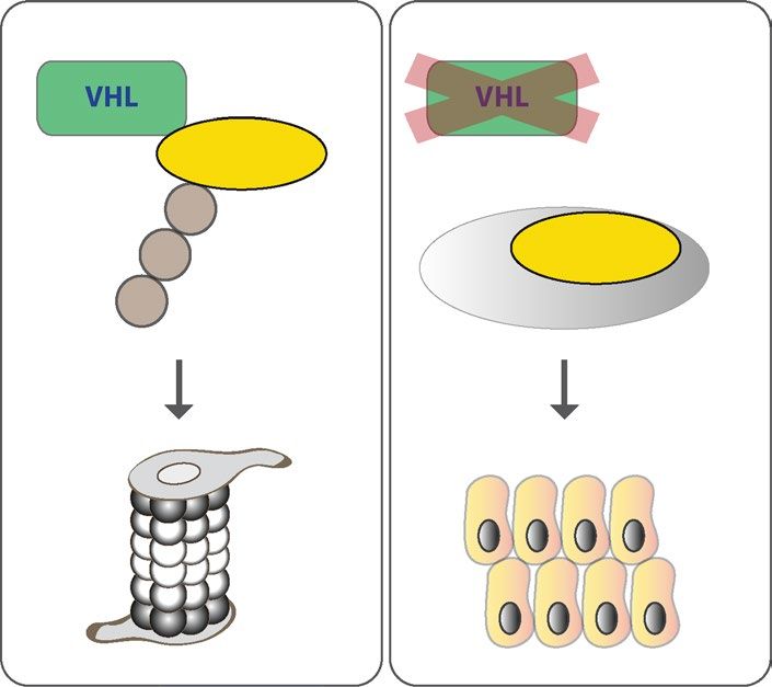
www.nature.com/scientificreports/
a b c
se
e
ra
as
i fe
V5 ifer
L
uc
H
L
uc
.V
H
.L
.V
.L
ag
ag
V5
Fl
Fl
EC
250 kD -
-A O
-7 4
-R 8
C
- Myc.RAPTOR - Flag.mTOR
9
-
PT
86
150 kD -
-4
C
-R
Lysates: anti-Myc Lysates: anti-Flag
250 kD - - mTOR
250 kD - - Flag.mTOR
150 kD - - Myc.RAPTOR
150 kD - - RAPTOR
IP: anti-Flag, WB: anti-Myc IP: anti-V5, WB: anti-Flag
- V5.Luciferase
75 kD - - γ-tubulin
- Flag.Luciferase 50 kD - 37 kD -
- heavy chain - β-actin
37 kD -
} VHL
37 kD - 37 kD - 25 kD -
- Flag.VHL - V5.VHL
IP: anti-Flag, WB: anti-Flag IP: anti-V5, WB: anti-V5
d e f
5
0
9
-2
L3
L1
O
H
H
.M
.V
.V
ag
ag
ag
Fl
Fl
Fl
CPTAC samples
RAPTOR expression
150 kD - - Myc.RAPTOR
G
ccRCC
L
Ig
VH
it
bb
ti-
Lysates: anti-Myc
an
ra
3
*** 150 kD - - Myc.RAPTOR
150 kD - - RAPTOR
2
IP: anti-Flag, WB: anti-Myc Lysates: anti-RAPTOR
Z-value
1 50 kD - - RAPTOR
0 - Flag.CTRL 150 kD -
37 kD - WB: anti-RAPTOR
-1 - Flag.VHL30 20 kD - IP
- VHL
-2
- light chain
-3
- Flag.VHL19 WB: anti-VHL
Normal Primary tumor 25 kD -
n=84 n=110
IP: anti-Flag, WB: anti-Flag
g
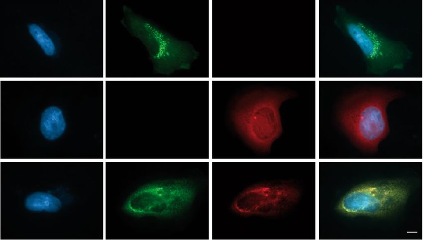
Hoechst GFP.RAPTOR VHL.RFP merge
GFP.
RAPTOR
VHL.
RFP
VHL.RFP
and
GFP.
RAPTOR
Scientific Reports | (2021) 11:14827 | https://doi.org/10.1038/s41598-021-94132-5 2
Vol:.(1234567890)www.nature.com/scientificreports/
◂Figure 1. VHL interacts and co-localizes with RAPTOR. (a) VHL binds RAPTOR. HEK293T cells were
co-transfected with Flag.VHL and Myc.RAPTOR, or Flag.Luciferase as control. Cell lysates were used for
detecting RAPTOR expression by anti-Myc antibody and for immunoprecipitation (IP) by anti-Flag antibody.
The lower panel shows the IP of Flag-tagged proteins. Co-immunoprecipitation of RAPTOR was detected by
anti-Myc (middle panel). kD, kilodalton. Full-length blots are presented in Supplementary Fig. 4. (b) VHL
interacts with mTOR. Flag.mTOR and V5.VHL were transiently co-expressed in HEK293T cells. V5.Luciferase
was used as control. After immunoprecipitation (IP) with anti-V5 antibody, the immobilized mTOR was
detected by Western blot (WB) analysis using anti-Flag antibody in the precipitate containing VHL, but not
Luciferase (middle panel). Full-length blots are presented in Supplementary Fig. 4. (c) RAPTOR expression
in upregulated in VHL-deficient ccRCC cell lines. Cell lysates of RCC4, 786–O, A-498 and RPTEC cells were
analyzed by Western blot using anti-RAPTOR and anti-mTOR antibodies. Equal concentrations of total protein
were determined by Bradford assay. β-actin and γ-tubulin levels were used as a loading control. Full-length blots
are presented in Supplementary Fig. 4. (d) RAPTOR expression data from the UALCAN database comparing
ccRCC to normal tissue, ***p ≤ 0.001. (e) VHL30 but not VHL19 interacts with RAPTOR. Myc.RAPTOR and
Flag.VHL30 or Flag.VHL19 were transiently co-expressed in HEK293T cells. Flag.MO-25 was used as control.
After immunoprecipitation (IP) with anti-Flag antibody, the immobilized RAPTOR was detected by Western
blot (WB) analysis using anti-Myc antibody in the precipitate containing VHL30, but not VHL19 (middle
panel). Full-length blots are presented in Supplementary Fig. 4. (f) Endogenous RAPTOR interacts with VHL.
Endogenous VHL was immunoprecipitated (IP) from HeLa cell lysates by anti-VHL antibody (lower panel).
Endogenous RAPTOR co-precipitating with VHL was detected by Western blot analysis using anti-RAPTOR
antibody (middle panel). Full-length blots are presented in Supplementary Fig. 4. (g) Co-localization of VHL
and RAPTOR. HeLa cells were transfected with GFP-tagged RAPTOR (green, upper panel) and RFP-tagged
VHL (red, middle panel). Co-expression of VHL and RAPTOR revealed co-localization in peri-nuclear foci
(lower panel). Fluorescent confocal microscopy images of HeLa cells. Hoechst (blue) was used for nuclei
staining. Scale bar 10 µm.
The mechanisms underlying increased mTOR signaling activity in ccRCC have remained unclear. Genomic
studies have found that ~ 26% of ccRCC harbor mutations in a number of PI3K-AKT-mTORC1 pathway genes2,5.
Genetic alterations thus likely contribute to mTORC1 activation in ccRCC. Further integrated molecular studies
of ccRCC revealed high levels of AKT-mTORC1 signaling also without an associated genetic alteration11 sug-
gesting that additional molecular players and upstream signals are involved. Notably, VHL was recently found
to directly suppress AKT activity, and in VHL-deficient cells AKT was activated promoting cell survival and
tumorigenesis12. HIF is generally thought to inhibit mTORC1 in hypoxia by activating the expression of its down-
stream target gene REDD1, which subsequently activates the mTORC1 repressor tuberous sclerosis c omplex13.
However, mTOR can evade inhibition by REDD114. On the other hand HIF2α increases mTORC1 activity under
low amino acid availability by increasing the expression of the SLC7A5 amino acid c arrier15. It was also shown
that suppression of the mTOR inhibitor DEPTOR in VHL-deficient ccRCC accelerated tumor cell p roliferation16.
This study reveals new mechanistic insights into deregulation of mTORC1 in ccRCC. Combining cellular
models for ccRCC and the C. elegans system we identify an additional layer of interaction of VHL and the PI3K-
mTORC1 pathway and directly link VHL to control of RAPTOR, the essential scaffolding protein of mTORC1.
Results
VHL interacts with the mTORC1 subunit RAPTOR. mTORC1 is frequently hyperactivated in ccRCC
and accelerates tumor progression. mTORC1 consists of the three core components mTOR, RAPTOR, and
LST8. To test whether VHL can physically interact with one or more of mTORC1 core proteins, HEK293T cells
were co-transfected with tagged expression constructs of VHL, RAPTOR, mTOR, and LST8 respectively. Immu-
noblotting analysis showed association of VHL with RAPTOR, mTOR (Fig. 1a,b), and LST8 (Supplementary
Fig. 1a). Binding of VHL to the inhibitory subunits DEPTOR (DEP domain-containing mTOR-interacting pro-
tein) and PRAS40 (proline-rich Akt substrate of 40 kDa) was not observed (Supplementary. Figure 1b). As we
noticed constant reduction of RAPTOR in the presence of VHL in our cell lysates by immunoblotting, we evalu-
ated RAPTOR protein levels in different ccRCC cell lines. Interestingly, RAPTOR expression was increased in
VHL-deficient ccRCC cell lines compared to human renal proximal tubular epithelial cells (RPTECs) (Fig. 1c).
Consistent with these observations, bioinformatic analysis using the online web portal UALCAN (http://ual-
can.path.uab.edu) revealed a significant upregulation of RAPTOR protein in tumor tissues of VHL-dependent
ccRCCs when compared to normal tissues (Fig. 1d) according to the CPTAC mass-spectrometry-based prot-
eomic tumor dataset17. These findings led us to investigate the interaction between VHL and RAPTOR in more
detail.
The VHL gene encodes two biologically active isoforms, full length VHL consisting of 213 amino acid resi-
dues with a molecular mass of 30 kDa (VHL30) and an internally translated form corresponding to amino
acid residues 54–213 with a molecular mass of 19 kDa (VHL19) (Supplementary Fig. 1c)18. Both isoforms act
similarly to promote HIFα degradation and so appear to retain tumor suppressor activity, yet have isoform-
specific functions19,20. HEK293T cells were transiently transfected with Myc-tagged RAPTOR and Flag-tagged
VHL30 or VHL19. Following immunoprecipitation with Flag-antibody, immunoblotting analysis revealed that
the full length VHL30 efficiently immunoprecipitates RAPTOR, while the shorter VHL19 is able to immuno-
precipitate only a very small amount of RAPTOR protein, despite being expressed at similar levels to VHL30
(Fig. 1e). A similar experiment was conducted with the N-terminal fragment of VHL consisting of amino acid
residues 1–53 (VHL(AA1-53)). Again, immunoprecipitation of VHL30 led to co-precipitation of RAPTOR,
Scientific Reports | (2021) 11:14827 | https://doi.org/10.1038/s41598-021-94132-5 3
Vol.:(0123456789)www.nature.com/scientificreports/
a b c
- endogenous 150 kD - - Myc.RAPTOR
150 kD - - Myc.RAPTOR 150 kD -
RAPTOR
- β-actin
- β-actin - β-actin 37 kD -
37 kD - 37 kD - 37 kD -
- Flag.VHL 30
37 kD - 37 kD -
- Flag.VHL - Flag.VHL
25 kD - - Flag.VHL 19
n.s.
pwww.nature.com/scientificreports/
◂Figure 2. VHL regulates RAPTOR protein abundance. (a) VHL reduced transiently expressed RAPTOR
protein. HEK293T cells were co-transfected with Myc.RAPTOR and Flag.VHL. Cell lysates were analyzed by
immunoblotting using Myc- and Flag-specific antibodies. Protein levels of RAPTOR were quantified using
LabImage 1D software and normalized to β-actin protein levels. The lower panel shows quantification of
relative RAPTOR protein levels from three independent experiments. Data are presented as mean ± SEM.
p < 0.05 by t test. kD, kilodalton. Full-length blots are presented in Supplementary Fig. 4. (b) VHL diminished
endogenous RAPTOR levels. HEK293T cells were transiently co-transfected with Flag.VHL and serum starved
overnight. Endogenous RAPTOR was detected by a specific anti-RAPTOR antibody. Lower panel shows
the quantification of RAPTOR levels from three independent experiments, p < 0.05 (t test). Full-length blots
are presented in Supplementary Fig. 4. (c) Full length VHL30 but not VHL19 regulates RAPTOR protein
abundance. Immunoblotting analysis of HEK293T cells co-transfected with expression vectors for RAPTOR
and control protein (Luciferase), VHL30 or VHL19. Lower panel shows the quantification of relative RAPTOR
levels from three independent experiments, p < 0.05; n.s., not significant (t test). Full-length blots are presented
in Supplementary Fig. 4. (d) RAPTOR is downregulated in VHL-reintroduced renal carcinoma cells. Cell
lysates were prepared from VHL-deficient 786-O ccRCC cells and cells transduced with VHL (786-O + VHL)
and assayed by immunoblotting after overnight serum withdrawal. Lower panel shows quantification of
relative RAPTOR levels from three independent experiments. p < 0.05 (t test). Full-length blots are presented in
Supplementary Fig. 4. (e) Differential regulation of RAPTOR by type 2A and 2B VHL mutants. HEK293T cells
were co-transfected with expression vectors for RAPTOR and VHL wt, VHL(Y98H), VHL(112H), VHL(Y98N),
and VHL(Y112N), respectively. Lower panel shows the quantification of relative RAPTOR levels from three
independent experiments, p < 0.05; n.s., not significant (t test). Full-length blots are presented in Supplementary
Fig. 4.
while VHL(AA1-53) did not associate with RAPTOR (Supplementary Fig. 1d). Together, these findings show
that the VHL30 N-terminal tail, although necessary, is not sufficient for RAPTOR binding. To further confirm
the interaction between VHL and RAPTOR we tested the association of endogenous proteins. HeLa cell extracts
were subjected to immunoprecipitation with anti-VHL antibodies and immunoblotting analysis with a RAPTOR-
specific antibody revealed the binding of endogenous RAPTOR to VHL (Fig. 1f). RAPTOR accumulates in small
granules in the cytoplasm that have previously been identified as Golgi and the endoplasmic reticulum (ER)21,22
(Fig. 1g). VHL is predominantly cytoplasmic and can shuttle to the nucleus. In the cytoplasm, VHL proteins
were shown to display ER localization23. Co-transfection of VHL and RAPTOR revealed a partial co-localization
in cytoplasmic foci near the nucleus potentially relating to the ER (Fig. 1g).
VHL regulates RAPTOR protein abundance. Our experiments so far suggest that VHL may have an
impact on mTORC1 and most notably RAPTOR. Following this hypothesis we analyzed whether VHL alters
RAPTOR protein levels under different cellular conditions. As observed before, overexpressed VHL signifi-
cantly reduced the levels of RAPTOR in HEK293T cells (Fig. 2a). To confirm the effect of VHL on RAPTOR
protein abundance endogenous RAPTOR protein levels were also measured. Overexpression of VHL resulted in
reduced levels of endogenous RAPTOR (Fig. 2b). Since only full length VHL30 but not the short form VHL19
interacted with RAPTOR, we tested whether the VHL protein isoforms differ in their impact on RAPTOR pro-
tein abundance. Consistently, VHL30 markedly diminished RAPTOR protein levels, while VHL19 only had a
weak effect (Fig. 2c). Given the prominent regulation of RAPTOR by VHL, RAPTOR protein levels in 786-O
ccRCC cells were investigated. 786-O cells are a well-established cell model for ccRCC and have been previously
used to show that mTOR activation drives tumorigenesis in ccRCC7. We re-introduced a retrovirus encoding full
length VHL cDNA (VHL30) and observed downregulation of RAPTOR protein abundance as compared with
VHL-deficient 786-O tumor cells (Fig. 2d). Together, these observations indicate that VHL suppresses RAPTOR
protein abundance.
The risk for developing ccRCC seems to be linked to VHL genotype: type 1 mutations (large deletions and
premature stops) and type 2B missense mutations are associated with a high risk, while type 2A missense muta-
tions have lower risk24. To understand the basis for the suppression of RAPTOR by VHL we tested the ability of
the most frequent VHL type 2A (Y98H and Y112H) and type 2B (Y98N and Y112N) representative mutants25 to
regulate RAPTOR. The type 2A mutations Y98 H and Y112 H retained the ability to reduce RAPTOR protein lev-
els to the same extent as wildtype VHL (Fig. 2e). Likewise, the type 2B VHL(Y98N) mutant form still suppressed
RAPTOR protein levels, whereas overexpression of VHL(Y112N) mutant failed to reduce RAPTOR abundance
(Fig. 2e). Mutations in residues Y98 and Y112 have been shown to affect the interaction with the key cellular
substrate HIFα25, but we observed similar binding of VHL type 2B mutations to RAPTOR (Supplementary Fig. 2).
VHL impairs mTORC1 signaling. To further investigate the regulation of RAPTOR by VHL, HeLa cells
were infected with lenti-VHL shRNA or lenti-control shRNA. shRNA-mediated VHL knockdown efficiency
was verified by qPCR and, as expected, depletion of VHL increased HIF1α (Supplementary Fig. 3a,b). In VHL-
depleted cells RAPTOR protein levels were strongly upregulated (Fig. 3a). We also tested whether the changes in
RAPTOR levels were due to altered transcription. However, knockdown of VHL in HeLa cells and re-expression
of VHL in 786-O cells did not substantially change RPTOR mRNA expression (Fig. 3b,c). These results indicate
that VHL modulates RAPTOR at the post-transcriptional level.
mTORC1 directly phosphorylates and activates a set of well-characterized targets, most notably p70 S6 kinase
(p70 S6K). Upon shRNA-mediated knockdown of VHL an increase in p70 S6K phosphorylation was observed
(Fig. 3d). Immunoblotting analysis of VHL-deficient RCC4 cells lines confirmed the activation of mTORC1
Scientific Reports | (2021) 11:14827 | https://doi.org/10.1038/s41598-021-94132-5 5
Vol.:(0123456789)www.nature.com/scientificreports/
Figure 3. VHL modulates RAPTOR—mTORC1 signaling. (a) RAPTOR is upregulated in VHL-deficient cells. ▸
Representative blot showing RAPTOR expression in HeLa cells infected with lenti-control shRNA or lenti-
VHL shRNA. Quantification of 3 independent experiments (lower panel). p < 0.05 (t test). Full-length blots
are presented in Supplementary Fig. 4. (b) RPTOR mRNA levels are not affected by VHL. qPCR analysis of
HeLa cells expressing shRNA against control or VHL. Two different primer sets were used for RPTOR mRNA
analysis. n = 5 for each experiment. Data are mean ± SEM. t test. n.s., not significant. (c) Analysis of RPTOR
mRNA levels in 786-O cells and 786-O cells expressing VHL. n = 4 for each experiment. Data are mean ± SEM.
t test. n.s., not significant. (d) mTORC1 is activated in VHL-depleted cells. The levels and phosphorylation of
p70 S6 kinase in HeLa shRNA cells were determined by Western blotting. Quantification of three independent
experiments. t test; mean values ± SEM. Full-length blots are presented in Supplementary Fig. 4 (e) VHL
inhibits phosphorylation of p70S6K and S6 ribosomal protein in RCC4 cells. Cell lysates of VHL-deficient
RCC4 cells and RCC4 cells transduced with VHL were serum starved overnight and analyzed by western blot.
Quantification of three independent experiments. t test; mean values ± SEM. Full-length blots are presented in
Supplementary Fig. 4 (f) Overexpression of VHL reduces phosphorylation of p70 S6K. HEK293T cells were
transiently transfected with Flag.VHL. Quantification of phospho-p70 S6K/ p70 S6K from three independent
experiments; mean ± SEM, p < 0.05 (t test). Full-length blots are presented in Supplementary Fig. 4. (g) RAPTOR
protein and mRNA levels are independent of HIF2α inhibition. RCC4 cells were incubated with the HIF2α
inhibitor PT2385 for 72 h and lysates were assessed by immunoblotting with anti- RAPTOR and anti-HIF2α
antibody. Quantification of three independent experiments. t test; mean values ± SEM. Full-length blots are
presented in Supplementary Fig. 4. (h) RCC4 cells were incubated with the HIF2α inhibitor PT2385 for 72 h at
concentrations indicated. mRNA expression of RPTOR and PAI-1 were assessed by q-PCR relative to HSPCB.
Data are represented as mean ± SEM. **p < 0.01, n.s., not significant (t test).
signaling and re-expression of VHL significantly decreased phosphorylation of p70 S6K and its target ribosomal
protein S6 (Fig. 3e). Consistently, overexpression of VHL in HEK293T cells significantly reduced the phospho-
rylation of p70 S6K (Fig. 3f). Cell proliferation rates were not affected by manipulating VHL in different cell
lines (Supplementary Fig. 3c) indicating that proliferation does not account for the VHL-dependent RAPTOR
abundance and mTORC1 activation. Taken together, this experimental evidence supports the idea that VHL
associates with RAPTOR and impairs mTORC1 signaling.
To test whether the interplay between VHL and RAPTOR in our system was HIF-dependent, ccRCC cells
T238526.Treatment of RCC4 and 786-O cells with PT2385
were treated with the HIF2α isoform specific inhibitor P
significantly reduced HIF2α activity, as evidenced by decreased expression of the target gene PAI-116,27, but did
not alter RAPTOR protein levels or mRNA expression (Fig. 3g,h, and Supplementary Fig. 3d,e). HIF1α was
inhibited by IDF-1177428, again no significant differences in RAPTOR protein expression were observed (Sup-
plementary Fig. 3f.). Taken together, these results point to a HIF-independent link between VHL and RAPTOR.
In ccRCC, HIF-mediated upregulation of the mTORC1 inhibitor REDD1 has been described14. Consistent
with the literature, REDD1 levels were upregulated in VHL-deficient 786-O cells, but these cells still expressed
RAPTOR and showed high mTORC1 activity (Supplementary Fig. 3g)12,14 indicating that VHL suppresses RAP-
TOR in a REDD1-independent manner.
VHL‑1 impairs mTORC1 signaling in C. elegans. To gain further insight into the significance of the
interaction between VHL and mTORC1 signaling pathways in vivo, we used the genetically tractable model
C. elegans. Core components of the VHL and mTOR signaling pathways are highly evolutionary conserved in
C. elegans and this model organism has provided comprehensive insights into their fundamental f unctions29–32.
First, we analyzed whether the direct association between VHL and RAPTOR is conserved between species.
Co-immunoprecipitation experiments from HEK293T cells transiently transfected with DAF-15, the C. elegans
homolog of human RAPTOR, and VHL-1 were performed. Indeed, DAF-15/RAPTOR co-precipitated with
VHL-1 (Fig. 4a). To investigate the functional connection between VHL and mTORC1 signaling we analyzed
canonical mTORC1 downstream targets. Monitoring the phosphorylation status of p70 S6 kinase has been
previously utilized to characterize mTORC1 activity in C. elegans33,34. Inactivation of vhl-1 enhanced the phos-
phorylation level of p70 S6K compared to wild type (Fig. 4b) consistent with hyperactivation of mTORC1 in
vhl-1 mutants. We next examined whether VHL-1 might regulate daf-15/Raptor transcription by performing
quantitative PCR. The mRNA expression of daf-15/Raptor was not changed in vhl-1 and hif-1 mutants compared
to wild type (Fig. 4c). Hence, the activation of the mTORC1 pathway in vhl-1 mutants was not due alteration of
corresponding daf-15/Raptor mRNA levels.
mTORC1 is essential for larval growth and development in C. elegans and previous studies have shown that
loss of daf-15/Raptor causes developmental arrest at L3 larval-stage35. We inactivated daf-15/Raptor and vhl-
1 together and found that a substantial portion of larvae (approx. 42%) progressed through development to
adulthood, while all larvae from daf-15/Raptor deficient animals arrested at L3 stage (Fig. 4d, f, h). vhl-1 single
mutants did not display growth defects (Fig. 4g). The observation that mutation of vhl-1 suppresses the devel-
opmental defects associated with loss of daf-15/Raptor suggests that the genes function in the same pathway.
mTORC1 is a well-described inhibitor of autophagy, a cellular recycling pathway degrading cytoplasmic material.
LGG-1, the C. elegans homolog of mammalian LC3 has been widely used as an indicator of autophagy36,37. To
monitor the function of vhl-1 in autophagy we used a reporter strain expressing mCherry-tagged LGG-1 in the
intestine38. Upon induction of autophagy, mCherry::LGG-1 changes its diffuse cytoplasmic distribution pattern
to punctate structures reflecting autophagosomal localization of LGG-1. As expected, mCherry::LGG-1 was
diffusely localized in intestinal cells of fully fed wild type worms and relocalized into punctae after starvation, a
potent inducer of intestinal autophagosome formation (Fig. 4i,j,l). Well-fed vhl-1 mutants also displayed a diffuse
Scientific Reports | (2021) 11:14827 | https://doi.org/10.1038/s41598-021-94132-5 6
Vol:.(1234567890)www.nature.com/scientificreports/
a b c
n.s. n.s.
2,5 1,5
L
relative mRNA level RPTOR_primer 1
relative mRNA level RPTOR_primer 1
VH
rl
ct
A
A
N
N
2,0
R
R
sh
sh
1,0
1,5
150 kD - - RAPTOR
1,0
0,5
- β-actin
0,5
37 kD -
25 kD - 0,0 0,0
- VHL
L
L
rl
O
VH
VH
ct
6-
A
78
A
+
20 kD -
N
N
R
O
R
sh
6-
sh
78
n.s. n.s.
2,5 1,5
relative mRNA level RPTOR_primer 2
relative mRNA level RPTOR_primer 2
pwww.nature.com/scientificreports/
Figure 4. VHL-1 impairs mTORC1 signaling in C. elegans. (a) C. elegans VHL-1 interacts with DAF-15/ ▸
RAPTOR in HEK 293 T cells. Flag-tagged VHL-1 and EGFP-tagged DAF-15 were co-expressed in HEK293T
cells. After immunoprecipitation (IP) with anti-Flag antibody, the immobilized DAF-15/RAPTOR was detected
by Western blot (WB) analysis using anti-GFP antibody (middle panel). The control protein Luciferase failed
to bind DAF-15. Upper part shows expression of DAF-15.EGFP in cell lysates, the lower panel shows the
expression of Flag-tagged proteins. kD, kilodalton. Full-length blots are presented in Supplementary Fig. 4.
(b) VHL-1 impairs mTORC1 activity in C. elegans. Western blot analysis of phosphorylation of p70 S6 kinase
in wild type and vhl-1(ok161) mutant C. elegans animals (upper panel). The graph shows quantification of
phospho p70 S6K levels normalized to β-actin from three independent experiments. Mean ± SEM, p < 0.05 (t
test). kD, kilodalton. Full-length blots are presented in Supplementary Fig. 4. (c) daf-15/Raptor expression was
not altered in vhl-1 and hif-1 mutants. daf-15/Raptor mRNA expression in wild type animals, vhl-1(ok161), and
hif-1(ia4) mutants was analyzed by qPCR. Data are mean ± SEM of four independent experiments. t test. n.s.,
not significant. (d-h) Loss of vhl-1 suspends larval development defects of daf-15/Raptor deficient C. elegans
animals. rrf-3 control animals and rrf-3;vhl-1 mutants were fed with daf-15/Raptor RNAi and control RNAi
respectively. (d) Quantification of arrested L3 larvae. The total number of L3 larvae and the portion that
progressed development to adulthood was counted. n > 300 for each condition. Graph shows mean ± SEM. t
test, ***p < 0.001 versus rrf-3 on control RNAi. (e–h) Representative images, scale bar indicates 1 mm. (i-m)
Mutation of vhl-1 prevents autophagy. In wild type (WT) (j) and vhl-1 mutant (k) animals mCherry::LGG-1
is diffusely distributed in the cytosol. After starvation, the number of intestinal mCherry::LGG-1 punctae is
strongly increased in wild type (l). Loss of vhl-1 reduces intestinal LGG-1 foci (m). Representative fluorescence
images of young adult animals expressing mCherry::LGG-1 in the intestine. Scale bar indicates 10 µm. (i)
Quantification of mCherry::LGG-1 foci in intestinal cells. Box plot marks median and first and third quartiles,
whiskers extend to 10th and 90th percentile, and outliers are plotted as points. n > 50 for each condition.
***p < 0.001; n.s., not significant (ANOVA).
cytoplasmic LGG-1 distribution (Fig. 4k). Importantly, upon starvation we observed a significant decrease of
mCherry::LGG-1 positive foci in vhl-1 mutants compared to wild type (Fig. 4i,l,m). Together, these results pro-
vide strong evidence for the evolutionary conservation of the link between VHL and mTORC1 signaling and
indicate that VHL-1 serves as an inhibitor for mTORC1 in multiple species.
RAPTOR is a novel target of the VHL E3 ubiquitin ligase. Our results indicate that VHL regulates
RAPTOR protein abundance. Next, we sought to investigate the underlying mechanism. First, the decay kinetic
of RAPTOR protein after treatment with the translation inhibitor cycloheximide was analyzed. Upon VHL-
overexpression RAPTOR levels were significantly diminished with time by cycloheximide treatment, while
RAPTOR abundance was unchanged under basal conditions (Fig. 5a). Previous work has shown that AMPK
phosphorylates RAPTOR, induces binding to 14–3–3 and inhibits mTORC1 a ctivity39. VHL also associated
with AMPK (Supplementary Fig. 1b) but did not interfere with AMPK-mediated RAPTOR phosphorylation
(Fig. 5b). VHL functions as the substrate recognition subunit of an E3 ubiquitin ligase complex targeting HIFα
and other proteins for proteasomal degradation. To test whether the regulation of RAPTOR by VHL depends on
the function of the proteasome, RAPTOR levels following treatment with the proteasome inhibitor ALLN were
analyzed. While overexpression of VHL strongly suppressed RAPTOR protein levels, inhibition of the protea-
some by ALLN prevented VHL-mediated RAPTOR degradation (Fig. 5c). These data suggest that RAPTOR
could be a specific target of the VHL ubiquitin ligase complex. Therefore, we next examined whether RAPTOR is
ubiquitinated by VHL. Flag-tagged RAPTOR, V5-tagged VHL, and HA-ubiquitin were transfected into HEK293
cells. We detected RAPTOR polyubiquitination, and VHL overexpression further increased ubiquitination of
RAPTOR (Fig. 5d). Together our data reveal a novel mechanism where VHL increases ubiquitination and deg-
radation of RAPTOR and thereby limits mTORC1 signaling (Fig. 5e).
Discussion
Hyperactivation of mTORC1 signaling in VHL-deficient ccRCC is well described in the literature6,7 although
the functional inter-connection between VHL and mTORC1 and their mutual regulation are still ill defined.
Integrating biochemical assays with molecular renal cancer cell analyses we show here that VHL binds to the key
subunits of mTORC1, limits RAPTOR protein abundance, and suppresses mTORC1 signaling. Importantly, our
C. elegans analyses demonstrate that the link between VHL and RAPTOR/mTORC1 is conserved from nema-
todes to human. We propose a new mechanism by which VHL regulates mTORC1 signaling: VHL-mediated
ubiquitinylation and degradation of RAPTOR may result in impaired mTORC1 activity.
Our investigation identified RAPTOR as a new VHL substrate in ccRCC. The endogenous levels of RAPTOR
were elevated in VHL-deficient ccRCC cells and VHL shRNA cells (Figs. 2d, 3a). The in vivo significance of our
findings was corroborated by increased RAPTOR expression in ccRCC tumor samples (Fig. 1d). Conversely,
transient over-expression of VHL resulted in a concomitant decrease in RAPTOR protein (Fig. 2a-c). The near
complete depletion of RAPTOR following treatment with cycloheximide within 6 h strongly suggests that VHL
regulation occurs at the post-translational level (Fig. 5a), while mRNA expression of RPTOR was not affected
in different cell lines (Fig. 3b,c). Moreover the regulation of RAPTOR by VHL was HIFα independent as phar-
macological inhibition of HIFα did not alter RAPTOR abundance in ccRCC cells (Fig. 3g,h and Supplementary
Fig. 3d,e,f). Inhibiting the proteasome partially rescued RAPTOR levels (Fig. 5c). That RAPTOR is ubiquity-
lated by the VHL-associated E3 ligase is demonstrated by our finding that overexpression of VHL increases
its ubiquitylation (Fig. 5d). Our data demonstrate VHL’s ability to recognize and ubiquitinylate RAPTOR and
Scientific Reports | (2021) 11:14827 | https://doi.org/10.1038/s41598-021-94132-5 8
Vol:.(1234567890)www.nature.com/scientificreports/
Scientific Reports | (2021) 11:14827 | https://doi.org/10.1038/s41598-021-94132-5 9
Vol.:(0123456789)www.nature.com/scientificreports/
a b c
DMSO
DMSO
ALLN
- - - - + + + + Flag.VHL
+ + + Myc.RAPTOR
150kD - - phospho.RAPTOR
- Myc. 150 kD - - + + Flag.VHL
RAPTOR S792
150kD - - Myc.RAPTOR
37 kD - - β-actin 150 kD - - Myc.RAPTOR
37 kD - 37 kD - - β-actin
- Flag.VHL 37 kD -
- Flag.VHL
37 kD -
0 2 4 6 0 2 4 6 - Flag.VHL
- β-actin
cycloheximide (hours) 37 kD -
control
2.0
Flag.VHL
phospho RAPTOR/ Myc.RAPTOR
n.s.
relative RAPTOR levels
1.5 n.s.
relative band intensity,
normalized to β-actin
1,2 1,2 pwww.nature.com/scientificreports/
consequently to target it for proteasomal degradation. Of note, RAPTOR level has been shown previously to
correlate with mTORC1 a ctivity40,41. VHL has been reported to regulate additional oncogenic substrates besides
the well-established HIFs such as ZHX242, AKT12 and SFMBT143. Hence the function of VHL appears to be
multi-faced, including both E3 ligase-dependent and -independent functions.
Post-translational modification of mTOR pathway members by ubiquitinylation has been shown to modulate
mTOR signaling activity. mTOR, the essential kinase of both mTORC1 and mTORC2 complexes, is targeted for
ubiquitinylation and degradation by binding to the tumor suppressor FBXW744. In colorectal carcinoma, down-
regulation of the E3 ligase FBX8 correlated with enhanced mTOR activity and might thereby promote invasion
and metastasis45. RICTOR, the integral component of mTORC2, is also degraded through an FBXW7-mediated
ubiquitination/proteasome mechanism46. RAPTOR protein is reportedly modified by u biquitylation47–49, but
the mechanism how the ubiquitin pathway governs RAPTOR/mTORC1 activation remains unclear. The DDB1-
CUL4 ubiquitin ligase complex has been shown to interact with RAPTOR, but it seems to impact upon mTORC1
signaling indirectly through a non-degradative mechanism by affecting the assembly of the mTORC1 c omplex50.
In this study we uncovered a novel VHL-mediated regulation of mTORC1 by targeted RAPTOR ubiquitination
and degradation.
Our data support the notion that VHL30 and VHL19 have different functional specializations. We connect
full length VHL30 with RAPTOR and mTORC1 pathway regulation while VHL19 lacks this interaction (Fig. 2c).
Both isoforms act as tumor suppressor inhibiting cancer development when a wild type copy is reintroduced in
ccRCC, but isoform-specific VHL functions are emerging from the literature51. VHL30 has been shown to inter-
act with p53 and p 14ARF51,52 to control cell cycle and apoptosis. Moreover, VHL30 co-localizes predominantly
with cytoplasmic microtubules and alters microtubule d ynamics53. Our findings show that VHL30 executes
RAPTOR regulating functions, suggesting that VHL30 may contribute independently to tumor suppression in
specific contexts. Moreover, we found that VHL mutations with altered binding capacity to HIFα25,54 differentially
regulated RAPTOR protein levels. Particularly, the VHL mutant Y112N did not lead to RAPTOR destabiliza-
tion, in contrast to the VHL mutants Y98N, Y98H and Y112H which reduced RAPTOR protein comparable
to wild type VHL (Fig. 2e). The mutational hot spot residues Y98 and Y112 make important contributions to
HIFα binding55,56 and have been shown to affect substrate interactions to different extents with type 2B muta-
tions (Y98N and Y112N) causing more severe defects and reduced ubiquitylation activity25. The binding of other
ubiquitinylation targets to the same key surface of VHL may be affected likewise. In our study VHL mutants still
associated with RAPTOR (Supplementary Fig. 2) albeit overexpression of VHL mutants could mask differences
in binding affinity and activity. This remains to be determined in a more rigorous approach. Nevertheless, the
effect of VHL on RAPTOR/mTORC1 complex stability suggests an additional mechanism for tumorigenesis in
VHL-dependent ccRCC and part of the phenotypic variability observed in VHL disease may be due differential
impact of VHL mutations on oncogenic pathways.
Remarkably, the mechanism of RAPTOR/mTORC1 inhibition by VHL is conserved in evolution between
nematodes and mammals. Consistent with the hyperactivation of the mTORC1 pathway in VHL-deficient ccRCC
cells, vhl-1 C. elegans mutants displayed increased phosphorylation of ribosomal S6 kinase, which represents
the main activity of the mTORC1 pathway (Fig. 4b). The primary role of mTORC1 is to promote growth-related
processes and development as well as to inhibit autophagy. In fact developmental defects of Raptor/daf-15 defi-
cient worms were recovered by knockdown of vhl-1 (Fig. 4d-h) and starvation-induced autophagy was markedly
suppressed by inactivation of vhl-1 (Fig. 4i,l,m). Together, our C. elegans findings support the inhibitory function
of VHL in mTORC1 pathway regulation. The nematode C. elegans has been widely used to elucidate the molecular
function of mTOR signaling proteins and their hierarchical order in the p athways29. C. elegans overcomes many
problems of VHL-studies in cell culture and mouse models and offers a powerful tool in studying molecular
and genetic aspects of the VHL-mTOR network32. Exploiting the genetic screening possible in C. elegans might
provide further insight how mTOR influences phenotype and disease progression with VHL, and potentially
reveal novel concepts to treat VHL-driven cancer.
Studies during the last decade have highlighted that mTORC1 signaling is hyperactivated in ccRCC. mTORC1
hyperactivation correlates with clinico-pathological parameters and poor outcome in ccRCC patients. The induc-
tion of mTORC1 signaling in ccRCC may occur at several levels and by multiple mechanisms: First, genomic
studies have identified genetic alterations activating the mTORC1 pathway including MTOR, PTEN, AKT, and
PIK3CA in ~ 26% of ccRCC cases2. Second, HIF-mediated aberrant expression of mTORC1 upstream genes
including the mTOR inhibitor DEPTOR16, REDD113,14 and the amino acid carrier SLC7A515 has been shown to
modulate mTORC1 signaling activity. Next, essential HIF-independent mechanisms have also been described
recently. VHL can directly bind and inhibit AKT kinase activity. In VHL-deficient cells AKT was activated,
promoting cell survival and t umorigenesis12. In this study we describe a novel VHL mechanism for regulation
of mTORC1 signaling in vitro and in vivo by limiting RAPTOR protein abundance. The relative contribution of
these pathways to mTORC1 activation in ccRCC remains to be determined.
Material and methods
Reagents and plasmids. Cycloheximide was obtained from Sigma, ALLN from Calbiochem, MG-132
from Enzo Life Sciences, DMOG from Frontier Scientific. PT2385 was purchased from Abcam and IDF-11774
from Hycultec. All reagents were used at concentrations as indicated.
Myc.RAPTOR was a gift from David Sabatinini (Addgene plasmid # 1859)40 and RAPTOR cDNA was fused by
standard cloning techniques to a pcDNA6 vector encoding an N-terminal GFP or Flag-tag (Invitrogen). pcDNA3-
Flag mTOR wt was a gift from Jie Chen (Addgene plasmid # 26603)57. VHL30 was cloned into pcDNA6 vector
with N-terminal Flag-, GFP- and V5-tag, and C-terminal RFP-tag. VHL(AA1-53) and VHL19 were generated by
standard cloning techniques and fused to the appropriate vectors. Full-length human VHL(Y98N), VHL(Y112N),
Scientific Reports | (2021) 11:14827 | https://doi.org/10.1038/s41598-021-94132-5 11
Vol.:(0123456789)www.nature.com/scientificreports/
VHL(Y98H) und VHL(Y112H) were generated by site-directed mutagenesis. The resulting constructs were
sequence verified. For Flag-tagged and V5-tagged Luciferase the Luciferase cDNA (Addgene Plasmid #13458)
was cloned into Flag- and V5.pcDNA6 vector, respectively (Invitrogen). The Flag-tagged constructs for GFP and
MO-25 were previously d escribed58, as well as the constructs for HA.Ubiquitin and C
D2AP59. LST8, PRAS40,
AMPK and DEPTOR were subcloned by PCR from human cDNA (Agilent), and fused to the V5-tagged pcDNA6
vector (Invitrogen). DAF-15.pEGFP was kindly provided by Ralf Baumeister, C. elegans VHL-1 with Flag-tag in
pcDNA6 was cloned using cDNA from N2 animals.
Antibodies. Antibodies used in this study included antibody to Flag (Sigma), antibody to Myc (Clone 9E10
Santa Cruz Biotechnology), mouse antibody to GFP (B-2, Santa Cruz Biotechnology), rabbit antibody to GFP
(MBL), antibody to RAPTOR (Cell Signaling), rabbit antibody to VHL (Santa Cruz Biotechnology FL-181 and
Cell Signaling), antibody to β-actin (Sigma), antibody to γ-tubulin (Sigma), rabbit antibody to V5 (Millipore),
and mouse antibody to HA Clone12CA5 (Roche). The antibodies to mTOR, p70 S6 kinase, phospho-p70 S6
Kinase, phospho-S6 ribosomal protein, phospho-RAPTOR S792, REDD1 and phospho-drosophila p70 S6
(Thr398) kinase were all obtained from Cell Signaling, the antibody against HIF1α from BD Transduction Labo-
ratories and the antibody against HIF2α from Abcam.
Cell culture and transfections. HEK 293 T and HeLa cells were grown in Dulbecco’s modified Eagle’s
medium (DMEM) supplemented with 10% FBS. For 293 T cells, transient transfections were carried out using the
calcium phosphate method; and cells were lysed 24 h after transfection in IP-buffer containing 20 mM Tris·HCl
(pH 7,5), 1% Triton X-100, 50 mM NaF, 15 mM Na4P2O7, 0.1 mM EDTA, 50 mM NaCl, 2 mM Na3VO4, and
cOmplete protease inhibitor (Roche) as described in r eference60. HeLa cells were transfected with Lipofectamine
3000 (Invitrogen).
A498 and RPTEC cells were obtained from ATCC, RPTEC cells were cultured in DMEMF12 supplemented
with hTERT Immortalized RPTEC Growth Kit (ATCC) and G418. VHL-negative 786-O and RCC4 cells, as well
as 786-O and RCC4 cells expressing VHL were previously d escribed61,62 and were cultured in DMEM (RCC4)
or RPMI1640 (786-O) medium supplemented with 10%FBS and Geniticin 0.5 mg/ml.
For immunoprecipitation experiments, cells were transfected with the indicated plasmids, washed with PBS,
lysed in IP buffer and after centrifugation (13.000 rpm, 15 min, 4 °C followed by 43,000 rpm, 30 min, 4 °C) the
supernatants were incubated with anti-FLAG M2 affinity beads (Sigma) or V5 beads (Abcam) overnight at 4 °C.
For GFP-tagged protein, lysates were incubated with GPF antibody (MBL) overnight, followed by incubation with
Protein A beads (GE Healthcare) for 2 h at 4 °C. All the precipitates were washed extensively with IP buffer and
subjected to SDS-PAGE and immunoblotting analysis with anti-Flag, anti-myc, anti-V5 or anti-GFP antibodies.
Immunoprecipitation experiments from endogenous proteins were previously described63. Briefly, for detec-
tion of the interaction between VHL and RAPTOR, after lysis with IP-buffer four dishes of confluent HeLa cells
were pooled for each condition. After centrifugation (13.000 rpm, 15 min, 4 °C followed by 43,000 rpm, 30 min,
4 °C) the supernatants were incubated with anti-VHL antibody (Santa Cruz Biotechnology) or normal rabbit IgG
(Santa Cruz Biotechnology) overnight. After addition and incubation with Protein A beads (GE Healthcare) for
2 h, 4 °C, the precipitates were washed and analyzed by Western blot with antibodies against RAPTOR and VHL.
For the quantification of total amounts of the overexpressed and endogenous proteins, cells were split in
parallel, lysed in the buffer described above and analyzed by immunoblotting. The blots were scanned and bands
were quantified using LabImage 1D software. Data were expressed as means or as means ± SEM. The statistical
analysis was performed using SigmaPlot 11.0 software.
To analyze the turnover of RAPTOR, 24 h after transfection cells were treated with cycloheximide in DMEM
for the indicated time points followed by lysis in IP-buffer as described in reference60. Proteins were fractionated
by SDS/PAGE, and protein levels were analyzed by Western blot.
Ubiquitination assays were performed as previously d escribed59. Briefly, HEK 293 T cells were transfected
with the plasmids as indicated along with the HA-tagged ubiquitin construct and treated with MG-132 (5 µM,
overnight). 24 h after transfection, the cells were washed with PBS and lysed in RIPA buffer (1% Triton X-100,
0.5% sodium deoxycholate, 0.1% SDS, 150 mM NaCl, 50 mM NaF, 2 mM EDTA, 13.7 mM Na2HPO4, 6.3 mM
NaH2PO4). The lysates were clarified by ultracentrifugation and incubated with Flag-M2 affinity beads at 4 °C
for 2 h. After washing extensively with RIPA buffer, precipitates were subjected to SDS-PAGE and immunoblot-
ting analysis with anti-Flag, anti-V5 and anti-HA antibodies.
shRNA stable polyclonal cell line. To generate a HeLa cell line for tetracycline-inducible knockdown
of VHL, a lentivirus-based transduction system (pLVTH) was used as previously described64. The VHL shRNA
targeting sequence was 5’ ACACAGGAGCGCATTGCACAT3’, the control targeting sequence was 5′ GTACGC
GGAATACTTCGA3’.
Quantitative real‑time PCR. Total RNA was obtained from HeLa cells using RNeasy Mini Kit (Qiagen)
and reverse-transcribed using the SuperScript™ IV First-Strand Synthesis System (Invitrogen) according to the
manufacturer’s protocol. qRT–PCR was performed on a LightCycler 480 (LC 480, Roche). GAPDH or HSPCB
were used as normalization controls. Each biological replicate was measured in technical triplicates. The primers
used for qRT–PCR were: VHL: 5′CACAGCTACCGAGGTCAC3′ and 3′CTGAATTATTTGTGCCATCTCTCA
5′, RAPTOR_primer 1: 5′GATCGCATGTGGCTCCGT3′ and 3′TCAACAACATCAAGTACTACGACG5′, RAP-
TOR:_primer 2: 5′ACACCAGAATCTTCCAGAA3′ and 3′AGTCCTTCAACTCAATTCTTAC5′, PAI-1: 5′CCT
GGTTCTGCCCAAGTTCT3′ and 3′ ATCGAGGTGAACGAGAGTGG5′, HSPCB: 5′TCTGGGTATCGGAAA
Scientific Reports | (2021) 11:14827 | https://doi.org/10.1038/s41598-021-94132-5 12
Vol:.(1234567890)www.nature.com/scientificreports/
GCAAGCC3V and 3′CAAGATGCCTGAGGAAGTGCAC5′, GAPDH: 5′CATTTCCTGGTATGACAA3′ and
3′CAAGAGGAAGAGAGAGAC5′.
For C. elegans samples, total RNA was isolated from L4 worms using TRI Reagent (Sigma-Aldrich) and a
RNA clean and concentrator kit (Zymo Research Corp.). DNase treatment was performed using the on-column
DNase digestion (Qiagen). To generate cDNA 1 μg of RNA was reverse transcribed with oligo-dT primer and
SuperScript™ IV First-Strand Synthesis System (Invitrogen). qPCR reactions were performed in at least three
independent samples in triplicates. Results were normalized against endogenous cdc-42 and Y45F10D.4 expres-
sion. Primer sequences for daf-15 are: 5′TGAGTGGAAGAATGTCAT3′ and 3′CATCAAATGAGACTGCTC5′.
The quantification of changes in mRNA expression levels was based on 2 −ΔΔCt method.
Immunofluorescence and imaging. HeLa cells were seeded onto poly-l-lysine (Sigma) coated cover-
slips. 24 h after transfection the cells were fixed in 4% paraformaldehyde/PBS. After permeabilization and block-
ing in PBS containing 0,1% Triton X-100 and 1% fish gelatine cells were stained for 10 min with Hoechst 33,342
(Molecular Probes). Coverslips were mounted onto slides using ProLong Diamond Antifade Mountant (Invitro-
gen), and visualized under a Zeiss Axiovert 200 M2-microscope as previously d escribed60.
C. elegans growth conditions. Unless otherwise indicated C. elegans were cultured at 20 °C on stand-
ard NGM plates seeded with E. coli OP50. The strains used were as follows: wild type N2, CB5602 vhl-
1(ok161), NL2099 rrf-3(pk1426), ENH626 rrf-3(pk1426);vhl-1(ok161), ZG31 hif-1(ia4), BR7019 byIs205[Pnhx-
2::mCherry::lgg-1], ENH672 vhl-1(ok161);byIs205[Pnhx-2::mCherry::lgg-1].
RNAi. RNAi experiments were carried out as described65. HT115 bacteria transformed with daf-15/Raptor
RNAi or empty vector (pL4440) were grown overnight in 12.5 μg/ml tetracycline and 50 μg/ml ampicillin. The
following day, cultures were diluted 1:10 and grown to an OD600 of 0.8–1.0 and induced with 0.7 mM IPTG.
This culture was seeded on NGM plates containing tetracycline, ampicillin, and 1 mM IPTG. The RNAi plasmid
for knockdown of daf-15/Raptor was derived from the Ahringer RNAi library and confirmed by sequencing.
Larval arrest test. rrf-3 and rrf-3;vhl-1 mutants were fed with daf-15/Raptor or control L4440 RNAi. To
obtain synchronized progeny, adult animals were allowed to lay eggs for 2-4 h on RNAi plates. The total number
of eggs was counted after removing parents. Arrested L3 larvae and adult animals were counted after 72 h. All
experiments were performed at 20 °C.
Autophagy assay. A genomically integrated version of an intestinal mCherry::LGG-1 marker was used38,66.
Animals were raised at 20 °C until young adult. Worms were then either placed on empty NGM plates or kept on
OP50 seeded plates for 3 h. To assess autophagy, the posterior part of young adult animals was imaged with an
Axioplan 2 microscope at high magnification (630x). mCherry-positive punctae within one int9 cell per animal
were counted using ImageJ as described66. Three independent biological samples were analyzed for autophagic
events. ANOVA test was performed with SigmaStat 3.5.
Analysis of p 70 S6K phosphorylation in C. elegans. To prepare C. elegans proteins, L4 stage wild
type and vhl-1(ok161) animals were collected and mechanically disrupted using an electronic hand homog-
enizer (IKA T10, ULTRA-TURRAX) in lysis buffer (50mMTris-HCl [pH 7.5], 150 mM NaCl, 1 mM EDTA, 0.5%
NP-40, phosphatase inhibitors, and protease inhibitors). Phosphorylation of p70 S6K was detected by Western
blot and levels were analyzed using the drosophila phospho p70 S6 Kinase antibody (Cell Signaling)33. Anti-
actin (Sigma) was used to verify equivalent input of total protein.
Cell viability assay. 6 h after transient transfection, 293 T cells were counted and seeded in at least tripli-
cates in 96-well-plates (approx. 5 × 10⁴ cells/well). RCC4 cells were counted and seeded 24 h after last splitting
(approx. 1 × 1004 cells/well). Viability was assessed using the MTT Cell Proliferation Assay Kit (Cayman Chemi-
cal) performed according to the manufacturer’s protocol. Viability was calculated relative to control.
Received: 15 January 2021; Accepted: 25 June 2021
References
1. Global Burden of Disease Cancer, C., et al. Global, regional, and national cancer incidence, mortality, years of life lost, years lived
with disability, and disability-adjusted life-years for 29 cancer groups, 1990 to 2017: A systematic analysis for the global burden
of disease study. JAMA Oncol. 5, 1749–1768 (2019).
2. Clark, D.J., et al. Integrated proteogenomic characterization of clear cell renal cell carcinoma. Cell 179, 964–983 e931 (2019).
3. Ricketts, C. J. et al. The Cancer genome atlas comprehensive molecular characterization of renal cell carcinoma. Cell Rep. 23, 3698
(2018).
4. Cancer Genome Atlas Research, N. Comprehensive molecular characterization of clear cell renal cell carcinoma. Nature 499, 43–49
(2013).
5. Sato, Y. et al. Integrated molecular analysis of clear-cell renal cell carcinoma. Nat. Genet. 45, 860–867 (2013).
6. Pantuck, A. J. et al. Prognostic relevance of the mTOR pathway in renal cell carcinoma: Implications for molecular patient selection
for targeted therapy. Cancer 109, 2257–2267 (2007).
Scientific Reports | (2021) 11:14827 | https://doi.org/10.1038/s41598-021-94132-5 13
Vol.:(0123456789)www.nature.com/scientificreports/
7. Robb, V. A., Karbowniczek, M., Klein-Szanto, A. J. & Henske, E. P. Activation of the mTOR signaling pathway in renal clear cell
carcinoma. J. Urol. 177, 346–352 (2007).
8. Ilagan, E. & Manning, B. D. Emerging role of mTOR in the response to cancer therapeutics. Trends in cancer 2, 241–251 (2016).
9. Cornu, M., Albert, V. & Hall, M. N. mTOR in aging, metabolism, and cancer. Curr. Opin. Genet. Dev. 23, 53–62 (2013).
10. Liu, G. Y. & Sabatini, D. M. mTOR at the nexus of nutrition, growth, ageing and disease. Nat. Rev. Mol. Cell Biol. 21, 183–203
(2020).
11. Zhang, Y., et al. A Pan-Cancer Proteogenomic Atlas of PI3K/AKT/mTOR Pathway Alterations. Cancer cell 31, 820–832 e823 (2017).
12. Guo, J. et al. pVHL suppresses kinase activity of Akt in a proline-hydroxylation-dependent manner. Science 353, 929–932 (2016).
13. Brugarolas, J. et al. Regulation of mTOR function in response to hypoxia by REDD1 and the TSC1/TSC2 tumor suppressor complex.
Genes Dev. 18, 2893–2904 (2004).
14. Kucejova, B. et al. Interplay between pVHL and mTORC1 pathways in clear-cell renal cell carcinoma. Mol. Cancer Res. MCR 9,
1255–1265 (2011).
15. Elorza, A. et al. HIF2alpha acts as an mTORC1 activator through the amino acid carrier SLC7A5. Mol. Cell 48, 681–691 (2012).
16. Doan, H. et al. HIF-mediated suppression of DEPTOR confers resistance to mTOR kinase inhibition in renal cancer. iScience 21,
509–520 (2019).
17. Chen, F., Chandrashekar, D. S., Varambally, S. & Creighton, C. J. Pan-cancer molecular subtypes revealed by mass-spectrometry-
based proteomic characterization of more than 500 human cancers. Nat. Commun. 10, 5679 (2019).
18. Iliopoulos, O., Kibel, A., Gray, S. & Kaelin, W. G. Jr. Tumour suppression by the human von Hippel–Lindau gene product. Nat.
Med. 1, 822–826 (1995).
19. Kibel, A., Iliopoulos, O., DeCaprio, J. A. & Kaelin, W. G. Jr. Binding of the von Hippel–Lindau tumor suppressor protein to Elongin
B and C. Science 269, 1444–1446 (1995).
20. Iwai, K. et al. Identification of the von Hippel–Lindau tumor-suppressor protein as part of an active E3 ubiquitin ligase complex.
Proc. Natl. Acad. Sci. U.S.A. 96, 12436–12441 (1999).
21. Sancak, Y. et al. The Rag GTPases bind raptor and mediate amino acid signaling to mTORC1. Science 320, 1496–1501 (2008).
22. Yadav, R. B. et al. mTOR direct interactions with Rheb-GTPase and raptor: Sub-cellular localization using fluorescence lifetime
imaging. BMC Cell Biol. 14, 3 (2013).
23. Schoenfeld, A. R., Davidowitz, E. J. & Burk, R. D. Endoplasmic reticulum/cytosolic localization of von Hippel–Lindau gene prod-
ucts is mediated by a 64-amino acid region. Int. J. Cancer 91, 457–467 (2001).
24. Gossage, L., Eisen, T. & Maher, E. R. VHL, the story of a tumour suppressor gene. Nat. Rev. Cancer 15, 55–64 (2015).
25. Knauth, K., Bex, C., Jemth, P. & Buchberger, A. Renal cell carcinoma risk in type 2 von Hippel–Lindau disease correlates with
defects in pVHL stability and HIF-1alpha interactions. Oncogene 25, 370–377 (2006).
26. Choueiri, T. K. & Kaelin, W. G. Jr. Targeting the HIF2-VEGF axis in renal cell carcinoma. Nat. Med. 26, 1519–1530 (2020).
27. Wallace, E. M. et al. A small-molecule antagonist of HIF2alpha Is efficacious in preclinical models of renal cell carcinoma. Can.
Res. 76, 5491–5500 (2016).
28. Ban, H. S. et al. The novel hypoxia-inducible factor-1alpha inhibitor IDF-11774 regulates cancer metabolism, thereby suppressing
tumor growth. Cell Death Dis. 8, e2843 (2017).
29. Blackwell, T. K., Sewell, A. K., Wu, Z. & Han, M. TOR signaling in Caenorhabditis elegans development, metabolism, and aging.
Genetics 213, 329–360 (2019).
30. Muller, R. U. et al. The von Hippel Lindau tumor suppressor limits longevity. J. Am. Soc. Nephrol. 20, 2513–2517 (2009).
31. Mehta, R. et al. Proteasomal regulation of the hypoxic response modulates aging in C. elegans. Science 324, 1196–1198 (2009).
32. Ganner, A. & Neumann-Haefelin, E. Genetic kidney diseases: Caenorhabditis elegans as model system. Cell Tissue Res. 369, 105–118
(2017).
33. Heintz, C. et al. Splicing factor 1 modulates dietary restriction and TORC1 pathway longevity in C. elegans. Nature 541, 102–106
(2017).
34. Nakamura, S. et al. Mondo complexes regulate TFEB via TOR inhibition to promote longevity in response to gonadal signals. Nat.
Commun. 7, 10944 (2016).
35. Jia, K., Chen, D. & Riddle, D. L. The TOR pathway interacts with the insulin signaling pathway to regulate C. elegans larval devel-
opment, metabolism and life span. Development 131, 3897–3906 (2004).
36. Hansen, M. et al. A role for autophagy in the extension of lifespan by dietary restriction in C. elegans. PLoS Genet. 4, e24 (2008).
37. Feng, Y., He, D., Yao, Z. & Klionsky, D. J. The machinery of macroautophagy. Cell Res. 24, 24–41 (2014).
38. Gosai, S. J. et al. Automated high-content live animal drug screening using C. elegans expressing the aggregation prone serpin
alpha1-antitrypsin Z. PloS One 5, e15460 (2010).
39. Gwinn, D. M. et al. AMPK phosphorylation of raptor mediates a metabolic checkpoint. Mol. Cell 30, 214–226 (2008).
40. Sarbassov, D. D. et al. Rictor, a novel binding partner of mTOR, defines a rapamycin-insensitive and raptor-independent pathway
that regulates the cytoskeleton. Curr. Biol.CB 14, 1296–1302 (2004).
41. Dalle Pezze, P. et al. A dynamic network model of mTOR signaling reveals TSC-independent mTORC2 regulation. Sci. Signal. 5,
ra25 (2012).
42. Zhang, J. et al. VHL substrate transcription factor ZHX2 as an oncogenic driver in clear cell renal cell carcinoma. Science 361,
290–295 (2018).
43. Liu, X., et al. Genome-wide screening identifies SFMBT1 as an oncogenic driver in cancer with VHL loss. Molecular cell 77,
1294–1306 e1295 (2020).
44. Mao, J. H. et al. FBXW7 targets mTOR for degradation and cooperates with PTEN in tumor suppression. Science 321, 1499–1502
(2008).
45. Wang, F. F. et al. FBX8 is a metastasis suppressor downstream of miR-223 and targeting mTOR for degradation in colorectal
carcinoma. Cancer Lett. 388, 85–95 (2017).
46. Koo, J., Wu, X., Mao, Z., Khuri, F. R. & Sun, S. Y. Rictor undergoes glycogen synthase kinase 3 (GSK3)-dependent, FBXW7-mediated
ubiquitination and proteasomal degradation. J. Biol. Chem. 290, 14120–14129 (2015).
47. Choi, S. I., Maeng, Y. S., Kim, K. S., Kim, T. I. & Kim, E. K. Autophagy is induced by raptor degradation via the ubiquitin/protea-
some system in granular corneal dystrophy type 2. Biochem. Biophys. Res. Commun. 450, 1505–1511 (2014).
48. Hussain, S. et al. Ubiquitin hydrolase UCH-L1 destabilizes mTOR complex 1 by antagonizing DDB1-CUL4-mediated ubiquitina-
tion of raptor. Mol. Cell. Biol. 33, 1188–1197 (2013).
49. Wang, B. et al. TRAF2 and OTUD7B govern a ubiquitin-dependent switch that regulates mTORC2 signalling. Nature 545, 365–369
(2017).
50. Ghosh, P., Wu, M., Zhang, H. & Sun, H. mTORC1 signaling requires proteasomal function and the involvement of CUL4-DDB1
ubiquitin E3 ligase. Cell Cycle 7, 373–381 (2008).
51. Minervini, G. et al. Isoform-specific interactions of the von Hippel–Lindau tumor suppressor protein. Sci. Rep. 5, 12605 (2015).
52. Roe, J. S. et al. p53 stabilization and transactivation by a von Hippel–Lindau protein. Mol. Cell 22, 395–405 (2006).
53. Frew, I. J., Smole, Z., Thoma, C. R. & Krek, W. Genetic deletion of the long isoform of the von Hippel–Lindau tumour suppressor
gene product alters microtubule dynamics. Eur. J. Cancer 49, 2433–2440 (2013).
54. Cockman, M. E. et al. Hypoxia inducible factor-alpha binding and ubiquitylation by the von Hippel–Lindau tumor suppressor
protein. J. Biol. Chem. 275, 25733–25741 (2000).
Scientific Reports | (2021) 11:14827 | https://doi.org/10.1038/s41598-021-94132-5 14
Vol:.(1234567890)You can also read