Microcystin-LR ameliorates pulmonary fibrosis via modulating CD206+ M2-like macrophage polarization - Nature
←
→
Page content transcription
If your browser does not render page correctly, please read the page content below
Wang et al. Cell Death and Disease (2020)11:136
                                    https://doi.org/10.1038/s41419-020-2329-z                                                                                                              Cell Death & Disease

                                     ARTICLE                                                                                                                                                    Open Access

                                    Microcystin-LR ameliorates pulmonary fibrosis
                                    via modulating CD206+ M2-like macrophage
                                    polarization
                                    Jie Wang1,2, Lizhi Xu1,3, Zou Xiang4, Yan Ren1,3, Xiufen Zheng2, Qingya Zhao1,3, Qunzhi Zhou1, Yuefen Zhou1,
                                    Lin Xu2 and Yaping Wang1,3

                                      Abstract
                                      Idiopathic pulmonary fibrosis (IPF) is a group of chronic interstitial pulmonary diseases characterized by myofibroblast
                                      proliferation and extracellular matrix deposition with limited treatment options. Based on our previous observation, we
                                      hypothesized microcystin-leucine arginine (LR), an environmental cyanobacterial toxin, could potentially suppress
                                      pulmonary fibrosis. In this study, we first demonstrated that chronic exposure of microcystin-LR by oral for weeks
                                      indeed attenuated the pulmonary fibrosis both on bleomycin-induced rat and fluorescein isothiocyanate-induced
                                      mouse models. Our data further indicated that treatment with microcystin-LR substantially reduced TGF-β1/Smad
                                      signaling in rat pulmonary tissues. The experiments in vitro found that microcystin-LR was capable of blocking
                                      epithelial–mesenchymal transition (EMT) and fibroblast–myofibroblast transition (FMT) through suppressing the
                                      differentiation of CD206+ macrophages. Mechanically, microcystin-LR was found to bind to glucose-regulated protein
                                      78 kDa (GRP78) and suppress endoplasmic reticulum unfolded protein response (UPRER) signaling pathways. These
                  1234567890():,;
1234567890():,;
                  1234567890():,;
1234567890():,;

                                      events led to the modulation of M2 polarization of macrophages, which eventually contributed to the alleviation of
                                      pulmonary fibrosis. Our results revealed a novel mechanism that may account for therapeutic effect of microcystin-LR
                                      on IPF.

                                    Introduction                                                                                        process5–7. The nonspecific insults lead to the destruction
                                      Idiopathic pulmonary fibrosis (IPF), a disease of                                                  of alveolar architecture and induction of macrophage
                                    unknown etiology, is characterized by chronic inflam-                                                migration and polarization8–10. The classical activated
                                    mation, myofibroblast proliferation and exaggerated                                                  macrophages produce anti-microbial mediators, but the
                                    extracellular matrix (ECM) accumulation, which leads to                                             macrophage response can be converted from a pro-
                                    a progressive decline of lung function with limited ther-                                           inflammatory phenotype (M1) to an alternatively acti-
                                    apeutic options1–4. The pathogenesis of IPF is currently                                            vated condition that exhibits an anti-inflammatory phe-
                                    assumed to be the recurrent or persistent microinjuries to                                          notype (M2). The alternatively activated macrophages can
                                    the pulmonary alveolus, which drives microenvironmental                                             trigger a pathologic fibrotic-repair mechanism if this
                                    changes and provokes a dysregulated tissue repair                                                   irritant persists11–13. The pro-fibrotic roles of alternatively
                                                                                                                                        activated macrophages are mainly associated with
                                                                                                                                        recruitment and proliferation of fibroblasts, and induction
                                    Correspondence: Yaping Wang (wangyap@nju.edu.cn)
                                    1                                                                                                   of epithelial to mesenchymal transition (EMT) and
                                     Department of Medical Genetics, Nanjing University School of Medicine,
                                    Nanjing 210093, China                                                                               fibroblast to myofibroblast transition (FMT) through the
                                    2
                                     Jiangsu Key Laboratory of Molecular and Translational Cancer Research,                             secretion of fibrogenic mediators, in particular TGF-
                                    Jiangsu Cancer Hospital, Jiangsu Institute of Cancer Research, The Affiliated
                                                                                                                                        β114,15. Myofibroblasts are widely believed as the principal
                                    Cancer Hospital of Nanjing Medical University, Nanjing 210009, China
                                    Full list of author information is available at the end of the article.                             effector cells responsible for fibrosis.
                                    Edited by H.-U. Simon

                                    © The Author(s) 2020
                                                        Open Access This article is licensed under a Creative Commons Attribution 4.0 International License, which permits use, sharing, adaptation, distribution and reproduction
                                                        in any medium or format, as long as you give appropriate credit to the original author(s) and the source, provide a link to the Creative Commons licence, and indicate if
                                    changes were made. The images or other third party material in this article are included in the article’s Creative Commons licence, unless indicated otherwise in a credit line to the material. If
                                    material is not included in the article’s Creative Commons licence and your intended use is not permitted by statutory regulation or exceeds the permitted use, you will need to obtain
                                    permission directly from the copyright holder. To view a copy of this licence, visit http://creativecommons.org/licenses/by/4.0/.

                                    Official journal of the Cell Death Differentiation Association
Wang et al. Cell Death and Disease (2020)11:136                                                               Page 2 of 15

  Exaggerated TGF-β1 signaling is one of the most stu-          (Fig. 1b). Bleomycin-treated rats showed dense deposition
died mechanisms in IPF. When alveolar epithelium is             of collagen with destruction of normal tissue architecture
injured, TGF-β1 arisen from infiltrating macrophages and         and a relative increase in the number of inflammatory
activated fibroblast proliferation enhances synthesis of α       cells revealed by histological staining. Interestingly,
smooth muscle actin (αSMA) and collagen, and promotes           administration of microcystin-LR suppressed bleomycin-
EMT and FMT by Smad signaling pathway16,17. Increased           induced inflammation and collagen deposition (Fig. 1c-e).
abundance of ECM and enhanced stiffness of lung tissues,        The therapeutic effect of microcystin-LR on pulmonary
which leads to disturbance of the homeostatic micro-            fibrosis was also confirmed in a fluorescein isothiocyanate
environment and dysfunction of pulmonary alveolar sur-          (FITC)-induced mouse model (Supplementary Fig. S1).
factant, is a fatal hallmark in the pathogenesis of IPF18.
Targeting TGF-β1 signaling is thus considered as a              Microcystin-LR suppresses the TGF-β signaling pathway
therapeutic strategy for IPF, but no convincing treatment       and reduces the expression levels of fibrotic markers in
efficacy has been obtained19.                                    pulmonary tissues of the model rats
  Microcystins produced by cyanobacteria are a group of           TGF-β1 is the most abundant isoform of TGF-β and has
cyclic compounds containing seven peptide-linked amino          been well established as a key pro-fibrotic mediator in
acids. More than 80 different structural analogues of           fibrotic diseases13,23. As expected, we observed an ele-
microcystin have been identified. Of these, substitutions        vated expression of TGF-β1 in the tissues of bleomycin-
of the variable L-amino acids at positions 2 and 4 give rise    induced pulmonary fibrosis (Fig. 2a, Supplementary Fig.
to at least 21 known microcystin analogues. Microcystin-        S2). Reasonably, we also observed increased phosphor-
leucine arginine (LR) displays L-leucine and L-arginine at      ylation levels of Smad2 and Smad3, a measure of TGF-β
the two positions, respectively, and is the most abundant       signaling activity, and upregulation of the prolyl 4-
toxicant among microcystin variants. Microcystin-LR is          hydroxylase subunit αIII (P4HA3), a novel TGF-β1
broadly reported to pose a threat to aquatic animals and        downstream target gene, in fibrotic animals24. Con-
humans20,21.                                                    sistently, the administration of microcystin-LR could
  We previously explored the genetic toxicity of                suppress the expression of TGF-β1 and reduce activation
microcystin-LR in mice following oral exposure for twelve       of the TGF-β/Smad signaling pathway (Fig. 2a, Supple-
months22. In the following experiments, we unexpectedly         mentary Fig. S2). In addition, bleomycin-induced
found a lower expression of TGF-β1 in the lung tissues          expression of fibrotic markers, including fibronectin,
after a chronic exposure to microcystin-LR (data not            collagen 1α1 and αSMA in rat pulmonary tissues, was
published) and therefore hypothesized a possible sup-           significantly reduced following microcystin-LR treatment
pressive effect of microcystin-LR on fibrosis. In the cur-       at most of the observation points (Fig. 2b-e, Supplemen-
rent study, we have confirmed an anti-fibrotic activity of        tary Fig. S2).
microcystin-LR through antagonizing macrophage polar-
ization to M2-like phenotype in the animal models and           Residual accumulation of microcystin-LR in pulmonary
revealed a novel mechanism accounting for therapeutic           tissues after treatment does not compromise protein
effect of microcystin-LR on pulmonary fibrosis. To the           phosphatase activity
best of our knowledge, such an anti-fibrotic activity of           Microcystin-LR has been characterized as a specific
microcystin-LR has not been previously revealed.                inhibitor of protein phosphatase, mainly protein phos-
                                                                phatase 2A (PP2A) that has been demonstrated to be
Results                                                         involved in the regulation of most metabolic pathways and
Microcystin-LR exerts a therapeutic effect on pulmonary         the control of cell cycling25. To determine whether the
fibrosis in model animals                                        treatment with microcystin-LR could exert anti-fibrotic
  To investigate the possible effect of microcystin-LR on       activities through inhibition of PP2A, we measured the
progressive fibrosis, we exposed rats to microcystin-LR          PP2A activity in rat pulmonary tissues. Unexpectedly, we
through drinking water starting on days 7 (LR7), 14             failed to observe any differences in PP2A activity among
(LR14) or 28 (LR28) after intratracheal administration of       various treatments (Supplementary Fig. S3a). The liver is
bleomycin, corresponding to the early phase, transitional       regarded as the specific target organ of microcystin-LR.
phase of inflammation/fibrosis, and the late stage with           Consistently, we revealed a substantially lower
increased deposition of lung collagen, respectively (Fig.       microcystin-LR accumulation in the lungs than in the liver
1a). Fibrosis induction resulted in body weight losses at       (Supplementary Fig. S3b). Next, we demonstrated that this
the early inflammatory stage. However, the administration        residual amount of microcystin-LR in rat pulmonary tis-
of microcystin-LR improved the weight recovery, espe-           sues had no inhibition of PP2A activity and retardation of
cially if the treatment was initiated in earlier time points    cell yield in vitro (Supplementary Fig. S3c, d).

Official journal of the Cell Death Differentiation Association
Wang et al. Cell Death and Disease (2020)11:136                 Page 3 of 15

  Fig. 1 (See legend on next page.)

Official journal of the Cell Death Differentiation Association
Wang et al. Cell Death and Disease (2020)11:136                                                                                                   Page 4 of 15

  (see figure on previous page)
  Fig. 1 Microcystin-LR exerts an anti-fibrotic activity in rats with bleomycin-induced pulmonary fibrosis. Rats were intratracheally instilled with
  a single dose of bleomycin (5.0 mg/kg) on day 0 and received microcystin-leucine arginine (LR) (20 μg/L) in drinking water starting on day 7 (LR7), 14
  (LR14) or 28 (LR28). Rats were euthanized and samples were collected for analysis day 56 after receiving bleomycin intratracheal instillation. a
  Schematic representation of experimental design is shown. Data of two rats in Bleomycin group were excluded for further analysis due to animal
  dead. b Body weight gain after bleomycin treatment was calculated and shown as percentage increase over the baseline body weight. Data are
  expressed as mean ± SEM. *P < 0.05, **P < 0.01 determined by two-way ANOVA with Student–Newman–Keuls (S–N–K) post hoc analysis (treatment
  effect F4,28 = 15.299, P < 0.000; time effect F7,28 = 478.774, P < 0.000; interaction effect F28,28 = 2.814, P < 0.000). c Lung tissue sections were prepared
  and subjected to H&E staining and Masson’s trichrome staining. Scale bar: 2000 μm and 100 μm (insets). d The development of lesions revealed by
  H&E staining (left panel, F4,28 = 6.434, P = 0.001) and Masson’s trichrome (right panel, F4,28 = 29.255, P < 0.000) was scored by pathologists blind to the
  study design. e Lung tissue collagen content was determined by hydroxyproline analysis (F4,28 = 6.680, P = 0.001). *P < 0.05, **P < 0.01 determined by
  one-way ANOVA with S-N-K post hoc analysis (d, e). n = 5 (bleomycin) or n = 7 (all the other treatments) rats per group.

The blocking effect of microcystin-LR on EMT and FMT is                           fibrosis, whereas CD163 on M2c macrophages indicates
mediated by monocyte/macrophages                                                  immunosuppression and tissue repair. However,
  To investigate the effect of microcystin-LR treatment on                        microcystin-LR treatment did not exert significant impact
EMT and FMT responses, we cultured A549 (commonly                                 on the expression of CD86 and inducible nitric oxide
used as a model of human alveolar type II pulmonary                               synthase (iNOS), two markers of M1 macrophages (Fig.
epithelium), MRC5 (human fetal lung fibroblasts) and                               4). On the other hand, the cells with CD68+, a pan marker
NIH3T3 (mouse embryonic fibroblast cells) in the pre-                              for macrophage, were significantly increased in the lung
sence of TGF-β1 to establish EMT or FMT in vitro.                                 tissues of bleomycin-induced model rats, comparing with
Microcystin-LR did not modulate TGF-β1-induced                                    saline controls. But there is no statistical difference on
acquisition of mesenchymal characteristics or fibrotic                             CD68+ macrophages among the bleomycin-induced rats
markers in A549, MRC5 and NIH3T3 cells (Fig. 3a-c).                               with or without microcystin-LR treatment (Supplemen-
Interestingly, a microcystin-LR-associated inhibition of                          tary Fig. S6). Furthermore, microcystin-LR was observed
EMT or FMT was observed when A549, MRC5 or                                        to counteract, at the mRNA level, bleomycin-induced
NIH3T3 was cocultured with the murine monocyte/                                   upregulation of anti-inflammatory molecules of M2
macrophage cell line RAW264.7 that had been pretreated                            macrophages. However, microcystin-LR used in model
with IL-4 for inducing polarization (Fig. 3d-f). The effect                       animals did not significantly alter the mRNA expression
of microcystin-LR treatment on inhibition of EMT or                               of pro-inflammatory molecules characterized by M1
FMT was confirmed with the coculture system of IL-4-                               macrophages in the pulmonary tissues (Supplementary
induced M2 polarization from bone marrow-derived                                  Fig. S7). These results suggest that microcystin-LR could
macrophages (BMDM) and A549, MRC5 or NIH3T3                                       reduce the numbers of CD206 + M2-like macrophages.
cells (Supplementary Fig. S4a). However, no significant                            The amelioration of bleomycin-induced pulmonary
inhibitory effect of microcystin-LR was observed on EMT                           fibrosis by microcystin-LR treatment may be associated
and FMT of these epithelial or fibroblast cells when                               with an alteration of the macrophage polarization.
cocultured with M0 or LPS-induced M1 polarization of
RAW264.7 (Supplementary Fig. S4b). Additionally, we                               Microcystin-LR modulates macrophage polarization by
observed efficient uptake of microcystin-LR in                                     suppressing GRP78-mediated responses to endoplasmic
RAW264.7, but minimal uptake in MRC5 and hardly any                               reticulum stress
in A549 cells (Supplementary Fig. S5).                                              To explore the mechanisms of microcystin-LR-
                                                                                  mediated macrophage polarization, we set up an IL-4-
Microcystin-LR inhibits bleomycin-induced M2 polarization                         dependent monocyte/macrophage (RAW264.7) activation
of macrophages in pulmonary tissues                                               model. Microcystin-LR was found to be capable of redu-
  Immunohistochemistry staining was performed to make                             cing the portion of CD206+ pool in RAW264.7 cells by
an investigation on whether treatment with microcystin-                           flow cytometry (Fig. 5a). Next, we confirmed microcystin-
LR could alter bleomycin-induced macrophage responses.                            LR-mediated down-regulation of the transcriptional
The results showed that microcystin-LR inhibited the                              expression of CD206, TGF-β1, arginase-1 (Arg1) and
bleomycin-induced CD206+ macrophage differentiation                               chitinase-like 3 (Ym1) (Fig. 5b). Furthermore, treatment
but failed to impact on CD163+ macrophages in rat                                 with microcystin-LR could inhibit the level of p-PI3K, p-
pulmonary tissues (Fig. 4). Although both CD206 and                               AKT and p-STAT6 as evidenced by antagonizing IL4-
CD163 represent M2 markers, CD206 is positively                                   induced polarization to M2-like monocyte/macrophages
expressed on M2a subpopulations, mainly responsible for                           (Fig. 5c)26,27. Moreover, it is failed to observe any dis-
anti-inflammatory responses and induction of tissue                                cernible signs of regulation on cell cycling and apoptosis

Official journal of the Cell Death Differentiation Association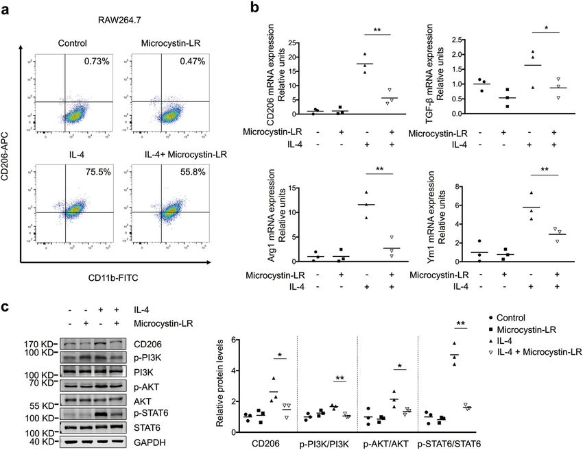
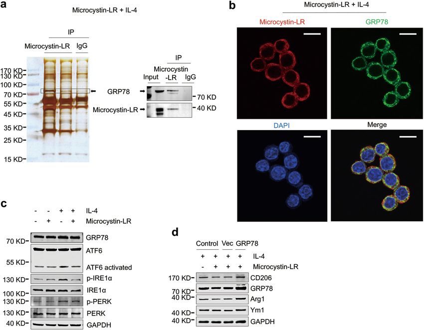
Wang et al. Cell Death and Disease (2020)11:136                                                                                               Page 5 of 15

  Fig. 2 Microcystin-LR antagonizes activation of the TGF-β signaling pathway and expression of fibrotic markers in rats with bleomycin-
  induced pulmonary fibrosis. Rats were treated as explained in Fig. 1. a Expression of TGF-β signaling pathway molecules, including TGF-β1, p-
  Smad2, Smad2, p-Smad3, Smad3 and prolyl 4-hydroxylase subunit αIII (P4HA3) in rat pulmonary tissues was measured by western blot (one-way
  ANOVA; TGF-β1: F4,10 = 3.726, P = 0.042; p-Smad2: F4,10 = 5.790, P = 0.011; p-Smad3: F4,10 = 5.720, P = 0.012; P4HA3: F4,10 = 3.861, P = 0.038; n = 3). b
  Protein levels of the fibrotic markers, including fibronectin, collagen 1α1 and α-smooth muscle actin (αSMA) in rat pulmonary tissues was measured
  by western blot (one-way ANOVA; fibronectin: F4,10 = 3.742, P = 0.041; collagen 1α1: F4,10 = 5.808, P = 0.011; αSMA: F4,10 = 4.722, P = 0.021; n = 3). c–
  e Expression of fibronectin, collagen 1α1 and αSMA at the mRNA level in rat pulmonary tissues was examined by quantitative RT-PCR (qRT-PCR). One-
  way ANOVA, fibronectin: F4,28 = 7.024, P < 0.000; collagen 1α1: F4,28 = 3.317, P = 0.024; αSMA: F4,28 = 2.841, P = 0.043. n = 5 (bleomycin) or n = 7 (all
  the other treatments). *P < 0.05, **P < 0.01 determined by one-way ANOVA with S-N-K multiple-comparison test.

of RAW264.7 with microcystin-LR treatment (Supple-                              stress facilitates macrophage acquisition of the M2-like
mentary Fig. S8).                                                               phenotype. As shown in Fig. 6c, microcystin-LR modu-
  With immunoprecipitation and protein mass spectro-                            lated ER stress in IL-4-induced RAW264.7 by reducing
metry techniques, we further identified that microcystin-                        the levels of the three activated UPRER signaling sensors,
LR has an interaction with glucose-regulated protein 78                         phosphorylated PKR-like endoplasmic reticulum kinase
(GRP78), a chaperonin and a master regulator of the                             (p-PERK), phosphorylated inositol requiring enzyme 1α
endoplasmic reticulum (ER) unfolded protein response                            (p-IRE1α) and cleaved activating transcription factor 6
(UPRER) in ER stress (Fig. 6a). We next observed a strong                       (ATF6 activated)28,29. Additionally, our data also revealed
overlap of GRP78 and microcystin-LR for the subcellular                         that GRP78 overexpression could reverse microcystin-LR-
localization in IL-4-induced RAW264.7 cells (Fig. 6b). ER                       mediated inhibition of CD206+ M2-like phenotype (Fig.

Official journal of the Cell Death Differentiation Association
Wang et al. Cell Death and Disease (2020)11:136                 Page 6 of 15

  Fig. 3 (See legend on next page.)

Official journal of the Cell Death Differentiation Association
Wang et al. Cell Death and Disease (2020)11:136                                                                                           Page 7 of 15

  (see figure on previous page)
  Fig. 3 Microcystin-LR targets macrophages to suppress EMT and FMT. a–c A549 (commonly used as a model of human alveolar type II
  pulmonary epithelium), MRC5 (human fetal lung fibroblasts) and NIH3T3 (mouse embryonic fibroblasts) were left alone or cultured for 48 h with TGF-
  β1 (5 ng/ml) to induce epithelial–mesenchymal transition (EMT) or fibroblast-to-myofibroblast transition (FMT). Some of the cells were also treated
  with 0.1 μM microcystin-LR as indicated. Protein expression in A549 (a), MRC5 (b) and NIH3T3 (c) were examined by western blot. d–f RAW264.7 cells
  (murine monocyte/macrophage cell line) were cultured for 48 h with IL-4 (5 ng/ml) to induce the polarization of macrophages in 6-well Millicell
  hanging cell culture inserts with a membrane pore size of 1.0 μm. Some of the cells were also treated with 0.1 μM microcystin-LR as indicated. Next,
  cell culture inserts containing the pretreated macrophages were transferred to the plates that were seeded with A549, MRC5 or NIH3T3 cells 24 h
  ago. After the cell coculture for 48 h, the expression levels of various proteins as indicated in A549 (d), MRC5 (e) and NIH3T3 (f) were examined by
  western blot (left panel) and immunofluorescence staining (right panel). Densitometric intensity of the bands was quantified (lower panel). *P < 0.05,
  **P < 0.01 determined by one-way ANOVA with S-N-K post hoc analysis (A549 E-cadherin: F3,8 = 13.624, P = 0.002, Vimentin: F3,8 = 8.323, P = 0.008;
  MRC5 fibronectin: F3,8 = 12.317, P = 0.002, αSMA: F3,8 = 7.736, P = 0.009; NIH3T3 fibronectin: F3,8 = 7.681, P = 0.01, αSMA: F3,8 = 13.681, P = 0.002).
  Scale bar: 100 μm.

6d). Next, immunofluorescence staining confirmed the                            twelve months22. Decreased expression of TGF-β1
localization of GRP78 with CD206 was overlapping in the                       appeared in the mice exposed to microcystin-LR equal to
pulmonary resident cells. As expected, GRP78 was                              or above 20 μg/L in drinking water (data not published).
abundantly expressed in CD206+ cells, but the numbers                         Therefore, 20 μg/L was selected as the dose of
of double-labeled cells were reduced in all the                               microcystin-LR in drinking water in our current research.
microcystin-LR treatment rats (Supplementary Fig. S9).                          Bleomycin-induced pulmonary fibrosis is the most
These results indicated that the interaction of                               commonly used model of fibrosis in rodents, and it
microcystin-LR with GRP78 might have a key regulatory                         manifests similar characteristics of IPF30,31. Most previous
role in the alleviation of ER stress and in the modulation                    works that focused on a beneficial antifibrotic substance
of CD206+ monocyte/macrophage differentiation.                                or molecule for bleomycin-induced pulmonary fibrosis
                                                                              was usually carried out in a preventive regimen, with the
The regimen of microcystin-LR does not cause functional                       drug given within 7 days after bleomycin administration32.
damage in bleomycin-instilled animals                                         In this work, we have provided compelling evidence
  To investigate whether the regimen of microcystin-LR                        demonstrating that microcystin-LR has a therapeutic
used in this study caused extra damage to rats with                           effect on the rats with bleomycin-induced pulmonary
bleomycin-induced pulmonary fibrosis, we performed                             fibrosis, especially when microcystin-LR was administered
blood tests and histological examination. Our data                            during early or inflammation/fibrosis transitional phase of
exhibited no observable adverse effect on serum bio-                          the fibrotic development. To confirm the therapeutic
chemistry with microcystin-LR treatment (Supplementary                        effect of microcystin-LR on pulmonary fibrosis indepen-
Table 1). The blood cell counting indicated that                              dent of bleomycin exposure, we treated FITC-induced
microcystin-LR could suppress bleomycin-associated                            pulmonary fibrosis with microcystin-LR in mice and
increases in neutrophils and monocytes (Supplementary                         observed a similar remission of fibrosis.
Fig. S10). Histological examination of liver and kidneys,                       Myofibroblasts, originated from exaggerated activa-
two susceptible organs to microcystin exposure, failed to                     tion and abnormal differentiation of fibroblasts via EMT
identify any tissue damage in the rats with microcystin-LR                    and FMT pathways, are widely considered as the effector
treatment (Supplementary Fig. S11). Based on these data,                      cells responsible for fibrosis33,34. A prolonged inflam-
it was believed that the regimen of microcystin-LR for                        mation, M2 macrophages differentiation and increased
developing a therapeutic strategy against pulmonary                           profibrotic factors, especially TGF-β1/Smad signaling
fibrosis would not be accompanied with substantial toxic                       form and maintain the microenvironment of fibrogen-
damage.                                                                       esis. Therapeutic approaches either by repressing TGF-
                                                                              β1/Smad signaling or via targeting M2 macrophages
Discussion                                                                    could generate a potential effect on alleviating pul-
   We conceived this study based on our previous obser-                       monary fibrosis35,36. Using a cell coculture system,
vation of a reduced expression of TGF-β1 in mouse lung                        A549, MRC5 and NIH3T3 cells cocultured with IL-4-
tissues after chronic exposure to microcystin-LR. Given                       induced macrophages (RAW264.7 or mouse BMDM),
that TGF-β1 is one of the most important profibrotic                           we revealed that microcystin-LR blocked the EMT or
cytokines, we hypothesized that microcystin-LR could                          FMT pathways through modulating M2 polarization of
possibly have an effect on pulmonary fibrosis. In the                          macrophages, rather than directly affecting epithelial
previous work, mice received microcystin-LR in drinking                       cells or the fibroblast phenotype. Given the number of
water at five concentrations (1, 5, 10, 20, and 40 μg/L) for                   CD206 + macrophages, but not CD163+, CD68+,

Official journal of the Cell Death Differentiation Association
Wang et al. Cell Death and Disease (2020)11:136 Page 8 of 15 Fig. 4 (See legend on next page.) Official journal of the Cell Death Differentiation Association
Wang et al. Cell Death and Disease (2020)11:136                                                                                                 Page 9 of 15

  (see figure on previous page)
  Fig. 4 Microcystin-LR alters bleomycin-induced macrophage polarization. Rats were treated as explained in Fig. 1. a Pulmonary tissue sections
  were examined for the expression of CD206 and CD163, markers of alternative activated macrophages, and CD86 and inducible nitric oxide synthase
  (iNOS), markers as classically activated macrophages using immunohistochemistry. Black arrows indicate CD206, CD163, CD86 or iNOS expression in
  macrophages. Scale bars: 100 μm. b Quantification of M1 and M2 macrophages in rat pulmonary tissues. **P < 0.01 determined by one-way ANOVA
  with S-N-K post hoc analysis (CD206: F4,28 = 7.867, P = 0.000; CD163: F4,28 = 0.441, P = 0.778; CD86: F4,28 = 1.865, P = 0.144; iNOS: F4,28 = 0.618, P =
  0.653). c Total protein was extracted from pulmonary tissues and examined for the expression of CD206, CD163, CD86 and iNOS by western blot.
  Densitometric intensity of the bands was quantified (right panel). *P < 0.05 determined by one-way ANOVA with S-N-K post hoc analysis (CD206: F4,10
  = 5.493, P = 0.013; CD163: F4,10 = 1.181, P = 0.376; CD86: F4,10 = 0.725, P = 0.594; iNOS: F4,10 = 0.437, P = 0.779).

  Fig. 5 Microcystin-LR has inhibitory effect on IL-4 induced M2 polarization of macrophages. RAW264.7 cells were left alone or treated with IL-4
  (5 ng/ml) for 48 h to induce polarization. In some of the assays, cells were also treated with 0.1 μM microcystin-LR for the same duration as indicated.
  a Percentages of CD206+/CD11b+ M2-like macrophages were analyzed by flow cytometry. b Relative expression of the M2-like macrophage markers
  CD206, TGF-β1, arginase-1 (Arg1) and chitinase-like 3 (Ym1) was analyzed using qRT-PCR. *P < 0.05, **P < 0.01 determined by one-way ANOVA with S-
  N-K post hoc analysis (CD206: F3,8 = 38.234, P < 0.000; TGF-β1: F3,8 = 6.900, P = 0.013; Arg1: F3,8 = 23.223, P < 0.000; Ym1: F3,8 = 15.990, P = 0.001; n =
  3). c Protein levels of CD206, p-PI3K/PI3K, p-AKT/AKT and p-STAT6/STAT6 signaling pathway molecules were determined by western blot. *P < 0.05,
  **P < 0.01 determined by one-way ANOVA with S-N-K post hoc analysis (CD206: F3,8 = 6.475, P = 0.016; PI3K: F3,8 = 9.546, P = 0.005; AKT: F3,8 = 6.813,
  P = 0.014; STAT6: F3,8 = 67.900, P < 0.000; n = 3).

CD86+ or iNOS+ macrophages, was markedly decreased                               macrophage differentiation. Human-derived macrophage
upon microcystin-LR treatment, it was reasonable to                              polarization will be used in the coculture system in our
assume that microcystin-LR could meliorate pulmonary                             future work although TGF-β1 shows a cross-species
fibrosis mainly by suppressing CD206+ M2a-like                                    activity.

Official journal of the Cell Death Differentiation Association
Wang et al. Cell Death and Disease (2020)11:136                                                                                            Page 10 of 15

  Fig. 6 Microcystin-LR inhibits the responses to endoplasmic reticulum stress by interaction with GRP78. RAW264.7 cells were treated as
  explained in Fig. 5. a Total protein extracts from RAW264.7 cells pretreated with IL-4 and microcystin-LR were immunoprecipitated with two
  independent microcystin-LR antibodies (left lane: MOB-647, Creative Biolabs; middle lane: ALX-804-320, Alexis Biochemicals). A ~ 78 kDa protein band
  was subsequently identified as glucose-regulated protein 78 kDa (GRP78) using mass spectrometry (left panel) and western blot (right panel). b
  Subcellular localization of microcystin-LR (red) and GRP78 (green) was examined using immunofluorescence staining. DAPI was used for nuclear
  staining (blue). Scale bar: 50 μm. c Cellular protein expression of GRP78 and its three transmembrane sensors including activating transcription factor
  6 (ATF6), inositol requiring enzyme 1α (IRE1α) and PKR-like endoplasmic reticulum kinase (PERK), together with their activated forms as indicated was
  measured by western blot. d GRP78 rescue assay was performed by transfecting RAW264.7 cells with the recombinant GRP78 expression plasmid
  (pcDNA3-GRP78). Control cells without transfection, vector (pcDNA3.1) and the pcDNA3-GRP78 transfected cells were treated as indicated for 48 h.
  The expressions of M2 macrophage markers, CD206, Arg1 and Ym1, and GRP78 were analyzed by western blot.

  We next attempted to clarify the issues involved in                          9S)-3-amino-9-methoxy-2-6-8-trimethyl 10-phenyldeca-4,6-
microcystin-LR-mediated macrophage differentiation. Using                      dienoic acid (Adda) side chains, which allows interaction
immunoprecipitation with two independent microcystin-LR                        with GRP7837,38. Chun et al. reported that the holoenzyme of
antibodies and protein mass spectrometry, we unexpectedly                      PP1γ2, a testis-specific isotype of protein phosphatase 1, is a
identified that microcystin-LR was capable of interacting                       heterotrimer-containing GRP78 subunit. Removing GRP78
with GRP78, a chaperonin associated with ER stress. This                       from the heterotrimer of PP1γ2 results in a significant loss of
result was confirmed by western blot. Knarr et al. developed                    enzymatic activity and renders PP1γ2 less sensitive to its
a computer program to predict GRP78 (also known as BiP)-                       inhibitor microcystin-LR39. Both the mentioned works also
binding sites in proteins by scoring amino acid sequences                      support a potential interaction between GRP78 and micro-
based on the segments of seven residues, and then test them                    cystin-LR, which we demonstrated in the current study.
with synthetic heptapeptides corresponding to the potential                    Increasing evidence suggests that the ER stress controls M2
binding sites. They confirmed that GRP78 is associated with                     phenotype differentiation of macrophages40,41. Under ER
a wide variety of target proteins and the binding peptides for                 stress, GRP78 frees its client proteins, which leads to phos-
GRP78 are enriched in hydrophobic residues. Microcystin-                       phorylation of PERK and IRE1α, and cleavage of ATF6, and
LR is a cyclic heptapeptides with hydrophobic (2S, 3S, 8S,                     contributes to the initiation of the UPRER signaling pathway.

Official journal of the Cell Death Differentiation Association
Wang et al. Cell Death and Disease (2020)11:136                                                                            Page 11 of 15

Our further investigation demonstrated that microcystin-LR
could suppress the UPRER signaling pathway through
downregulating p-PERK, p-IRE1α and the cleaved ATF6.
Overexpression of GRP78 antagonized microcystin-LR-
mediated inhibition of CD206+ macrophage polarization,
suggesting microcystin-LR could avert activation of GRP78.
Kaufman and Schroder reported a dynamic equilibrium
between monomeric and oligomeric GRP78 in cells42. In the
oligomeric status, the peptide-binding domain of GRP78 is in
phosphorylation and forms a protein complex with PERK,
IRE1α and ATF6. In ER stress, the oligomeric GRP78 can
dissociate into dephosphorylated monomer that binds to the
unfolded proteins, releasing three ER transmembrane sensors
to activate downstream signaling pathway molecules. Thus,
we speculate that the physical binding between microcystin-
LR and GRP78 may prevent the GRP78 protein from
dephosphorylation catalyzed by protein phosphatases, which
will in turn impede the regulation of GRP78 on UPRER sig-
naling pathway.
  Microcystin-LR is a well characterized environmental
toxicant absorbed by human body via the oral route and
accumulates in the liver, kidney, lung and other tissues. It
                                                                   Fig. 7 Model of the proposed mechanism by which microcystin-
was compulsory to assess whether the regimen of                    LR ameliorates pulmonary fibrosis. Microcystin-LR is selectively
microcystin-LR in the current study may cause additional           taken into macrophages in lung tissue and binds to GRP78, a master
lesion in the rats of bleomycin-induced pulmonary                  regulator of ER stress, resulting in attenuation of M2 polarization of
fibrosis. Fawell et al. implemented several sets of experi-         macrophages through reduction of p-PERK, p-IRE1α and cleaved ATF6.
ments to investigate the toxicity of microcystin-LR in             The macrophages with modulated ER stress decrease the expression
                                                                   of TGF-β1, which causes a repressed epithelial–mesenchymal
rodents. They demonstrated that the mice exposed to                transition and fibroblast–myofibroblast transition, and leads to the
microcystin-LR with the dose of 40 μg/kg body weight               amelioration of pulmonary fibrosis.
per day by gavage for 13 weeks have no observed adverse
effect level (NOAEL) for liver tissues43. In this study, we
treated rats with drinking water containing 20 μg/L
microcystin-LR, which is equivalent to 3 μg/kg/day intake         TGF-β1 expression, which would result in amelioration of
(calculated based on daily water consumption of 1.5 ml/           pulmonary fibrosis (Fig. 7). Our results provide a novel
10 g body mass) for 7, 6 and 4 weeks, respectively. We            insight on the therapeutic exploitation of microcystin-LR, an
previously described the damage of liver tissue in the mice       enigmatic natural molecule classically described as a cyto-
following chronic exposure to microcystin-LR for twelve           toxic agent, for the treatment of IPF.
months22. The duration of microcystin-LR treatment used
in the current study was much shorter compared with our           Materials and methods
previous experiments. Therefore, it is not surprising that        Chemicals
we failed to identify any discernable toxic effects of              Microcystin-LR was purchased from Alexis Biochem-
microcystin-LR on the model rats. Further investigations          icals (Lausen, Switzerland), while bleomycin was pur-
should be performed to identify the structural analogues          chased from Nippon Kayaku Co. (Tokyo, Japan). Human
with the same efficacy but lower toxicity in microcystins          TGF-β1, murine macrophage colony-stimulating factor
for the treatment of pulmonary fibrosis.                           (M-CSF) and IL-4 were purchased from Pepro Tech
  Microcystin-LR can attenuate macrophage polarization            (Rocky Hill, New Jersey, USA). FITC and LPS was pur-
toward a CD206+ M2-like phenotype, resulting in the inhi-         chased from Sigma-Aldrich (St. Louis, Missouri, USA).
bition of EMT or FMT signaling in epithelial cells and
fibroblasts. Furthermore, we demonstrated an interaction           Rat model
between microcystin-LR and GRP78, a master regulator of             Six-eight weeks old male SD rats (220–250 g in weight)
ER stress, which led to a blockage effect on UPRER signal         were provided by the Model Animal Research Center of
transduction in the stressed cells. This may well be a possible   Nanjing University (Nanjing, China). The animals were
mechanism underlying microcystin-LR-mediated attenuation          housed in a special pathogen-free, and temperature- and
of CD206+ macrophage differentiation and reduction of             humidity-controlled (23 °C ± 2 °C and 50% relative

Official journal of the Cell Death Differentiation Association
Wang et al. Cell Death and Disease (2020)11:136 Page 12 of 15
humidity) room with a 12-h light/dark cycle. Water and β-mercaptoethanol, 0.5 mg/ml bovine serum albumin).
rodent chow were freely available. All of the rats were Free phosphate, generated from a synthetic phospho-
acclimated for 7 days and then completely randomized threonine peptide RRA(pT)VA-specific for PP2A, was
into five groups. For the induction of pulmonary fibrosis, quantified by measuring the molybdate/malachite green/
rats were intratracheally instilled with a single dose of phosphate complex at 630 nm.
bleomycin (5.0 mg/kg) on day 0. Rats that received
intratracheal instillation of an equal volume of saline were Quantification of microcystin-LR in the lung and liver
used as control. In some of the rats with fibrosis induc- tissues
tion, the animals received microcystin-LR (20 μg/L) in The retention of microcystin-LR in the lung and liver
drinking water in three groups, starting on day 7 (LR7), 14 tissues was assessed by an ELISA kit (Alexis Biochemicals,
(LR14) or 28 (LR28), respectively. All rats were euthanized Lausen, Switzerland). Briefly, tissue samples were homo-
56 days after receiving bleomycin intratracheal instillation genized directly in a lysis buffer for protein extraction and
for further analysis (Fig. 1a). diluted ten-fold to fall within the microcystin-LR detec-
tion range according to the manufacturer’s instructions.
Mouse model Plates were read at 550 nm.
Six–eight weeks old male C57BL/6 mice (20–22 g) were
purchased from Model Animal Research Center of Histology and immunostaining
Nanjing University. The feeding environment and con- For histologic evaluation, the fixed right lung tissues
ditions of mice were the same as described in the rat were embedded in paraffin, sectioned (4 μm) onto glass
model. Mice were completely randomized into three slides and stained with H&E for structured observation
groups. Some mice were intratracheally instilled with a and Masson’s trichrome for detection of collagen depos-
single dose of FITC (3.5 mg/kg) on day 0 to induce pul- its. The development of pulmonary lesions was scored by
monary fibrosis. Saline was administered similarly as a two pathologists blind to the study design. For immuno-
control. For microcystin-LR treatment, the mice with histochemistry staining, the sections were permeabilized
fibrosis induction received microcystin-LR (20 μg/L) in in 1× PBS containing Triton X-100 (0.1%) for 10 minutes
drinking water starting on day 14. All animals were and then probed with antibodies against CD206 (ab64693,
euthanized on day 56 (Supplementary Fig. S1a). Tissue Abcam, Cambridge, Massachusetts, USA), CD163
samples were collected followed by fixation with a 10% (ab182422, Abcam), CD86 (BS9900M, Bioworld, St. Louis
neutralized formalin solution for 24 h at room tempera- Park, Minnesota, USA), iNOS (ab178945, Abcam), CD68
ture and the remaining tissues were kept at −80 °C. (ab213363, Abcam), TGF-β1 (ab215715, Abcam) and
αSMA (ab32575). For the immunofluorescence assay,
Hydroxyproline assay slides were first labeled with a mouse-derived antibody
The collagen content in lung homogenates was eval- against CD206 (ab8918, Abcam) and a rabbit-originated
uated by a hydroxyproline (HYP) assay using a kit from GRP78 (ab21685, Abcam) antibody followed by staining
Nanjing JianCheng Bioengineering Institute (Nanjing, with an Alexa Fluor 595-labelled anti-mouse and Alexa
China). The HYP assay was conducted essentially Fluor 488-conjugated anti-rabbit antibodies (Thermo
according to the manufacturer’s instruction. The absor- Fisher Scientific, Inc., Waltham, Massachusetts, USA).
bance of each sample was read at 550 nm in a SpectraMax Images were captured using a Zeiss Axio upright fluor-
M2e microplate reader (Molecular Devices, Sunnyvale, escent microscope.
California, USA). The experiment was performed in tri-
plicate and data presented as μg/mg protein of the lung Cell culture
tissues. Cell lines including L02, A549, MRC5, NIH3T3 and
RAW264.7 were obtained from Cell Bank of Typical
PP2A phosphatase activity assay Culture Preservation Commission, Chinese academy of
PP2A activity was determined by a serine/threonine Sciences. Three human-derived cell lines (L02, A549 and
phosphatase assay kit in accordance with the manu- MRC5) were authenticated by STR profiling. All cells
facturer’s protocols (Promega, Madison, Wisconsin, were tested negative for mycoplasma contamination using
USA). The total proteins of the lung tissues or the cul- MycoBlueTM mycoplasma detector (Vazyme, Nanjing,
tured L02 cells were extracted in a phosphatase lysis China). L02, NIH3T3 and RAW264.7 cells were cultured
buffer (50 mM Tris-HCl pH 7.5, 10% glycerol, 0.05% in DMEM medium (Thermo Fisher Scientific). A549 was
β-mercaptoethanol, 0.1 mM EDTA, 0.05% Triton X-100, maintained in RPMI 1640 medium (Thermo Fisher Sci-
0.5 mM PMSF, phosphatase inhibitor cocktail) and mea- entific) and MRC5 in low-glucose DMEM medium
sured for phosphatase activity using a PP2A-type specific (Thermo Fisher Scientific). Bone marrow mononuclear
buffer (250 mM imidazole pH 7.2, 1 mM EGTA, 0.1% cells from C57BL/6 mice were isolated and treated with
Official journal of the Cell Death Differentiation AssociationWang et al. Cell Death and Disease (2020)11:136 Page 13 of 15
M-CSF (20 ng/ml) in DMEM medium for differentiation NaOH, 140 mM NaCl, 2.5 mM CaCl2, 5 mM KCl, pH 7.4)
into macrophages prior to experiments. All cell media and stained with FITC-conjugated AnnexinV and PI
were supplemented with 10% fetal bovine serum (Thermo (50 μg/ml) for 30 min at room temperature in the dark.
Fisher Scientific). Cells were cultured at 37 °C in a 5% CO2
humidified incubator to reach 80% of confluence. Plasmid recombination and cell transfection
The expression plasmid pcDNA3.1 was purchased from
Treatment of the cultured cells and coculture Thermo Fisher Scientific. The recombinant GRP78 expres-
A549, MRC5 or NIH3T3 cells with a seeding density of sion plasmid (pcDNA-GRP78), containing full-length rat
1×10⁵ cells/ml in six-well plates were cultured in medium GRP78 cDNA, was constructed. When the cultured cells
containing TGF-β1 (5 ng/ml) for 48 h to induce EMT or reached 60–80% of confluence, the expression vectors were
FMT in vitro. Microcystin-LR (0.1 μM) was synchro- transiently transfected into the indicated cells with lipo-
nously used to observe the intervention effect on the fectamine 3000 (Thermo Fisher Scientific) following the
treated cells. RAW264.7 cells or mouse BMDM seeded at manufacturer’s instructions. The transfected cells were then
a density of 1×10⁵ cells/ml were exposed to LPS (10 ng/ cultured for 48 h and harvested for analysis.
ml) for 48 h to induce M1- or exposure to IL-4 (5 ng/ml)
for 48 h to induce M2-like differentiation. Some of the Immunoprecipitation and mass spectrometry
cells were also treated with 0.1 μM microcystin-LR. To RAW264.7 cells were treated with IL-4 (5 ng/ml) and
establish the coculture with M1 or M2-like macrophages, microcystin-LR (0.1 μM) for 48 h followed by harvesting.
we transferred the cell culture inserts containing LPS or The cells were lysed using immunoprecipitation lysis/
IL-4 pretreated macrophages (as mentioned above) to the wash buffer (Thermo Fisher Scientific) containing a Halt
plates that had been seeded with A549, MRC5 or NIH3T3 protease and phosphatase inhibitor cocktail (Thermo
cells (5×10⁴ cells/ml) for culturing 24 h. After 48 h of Fisher Scientific). The cell extracts left on a rocking
coculture, the cells at the bottom of the plates were har- platform for 1 h, protein concentration was determined
vested for further experiments. using the pierce BCA protein assay (Thermo Fisher Sci-
entific). Protein from 500 μg to 1 mg was incubated with
Cell proliferation assay two clones of microcystin-LR antibodies separately
Cell yield was measured in real time using xCELLigence (MOB-647, Creative Biolabs, Shirly, New York, USA and
system (ACEA Biosciences Inc., San Diego, California, ALX-804–320, Alexis Biochemicals) overnight at 4 °C.
USA). Firstly, 50 μl medium was added to each well of E- The protein A/G PLUS-Agarose beads (Santa Cruz Bio-
Plate for the impedance baseline measurement. Next, the technology Inc., Santa Cruz, California, USA) were sub-
final volume was adjusted up to 150 μl by the addition of sequently applied for 2 h at 4 °C. After centrifugation, the
100 μl culture medium containing 5000 cells in each well. beads were collected and washed 5 times with IP Lysis
The E-Plate was incubated at 37 °C with 5% CO2 for 20 h Buffer. The samples were then boiled in 1× SDS solution
and then microcystin-LR was added to the wells at the at 95 °C for 5 min to elute the immunocomplexed pro-
indicated concentrations. The E-Plate continued to be teins. The eluted products were separated in SDS-PAGE,
incubated and cell yield was monitored at a 15-minute followed by silver staining (Thermo Fisher Scientific)
interval for up to 48 h. according to the manufacturer’s protocol. Candidate
protein bands were carefully cut for mass spectrometry
Flow cytometry analysis provided by Hoogen Biotech (Shanghai, China).
RAW264.7 cells were left alone or cultured with IL-4
(5 ng/ml) for 48 h. Some of the cells were also treated with Western blot
0.1 μM microcystin-LR. Cells were harvested and washed The cells and tissues were homogenized and lysed in the
twice with 3 ml 0.5% BSA-PBS by centrifuging at 500 g for RIPA buffer supplemented with proteinase inhibitors.
5 min at 4 °C. The cell pellet was suspended in 1 ml 0.5% Equal amounts of proteins were loaded and separated on a
BSA-PBS and the total number was calculated by a cell 10% SDS-PAGE gel. Following electrophoresis, the pro-
counter. Following preincubation with mouse Fc block teins were transferred to a PVDF membrane, blocked in
(eBioscience, San Diego, California, USA) for 15 min, the 5% (w/v) non-fat milk and incubated with the primary
cell suspension was stained with anti-mouse CD11b-FITC antibodies overnight. The sources of primary antibodies
(eBioscience) and CD206-APC (eBioscience) at 4 °C for were: TGF-β1 (ab215715, Abcam), αSMA (ab32575,
30 min in the dark. After washing and resuspending in Abcam), Fibronectin (ab32419, Abcam), Collagen 1α1
PBS, the cells were analyzed by a flow cytometer (ab138492, Abcam), Smad2/3 (3102, Cell Signaling
(FACSCalibur, BD Biosciences, San Jose, California, USA). Technology, Beverly, Massachusetts, USA), p-Smad2
For PI and Annexin-V double staining, cells were sus- (ab53100, Abcam), p-Smad3 (ab138659, Abcam), P4HA3
pended in 100 μl of a buffer solution (10 mM HEPES/ (ab101657, Abcam), E-cadherin (3195S, Cell Signaling
Official journal of the Cell Death Differentiation AssociationWang et al. Cell Death and Disease (2020)11:136 Page 14 of 15
Technology), Vimentin (5147S, Cell Signaling Technol- Hematological analysis and serum biochemistry
ogy), CD163 (BS71200, Bioworld), CD206 (ab125028, Blood samples were collected from the rats for mea-
Abcam), CD86 (BS9900M, Bioworld), iNOS (ab178945, suring the hematological parameters. Blood cells were
Abcam), PI3K (4249T, Cell Signaling Technology), p- differentially counted with an Automatic Blood Analyzer
PI3K (4228T, Cell Signaling Technology), AKT (A5031, (Sysmex, Kobe, Japan). Serum parameters, including AST,
Selleckchem, Houston, Texas, USA), p-AKT (A5030, ALT, BUN, and CRE, and the lipid profile, were assayed
Selleckchem), p-STAT6 (56554S, Cell Signaling Tech- by an Auto-dry Chemistry Analyzer (Kehua Bioengi-
nology), STAT6 (5397S, Cell Signaling Technology), neering, Shanghai, China).
microcystin-LR antibody (ALX-804-320, Alexis Bio-
chemicals), GRP78 (ab21685, Abcam), PERK (BS2156, Statistical evaluation
Bioworld), p-PERK (YP1055, ImmunoWay Biotechnology, Statistical analysis was carried out using SPSS 24.0
Planto, Texas, USA), IRE1α (ab37073, Abcam), p-IRE1α (SPSS Inc., Chicago, Illinois, USA). Data were expressed
(ab226974, Abcam), ATF6 (BS6476, Bioworld), Arg1 using plotting individual value. Normal distribution of the
(ab60176, Abcam) and Ym1 (ab93034, Abcam). The data was tested. All two-sample and multiple-comparison
membranes were washed three times and incubated in a tests were two-tailed. Differences in body weight gain
0.1% tween phosphate buffer solution (PBST) for 10 min ratios in experimental animals that received various
followed by probing with goat anti-mouse or goat anti- treatments were assessed by repeated measure ANOVA.
rabbit IR-Dye 800cw for 1.5 h at room temperature. The Statistical significance for multiple comparisons was
signal was detected using an Odyssey scanner (Li-Cor evaluated by one-way ANOVA with S–N–K post hoc
Biosciences, Lincoln, Nebraska, USA). analysis. For comparisons of microcystin-LR concentra-
tion in rat liver and lung tissues, data were analyzed using
Quantitative RT-PCR analysis paired Student’s t test. P < 0.05 was considered statistically
Total RNA was isolated from cells and pulmonary tissues significant. The sample size for rat model was calculated
using TRIzol reagent (Thermo Fisher Scientific), and qRT- using power analysis based our pilot study. Type I error is
PCR was performed to examine the target RNA using Fast fixed at the level of 5% (P = 0.05). Power is kept at 90%.
SYBR® Green Master Mix (Thermo Fisher Scientific) on a Supposed sample size was estimated by formula (com-
QuantStudio6 (Applied Biosystems, Foster City, California, pletely randomized design for multiple means compar-
USA) as directed by the manufacturer. Each sample was ison). The sample size for FITC-induced mice pulmonary
tested in triplicate, and at least two biological samples were fibrosis was determined as previously described44.
included in each assay. Results of mRNA quantification were
normalized against GAPDH and calculated using the ΔΔ Study approval
cycle threshold (Ct) method. Forward and reverse primer The animal care and the study procedures were
sequences for specific genes are listed in Supplementary approved by the Ethics Committee for Animal Research in
Table 2. Medical School of Nanjing University.
Immunofluorescence assay
Acknowledgements
Cells were fixed with 4% paraformaldehyde (Thermo The authors would like to express our sincere thanks to Dr. Lei Fang (Nanjing
Fisher Scientific) in PBS, permeabilized with 0.1% Triton University School of Medicine) for discussion and suggestion on
immunoprecipitation and mass spectrometry, and thanks to Dr. Yimei Fan
X-100 (Thermo Fisher Scientific) in PBS, and then
(Nanjing University School of Medicine) for discussion on statistical analysis.
blocked in 3% BSA (Sigma-Aldrich). Subsequently, cells This work was supported by the National Natural Science Foundation of China
were incubated with specific primary antibodies overnight (81270152, 81771504 and 81501977), Human Resource Summit Grant of
Jiangsu Province (WSN-043) and The Young Talents Program of Jiangsu Cancer
at 4 °C. The following primary antibodies at indicated
Hospital (QL201807).
dilutions were used: microcystin-LR (Creative Biolabs,
1:50), Vimentin (Cell Signaling Technology, 1:200), E-
cadherin (Cell Signaling Technology, 1:200), Fibronectin Author details
1
Department of Medical Genetics, Nanjing University School of Medicine,
(Abcam, 1:200), αSMA (Abcam, 1:200), GRP78 (Abcam, Nanjing 210093, China. 2Jiangsu Key Laboratory of Molecular and Translational
1:200). Cells were stained with Alexa Fluor 488- and Alexa Cancer Research, Jiangsu Cancer Hospital, Jiangsu Institute of Cancer Research,
Fluor 549-labeled secondary antibodies (Thermo Fisher The Affiliated Cancer Hospital of Nanjing Medical University, Nanjing 210009,
China. 3Jiangsu Key Laboratory of Molecular Medicine, Nanjing University
Scientific, 1:300) for 1 h at room temperature in the dark. School of Medicine, Nanjing 210093, China. 4Department of Health
Nuclear staining was performed with DAPI (Thermo Technology and Informatics, Faculty of Health and Social Sciences, The Hong
Fisher Scientific, 1 μg/ml in PBS) for 10 minutes at room Kong Polytechnic University, Hung Hom, Kowloon, Hong Kong, China
temperature. The images were observed on an Olympus
confocal laser scanning microscope imaging system and a Conflict of interest
Zeiss fluorescent microscope. The authors declare that they have no conflict of interest.
Official journal of the Cell Death Differentiation AssociationWang et al. Cell Death and Disease (2020)11:136 Page 15 of 15
Publisher’s note 21. Dittmann, E., Fewer, D. P. & Neilan, B. A. Cyanobacterial toxins: biosynthetic
Springer Nature remains neutral with regard to jurisdictional claims in routes and evolutionary roots. FEMS Microbiol. Rev. 37, 23–43 (2013).
published maps and institutional affiliations. 22. Li, X., Zhao, Q., Zhou, W., Xu, L. & Wang, Y. Effects of chronic exposure to
microcystin-LR on hepatocyte mitochondrial DNA replication in mice. Environ.
Supplementary Information accompanies this paper at (https://doi.org/ Sci. Technol. 49, 4665–4672 (2015).
10.1038/s41419-020-2329-z). 23. Xaubet, A. et al. Transforming growth factor-beta1 gene polymorphisms are
associated with disease progression in idiopathic pulmonary fibrosis. Am. J.
Respir. Crit. Care Med. 168, 431–435 (2003).
Received: 26 September 2019 Revised: 4 February 2020 Accepted: 5
24. Luo, Y. et al. A novel profibrotic mechanism mediated by TGFβ-stimulated
February 2020
collagen prolyl hydroxylase expression in fibrotic lung mesenchymal cells. J.
Pathol. 236, 384–394 (2015).
25. Xing, Y. et al. Structure of protein phosphatase 2A core enzyme bound to
tumor-inducing toxins. Cell 127, 341–353 (2006).
26. Vergadi, E., Ieronymaki, E., Lyroni, K., Vaporidi, K. & Tsatsanis, C. Akt signaling
References
pathway in macrophage activation and M1/M2 polarization. J. Immunol. 198,
1. Raghu, G. et al. Idiopathic pulmonary fibrosis in US medicare beneficiaries
1006–1014 (2017).
aged 65 years and older: incidence, prevalence, and survival, 2001-11. Lancet
27. Murray, P. J. et al. Macrophage activation and polarization: nomenclature and
Respir. Med. 2, 566–572 (2014).
experimental guidelines. Immunity 41, 14–20 (2014).
2. Cottin, V. & Richeldi, L. Neglected evidence in idiopathic pulmonary fibrosis
28. Hetz, C. & Papa, F. R. The unfolded protein response and cell fate control. Mol.
and the importance of early diagnosis and treatment. Eur. Respir. Rev. 23,
Cell 69, 169–181 (2018).
106–110 (2014).
29. Maurel, M., Chevet, E., Tavernier, J. & Gerlo, S. Getting RIDD of RNA: IRE1 in cell
3. Raghu, G. Idiopathic pulmonary fibrosis: lessons from clinical trials over the
fate regulation. Trends Biochem. Sci. 39, 245–254 (2014).
past 25 years. Eur. Respir. J. 50, 1701209 (2017).
30. Chaudhary, N. I., Schnapp, A. & Park, J. E. Pharmacologic differentiation of
4. Kim, H. J., Perlman, D. & Tomic, R. Natural history of idiopathic pulmonary
inflammation and fibrosis in the rat bleomycin model. Am. J. Respir. Crit. Care
fibrosis. Respir. Med. 109, 661–670 (2015).
Med. 173, 769–776 (2006).
5. Fernandez, I. E. & Eickelberg, O. New cellular and molecular mechanisms of
31. Chua, F., Gauldie, J. & Laurent, G. J. Pulmonary fibrosis: searching for model
lung injury and fibrosis in idiopathic pulmonary fibrosis. Lancet 380, 680–688
answers. Am. J. Respir. Cell Mol. Biol. 33, 9–13 (2005).
(2012).
32. Moeller, A., Ask, K., Warburton, D., Gauldie, J. & Kolb, M. The bleomycin animal
6. Selman, M. & Pardo, A. Revealing the pathogenic and aging-related
model: a useful tool to investigate treatment options for idiopathic pulmonary
mechanisms of the enigmatic idiopathic pulmonary fibrosis. an integral
fibrosis? Int. J. Biochem. Cell Biol. 40, 362–382 (2008).
model. Am. J. Respir. Crit. Care Med. 189, 1161–1172 (2014).
33. Shu, D. Y. & Lovicu, F. J. Myofibroblast transdifferentiation: the dark force in
7. Martinez, F. J. et al. Idiopathic pulmonary fibrosis. Nat. Rev. Dis. Prim. 3, 17074
ocular wound healing and fibrosis. Prog. Retin. Eye Res. 60, 44–65 (2017).
(2017).
34. Yao, L. et al. Paracrine signalling during ZEB1-mediated
8. Duffield, J. S., Lupher, M., Thannickal, V. J. & Wynn, T. A. Host responses in tissue
epithelial–mesenchymal transition augments local myofibroblast differentia-
repair and fibrosis. Annu. Rev. Pathol. 8, 241–276 (2013).
tion in lung fibrosis. Cell Death Differ. 26, 943–957 (2019).
9. Wynn, T. A. & Barron, L. Macrophages: master regulators of inflammation and
35. Byrne, A. J., Maher, T. M. & Lloyd, C. M. Pulmonary macrophages: a new
fibrosis. Semin. Liver Dis. 30, 245–257 (2010).
therapeutic pathway in fibrosing lung disease? Trends Mol. Med. 22, 303–316
10. Lupher, M. L. Jr. & Gallatin, W. M. Regulation of fibrosis by the immune system.
(2016).
Adv. Immunol. 89, 245–288 (2006).
36. Byrne, A. J., Mathie, S. A., Gregory, L. G. & Lloyd, C. M. Pulmonary
11. Mantovani, A., Sica, A. & Locati, M. Macrophage polarization comes of age.
macrophages: key players in the innate defence of the airways. Thorax
Immunity 23, 344–346 (2005).
70, 1189–1196 (2015).
12. Wynn, T. A., Chawla, A. & Pollard, J. W. Macrophage biology in development,
37. Knarr, G., Gething, M. J., Modrow, S. & Buchner, J. BiP binding sequences in
homeostasis and disease. Nature 496, 445–455 (2013).
antibody. J. Biol. Chem. 270, 27589–27594 (1995).
13. Wynn, T. A. & Ramalingam, T. R. Mechanisms of fibrosis: therapeutic translation
38. Knarr, G., Modrow, S., Todd, A., Gething, M. J. & Buchner, J. BiP-binding
for fibrotic disease. Nat. Med. 18, 1028–1040 (2012).
sequences in HIV gp160. Implications for the binding specificity of bip. J. Biol.
14. Murray, P. J. & Wynn, T. A. Protective and pathogenic functions of macrophage
Chem. 274, 29850–29857 (1999).
subsets. Nat. Rev. Immunol. 11, 723–737 (2011).
39. Chun, Y. S. et al. Role of the 78-kDa glucose-regulated protein as an activity
15. Wei, Y. et al. Fibroblast-specific inhibition of TGF-β1 signaling attenuates lung
modulator of protein phosphatase1gamma2. Biochem. Biophys. Res. Commun.
and tumor fibrosis. J. Clin. Invest. 127, 3675–3688 (2017).
259, 300–304 (1999).
16. Li, M. et al. Epithelium-specific deletion of TGF-β receptor type II protects mice
40. Oh, J. et al. Endoplasmic reticulum stress controls M2 macrophage differ-
from bleomycin-induced pulmonary fibrosis. J. Clin. Invest. 121, 277–287
entiation and foam cell formation. J. Biol. Chem. 287, 11629–11641 (2012).
(2011).
41. Burman, A., Tanjore, H. & Blackwell, T. S. Endoplasmic reticulum stress in
17. Noble, P. W., Barkauskas, C. E. & Jiang, D. Pulmonary fibrosis: patterns and
pulmonary fibrosis. Matrix Biol. 68-69, 355–365 (2018).
perpetrators. J. Clin. Invest. 122, 2756–2762 (2012).
42. Schroder, M. & Kaufman, R. J. The mammalian unfolded protein response.
18. Fernandez, I. E. & Eickelberg, O. The impact of TGF-β on lung fibrosis: from
Annu. Rev. Biochem. 74, 739–789 (2005).
targeting to biomarkers. Proc. Am. Thorac. Soc. 9, 111–116 (2012).
43. Fawell, J. K., Mitchell, R. E., Everett, D. J. & Hill, R. E. The toxicity of cyanobacterial
19. Akhurst, R. J. & Hata, A. Targeting the TGFβ signalling pathway in disease. Nat.
toxins in the mouse: I microcystin-LR. Hum. Exp. Toxicol. 18, 162–167 (1999).
Rev. Drug Discov. 11, 790–811 (2012).
44. McMillan, T. R. et al. Exacerbation of established pulmonary fibrosis in a murine
20. Zheng, C. et al. Serum microcystin levels positively linked with risk of hepa-
model by gammaherpesvirus. Am. J. Respir. Crit. Care Med. 177, 771–780
tocellular carcinoma: a case-control study in southwest China. Hepatology 66,
(2008).
1519–1528 (2017).
Official journal of the Cell Death Differentiation AssociationYou can also read