Identification of the feline foamy virus Bet domain essential for APOBEC3 counteraction
←
→
Page content transcription
If your browser does not render page correctly, please read the page content below
Slavkovic Lukic et al. Retrovirology 2013, 10:76
http://www.retrovirology.com/content/10/1/76

 RESEARCH                                                                                                                                       Open Access

Identification of the feline foamy virus Bet
domain essential for APOBEC3 counteraction
Dragana Slavkovic Lukic1, Agnes Hotz-Wagenblatt2, Janet Lei1, Ann-Mareen Räthe1, Michael Mühle3,
Joachim Denner3, Carsten Münk4 and Martin Löchelt1*

  Abstract
  Background: APOBEC3 (A3) proteins restrict viral replication by cytidine deamination of viral DNA genomes and
  impairing reverse transcription and integration. To escape this restriction, lentiviruses have evolved the viral
  infectivity factor (Vif), which binds A3 proteins and targets them for proteolytic degradation. In contrast, foamy
  viruses (FVs) encode Bet proteins that allow replication in the presence of A3, apparently by A3 binding and/or
  sequestration, thus preventing A3 packaging into virions and subsequent restriction. Due to a long-lasting FV-host
  coevolution, Bet proteins mainly counteract restriction by A3s from their cognate or highly related host species.
  Results: Through bioinformatics, we identified conserved motifs in Bet, all localized in the bel2 exon. In line with
  the localization of these conserved motifs within bel2, this part of feline FV (FFV) Bet has been shown to be
  essential for feline A3 (feA3) inactivation and feA3 protein binding. To study the function of the Bet motifs in detail,
  we analyzed the ability of targeted deletion, substitution, and chimeric FFV-PFV (prototype FV) Bet mutants to
  physically bind and/or inactivate feA3. Binding of Bet to feA3Z2b is sensitive to mutations in the first three
  conserved motifs and N- and C-terminal deletions and substitutions across almost the complete bel2 coding
  sequence. In contrast, the Bel1 (also designated Tas) domain of Bet is dispensable for basal feA3Z2b inactivation
  and binding but mainly increases the steady state level of Bet. Studies with PFV Bel1 and full-length FFV Bel2
  chimeras confirmed the importance of Bel2 for A3 inactivation indicating that Bel1 is dispensable for basal feA3Z2b
  inactivation and binding but increases Bet stability. Moreover, the bel1/tas exon may be required for expression of a
  fully functional Bet protein from a spliced transcript.
  Conclusions: We show that the Bel2 domain of FV Bet is essential for the inactivation of APOBEC3 cytidine
  deaminase restriction factors. The Bel1/Tas domain increases protein stability and can be exchanged by related
  sequence. Since feA3 binding and inactivation by Bet are highly correlated, the data support the view that FV Bet
  prevents A3-mediated restriction of viral replication by creating strong complexes with these proteins.
  Keywords: APOBEC3, Retrovirus, Foamy virus, Antiviral restriction factors, Bet protein, Host-virus interaction,
  Virus defence protein

Background                                                                           characterized in detail: APOBEC3, Trim5α, tetherin and
Cellular restriction factors are key players of intrinsic                            SAMHD1 [8-12]. In addition, several other restriction fac-
immunity, which acts against viruses immediately after in-                           tors have been detected by genome-wide screens but
fection [1]. Restriction factors are constitutively expressed                        require additional experimental characterization [13,14].
in cells but their expression can be increased by inter-                             Restriction factors interfere with defined steps in the viral
ferons produced upon viral infection [2-7]. So far, four                             life cycle, leading to attenuation or complete suppression
anti-retroviral restriction factors have been functionally                           of virus replication. By coevolution with host-encoded re-
                                                                                     striction factors, current viruses have achieved mecha-
* Correspondence: m.loechelt@dkfz.de                                                 nisms to circumvent this inhibitory activity. Some viruses,
1
 Research Program Infection and Cancer, Department Genome Modifications              for instance, have acquired special proteins that directly
and Carcinogenesis, German Cancer Research Center (DKFZ), Heidelberg,
Germany
                                                                                     counteract restriction factors [1,2,8,12,15]. Due to the
Full list of author information is available at the end of the article               interaction of viral proteins with restriction factors, both
                                      © 2013 Slavkovic Lukic et al.; licensee BioMed Central Ltd. This is an Open Access article distributed under the terms of the
                                      Creative Commons Attribution License (http://creativecommons.org/licenses/by/2.0), which permits unrestricted use,
                                      distribution, and reproduction in any medium, provided the original work is properly cited.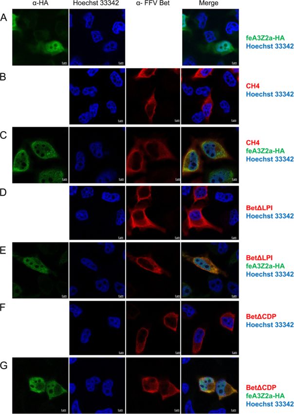
Slavkovic Lukic et al. Retrovirology 2013, 10:76                                                                       Page 2 of 20
http://www.retrovirology.com/content/10/1/76

viral and host-encoded proteins are under constant posi-          substitution of only a single amino acid in human A3G re-
tive selection to evade or strengthen, respectively, this         sults in a mutant that is no longer targeted by HIV Vif [42].
functional interaction [16]. Host-virus coevolution directly         The FV bet genes are expressed by all known exogenous
impacts the species-specificity of a given pathogen and is        FVs but are also present in the sloth endogenous FV
considered as one of the main factors preventing interspe-        (SloEFV) genome, which is at least 100 million years old
cies virus transmission [2].                                      [43]. Feline and prototype/primate/human FV (PFV) Bet
   APOBEC3 (A3) cytidine deaminases are potent anti-              have been recently shown to counteract defined A3 pro-
viral restriction factors. These host proteins deaminate          teins of feline and human/non-human primate (NHP) ori-
cytidine residues in single-stranded (ss) DNA intermedi-          gin, resp., and protect FV replication in A3-positive cells
ates generated during retroviral reverse transcription,           [15,44,45]. There is no sequence homology between Bet
creating hypermutated genomes with uridine residues [17].         and Vif and Bet does not contain the SOCS box motif re-
Although considered to deaminate only ssDNA, A3 pro-              quired for E3 ubiquitin ligase complex interactions [46]. In
teins are also active against double-stranded DNA viruses         contrast to lentiviral Vif, Bet does not induce A3 degrad-
such as papillomaviruses, probably due to a transient ex-         ation [15,44,47]. Bet is thought to directly bind and pos-
posure of ssDNA during replication and/or transcription           sibly sequester A3 proteins, preventing their incorporation
[18]. In addition, it has been shown that A3s deaminate ge-       into viral particles [15,44]. In line with this, Bet is expressed
nomes of hepadnaviruses, endogenous retroviruses, retroid         at high levels in infected cells and animals [48] which may
elements and possibly even cellular genes [8,19-22]. To           be a prerequisite for such a stoichiometric reaction.
restrict retroviral replication, A3 restriction factors must be      Bet is an accessory protein of FVs, viruses that display
incorporated into viral particles to edit the forming proviral    a complex genetic organization. Together with the essen-
DNA in the newly infected cell [23]. In contrast, A3s deam-       tial Bel1/Tas transactivator of both FV promoters, Bet is
inate foamy virus (FV) genomes not only in newly infected         mainly expressed from the internal promoter located in
cells, but also in virus-producing cells [15] as reverse tran-    the 3′end of env [49]. Bet is the product of a splicing event
scription of FVs may already occur in virus-producing cells       that fuses the 5′ domain of bel1 to the complete bel2 open
[24]. In addition, it has been shown that some human A3s          reading frame (ORF). All known FVs encode bet and, as
have deaminase-independent activities such as blocking            shown by genome localization, corresponding bet genes
reverse transcription and integration [25-31]. In fact, A3        are also present in endogenous FVs [43,50]. A protein
proteins with mutated catalytic sites have been shown to be       consisting of bel2 only is not expressed in vitro; whether it
active against HIV-1 [25,31,32].                                  is expressed in vivo is still unknown.
   The dynamic coevolution of host defences and virus                Although it has been shown that FFV Bet binds to all
counter-defence has resulted in a significant expansion           known feA3 proteins, the amino acid residues involved
of the A3 locus in higher mammals by distinct gene du-            in binding have yet to be identified. It has been shown,
plication events. In humans, this has led to seven dis-           however, that FFV Bet-MCS, with a mutation in the Bel2
tinct genes/proteins (A3A, B, C, DE, F, G, H), while only         domain of Bet, is incapable of counteracting feA3 and
four single-domain A3 genes are present in cats [33-37].          cannot replicate in A3-positive CrFK cells [15,47,51]. In
Additional complexity of the A3 repertoire in cats is             this FFV proviral genome, a multiple cloning site (MCS)
achieved by alternative and complex splicing events of            had been introduced inside the bel2 ORF, leading to the
these genes leading to one two-domain feline A3 (feA3)            alteration of E117L118L119 residues to ASVRRGP [51]. Des-
proteins; for details, see refs. [33,37,38].                      pite complete sequence conservation in the rest of Bet,
   Two classes of retroviral counter-defence proteins             Bet-MCS does not bind or inactivate feA3s, indicating that
against A3s have been described so far: lentivirus Vif            the mutated region is important for A3 counteraction
and FV Bet. Data on how other retroviruses replicate in           [15,47]. Not surprisingly, the replication of FFV-BBtr,
the presence of A3 restriction factors is scarce, but indi-       which contains only a truncated bet gene, is likewise
cate that these viruses have developed other means to             strongly impaired in the presence of A3s [15,47,51].
avoid A3 packaging [27,39].                                          FVs are retroviruses that differ from other viruses of this
   The mechanism of A3 inactivation by lentiviral (HIV) Vif       group in many aspects such as protein processing, mor-
is well characterized. Vif acts as an adaptor protein that        phogenesis, gene expression, and replication [49,52,53].
binds to both A3 and ubiquitin ligase complexes consisting        FVs have not yet been associated with any disease and are
of cullin-5, RING-box 1 and elongins B and C [40]. Thus,          considered apathogenic [54]. This feature makes FVs po-
Vif induces the ubiquitination and subsequent degradation         tential vectors for gene delivery and vaccination [53]. The
of A3 by the proteasome [40]. It has been shown that N-           known human FV isolates are results of zoonotic trans-
terminal regions of Vif bind to huA3G and huA3F while a           missions of diverse simian FVs to humans [55]. With re-
SOCS box motif mediates ubiquitin ligase complex binding          spect to virus-host coevolution, FV show a very strong
[40,41]. Binding of Vif to A3G is species-specific and            coevolution with their host and related species [56,57].
Slavkovic Lukic et al. Retrovirology 2013, 10:76                                                                                           Page 3 of 20
http://www.retrovirology.com/content/10/1/76

At current, FV research focuses mostly towards all as-                          [49]. To identify any possible conserved motifs amongst
pects of vector development, host-virus coevolution and                         different Bet proteins, we performed bioinformatics using
the potential of interspecies transmission to other hosts,                      the MEME program, which searches for repeated amino
including humans [54].                                                          acid patterns in given sequences [58]. Six conserved motifs,
  In this study, we analyzed the functional interaction                         all localized within Bel2, were identified in the Bet proteins
between FFV Bet and feA3 restriction factors as a model                         of bovine (BFV), equine (EFV), simian (SFV) FVs, FFV,
for the situation in humans and NHPs, since the feA3                            PFV and SloEFV (Figure 1). MEME detected four motifs
repertoire is less complex than that of these species.                          with standard settings (shown in black, motifs 1, 2, 3, 5);
Using bioinformatics and reverse genetics, we identified                        two additional motifs (4 and 6, marked in gray letters) were
conserved motifs in Bet and tested their importance. We                         identified by searching with only the bel2 sequences and
identified the bel2 sequence of Bet as the essential deter-                     using slightly modified parameters. The first five motifs are
minant for A3 inactivation. Moreover, this study shows                          also conserved in the predicted Bet sequence of SloEFV,
that nearly the entire FFV Bel2 domain is required for                          although the third and fifth motifs from SloEFV appear to
feA3 binding and inactivation, supporting the view that                         be shorter than the corresponding motifs in other Bet
Bet inactivates feA3 by creating strong complexes with                          proteins (Figure 1). In EFV Bet, the positions of the second
this restriction factor [44,45,47].                                             and the third motifs are swapped with respect to the corre-
                                                                                sponding motifs in the other FV Bet proteins.
Results
Bet contains six conserved motifs                                               The Bel2 part of FFV Bet is sufficient for feA3Z2b binding
All known FV Bet proteins consist of the N-terminus of                          and inactivation
Bel1 and the complete Bel2 sequence. However, sequence                          It was previously shown that FFV Bet binds and inacti-
homology between Bet proteins of different FVs is very low                      vates diverse feA3 proteins [15,47]. To determine the

  A            U3       R   U5                                                                        U3      R    U5
                                      gag                               env                  bel2

                                                        pol                       bel1

                                                                                             bel2

                                                                                   bet

  B

                            1                 2                     3                      4                   5                     6
  Bet         Bel1                                                              Bel2

        FFVBet (67)     LPILSPYVM   (121)   PGIGLVQI   (151)   DPDCDPLFCKLLCW   (258)    FKASHFDI   (272)   NSEERVSWA   (345)   FPKGTKVILPDGRKF
        BFVBet (70)     LPVLSPYPL   (125)   PGFGQVMI   (154)   DMCCGPPVCYGIFW   (234)    WKSLKFQA   (274)   SRQERVWWE   (334)   LPYGWSLMDPLGNRF
        EFVBet (76)     LPVISLFPI   (255)   PRIGQAGI   (159)   DYCCGPASCYTIVW   (239)    IKAIRWHK   (279)   GKKERVSWN   (337)   LPPGWCIVRPEGRTY
        PFVBet (131)    LPVVTPWPM   (190)   PPLGQVNI   (234)   DVWCSPSLCFKVIY   (321)    YKALQFHR   (361)   SNEERVWWN   (428)   LPYGWKVVTESGNDY
        SFVBet (134)    LPVVTPWPL   (193)   PPLGLVKI   (237)   RIACDPVYCVKIVW   (325)    VKALHFHR   (365)   SAEQRVWLI   (432)   LPRGASIVTADGNRY
        SloEFVBet(76)   IPATTLFPL   (128)   PGLGDVVM   (161)     KCNPKQCFAVYY   (253)    LKGQEFQR   (295)   NQWERVSQ

        Consensus       LPVVSPYPL           PGLGQVxI           DxCCDPxxCYKIVW            xKALHFHR           SxEERVWWN           LPYGWSIVTPDGNRY
        Sequence           LT F M            PI                    G    F V                                       S                  KV     R F
                           I W

 Figure 1 Bet proteins of different FVs contain six conserved motifs. (A) Genome organisation of FVs. The regulatory and accessory bel1 and
 bel2 genes are localized between env and the 3′LTR. The Bet protein is a product of a spliced transcript and consists of Bel1 and Bel2 parts.
 (B) Bioinformatics using the MEME program identified six conserved motifs localized in Bel2. Motifs are represented as gray boxes in the figure
 and the sequences are given below as well as the consensus sequence. Numbers in brackets indicate the position of the first amino acid of the
 motifs. In EFV Bet motifs 2 and 3 are found in reversed order. Motifs and residues that were not experimentally studied due to low degrees of
 conservation, such as motifs 4 and 6 and residues flanking motif 3, are represented in light shading and font, respectively. FFV, feline FV; BFV,
 bovine FV; EFV, equine FV; PFV, prototype/primate/human FV; SFV, simian FV; SloEFV, sloth endogenous FV.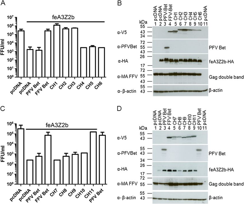
Slavkovic Lukic et al. Retrovirology 2013, 10:76                                                                                     Page 4 of 20
http://www.retrovirology.com/content/10/1/76

minimal FFV Bet sequence required for these two func-                        We tested the ability of the mutants to bind to and/or
tions, N-terminal Bet deletion mutants were constructed                    inactivate feA3Z2b, the feA3 with the highest restriction
(Figure 2A). Eukaryotic expression clone Bel2ORF en-                       potential against Bet-deficient FFV (FFV-BBtr) [33,47,51].
compasses the whole bel2 ORF, while Bel2ATG starts                         HEK293T cells were cotransfected with pCF-BBtr, the
with the first start codon inside bel2 which is not con-                   empty vector pcDNA, or pcfeA3Z2b-HA, and increasing
served among known FVs. Bel2ORF contains all six con-                      amounts of plasmids encoding either wt or mutant Bet
served motifs, while Bel2ATG and mutants BetΔN82                           (Figure 2B). Two days post transfection (d.p.t.), viral
and BetΔN92 lack the first motif.                                          infectivity was determined by titration using FeFab cells.

 Figure 2 Bet and Bel2ORF suppress feA3Z2b-mediated restriction. (A) Schematic representation of full-length Bet and N-terminal Bet
 deletion mutants. Dark and light gray boxes represent the different MEME motifs in Bet. Bet and Bel2ORF contain all six conserved motifs, while
 downstream N-terminal Bet deletion mutants do not contain the first conserved motif. Numbers indicate the first and the last amino acids of the
 deletion mutants. (B) HEK293T cells were cotransfected with 4 μg of pCF-BBtr, 0.8 μg pcfeA3Z2b-HA, and increasing amounts of wt Bet and Bet
 deletion mutant expression plasmids as indicated in the legend. pcDNA was used to compensate for different plasmid amounts. Viral titers were
 determined in triplicate using the FeFab titration assay and are presented as mean values of three measurements; error bars represent standard
 deviations. Labels below the columns indicate cotransfected clones. The line above the columns indicates the presence of feA3Z2b. The first
 column shows the viral titer in the absence of feA3Z2b, the second, in the presence of feA3Z2b. The other columns show the titer in the
 presence of feA3Z2b and the coresponding Bet clones, as indicated in the figure. 0.7 μg of Bet expression plasmid and 5.6 μg of Bel2ORF
 expression plasmid yielded similar levels of feA3Z2b counteraction. In both cases, the titer increased more than 1 log. The other N-terminal
 deletion mutants did not counteract feA3Z2b-mediated restriction. (C) 40 μg of protein of transfected HEK293T cells was used for
 immunoblotting. Bet proteins were detected with the FFV Bet- specific serum. Proper loading was confirmed by detecting β-actin (data not
 shown). (D) Densitometric analysis of the relative levels of Bet protein expression. Wt Bet at 5.6 μg of transfected DNA was set to 100%. The
 legend indicates the amount of expression plasmid used to obtain corresponding protein amounts.
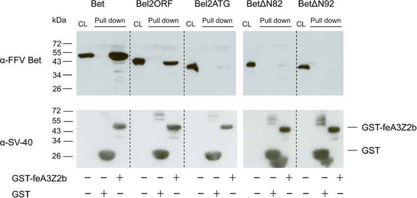
Slavkovic Lukic et al. Retrovirology 2013, 10:76                                                                                  Page 5 of 20
http://www.retrovirology.com/content/10/1/76

Figure 2B shows that feA3Z2b resulted in a 3-log decrease                proteins were precipitated using glutathione-coupled beads
of viral titer. Wt Bet efficiently counteracted this restric-            and detected by immunoblotting. In addition to wt Bet,
tion in a dose-dependent manner. The highest concentra-                  known to bind to feA3Z2b [15,47], Bel2ORF was efficiently
tion of Bel2ORF partially increased viral titer in the                   precipitated with feA3Z2b (Figure 3). Although low inten-
presence of feA3Z2b, while shorter deletion mutants did                  sity bands are present in the case of Bel2ATG and
not have any effect on the viral titer. The presence of Bet              BetΔN82, they were not reproducible and therefore con-
in cell lysates was confirmed by immunoblotting with an                  sidered to be unspecific. To confirm proper setup of the
FFV Bet-specific serum (Figure 2C). Levels of Bet deletion               assays, the presence of similar amounts of GST and GST-
mutants were lower relative to the level of wt Bet, indica-              feA3Z2b was confirmed with an SV40 tag-specific antibody
ting instability of the deletion mutants (Figure 2C and D).              (Figure 3 lower panel).
As inactivation of feA3Z2b by Bet is concentration-
dependent, the lower potential of Bel2ORF to counteract                  C-terminal Bet deletion mutants do not counteract
feA3Z2b may be due to the lower steady state of deletion                 feA3Z2b-mediated restriction
mutants. As shown in Figure 2 panels B and D, at 0.7 and                 To determine the minimal Bet sequence required for A3
5.6 μg transfected plasmid encoding Bet or Bel2ORF, re-                  inactivation, C-terminal deletion mutants were constructed
spectively, slightly increased amounts of Bel2ORF protein                (Additional file 2A). In transfected cells, the engineered
resulted in comparable restoration of infectivity of the bet-            proteins were present at only very low levels and thus,
deficient FFV genome in the presence feA3Z2b. Both wt                    functional studies were conducted in the presence and
and truncated Bet were detected with similar efficacy by                 absence of the proteasome inhibitor ALLN (Additional
the Bet antiserum and the V5 tag antibody, shown by                      file 2B). HEK293T cells were transfected with pCF-BBtr,
comparative immunoblotting (Additional file 1).                          feA3Z2b, and wt or mutant Bet constructs. Cultures were
  The ability of the truncated Bet proteins to bind feA3Z2b              treated for approximately 24 h with 25 μM of the prote-
was tested in pulldown assays with bacterially expressed                 asome inhibitor ALLN and tested for their ability to coun-
glutathione-S-transferase (GST) or GST-tagged feA3Z2b                    teract feA3Z2b restriction. Both V5-tagged and untagged
and lysates from HEK293T cells expressing wt or mutant                   full-length Bet but none of the C-terminal Bet deletion
Bet, as described previously [47]. Levels of N-terminal Bet              mutants were able to inactivate feA3Z2b in the presence of
deletion mutants were increased by supplementing the cell                ALLN or DMSO (solvent control) (Additional file 2B). The
culture medium with 8 mM sodium butyrate. Bound                          levels of wt Bet and wt Bet-V5 proteins did not change in

 Figure 3 Bet and Bel2ORF bind to feA3Z2b. Bet and Bet N-terminal deletion mutants were expressed in 293T cells and pulled down either
 with GST or GST-feA3Z2b. Immunoblot analysis was performed with Bet-specific serum (upper panel) and an SV40 tag-specific antibody to detect
 the GST and GST-feA3Z2b fusion proteins (lower panel). Cell lysates (CL) of transfected cells were analyzed in parallel to confirm proper
 expression of wt and mutant Bet proteins. In addition to wt Bet, Bel2ORF was pulled down with GST-feA3Z2b. Due to high protein levels, some
 bands have ‘shades’ (lower panel). Symbols below the image indicate the presence (+) or absence (−) of GST or GST-feA3.
Slavkovic Lukic et al. Retrovirology 2013, 10:76 Page 6 of 20
http://www.retrovirology.com/content/10/1/76
the presence of ALLN, while the level of C-terminal de- Two d.p.t., co-IP was performed with monoclonal anti-
letion mutants increased due to proteasome inhibition HA IgG. Precipitated proteins were detected by immuno-
(Additional file 2C). blotting. Unlike FFV Bet, PFV Bet did not
The feA3Z2b binding ability of the C-terminal deletion coimmunoprecipitate with feA3Z2b-HA (Figure 4B and C).
mutants was also tested in pulldown assays. None of the Immunoblotting with anti-HA IgG showed the presence of
C-terminal deletions bound to feA3Z2b, despite increased feA3Z2b-HA and an unspecific band of lower molecular
expression levels in transfected cells due to sodium butyr- mass (Figure 4B and C, lower panel). Incubation of cell ly-
ate addition (Additional file 2D). sates with beads only (without antibody, mock co-IP) did
not result in unspecific feA3Z2b-HA precipitation.
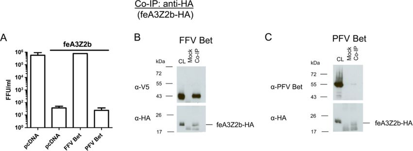
PFV Bet does not counteract or bind to feline A3Z2b
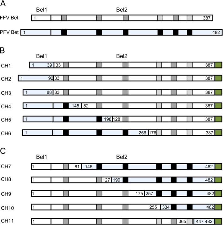
To circumvent the problems of the low stability of trun- Chimeric FFV-PFV Bet proteins containing almost the
cated FFV Bet proteins, we constructed chimeric FFV- entire FFV Bel2ORF bind and inactivate feA3Z2b
PFV proteins. Bet proteins from different FVs are known To stabilize FFV Bet deletion mutants, eleven chimeric
to counteract A3s from cognate species but not from dis- FFV-PFV Bet proteins, equivalent to the Bet deletion mu-
tantly related species. For instance, it has previously been tants described above, were constructed. In these chimeric
shown that FFV Bet does not bind to human A3s [15] and proteins, deleted parts of FFV Bet were substituted by the
that PFV Bet does not inactivate murine A3 [44,45]. We equivalent parts of PFV Bet (Figure 5).
therefore first determined whether PFV Bet binds or inac- Both PFV and FFV Bet consist of Bel1 and Bel2 regions
tivates feA3Z2b. HEK293T cells were cotransfected with (Figure 5A), though the sizes of Bel1 regions of FFV and
pCF-BBtr, pcfeA3Z2b-HA or pcDNA, and plasmids ex- PFV Bet differ greatly, between 31 and 88 amino acids. In
pressing FFV or PFV Bet. As shown in Figure 4A, viral ti- order to mimic this situation, three chimeric proteins
ters determined two d.p.i. decreased in the presence of containing the full FFV Bel2ORF and different lengths of
feA3Z2b and were completely rescued by FFV Bet while, PFV Bel1 were cloned as described in Methods. FFV-PFV
as anticipated, PFV Bet did not show anti-feA3 activity. CH1 contains the first N-terminal 39 amino acids of PFV
Since PFV Bet bound to glutathione beads incubated Bel1 fused to the FFV Bel2ORF and is still 8 amino acids
with GST (data not shown), the pulldown assay described larger than the FFV Bel1 part but maintains a predicted
above was not suitable for studying PFV Bet–feA3Z2b extended secondary structure in the PFV sequence (data
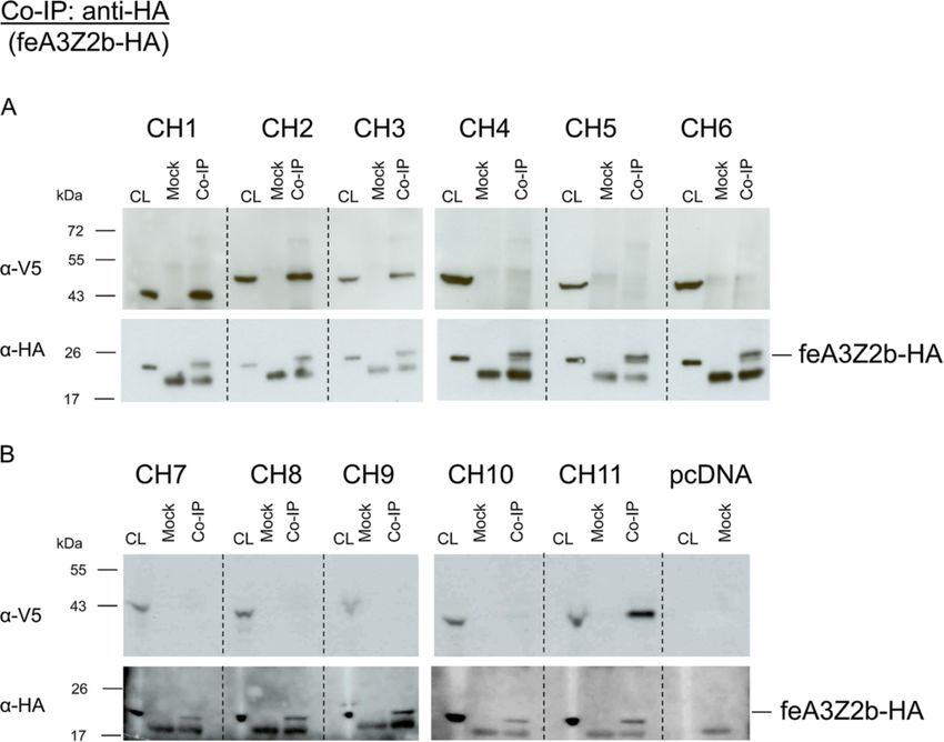
interactions, we thus used coimmunoprecipitation (co-IP) not shown). FFV-PFV CH2 contains PFV Bel1 and four
assays instead. HEK293T cells were cotransfected with additional residues from PFV Bel2 to avoid deletion of
pcfeA3Z2b-HA and plasmids expressing PFV or FFV Bet. a predicted alpha helix in PFV Bet (data not shown).
Figure 4 PFV Bet does not counteract feA3Z2b. (A) HEK293T cells were cotransfected with 4 μg of pCF-BBtr, 0.8 μg pcDNA or pfeA3Z2b, and
6 μg of plasmid expressing wt FFV or PFV Bet. Viral titers were determined two d.p.t. In the presence of feA3Z2b, FFV-BBtr titers decreased about
3 logs. Unlike FFV Bet, which completely rescues the viral titer, PFV Bet did not counteract feA3Z2b activity. The line above the graph indicates
the presence of feA3Z2b. For co-IP experiments, 293T cells were cotransfected with 6 μg of plasmid expressing feA3Z2b-HA and 12 μg of FFV Bet
(B) or PFV Bet (C). Two d.p.t., cell lysates (CL) were subjected to co-IP with monoclonal mouse anti-HA IgG or beads only (mock). Precipitated FFV
Bet but not PFV Bet proteins were detected by immunoblotting using monoclonal V5 tag-specific antibody and polyclonal hyperimmune sera
against PFV Bet, respectively. feA3Z2b-HA, along with a lower molecular weight band that also appeared in mock samples, was detected in all
samples using anti-HA IgG.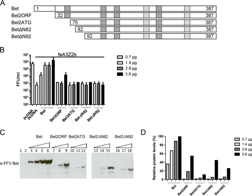
Slavkovic Lukic et al. Retrovirology 2013, 10:76                                                                                       Page 7 of 20
http://www.retrovirology.com/content/10/1/76

 Figure 5 Chimeric FFV/PFV Bet proteins. (A) Schematic structure of FFV Bet and PFV Bet (blue shading). Conserved motifs are represented as
 gray boxes for FFV Bet and black boxes for PFV Bet. The Bel1part consists of 31 amino acids in FFV Bet and 88 amino acids in PFV Bet. FFV Bet
 contains 387 amino acids and PFV Bet, 482 amino acids. (B) FFV-PFV CH1 to CH6 are chimeras of N-terminal parts of PFV Bet and central and
 C-terminal parts of FFV Bet. (C) FFV-PFV CH7 to 11 are chimeras of N-terminal parts of FFV Bet and central and C-terminal parts of PFV Bet. White
 shading represents FFV Bet-derived sequences and blue shading, PFV Bet. Numbers indicate positions of the first and the last amino acid derived
 from PFV and FFV Bet. For example, CH1 is composed of the first 39 residues of PFV Bet and residues 33–378 of FFV Bet. A green box at the end
 of each protein represents the V5 tag.

FFV-PFV CH3 contains the entire PFV Bel1 part fused                         different PFV Bel1 sequences, fully restored FFV titers to
directly to FFV Bel2ORF. FFV-PFV CH4 to 6 contain                           levels similar to wt Bet (Figure 6A). Bet chimeras 4 to 6,
shorter C-terminal FFV Bet and longer N-terminal PFV                        with N-terminal FFV Bel2 sequences replaced by those
Bet segments, while chimeric proteins CH7 to 11 are equiva-                 from PFV, were non-functional and did not suppress
lent to FFV Bet C-terminal deletion mutants (Figure 5C).                    feA3Z2b restriction.
All chimeric proteins were V5-tagged to facilitate proper                     FFV-PFV chimeric proteins CH7 to 11, carrying C-
protein detection (Figure 5B and C).                                        terminal PFV sequences of decreasing size were charac-
  To determine whether chimeric proteins CH1 to 6 in-                       terized as described above. Only FFV-PFV Bet CH11,
activate feA3Z2b, HEK293T cells were cotransfected with                     carrying a short and obviously non-conserved C-terminal
pCF-BBtr, pcfeA3Z2b-HA, and plasmids encoding either                        PFV Bet fragment, was functionally active against feA3Z2b.
wt PFV Bet, wt FFV Bet, or one of the chimeric FFV-PFV                        The presence of proteins in cell lysates was confirmed
Bet CH1 to CH6 proteins. Viral titers determined 2 d.p.t.                   by immunoblotting with the V5 antibody. Figure 6B and
are shown in Figure 6A. As shown before, PFV Bet did                        6D show that the levels of chimeric proteins were com-
not counteract feA3Z2b activity, while chimeric proteins                    parable or slightly lower than those of wt FFV Bet. The
CH1 to 3, containing the full-length FFV Bel2ORF and                        PFV Bet antiserum efficiently detected full-length Bet but
Slavkovic Lukic et al. Retrovirology 2013, 10:76                                                                                     Page 8 of 20
http://www.retrovirology.com/content/10/1/76

 Figure 6 The Bel1 domain and the C-terminal 22 amino acids of FFVBet can be replaced by PFV Bet sequences without loss of
 function. HEK293T cells were cotransfected with 4 μg pCF-BBtr, 0.8 μg pcDNA or pfeA3Z2b, and 6 μg of plasmid expressing wt FFV Bet, PFV Bet
 or chimera, as indicated in the picture. (A, C) Two d.p.t., titration was performed and viral titers are represented as mean values of three
 independent experiments. Error bars represent standard deviations. In the presence of feA3Z2b, the FFV-BBtr titer decreased more than two logs.
 Unlike FFV Bet, which completely rescued titers, PFV Bet did not counteract feA3Z2b activity. Chimera CH1, 2, 3, and 11 completely restored the
 viral titer in the presence of feA3Z2b (indicated by the line above the graphs). (B, D) FFV Bet and chimeric Bet were detected with anti-V5 IgG
 while PFV Bet was detected with a PFV Bet-specific serum. Anti-HA IgG was used for feA3Z2b-HA detection, rabbit anti-matrix serum for Gag
 detection, and anti-β-actin IgG as a loading control.

not chimera CH1 to CH5 and CH10 and CH11. In con-                          Identification of critical Bet residues by alanine scanning
trast, CH6 to CH9 were detectable only upon extended                       mutagenesis of conserved motifs
exposure (not shown).                                                      To identify functionally important amino acids in the
  Binding of chimeric FFV-PFV Bet proteins to feA3Z2b                      conserved Bet motifs, site-directed alanine scanning mu-
was studied by co-IPs as described above. As shown in                      tagenesis was performed. Amino acids in the conserved
Figure 7A and B, only FFV-PFV chimeric proteins CH1,                       motifs or in flanking sequences were substituted by ala-
2, 3 and 11 coimmunoprecipitated with feA3Z2b-HA.                          nine as described in Methods and indicated in Figure 8,
Importantly, unspecific binding to the beads (mock co-                     resulting in 20 FFV Bet mutants carrying one to three
IP) was not observed. Proper set up of the assay was                       amino acid substitutions each. Although these Bet mu-
confirmed by detection of feA3Z2b-HA (Figure 7, la-                        tants are not classical deletion mutants, Δ symbols were
belled α-HA in parts A and B).                                             used to facilitate labelling of these substitutions.
Slavkovic Lukic et al. Retrovirology 2013, 10:76                                                                                     Page 9 of 20
http://www.retrovirology.com/content/10/1/76

 Figure 7 FFV-PFV CH1, CH2, CH3 and CH11 bind to feA3Z2b-HA. 293T cells were cotransfected with 6 μg of plasmid expressing feA3Z2b-HA
 and 12 μg of one of the chimeric protein expression plasmids (A, N-terminal chimera CH1 to CH6; B, C-terminal chimera CH7 to CH11) or pcDNA,
 as indicated in the picture. Two d.p.t., cell lysates (CL) were subjected to co-immunoprecipitation (co-IP) with monoclonal anti-HA IgG or beads
 only (mock). Precipitated proteins were detected by immunoblotting. feA3Z2b-HA was detected with an anti-HA monoclonal antibody in each
 sample, as expected. A lower molecular weight band that was also detected is considered unspecific, as it also appeared in mock samples.
 Hatched lines mark empty lanes used to separate individual experiments. Chimeric proteins were detected with anti-V5 IgG. In addition to FFV
 Bet, CH1, 2, 3, and 11 were coimmunoprecipitated by feA3Z2b.

  The second conserved FFV Bet motif is localized dir-                     first, second, or third motifs were detrimental to both Bet
ectly behind the mutated part of FFV Bet-MCS, in which                     functions, while all 3 amino acid replacement mutants of
an MCS had been inserted inside bel2 [51]. The MCS                         the fifth motif displayed a wt phenotype with respect to
introduction resulted in the substitution of three and                     feA3 inactivation and binding. Moreover, FFV Bet func-
addition of four amino acids. Although the rest of Bet                     tion was not impaired by single amino acid changes, while
remained unchanged, Bet-MCS did not have anti-feA3                         double amino acid mutations in motif 2 resulted in loss of
activity and did not bind to feA3 [15,47,51]. We there-                    function (Figure 8 and Additional file 3). In fact, BetΔV
fore proceeded to more carefully characterize the second                   and BetΔP were both functionally active but the double
conserved motif together with these flanking sequences.                    mutant BetΔVP, in which both amino acids are replaced
  Functional feA3 inactivation and binding studies by                      by alanine, was completely incapable of binding and
GST-feA3Z2b pulldown were performed to determine                           counteracting feA3Z2b. As indicated in Figure 8 and
whether mutant Bet proteins inactivate and/or bind                         shown in the Additional file 4, all mutants capable of
feA3Z2b, as described above. The results are summarized                    functionally inactivating feA3Z2b also bound to this re-
in Figure 8 and provided in Additional files 3 and 4. As                   striction factor in pulldown assays. In addition, BetΔLTM
indicated in Figure 8, most mutations in or close to the                   where mutations are outside of motif 2 and BetΔI where
Slavkovic Lukic et al. Retrovirology 2013, 10:76 Page 10 of 20
http://www.retrovirology.com/content/10/1/76
Figure 8 Site-directed mutagenesis of the first, second, third and fifth conserved Bet motif. FFV Bet contains 387 amino acids and consists
of Bel1 and Bel2 sequences as indicated. Conserved motifs are represented as gray boxes and conserved motif 1, 2, 3 and 5 are underlined.
Motifs 4 and 6 were not analyzed. Alanine residues that substitute the original residues are marked in bold. Numbers in brackets indicate the
positions of the first amino acid in a given sequence. Mutations that did not impair Bet functions are marked with (+) and bolded names. Grey
(+) signs indicates incomplete restoration of FFV titers by certain mutants.
only the last amino acid of motif 2 were exchanged bound without any indication of a Bet- or feA3Z2b-mediated
to feA3Z2b and partially inactivated this restriction factor. relocalization of their corresponding binding partner.
This attenuated phenotype was reproducibly detectable This (apparent) colocalization pattern indicates that the wt
(data not shown). Moreover, BetΔGPL induced only a Bet–A3 complexes do not aggregate in specific regions of
minor increase of the FFV titer (Additional file 3C) and the cell but are rather evenly distributed throughout the
it was not pulled down with GST-feA3Z2b (Additional cytoplasm. Nonfunctional mutant Bet proteins (BetΔLPI,
file 4), which may indicate the low sensitivity of the pull BetΔCDP, FFV/PFV Bet CH4) were, similarly to wt Bet,
down assay used. However, immunoblotting data show also evenly distributed in the cytoplasm. In cells co-
that protein expression levels of the Bet mutants were expressing these nonfunctional Bet mutants and feA3,
comparable to or slightly lower than wt Bet (Additional there are regions of strong colocalization. However,
file 3D, E and F). Therefore, the lack of feA3Z2b coun- colocalization detected by confocal microscopy does not
teraction by non-functional Bet mutants (in particular prove the interaction of the two proteins but indicates
BetΔGPL) was not simply the consequence of low pro- that the two proteins have the same spatial occupancy
tein amounts. within the cytoplasm. Based on these data, we conclude
In general, the binding properties of Bet substitution that the lack of feA3 inactivation by Bet mutants is not
mutants were found to correlate well with their ability to the consequence of the physical distance between both
inactivate feA3Z2b, although there were differences in proteins.
the potential of individual mutants to inactivate feA3Z2b Since feA3Z2a had been shown in independent studies
(Additional file 4). to localize to the nucleus and the cytoplasm, we analyzed
the colocalization of this feA3 isoform with wt Bet and
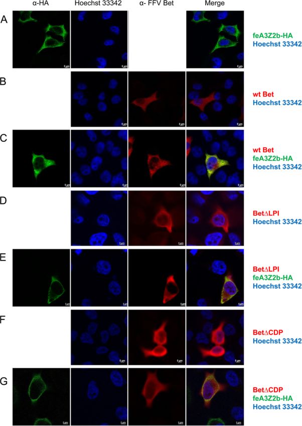
Mutant and wt Bet are localized in the cytoplasm and some of the Bet mutants (Figures 10 and 11). In cells
colocalize with feA3Z2b and feA3Z2a transfected only with V5-tagged wt Bet, Bet is predomi-
Subcellular localization of mutant Bet proteins and their nantly found in the cytoplasm (Figure 10B), while in cells
potential colocalization with feA3s were studied by in- transfected with feA3Z2a, feA3Z2a localizes in both the
direct immunofluorescence (IIF) of paraformaldehyde- nucleus and in the cytoplasm (Figure 10A). In most cells
fixed HeLa cells using confocal microscopy. Bet was that express both wt Bet and feA3Z2a, these two pro-
detected with an FFV Bel2-specific serum and is shown teins consistently colocalize in both compartments,
in red, while feA3Z2b was labeled with monoclonal meaning that Bet is recruited to the nucleus by feA3Z2a
anti-HA IgG and is shown in green (Figure 9). Both (Figure 10C). In addition, in some cells expressing both
feA3Z2b-HA and wt Bet predominantly localize to the proteins, feA3Z2a and wt Bet predominantly localized
cytoplasm and, in cells coexpressing both proteins, in the cytoplasm (Figure 10D). Functional FFV-PFV Bet
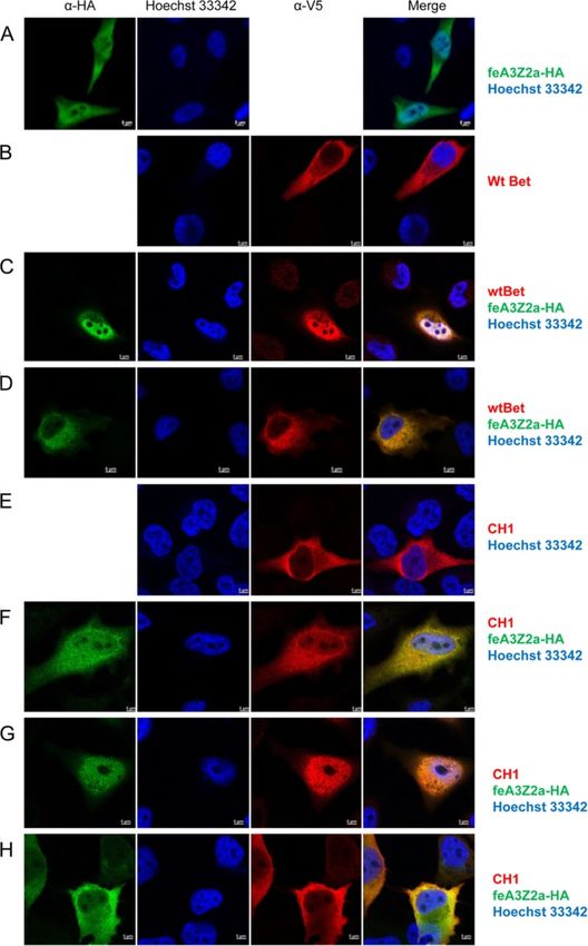
there is strong colocalization throughout the cytoplasm CH1 showed similar colocalization with feA3Z2a as wtSlavkovic Lukic et al. Retrovirology 2013, 10:76 Page 11 of 20
http://www.retrovirology.com/content/10/1/76
Figure 9 Colocalization of feA3Z2b with wt and mutant Bet proteins. HeLa cells were transfected with plasmids encoding HA-tagged
feA3Z2b-HA (A) and/or Bet expression plasmids (B – G) as indicated on the right side of the image. feA3Z2b-HA was detected with HA tag-
specific antibody (green), wt and mutated FFV Bet were detected by an FFV Bet-specific serum (red) and nuclei were stained with Hoechst 33342
(blue). The merge of feA3Z2b and Bet staining is shown in the right-hand column. feA3Z2b (A, C, E, G), wt (B, C) and mutant Bet proteins
(D – G) are predominantly localized in the cytoplasm. Inserted bars represent 5 μm.
Bet (Figure 10E-H). However, the non-functional Bet proteins that counteract A3 restriction factors have
mutants tested were not recruited to the nucleus by been described so far in detail: lentiviral Vif and FV Bet
feA3Z2a (Figure 11). [8,15,44]. These two proteins counteract the A3 activity
in different ways. While Vif induces proteasomal deg-
Discussion radation of A3s [40,59-62], FV Bet does not change the
A3 proteins are cellular restriction factors that deaminate steady state level of A3s [15,44,47]. It is assumed that
cytidine residues in ssDNA [8,19,34]. Two retroviral Bet counteracts the activity of A3s by forming stableSlavkovic Lukic et al. Retrovirology 2013, 10:76 Page 12 of 20 http://www.retrovirology.com/content/10/1/76 Figure 10 Colocalization of feA3Z2a with functional Bet proteins. HeLa cells were transfected with a plasmid encoding HA-tagged feA3Z2a- HA (A) and/or Bet expression plasmids (wt Bet or FFV-PFV Bet CH1, B – H), as indicated on the right. feA3Z2a-HA was detected with HA tag- specific antibody (green), FFV Bet was detected by an anti-V5 tag antibody (red) and nuclei were stained with Hoechst 33342 (blue). The merge of feA3Z2a and Bet staining is shown in the right-hand column. Wt Bet and FFV-PFV CH1 are localized in the cytoplasm (B, E) while feA3Z2a is localized both in the cytoplasm and the nucleus (A). In some cells coexpressing feA3Z2a and functional Bet proteins, wt Bet and CH1 relocalize to the nucleus (C, F, G). In other cells, feA3Z2a is recruited to the cytoplasm (D, H). Inserted bars represent 5 μm.
Slavkovic Lukic et al. Retrovirology 2013, 10:76                                                                                     Page 13 of 20
http://www.retrovirology.com/content/10/1/76

 Figure 11 Colocalization of feA3Z2a with nonfunctional Bet proteins. HeLa cells were transfected with a plasmid encoding HA-tagged
 feA3Z2a-HA (A) and/or Bet expression plasmids (B – F), as indicated on the right. feA3Z2a-HA was detected with HA tag-specific antibody
 (green), Bet proteins were detected by an FFV Bet-specific serum (red) and nuclei were stained with Hoechst 33342 (blue). The merge of the
 feA3Z2a and Bet staining is shown in the right-hand column. Nonfunctional Bet proteins are localized in the cytoplasm (B, D, F) while feA3Z2a is
 localized both in the cytoplasm and the nucleus (A). In cells coexpressing feA3Z2a and a nonfunctional Bet mutant, there is no relocalization of
 proteins (C, E, G). Inserted bars represent 5 μm.

complexes with A3s, thus preventing them from being                        but it is not essential for the A3-inactivating function of
incorporated into virions [15,45,47].                                      Bet and can be exchanged between PFV and FFV Bet
  Our results show that the Bel2 part of FFV Bet protein                   proteins without detectable loss of function. Alterna-
is essential for species-specific feA3Z2b inactivation and                 tively, bel1 RNA sequences or the splice junction may
that this domain contains the feA3Z2b interaction site.                    stabilize the transcripts or induce enhanced cytosolic
In contrast, the Bel1 domain increases protein stability                   protein biosynthesis. There is no sequence homology
Slavkovic Lukic et al. Retrovirology 2013, 10:76                                                                 Page 14 of 20
http://www.retrovirology.com/content/10/1/76

between PFV and FFV Bel1 (data not shown), leading to           all conserved bel2 motifs. This mode of expression may
the question of whether the presence of any protein             be an efficient way to generate high amounts of Bet,
domain appended to the N-terminus of Bel2ORF would              since the internal promoter (IP) is very active upon full
lead to the same increase of protein stability. However,        transactivation [49]. In addition, Bel1/Tas and Bet ex-
since higher amounts of Bel2ORF than wt Bet are                 pression via alternative splicing of a single transcription
needed for the same level of A3 counteraction (Figure 2),       unit may be an important regulatory mechanism of FV
it might be that the Bel1 domain contributes to anti-A3         replication. Through regulated splicing of IP transcripts,
activity, for instance, by interacting with some cellular       early expression of Bel1/Tas may initiate and maintain
effector protein.                                               high levels of viral gene expression, while later expres-
   In line with functional and binding studies, bioinfor-       sion of Bet protects the viral genome from A3 editing
matics showed that the Bel2 parts of all FVs contain            during and after particle formation and reverse tran-
conserved motifs. Without knowing the protein struc-            scription. Expression of Bet during the late phase of
ture of Bet, it is difficult to evaluate whether these motifs   replication and progeny virus production may therefore
are localized on the protein surface and may thus               be the result of FV evolution to avoid A3 incorporation
directly comprise the feA3 binding site. By molecular           into virus particles during replication in A3-positive
modelling using the Robetta full-chain protein structure        cells. It may be even more important for FVs due to the
prediction server [63,64], we obtained hypothetical three-      fact that reverse transcription may already occur in the
dimensional models of Bet. In the most probable model           virus-producing cells [52].
according to QMEAN and ProQservers [65,66], conserved             For all tested mutants, binding to feA3 correlated with
motifs 1, 2 and 3, which have been shown here to be abso-       the inactivation of this restriction factor. The tight asso-
lutely essential for Bet function, are localized on one side    ciation between feA3 binding and inactivation by FFV
of the Bet protein surface (data not shown). Provided that      Bet supports the hypothesis that Bet inactivates A3s by
this model reflects the real Bet structure, the feA3Z2b         creating strong complexes [15,45]. This mechanism of
binding domain may consist of a combination of these            A3 inactivation requires high amounts of Bet proteins
motifs. Experimental structure determination of Bet, for        and may be the reason for high expression levels of Bet
instance by crystallography, is required to solve these is-     in FV-infected cells and animals [48,67,68]. In line with
sues. For instance, it is currently not known whether EFV       this, results of this study show that, for efficient inactiva-
Bet has a similar structure to the other Bet proteins, since    tion of A3s, the amount of Bet must be at a sufficiently
the positions of the second and third conserved motifs are      high level; small to modest amounts of Bet do not or
inverted relative to the other known FVs Bet proteins.          only partially counteract A3 restriction.
   Considering the strong coevolution of FVs with their           Hypothetically, binding of Bet to feA3Z2b could mask
hosts including Bet and A3 proteins [2,56], the binding         the Gag binding site on feA3Z2b, thus preventing its in-
sites to the species-specific A3 forms may not be con-          corporation into viral particles. Results presented here
served between Bet proteins of different FVs. Alterna-          indicate that almost the whole Bel2ORF is important for
tively, one can imagine that the A3 binding sites may be        feA3Z2b binding and suggest that Bel2ORF may wrap
under highly dynamic, positive evolution. Thus, the A3          around feA3Z2b and prevent its interaction with Gag. Al-
binding site may be highly divergent and their primary          ternatively, binding of Bet to A3s could block transport of
sequence/structure is likely not maintained. Following          A3 into viral particles or interfere with some cellular fac-
this idea, conserved motifs of Bet may be more import-          tor(s) [15] crucial for A3 packaging into viral particles.
ant for maintaining the tertiary structure of Bet and for         Confocal microscopy indicates that both Bet and
proper presentation of the species-specific binding site.       feA3Z2b are apparently uniformly distributed in the
The results of this study support both models, since most       cytoplasm and thus do not allow detection of protein
mutations in these motifs and in flanking sequences im-         relocalization upon complex formation. In contrast,
paired the function of FFV Bet.                                 feA3Z2a, which also restricts FFV and is inhibited by
   There is no start codon that would enable expression         Bet binding [33,47], is also present in the nucleus. Wt
of the full-length motif 1 to 6-containing Bel2 protein         Bet and functional Bet mutants colocalize to the nucleus
and there is no conserved ATG start coding in the 5′            in the presence of this restriction factor. Surprisingly, in
end of bel2. The first ATG in FFV bel2 is, for instance,        some cells, feA3Z2a seems to retarget Bet which is
already downstream of the first essential motif, and such       somewhat counterintuitive since the smaller protein
a Bel2-only protein (Bel2ATG) does not have the cap-            dictates sub-cellular localization of its larger binding
acity to protect against A3 editing, as shown here. Since       partner. Why not all cells show this phenotype remains
the bet splice acceptor is located at the 5′end of the bel2     to be determined.
ORF of all FVs examined [49], Bet expression via a                Bet is a highly expressed viral protein and a diagnostic
spliced bel1-bel2 fusion transcript allows expression of        marker for FV infection [51,67]. All known FVs express
Slavkovic Lukic et al. Retrovirology 2013, 10:76                                                           Page 15 of 20
http://www.retrovirology.com/content/10/1/76

Bet, strongly supporting the importance of this protein     Plasmids and DNA transfection
for efficient FV replication. Sequences corresponding to    pFeFV-BBtr and pcfeA3 plasmids have been already
bet/bel2, as determined by localization and sequence        described [33,51,69]. The bacterial expression vector for
homology, have also been identified in the SloEFV, active   glutathione-S-transferase (GST)-tagged feA3Z2b has been
more than 100 million years ago [43], indicating that Bet   described recently [47].
is an ancient gene. In addition, a sequence correspond-
ing to bel2 is even present in coelacanth endogenous FV     Molecular cloning of FFV Bet mutants
[50], though it contains only the first conserved motif     To construct Bet N-terminal deletion mutants, the cor-
(data not shown). This may be a result of the accumula-     responding bel2 sequences were amplified by Bel2ORF,
tion of mutations from the lack of selective pressure       Bel2ATG, FFVBetΔN82, or FFVBetΔN92 sense primers
leading to truncations of the parental bel2 ORF.            (containing a HindIII site) and FFVbel2as antisense pri-
   Although Bet and Vif counteract A3 restriction fac-      mer using plasmid pBC-FFV-Bet as template [51]. All
tors, their localization in viral genomes and their fun-    reactions were performed with Phusion™ High-Fidelity
damentally different mechanisms of A3 counteraction         DNA Polymerase (NEB, Germany). Blunt-ended prod-
suggest that these two proteins may have evolved inde-      ucts were digested with HindIII and inserted into
pendently from each other. The ancestral protein(s) of      HindIII/SmaI-digested pBC12CMV.
Vif and Bet may have been cellular A3 binding pro-             To generate a V5-tagged Bet, bet was amplified with
teins with regulatory functions. In this model, there       Bet sense and Bet-V5 antisense primers as above and
was no need for high level of vif expression in lentivi-    cloned into HindIII/SmaI-digested pBC12CMV. To intro-
ruses, since Vif acts as an adaptor for a catalytic         duce V5 into Bet N-terminal deletion mutants, AccI/XmaI
degradation of A3 proteins. In contrast, the internal       fragments of pBC-Bel2ORF, pBC-Bel2ATG, pBC-BetΔN82
promoter (IP) of FVs provides high levels of Bet for ef-    and pBC-BetΔN92 were replaced by the corresponding
ficient inactivation of A3s simply by binding. Alterna-     fragment of pBC-FFVBetV5.
tively, FVs might have developed an IP to increase the         V5-tagged C-terminal Bet deletion mutants were con-
expression of Bet, since Bet does not recruit the cellu-    structed by amplification of corresponding bet sequences
lar degradation machinery.                                  with Bet sense and one of three different antisense
                                                            primers (dC1-V5, dC2-V5 or dC3-V5, Table 1) with a V5
                                                            epitope. PCR products were cloned into HindIII/SmaI-
Conclusions
                                                            digested pBC12CMV.
The Bel2 domain of FFV Bet contains conserved motifs
                                                               To construct chimeric Bet proteins, corresponding
and is essential for inactivation of feA3s. Although the
                                                            parts of PFV and FFV bet were fused in-frame by fusion
Bel1 part is not directly involved in binding and inacti-
                                                            PCR [70]. Corresponding parts of each gene were ampli-
vation of these restriction factors, it is important for
                                                            fied in individual PCR reactions. For instance, for clon-
expression of full-length Bel2ORF and protein stability.
                                                            ing of FFV-PFV CH1, PCR1 with chimera-s1 and ch1-as
The absolute correlation between binding and inactiva-
                                                            using pBC-FFV-Bet as template and PCR2 with ch1-s2
tion of feA3Z2b by Bet mutants suggests that Bet
                                                            and Bet V5 as primers and pBC-PFV-Bet as template were
inactivates A3s simply by creating strong complexes.
                                                            performed as given above. Finally, the amplicons were fused
Such a strong feA3Z2b-Bet binding is most probably a
                                                            in PCR3 using chimera-s1 and Bet-V5 antisense primers.
result of a very long coevolution of these two proteins.
                                                            Blunt-ended PCR products were digested with HindIII and
Considering that sloth and coelacanth FVs contain
                                                            cloned into HindIII/SmaI-digested pBC12CMV. To con-
sequences that correspond to bet [43,50] and its con-
                                                            struct FFV-PFV CH7 to CH11, FFV Bet was amplified with
served motifs, it is possible that this coevolution be-
                                                            generic ch-sense1-bspe1 and a mutant-specific as1 primer.
tween Bet and A3s or their progenitors extends far back
                                                            PFV Bet was amplified with the corresponding s2 primer
in vertebrate evolution.
                                                            and generic chimera-as primer. Products of both PCRs
                                                            were fused in a third PCR reaction with ch-sense1-bspeI
Methods                                                     and chimera-as primers. Final products were cloned into
Cell culture and virological methods                        BspEI/SmaI digested pBC-Bet-V5.
HEK293T and FeFab cells were propagated as described           Alanine scanning mutagenesis of the second conserved
before [69]. HEK293T cells were seeded in 6 cm or           motif of FFV Bet and flanking residues was performed
10 cm dishes and transfected using a modified calcium       by amplifying bet between the unique HindIII and KpnI
phosphate method [69]. FFV titers were determined by a      sites using Bet-sense and one of three antisense primers
β-galactosidase assay using FFV-FAB (FeFab) cells grown     (BetAlaRI, BetAlaRII, BetAlaRIII for ΔELL, ΔLTR and
in 96-well plates [69]. FFV-containing supernatants were    ΔLTM, respectively). Products were cloned into HindIII/
serially diluted 1:5, titrations were done in triplicate.   KpnI-digested pBC-Bet. Residues downstream of KpnI
Slavkovic Lukic et al. Retrovirology 2013, 10:76 Page 16 of 20 http://www.retrovirology.com/content/10/1/76 Table 1 Primers used for cloning and site-directed mutagenesis Name Sequence (5′-3′ direction) Cloning of N-terminal deletion mutants of FFV Bet FFVbel2ORF ATCCCAAGCTTGCCACCATGGTCGGAAAGAATCCGGAAC (HindIII) FFVbel2ATG ATCCCAAGCTTGCCACCATGGCTTGGGACAACCCTC (HindIII) FFVBetΔN82 ATATCCCAAGCTTGCCACCATGGTGGTCACACGTCTGGTG (HindIII) FFVBetΔN92 ATATCCCAAGCTTGCCACCATGGAATCATGGAAGAAGTATC (HindIII) FFVbel2as GGGTCACTCGAGCTATTCAGAGTCAGATGACTC (SmaI) Cloning of V5 tagged FFV Bet and C-terminal deletion mutants of Bet Bet-sense CTCCCCTCGAAGCTTTCTGGGATATGTAAAACC (HindIII) dC1-V5 GGGTCAGGTGCTGTCCAGGCCCAGCAGGGGGTTGGGGATGGGCTTGCCATCATCAGCTTGTGCTCTCC (SmaI) dC2-V5 GGGTCAGGTGCTGTCCAGGCCCAGCAGGGGGTTGGGGATGGGCTTGCCCAGCAGAGAGTATTCTCCTC (SmaI) dC3-V5 GGGTCAGGTGCTGTCCAGGCCCAGCAGGGGGTTGGGGATGGGCTTGCCTCTTCCATCAGGAAGTATCAC (SmaI) Bet-V5 antisense GGGTCAGGTGCTGTCCAGGCCCAGCAGGGGGTTGGGGATGGGCTTGCCTTCAGAGTCAGATGACTCAG (SmaI) Alanine scanning mutgenesis BetAlaRI ATGCAAGATGATGGTACCGCAGCGGCTCTAGTTAGCATAGTCAAATC (KpnI) BetAlaRII ATGCAAGATGATGGTACCAACAATTCGGCAGCGGCCATAGTCAAATCCCTCTC (KpnI) BetAlaRIII ATGCAAGATGATGGTACCACAATTCTCTAGTTAGGGCAGCGGCATCCCTCTCCCCACAATC (KpnI) BetAlaFI ATGCAAGATGATGGTACCAGCCGCTGCCCTGGTACAAATCGCCGCTAC (KpnI) BetAlaFII ATGCAAGATGATGGTACCAGGAATAGGCGCCGCTGCCATCGCCGCTACACTTAC (KpnI) BetAlaFIII ATGCAAGATGATGGTACCAGGAATAGGCCTGGTACAAGCCGCCGCTACACTTACTAAAACC (KpnI) BetAlaR2 AGGTAAAAGATTCCTATGTCTACGCACAATC (AccI) R-XhoI ATGCAAGATGATGGTACCAACAATTCTCGAGTTAGCATAGTC (KpnI) ForwardVP 5′ATGCAAGATGATCTCGAGAATTGTTGGCTGCCGGAATAGGCCTGGTAC (XhoI) ForwardV 5′ATGCAAGATGATCTCGAGAATTGTTGGCCCCAGGAATAGGCCTGGTAC (XhoI) ForwardP ATGCAAGATGATCTCGAGAATTGTTGGTAGCCGGAATAGGCCTGGTAC (XhoI) dGPL-AS1 TGGAAGGGCAGCGGCGGGAACATCCTGCTTCTTG dGPL-S2 GCCGCTGCCCTTCCAATTCTGAGTCCG dLPI-AS1 ACTCAGGGCAGCGGCGAGTGGGCCGGGAACATCC dLPI-S2 GCCGCTGCCCTGAGTCCGTATGTAATGG dLSP-AS1 TACATAGGCAGCGGCAATTGGAAGGAGTGG dLSP-S2 GCCGCTGCCTATGTAATGGCTTGGGACAACC dYVM-AS1 CCAAGCGGCAGCGGCCGGACTCAGAATTGGAAG dYVM-S2 GCCGCTGCCGCTTGGGACAACCCTCAG dDPD-AS1 ATCACAGGCAGCGGCGGTTCTAGAACCTGTAATAC dDPD-S2 GCCGCTGCCTGTGATCCTTTGTTCTGTAAG dCDP-AS1 GAACAAGGCAGCGGCATCTGGGTCGGTTCTAGAAC dCDP-S2 GCCGCTGCCTTGTTCTGTAAGTTGTTATGC dLFC-AS1 CAACTTGGCAGCGGCAGGATCACAATCTGGGTCGGTTC dLFC-S2 GCCGCTGCCAAGTTGTTATGCTGGAAAC dKLL-AS1 CCAGCAGGCAGCGGCACAGAACAAAGGATCACAATC dKLL-S2 GCCGCTGCCTGCTGGAAACAAAATATAC dNSE-AS1 CCTCTCGGCAGCGGCCCCAGAGGCACTTCCAAATATG dNSE-S2 GCCGCTGCCGAGAGGGTGTCATGGGCCAAAG dERV-AS1 CCATGAGGCAGCGGCCTCACTGTTCCCAGAGGCAC dERV-S2 GCCGCTGCCTCATGGGCCAAAGAGAATTC
Slavkovic Lukic et al. Retrovirology 2013, 10:76                                                                                                    Page 17 of 20
http://www.retrovirology.com/content/10/1/76

Table 1 Primers used for cloning and site-directed mutagenesis (Continued)
dSW-AS1                                       CTCTTTGGCAGCGGCCACCCTCTCCTCACTGTTCC
dSW-S2                                        GCCGCTGCCAAAGAGAATTCTCACAGAG
AS2-XmaI                                      AGTGTAAGTTCACCCGGGTCACTCGAGCTATTCAGAGTCAGATGACTC (SmaI, XmaI)
Cloning of chimeric FFV/PFV Bet fusion proteins
chimera-s1                                    ATGCAAGATGATAAGCTTTAGCTGCAGCAACAAAG (HindIII)
ch1-as1                                       GTGTTCCGGATTCTTTCCAGCAATAGTCAGCTCTCC
ch1-s2                                        GGAGAGCTGACTATTGCTGGAAAGAATCCGGAACAC
ch2-as1                                       GTGTTCCGGATTCTTTCCCTTCTGAGCAATCATTTC
ch2-s2                                        GAAATGATTGCTCAGAAGGGAAAGAATCCGGAACAC
ch4-as1                                       CCACGTGTGACCACGTTTGCATAGTGATCCTGGCTC
ch4-s2                                        GAGCCAGGATCACTATGCAAACGTGGTCACACGTGG
ch5-as1                                       GTAAGTGTAGCGGCGATCTGAATGTTCACCTGACC
ch5-s2                                        GGTCAGGTGAACATTCAGATCGCCGCTACACTTAC
ch6-as1                                       CAGGCACCATTCTTCTAGTTGCTTTTGGCCCATTGC
ch6-s2                                        GCAATGGGCCAAAAGCAACTAGAAGAATGGTGCCTG
ch3-as1                                       GTGTTCCGGATTCTTTCCCATTTCCTCTGGTGTGGGGATCC
ch3-s2                                        GGATCCCCACACCAGAGGAAATGGGAAAGAATCCGGAACAC
ch-sense1- bspei                              ATGCAAGATGATTCCGGAACACCCAAGACGGATC (BspEI)
ch7-as1                                       GATTCCAAAGAGGGTTGGCTGAGGGTTGTCCCAAGC
ch7-s2                                        GCTTGGGACAACCCTCAGCCAACCCTCTTTGGAATC
ch8-as1                                       CTGATAATTCTTATAAAATTGTACCAGGCCTATTCC
ch8-s2                                        GGAATAGGCCTGGTACAATTTTATAAGAATTATCAG
ch9-as1                                       GTCTGCACAGCCAGGTTTTTTGGTTACACTCTCTAGGGTC
ch9-s2                                        GACCCTAGAGAGTGTAACCAAAAAACCTGGCTGTGCAGAC
ch10-as1                                      AATCCTAGGATTGGTGAAGACTCTGGGATACAGGGAAGC
ch10-s2                                       GCTTCCCTGTATCCCAGAGTCTTCACCAATCCTAGGATT
ch11-as1                                      CTGTCAATGTTCTGATCTTCTCAGGATCACAGGCTATG
ch1-s2                                        CATAGCCTGTGATCCTGAGAAGATCAGAACATTGACAG
chimera-as                                    GGGTCAGGTGCTGTCCAGGCCCAGCAGGGGGTTGGGGATGGGCTTGCCGAAGGGTCCATCTGAGTC (SmaI)
All primer sequences are in the 5′ to 3′ orientation. Sequences in italics correspond to restriction enzyme recognition sites that were used for cloning; the names
of the enzymes are given in brackets. Underlined sequences mark the mutations introduced. Sequences encoding the V5 tag are given in bold-face letters.

were mutated by amplifying bet from KpnI to AccI using                              site (e. g. fusion PCR with Bet-sense, dGPL-AS1, dGPL-S2,
sense primers (BetAlaFI, BetAlaFII and BetAlaFIII for                               AS-XmaI primers for ΔGPL). The two fragments were
ΔGIG, ΔLVQ and ΔI, respectively) and BetAlaR2 as gen-                               fused by PCR using Bet-sense and AS-XmaI. The final
eral antisense primer. Products were cloned into pBC-                               product was cloned into pBC12-CMV using HindIII
Bet with KpnI and AccI. Using V, P, or VP sense primers,                            and XmaI.
the BetAlaR antisense primer, and Bet-XhoI (in which an
XhoI site was silently introduced with Bet sense and R-                             Coimmunoprecipitation (co-IP) and immunoblotting
XhoI antisense primer) as template, corresponding parts                             To study the interaction between proteins expressed in
of Bet were amplified and the indicated codons ex-                                  eukaryotic cells, HEK293T cells were seeded in 10 cm
changed by alanine codons. PCR products were cloned                                 dishes and transfected with 6 μg of pcfeA3Z2b-HA and
into pBC-Bet-XhoI using XhoI and AccI.                                              12 μg of PFV Bet, FFV Bet, or chimeric FFV/PFV Bet
  Residues in the first, third and fifth conserved motifs                           expression plasmid. Two d.p.t., cells were lysed in TLB
were exchanged by fusion PCR. Bet was amplified with                                (20 mM Tris, pH 7.4, 137 mM NaCl, 10% glycerol, 2 mM
Bet-sense and AS1 antisense primers. The second PCR                                 EDTA, 1% Triton X-100 and protease inhibitor). Lysates
was performed with S2 sense primers and AS-XmaI pri-                                were cleared by 5 min centrifugation at 500 × g. 100 μl of
mer, covering the bet stop codon and carrying an XmaI                               the cell lysate was incubated with monoclonal mouse anti-
You can also read