Neuroregenerative Medicine
←
→
Page content transcription
If your browser does not render page correctly, please read the page content below
Neuroregenerative Medicine

         MAYO CLINIC | Neuroregenerative Medicine   1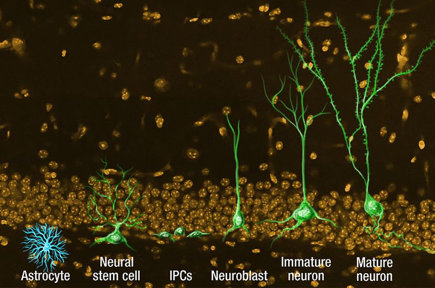
People are living longer than ever before, in part due to medical advances that
                                   have saved more people from life-threatening diseases, injuries and congenital
                                   conditions. But as people live longer, they’re more likely to develop age-related
                                   conditions or chronic diseases. After the onset of most chronic diseases or injuries,
                                   the underlying damage remains, and patients are left to manage the symptoms.
                                   Many of these patients, having so-called degenerative diseases, are particularly well-
                                   suited to treatment with regenerative medicine solutions.
                                       At Mayo Clinic, we have embraced regenerative medicine as a strategic
                                   investment in the future of health care. The quest for innovative solutions to meet the
                                   changing needs of our patients is instilled in our mission and values, as we seek
                                   to treat the underlying causes of symptoms and find innovative and affordable
                                   health care solutions.
                                       The Mayo Clinic Center for Regenerative Medicine consists of several focus areas,
                                   including neuroregeneration, each encompassing the discovery, development and
                                   delivery of next-generation patient care. In the center’s Neuroregeneration Program,
                                   clinicians, scientists, engineers and other specialists take a multidisciplinary
                                   integrative approach to neuroregeneration for a number of devastating neurological
                                   conditions. The research is multifaceted, ranging from basic science discovery to
                                   clinical applications. Throughout these pages, you will find examples of several
                                   team-based innovations in the field of neuroscience and the practice of neurology.
                                       As director of Mayo Clinic’s Center for Regenerative Medicine, I see firsthand the
                                   remarkable collaboration between physicians and scientists across disciplines. In
                                   this new era of medicine, we are working together to use advanced regenerative
                                   technologies and therapies to essentially teach the body to heal from within. And
                                   we are unrelenting in our mission to target the root cause of disease and offer the
                                   prospect of cures — all with the goal of advancing the science and improving health
                                   care across our patients’ lifespans.

                                   Andre Terzic, M.D., Ph.D.
                                   Michael S. and Mary Sue Shannon Director, Mayo Clinic Center for Regenerative Medicine
                                   Marriott Family Professor in Cardiovascular Diseases Research

                                   Science Writer: Barbara J. Toman

2   MAYO CLINIC | Neuroregenerative Medicine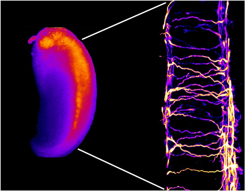
Harnessing the Body’s Healing Power
The complex, delicate structures comprising the nervous           Medicine at Mayo Clinic are pursuing a new strategy:
system — the brain, spinal cord and nerves — are                  regenerative treatments aimed at fully healing damage to
susceptible to injury as varied as trauma, cancer and             the nervous system. In this model of care, neurological
neurodegenerative diseases. Unfortunately, because of             damage is not simply managed but repaired through
the complexity of the brain and spinal cord, very little          growth of new nerves. Mayo’s multi-faceted research,
spontaneous nerve regeneration or healing occurs.                 ranging from basic scientific discovery to clinical
Injuries to the spinal cord and peripheral nerves are             application, offers hope for people who have neurological
often permanent and incapacitating. Although signs and            injury that today is beyond repair.
symptoms of Alzheimer’s disease, Parkinson’s disease                  “Neuroregeneration is the next step in the evolution
and amyotrophic lateral sclerosis (ALS) can be                    of neurology and neurosurgery,” says Fredric B. Meyer,
somewhat managed, these diseases cause progressive                M.D., chair of the Department of Neurologic Surgery at
deterioration that the body cannot repair.                        Mayo Clinic in Rochester, Minn. “Historically, neuro-
   Physicians and scientists at the Center for Regenerative       surgery has its origins in excising lesions to help patients.

Fluorescent spinal neurons in the developing aquatic frog (Xenopus) embryo.

                                                                                MAYO CLINIC | Neuroregenerative Medicine     3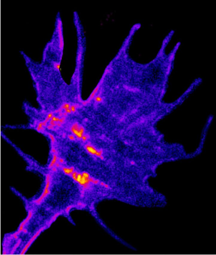
Recent technological advances have led to the develop-        Scarisbrick, Ph.D., leads a laboratory team at Mayo in
ment of minimally invasive procedures or device               Minnesota that is uncovering the physiologic changes
implantation. Our goal now is to harness the regenerative     unleashed by damage to the brain and spinal cord that
capacity of neurogenesis to improve treatment options for     create this inhibitory microenvironment. The work
brain, spine and nerve diseases and injuries.”                potentially may lead to new therapies for neurological
                                                              conditions such as multiple sclerosis and spinal cord
Discovering how neurons grow                                  injury (page 8).
The highly specialized cells that make up the nervous
system were once thought to be incapable of growing.          Clinical trials
Now they are known to be able to remodel and to               Early but unsuccessful efforts at neuroregeneration
possess some ability to self-heal. At Mayo, paradigms of      focused on creating antibodies to counteract inhibitory
neural regeneration take many forms — from                    factors. Mayo took a different approach. Led by Anthony
re-engineering a patient’s stem cells to promote neural       J. Windebank, M.D., a neurologist and molecular neuro-
growth to using electronic devices to bypass severed          scientist at Mayo in Minnesota, researchers pioneered
nerves and restore limb function after spinal injury.         ways of re-engineering mesenchymal stem cells to
    Fundamental to these efforts is a basic understanding     enhance their ability to produce growth factors. Dr.
of how neurons grow and, equally importantly, become          Windebank’s theory was that re-engineered stem cells
functionally integrated into the nervous system. In the       that were reimplanted in the patient’s body could serve
developing nervous system, slender axons projecting           as delivery vehicles for neural growth factors, promoting
from neurons extend and retract, “sniffing out” the           nerve regeneration. Based on this work, he and
molecules needed to help the nerve growth cones reach         colleagues are currently conducting a clinical safety trial
and connect with their targets in surrounding tissue. A       of stem cell therapy to treat ALS (page 11).
complex array of molecular guidance cues tell them                Less common than ALS, multiple system atrophy
whether to continue on their path or to turn left or right.   (MSA) is a devastating neurodegenerative disease that is
    John R. Henley, Ph.D., a Mayo Clinic molecular            generally fatal within three years of diagnosis. Phillip A.
neuroscientist and director of the Developmental and          Low, M.D., a neurologist at Mayo in Minnesota and
Regenerative Neurobiology Laboratory at Mayo in               founder of Mayo’s autonomic testing laboratory, is
Minnesota, has devoted his career to identifying and          leading a clinical safety trial of stem cell therapy to treat
manipulating these guidance cues. Growing nerve tips          MSA (page 12).
face a hostile environment, in which substances that              Yet another application of this stem cell work involves
may work to prevent random spinal growths also inhibit        injuries to the peripheral nervous system. These injuries
neural growth. Dr. Henley’s work is directed at priming       are particularly challenging: inhibitory factors can
nerves to grow in this hostile environment by altering        overwhelm the nerve tips’ efforts to grow across the gap
not only the external molecular environment but also          created by injury. To provide a friendlier environment,
the intrinsic state of a neuron — something previously        Mayo scientists have developed a synthetic tube that
not thought possible. Building on this work, Dr. Henley’s     eventually could house growth factors to promote
research team has succeeded in restoring function to lab      neuroregeneration. The tube will soon enter a clinical
animals with injured spines (page 5).                         safety trial (page 13).
    Working on a similar scale, Mi Hyeon Jang, Ph.D.,
a molecular biologist at Mayo in Minnesota, is investi-       Additional approaches
gating neurogenesis in the hippocampus, the portion           In addition to therapies to promote neuron growth, Mayo
of the brain associated with memory. Among other              is pursuing a vascular approach to neuroregeneration.
discoveries, Dr. Jang’s team has shown that exercise          Guojun Bu, Ph.D., and Leonard Petrucelli, Ph.D., both
increases neurogenesis. Her work has important                neuroscientists at Mayo Clinic in Jacksonville, Fla., are
implications for treating a range of neuropathological        investigating whether patient-derived stem cells can
conditions, including epilepsy, Alzheimer’s disease and       improve blood flow to the brain and thus slow or prevent
brain injury (page 7).                                        neurodegenerative disease (page 15).
    Other Mayo researchers are working to overcome the            Mayo’s work with stem cells also has potential to
hostile environment that inhibits nerve repair. Isobel A.     eventually improve treatment of brain cancer. Stem cells

4           MAYO CLINIC | Neuroregenerative Medicine
collected, processed and stored by Mayo’s Regenerative          From scientific discovery to patient care
Medicine Biotrust are facilitating research into why            Mayo’s commitment to translational medicine — moving
various types of brain tumors behave differently (page 17).     scientific discoveries from the lab bench to the patient’s
    Beyond stem cells, Mayo researchers are also exploring      bedside — makes it ideally suited to lead efforts in the
device-based regenerative therapies. Led by neurologist         growing field of regenerative medicine. Interaction
Ryan J. Uitti, M.D., and neurosurgeon Robert E. Wharen Jr.,     between lab scientists and clinicians is a constant at Mayo.
M.D., a pilot study at Mayo in Florida is investigating             “There are few barriers to translating scientific
whether deep brain stimulation can lessen cognitive             laboratory discovery to the clinical treatment of
decline in patients with Parkinson’s disease (page 17). At      patients,” Dr. Meyer says. “Because we’re a premier
Mayo in Minnesota, Kendall H. Lee, M.D., Ph.D., is              clinical institution working in an environment of
conducting laboratory tests that eventually may result in       collaboration, we can rapidly and safely apply novel
electronic devices that can bypass a severed spinal cord        treatments to help patients.
and restore function to patients with paralysis (page 19).          “Neuroregeneration is in its infancy,” Dr. Meyer adds.
                                                                “But its potential for medicine and mankind is huge.”

Axonal Growth and Spinal Recovery
One of the star players in Mayo Clinic’s neurogenesis
work measures all of 1.5 inches long. The zebrafish, a
tropical freshwater member of the minnow family, is
being used as an animal model to investigate how nerve
regeneration might be improved after spinal cord injury.
Preliminary findings indicate significant promise.                     Dendrites                    Axon
    Researchers at the Mayo Clinic Developmental and
Regenerative Neurobiology Laboratory in Rochester,
Minn., have developed a therapy that, in laboratory tests,
is given to zebrafish with spinal cord injuries.
Behavioral testing after treatment has demonstrated
dramatic functional recovery in the injured fish.                                      Soma
    “After injury, the fish don’t swim very well or very far.
After treatment, they have recovered,” says John R. Henley,     Figure 1. Axon projecting from the cell body (soma). Dendrites
                                                                conduct electrochemical stimulation received from other nerve
Ph.D., a molecular neuroscientist who directs the lab.          cells. The axon conducts electrical impulses away from the soma.

Steering decisions for neurons
In the developing nervous system, axons extend and              cells are grown (Figure 2) in defined cultures. “We apply
retract from neurons, seeking the molecules needed to           different environmental factors that are thought to
help the nerve growth cones connect with their targets          influence these steering decisions in vivo, and then
in surrounding tissue (Figure 1). A complex array of            measure these turning responses in a defined,
molecular cues guides axonal growth.                            quantitative assay,” Dr. Henley says.
    “A lot of the extracellular cues have been understood           The system also must overcome factors that inhibit
for a while,” Dr. Henley notes. “But the signals                neural growth. In research published in the July 2010
functioning inside the cell are the black box. We’ve been       issue of Nature Neuroscience, Dr. Henley and colleagues
studying these basic mechanisms underlying how nerve            discovered that a major component of myelin prevents
cells grow and are guided to synaptic targets.”                 growing nerve tips from producing the adhesions needed
    The work involves in vitro systems in which nerve           to form anchoring sites with surrounding tissue and the

                                                                               MAYO CLINIC | Neuroregenerative Medicine        5
a camera that records one frame per
millisecond, the researchers document the
response of zebrafish to a vibration against
their dish. “Within 10 to 12 milliseconds, the
fish start to bend. After another 10 to 12
milliseconds, the fish are in a very sharp C
bend and then swim away,” Dr. Henley says.
A fish that is injured and then left for
three days can only wiggle faintly. Its
reaction time is twice that of the control
fish. “But when we do our treatment, the
fish is able to bend back into its classic C,
and the latency period comes back down,”
Dr. Henley says. “We can measure this
quantitatively, in terms of the angle,
velocity and latency time.”
Dr. Henley’s lab team is investigating
other factors thought to be barriers to
neuroregeneration as well as working to
develop a more sophisticated animal model.
As always at Mayo, the ultimate goal is to
translate these molecular discoveries into
treatments that can help people recover
from spinal cord injury.
Cancer research
Figure 2. Nascent endocytic vesicles in the nerve growth cone labeled with a A related topic under investigation in the
fluorescent membrane dye. developmental and regenerative neurobiology
lab is the molecular mechanisms that control
traction to move forward. These inhibitory factors are the growth of glioblastoma multiforme (GBM), the most
released into cerebral spinal fluid after spinal injury. common and aggressive primary brain tumor. GBM cells
“At least one growth inhibitory factor actually causes a can rapidly infiltrate the brain, making GBM incurable by
loss of these adhesions at the surface of the nerve cell,” surgical resection. The invasion pattern follows selective
Dr. Henley says. “Our model of treatment is that either by tracts, suggesting that molecular guidance cues may
activating these adhesion proteins and/or by blocking regulate GBM cell migration.
this removal process, we might preserve the capacity of By investigating the specific biological cues that
an axon to be stimulated to grow even in the presence of control the invasiveness of these brain tumors, Dr. Henley
this inhibitory environment.” and colleagues hope to contribute to patient treatment.
Just as understanding the steering decisions of axons may
Measuring fish behavior ultimately lead to therapy for spinal cord injury so, too,
In 2011, the research moved from the petri dish to the might deciphering GBM migration cues pave the way for
zebrafish. “The zebrafish is a powerful in vivo model new cancer treatments.
because it’s at least somewhat permissive for regeneration.
Many, but not all, neurons will regenerate after injury,” For more information
Dr. Henley says. “It’s also a high-throughput system. We Hines JH, et al. Asymmetric endocytosis and remodeling
can measure functional recovery three days after injury.” of beta1-integrin adhesions during growth cone
Those measurements are facilitated by stereotypical chemorepulsion by MAG. Nature Neuroscience. 2010;13:829.
zebrafish behavior. When threatened, zebrafish quickly
curl into a sharp C shape before swimming away. Using
6 MAYO CLINIC | Neuroregenerative MedicineHippocampal Neurogenesis and Cognitive Function
Adult neurogenesis plays a critical role in brain neurogenesis and related behavioral responses that are
plasticity, learning and memory. “Newborn” neurons altered in neuropathological conditions. “We want to
have the ability to become functionally integrated into investigate the function of neurogenesis and the role of
the hippocampus, the part of the brain associated with neurogenesis in pathological diseases,” Dr. Jang says.
memory, and to contribute to specific cognitive functions
(Figure). Understanding the molecular mechanisms Integrating newborn neurons
of adult neurogenesis has important implications for One specific area involves exploring the intricate
a range of neuropathological conditions, including cellular and molecular mechanisms that regulate the
epilepsy, Alzheimer’s disease and brain injury. production, maturation and integration of new neurons
Researchers in the laboratory of Mi Hyeon Jang, in the circuitry. Using a retroviral system and unique
Ph.D., a molecular biologist at Mayo Clinic in Rochester, staining techniques to label neuronal progenitors,
Minn., are working to utilize the regenerative capacity of Dr. Jang and her colleagues were able to characterize
adult neurogenesis with the ultimate goal of improving the developmental stages of newborn neurons from a
treatment options for patients. “Much progress has been neural stem cell to a fully formed neuron with synapse
made in this area in the last 10 years,” Dr. Jang says. formation, a process that takes two to four weeks.
The central focus of her research team’s efforts “With retroviruses we can characterize morphology
is to increase understanding of the molecular at the single-cell level,” Dr. Jang notes.
targets involved in regulation of adult hippocampal The process of neurogenesis is modulated by various
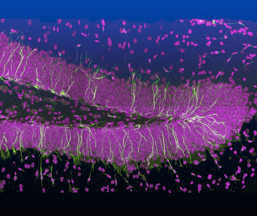
Figure. Model of the sequential process of adult neurogenesis within the dentate gyrus of the hippocampus. The neural stem cell can
remain quiescent, self-renew or proliferate to give rise to the astrocyte or intermediate progenitor cells (IPCs). The IPC differentiates into
a neuroblast, which migrates and integrates itself into the inner granule layer as an immature neuron. It then differentiates into the mature
granule cell, which extends its axon and dendrites — now comprising spines — to achieve functional integration.
MAYO CLINIC | Neuroregenerative Medicine 7physiological and pathological stimuli, with the niche the researchers identified a significant association of
mechanisms largely unknown. However, in a study three genetic variations in human sFRP3 with early
published in the Feb. 7, 2013, issue of Cell Stem Cell, Dr. responses to antidepressants in a clinical cohort. The
Jang and colleagues identified secreted frizzled-related results of both studies suggest that targeting sFRP3
protein 3 (sFRP3) as a neuronal activity-regulated factor may provide a novel therapeutic approach for treatment
that helps control multiple steps of adult hippocampal of depression.
neurogenesis, including the activation of quiescent adult Dr. Jang is also intrigued by factors that appear to
neural stem cells as well as the maturation, dendritic stimulate neurogenesis — such as exercise and electro-
development and spine formation of newborn dentate shock therapy — and those that apparently inhibit
granule neurons. Neuronal activity decreases the neurogenesis, such as stress and aging. She notes that
expression of sFRP3, a naturally secreted inhibitor of Wnt deep brain stimulation (DBS), which appears to stimulate
proteins, whose signaling is necessary for neurogenesis. neurogenesis, offers interesting therapeutic possibilities.
“DBS can target specific areas of the brain, which may
Potential for treating depression have applications for distinct pathologies,” Dr. Jang says.
A key strand of Mayo’s neurogenesis research is the role Translation of basic science to treatment for patients is
of adult neurogenesis in causing neurodevelopmental the overriding goal. “One of the benefits of Mayo is that
disorders. Dr. Jang’s discoveries concerning sFRP3 as a molecular biologist, I can closely collaborate with
suggest that antidepressants may ease patients’ symptoms physicians,” Dr. Jang says. “As a lab scientist, I want to
by promoting neurogenesis. In a letter to the editor in the contribute to the level of clinical care.”
September 2013 issue of Molecular Psychiatry, Dr. Jang
noted that electroconvulsive stimulation significantly For more information
reduces sFRP3 levels in the mouse hippocampus. Jang M-H, et al. Secreted frizzle-related protein 3 regulates
Chronic treatment with the antidepressants fluoxetine activity-dependent adult hippocampal neurogenesis. Cell
or imipramine also significantly suppresses sFRP3 Stem Cell. 2013;12:215.
expression in the mouse hippocampus.
In experiments with lab mice, Dr. Jang and colleagues Jang M-H, et al. Secreted frizzle-related protein 3 (sFRP3)
found that sFRP3 knockout mice exhibited less depression- regulates antidepressant responses in mice and humans.
like behaviors than did normal mice. In a parallel study, Molecular Psychiatry. 2013;18:957.
Preventing Environmental Damage
in the Central Nervous System
One of the most significant challenges to repairing the “environmental damage” occurs. These discoveries have
central nervous system (CNS) is the hostile environment the potential to spawn new therapies for a range of
unleashed when the brain or spinal cord suffers damage. neurological conditions, particularly those involving
In cases of injury and disease, the precisely controlled damage to myelin, including multiple sclerosis (MS) and
microenvironment of the CNS is greatly disrupted, spinal cord injury.
contributing directly to tissue damage and a lack of “Our goal is to bring the CNS microenvironment back
significant functional repair and nerve regeneration. under control,” says Isobel A. Scarisbrick, Ph.D., director of
Myelin, the sheath that insulates and protects axons, is the CNS Injury and Neurorehabilitation Laboratory at
particularly vulnerable. Mayo in Minnesota. “We hope to make this environment
A promising avenue of research at Mayo Clinic in one that is conducive to innate repair and that facilitates
Rochester, Minn., is uncovering precisely how this therapeutic interventions such as stem cell therapies.”
8 MAYO CLINIC | Neuroregenerative Medicinemyelin. They also signal to the
oligodendrocytes to stop making
myelin,” Dr. Scarisbrick says. “The
oligodendrocyte precursor cells are
ready; they want to remyelinate the
denuded axons. But they are
inhibited from doing so.”
Fortunately, protease activated
receptors are known to be potent
drug targets. “Because of their
location, partly inside and partly
outside the cell, they are highly
‘druggable,’” Dr. Scarisbrick says.
“It’s difficult to target all the
multiple proteases. But we can go
after the receptors. They may be
a common pathway to block the
multiple effects of the proteases.”
Figure 1. Photomicrographs showing immunoreactivity for three unique kallikreins in Success in vitro
a case of human vertebral fracture dislocation resulting in tetraplegia. High levels of
kallikrein staining, as indicated by arrows, were seen in association with swollen axons In a study published in the Sep-
and retraction bulbs visualized in the adjacent section stained using the Bielschowsky tember 2013 issue of Glia, the Mayo
silver method (BIELS).
researchers reported that over-
activating the PAR1 receptors in
Cascade of damage mouse oligodendrocyte cultures caused the cells to stop
When the CNS is injured by trauma or disease, a expressing myelin genes. When a PAR1 inhibitor was
cascade of secondary damage ensues. Vascular, cellular added to the culture, the cells resumed myelin production.
and chemical responses to the injury include tissue “We have shown that blocking the kallikrein-PAR
inflammation, reduced blood flow and scar formation. pathway can result in remyelination — in a dish,” Dr.
Demyelination occurs on injured axons, slowing the Scarisbrick notes. “This is very exciting, but now we
conduction of nerve impulses and stripping axons of want to translate that work into animal models of MS
protection against further damage. as well as spinal cord injury. Eventually, of course, the
These changes are brought about in part by multiple goal is to translate this into therapies for patient use. If
proteases, notably the kallikrein family of enzymes we continue the progress we have had until now, we’re
(Figure 1). Kallikreins are increasingly associated with very optimistic that could happen in a few years.”
neurological conditions including Alzheimer’s disease, Indeed, in a study published in the November 2013
Parkinson’s disease and frontotemporal dementia. Dr. issue of the Journal of Neuropathology & Experimental
Scarisbrick’s lab discovered one member of the Neurology, Dr. Scarisbrick and colleagues demonstrated
kallikrein family, called kallikrein 6. in postmortem human tissue the contribution of
In laboratory studies, the Mayo researchers further kallikreins to the pathophysiology of spinal cord injury.
found that kallikreins wreak neurologic havoc through Dr. Scarisbrick notes that in cases of disease and
a limited set of receptors, known as protease activated spinal cord injury, there is likely to be an early window
receptors (PAR). Specifically, aberrant activation of these of opportunity for treatment aimed at halting environ-
receptors promotes damage to the axonal wires that mental damage in the CNS. But lab tests indicate that it
conduct electrical impulses across the brain and spinal may also be possible to promote remyelination at sites
cord as well as to oligodendrocytes, the cells that where damage occurred previously.
produce myelin (Figure 2). “It would make a lot of sense to target these proteases
The kallikrein-PAR axis in fact delivers a one-two early,” Dr. Scarisbrick says. “But we hope there is an
punch. “Some of these enzymes not only degrade opportunity to target the same protease-PAR axis and
MAYO CLINIC | Neuroregenerative Medicine 9Figure 2. Lab cultures showing myelin-producing oligodendrocytes (stained green) and astrocytes (stained red) derived from a
mouse brain. The culture system allows researchers to determine the impact of altered levels of microenvironmental factors, such as
kallikreins, at sites of CNS injury. Elevations in KLK6 were shown to impede the ability of oligodendrocytes to extend processes and to
wrap and myelinate axons.
promote repair in patients with chronic MS lesions and For more information
spinal cord injury.” Radulovic M, et al. Kallikrein cascades in traumatic
Although myelin is associated most commonly with spinal cord injury: In vitro evidence for roles in
MS and spinal cord injury, myelin regeneration has axonopathy and neuron degeneration. Journal of
therapeutic applications for other neurological conditions. Neuropathology & Experimental Neurology. 2013;72:1072.
“The biology that we uncover in an MS lesion looks very
similar to what we see in spinal cord injury, and is likely Burda JE, et al. Critical role for PAR1 in kallikrein
to play parallel roles in stroke and other CNS disorders, 6-mediated oligodendrogliopathy. Glia. 2013;61:1456.
as well as glioblastoma multiforme,” Dr. Scarisbrick says.
“For conditions as complicated as MS and spinal Yoon H, et al. Kallikrein 6 signals through PAR1 and
injury and stroke, I don’t think there ever will be a single PAR2 to promote neuron injury and exacerbate glutamate
magic bullet,” she adds. “But remyelination is going to be neurotoxicity. Journal of Neurochemistry. 2013;127:283.
a very important piece of the puzzle.”
Scarisbrick IA, et al. Kallikrein 6 regulates early CNS
demyelination in a viral model of multiple sclerosis.
Brain Pathology. 2012;22:709.
10 MAYO CLINIC | Neuroregenerative MedicineStem Cell Treatment for ALS
Also called Lou Gehrig’s disease, amyotrophic lateral Each patient’s stem cells are then injected into his or
sclerosis (ALS) is a neurodegenerative disease that affects her central nervous system via spinal tap. The stem cells
motor neurons, causing muscle weakness, paralysis and spread through the patient’s cerebral spinal fluid and
eventually death. Today, there is no effective treatment. along the membranes that protect the spinal cord and
Therapies that showed promise in preclinical models nerve routes. “The stem cells are then in a position to
have performed poorly in clinical trials. secrete the growth factors that protect nerve cells,” Dr.
Researchers at Mayo Clinic in Rochester, Minn., are Windebank says.
testing a different approach: a stem cell-based therapy for The study participants have been divided into groups,
ALS. A clinical trial of the therapy, which uses adipose- with each group receiving an escalating dose of stem
derived mesenchymal stem cells from the patient’s own cells. “So far, we’ve seen no adverse effects related to the
body to promote neuron regeneration, is underway. treatment. That really is the main goal of this safety
“A very powerful tool in the regenerative medicine study,” Dr. Windebank says. The escalating dosages may
kit is the ability to take stem cells from a person’s skin provide some indication of efficacy, but those results
or adipose (fat) tissue, and, turn them into stem cells won’t be available for another one to two years.
that can do anything. This is opening up a whole new
field of healing possibilities,” says Anthony J. Windebank, Additional trial planned
M.D., a neurologist and molecular neuroscientist at Mayo Dr. Windebank’s lab already is planning a second study, in
in Minnesota who leads the research. collaboration with colleagues at Hadassah Medical Center
The clinical safety trial, which began in 2012, involves in Israel. That trial will involve modifying stem cells in the
25 patients with ALS. A biopsy of adipose tissue is taken laboratory to enhance their ability to produce nerve
from each participant. In the lab, stem cells are isolated growth factors before they are injected back into the
from the adipose tissue. Certain stem cells are then patient’s spine. Eventually, the researchers plan to
selected, based on their ability to produce growth factors. investigate modifying genetic factors within the stem cells
The selected cells are expanded into billions of stem cells, to further promote neurogenesis in patients with ALS.
a process that takes several weeks. “Our goal is to preserve function and, we hope, arrest
“At the end of all this, the population of cells from or slow the progress of this devastating disease,” he says.
each patient is making these growth factors that we Mayo’s cell-technology platform may have implications
know have the potential to protect nerve cells,” Dr. for a range of neurodegenerative diseases. “The stem cells
Windebank says. can be tailored to make different factors,” Dr. Windebank
explains. “There may be a factor that’s good for ALS or
Alzheimer’s disease or Parkinson’s disease. We think this
technology will have a huge impact.”
As Dr. Windebank points out, regenerative medicine
relies on the body’s ability to heal itself. “Almost every
organ of the body has some potential to regenerate, and
almost always that’s through stem cells that actually live
in that organ,” he says. Through their innovative work,
Mayo researchers are making substantial progress
toward regenerating nerves that were once considered
impossible to save.
Spinal neural growth cones.
MAYO CLINIC | Neuroregenerative Medicine 11Stem Cell Treatment for MSA
Multiple system atrophy (MSA) is a devastating neuro- “This initial clinical trial is an important study to
degenerative disorder, with death usually occurring determine whether stem cell therapy is safe for
within three years of diagnosis. The hallmark of the treatment of MSA. If it is, we would be in a good
disease is glial cytoplasmic inclusions of the protein alpha- position to launch a treatment trial focused specifically
synuclein in the brain (Figure), which in turn lead to a on efficacy,” Dr. Low says.
Crossing the blood-brain barrier
Previous studies have implicated growth factor
deficiencies as a possible cause of MSA. Mayo’s cell-
technology platform involves the use of a patient’s own
stem cells to provide and deliver growth factors that
promote neurogenesis.
Data from laboratory-animal studies and a clinical
trial in South Korea suggest that stem cell therapy can
delay progression of MSA. But the Korean trial, which
involved infusing stem cells into the carotid arteries of
participants, raised concerns in the U.S. about safety and
access across the blood-brain barrier. The Mayo
researchers hope to improve on that work by injecting
stem cells into cerebrospinal fluid.
“We’ve designed a study that avoids the risks of
intercarotid injections and provides a more efficient
passage for the stem cells across the blood-brain
barrier,” Dr. Low says.
The study involves 24 patients being treated for MSA
at Mayo. Eight patients will receive a low dose of the
stem cell therapy, eight will receive a moderate dose,
Figure. Post-mortem midbrain image showing glial cytoplasmic
inclusions indicative of MSA. Dark staining indicates the and eight will receive a high dose. The patients will be
presence of alpha-synuclein in glial cells. monitored for 12 months using clinical scores and MRI
morphometry to measure tissue loss.
cascade of events, including microglial activation, “Although our study is primarily a safety and
inflammation and neuronal degeneration. Clinically, the tolerability study, we have built into it a component of
cell degeneration causes problems with movement, efficacy by using three different doses,” Dr. Low says.
balance and autonomic functions of the body, such as “If stem cells are efficacious, we should see a dose-
bladder control and blood pressure regulation. response relationship.”
Mayo Clinic in Rochester, Minn., has a distinguished
history of research on this disorder. Phillip A. Low, M.D., The search for biomarkers
a neurologist at Mayo in Minnesota and founder of Dr. Low’s research has been continuously funded by the
Mayo’s autonomic testing laboratory, heads a program NIH for the past 30 years. As a result, much progress has
funded by the National Institutes of Health (NIH) on the been made in terms of understanding the pathogenesis
cause and treatment of MSA. of MSA. An animal model of the human disease has
Led by Dr. Low and Wolfgang Singer, M.D., Mayo been developed, allowing researchers to study the
researchers are now conducting a clinical trial of stem fundamental changes that occur in a brain affected by
cell therapy for MSA. The trial has a major goal of MSA — and even to stop those changes.
finding ways to slow progression of MSA, possibly by “But that ability does not translate well to humans,”
supplementing deficient growth factors such as BDNF Dr. Low says. “The reason probably is that in humans,
and GDNF. the disease is more advanced than in the animal model.
12 MAYO CLINIC | Neuroregenerative MedicineBy the time we see and diagnose patients, they are at the which can evolve into MSA but may take decades to do
late stage of the disease. We really want to recognize the so,” Dr. Low says. “If we can recognize these diseases
disease early.” early, then treatment may be more effective.”
To that end, Dr. Low’s research team is developing
biomarkers for MSA. They include two substances For more information
involved in metabolism — dihydroxyphenylglycine Lee PH, et al. A randomized trial of mesenchymal stem
(DHPG) and dihydroxyphenylacetic acid (DOPAC) — cells in multiple system atrophy. Annals of Neurology.
whose presence is reduced in MSA to levels lower than 2012;72:32.
those seen in Parkinson’s disease. This sophisticated
panel of autonomic biomarkers can thus differentiate Low PA, et al. Are trials of intravascular infusions
MSA from Parkinson’s disease. of autologous mesenchymal stem cells in patients
“It looks as though these biomarkers may even be able with multiple system atrophy currently justified,
to detect a relative of MSA called ‘pure autonomic failure,’ and are they effective? Annals of Neurology. 2012;72:4.
Peripheral Nerve Repair: Bridging the Gap
Despite advances in microsurgical techniques and A growth scaffold for neurons
instrumentation, functional recovery after nerve repair Mayo researchers have developed just such a guidance
is far from perfect. Time is the major challenge. Axons system — synthetic tubing that provides a biodegradable
regenerate at rates of just 1 to 3 millimeters daily yet scaffold between severed axons (Figure). Within the
must reach their targets across relatively long injury sites. tube, neural growth factors, signaling molecules and
An array of intrinsic cellular and extrinsic molecular
mechanisms that inhibit or misdirect axonal projection
creates an environment that is hostile to regeneration in
the peripheral nervous system (PNS).
Researchers at Mayo Clinic in Rochester, Minn.,
are pursuing a novel strategy to widen the window
of opportunity for optimal axonal regeneration and
improve functional recovery after peripheral nerve
injury. The research is an outgrowth of Mayo’s work
demonstrating the ability of stem cells to be re-engineered
to produce enhanced growth factors for neurons.
Application to the PNS required some means to create a
permissive environment that could sustain growth and
enable axons to more quickly find and connect with
appropriate targets.
“Peripheral nerves can repair themselves, but they
need guidance to do it,” notes Anthony J. Windebank,
M.D., a neurologist and molecular neuroscientist at
Mayo in Minnesota who has led the research underlying Figure. Synthetic tubing being implanted in the sciatic nerve of a
the stem cell technology platform. lab animal.
MAYO CLINIC | Neuroregenerative Medicine 13guidance cues can sustain new growth and axonal Collaboration on animal models
projection. The structure also provides physical channels Another unique aspect of this project is Mayo’s collabor-
through which axons can extend more readily, helping ation with other centers on the development of an animal
to prevent undirected peripheral nerves from forming model for pre-clinical testing. Scientists at Massachusetts
neuromas. The scaffold degrades naturally when axons General Hospital, Massachusetts Institute of Technology,
reconnect, a process that can take weeks to months. Cleveland Clinic and Rutgers University submitted their
The scaffold will soon enter a clinical trial at work in this area to Mayo researchers, who added their
Mayo in Minnesota to determine its safety for own and then tested various models to find the most
human use. Robert J. Spinner, M.D., a neurosurgeon successful one. The results were independently validated.
with peripheral nerve expertise, is implanting the “This means of collaboration between laboratories has
scaffold in 20 patients who require nerve biopsy. never been done before,” Dr. Windebank says. “It really
“We are translating the basic science done at Mayo pushes the field forward when people can work together
into clinical applications. From the clinical perspective, like that.”
this model of care is very elegant,” Dr. Spinner says.
Bridging the injured nerve site with an artificial Widening the gap
structure has potential not only to improve nerve Existing technology can promote growth of small
growth but also to avoid morbidity associated with sensory nerves, such as those in fingers. “These injury
current treatment options, which generally involve sites present with relatively small gaps; in general, up to
taking a nerve graft from elsewhere in the patient’s 3-centimeter defects can be reconstructed with current
body. “The ramifications are huge,” Dr. Spinner says. technology,” Dr. Spinner says. The clinical trial at Mayo
is a safety trial involving a slightly bigger skin nerve in
Treating injured veterans the leg and a 6-centimeter gap.
The genesis of this Mayo work was the injuries “Sensory nerve function is important — it provides
sustained by military personnel from improvised feeling in the fingers, for example — but the bigger
explosive devices in Afghanistan and Iraq. Michael demand is motor function,” Dr. Spinner notes. “Those
J. Yaszemski, M.D., Ph.D., an orthopedic surgeon nerve injuries, which can result from car accidents, for
and biomedical engineer at Mayo in Minnesota, is a example, involve bigger nerves and have very long gaps,
brigadier general in the U.S. Air Force Reserves. As a at times 15 centimeters or more. In the longer term, we
deputy commander of the hospital at Balad Air Base hope to be able to use this type of treatment for injuries
north of Baghdad, Dr. Yaszemski had direct experience to major motor nerves. It’s an exciting future.”
with the extensive limb wounds of soldiers in Iraq and
Afghanistan. He and Dr. Windebank have served as For more information
co-directors for nerve injury research in the Armed Rui J, et al. Controlled release of vascular endothelial
Forces Institute of Regenerative Medicine, a Department growth factor using poly-lactic-co-glycolic acid micro-
of Defense-funded consortium of 16 institutions to spheres: In vitro characterization and application in
generate new treatments for war-wounded people. polycaprolactone fumarate nerve conduits. Acta
The synthetic tubing was developed in Dr. Biomaterialia. 2012;8:511.
Yaszemski’s laboratory. Made of a copolymer called
polycaprolactone fumarate, the structure joins two De Ruiter GC, et al. Designing ideal conduits for
compatible polymers never before brought together. Dr. peripheral nerve repair. Neurosurgical Focus. 2009;26:E5.
Windebank notes that Mayo has the capacity to build
the tubing in-house rather than licensing the technology
to an outside company — a further example of the
breadth of Mayo’s expertise in regenerative medicine.
14 MAYO CLINIC | Neuroregenerative MedicineVascular Regenerative Therapy
for Alzheimer’s and Parkinson’s
Alzheimer’s disease (AD) is the major cause of dementia AD. Decreased cerebral blood flow can be detected
in the elderly, with progressive loss of neurons in areas before the onset of AD, and the levels of circulating
of the brain responsible for learning and memory. vascular progenitor cells are decreased in AD patients.
The accumulation, aggregation and deposition of “Any kind of vascular defect reduces the blood
beta-amyloid and tau in the brain are central events flow to the brain, which can compromise neuronal
in the pathogenesis of AD. In late-onset AD, which health,” notes Guojun Bu, Ph.D., a molecular neuro-
accounts for more than 99 percent of cases, the scientist at Mayo Clinic in Jacksonville, Fla.
clearance of beta-amyloid from the brain is impaired. He and Leonard Petrucelli, Ph.D., also a molecular
Characterized in the early stages by loss of motor neuroscientist at Mayo in Florida, are investigating
function, Parkinson’s disease (PD) is the second most whether stem cell technology can improve cerebro-
common neurodegenerative disorder after AD. The vascular function and thus slow or prevent neuro-
accumulation of the protein alpha-synuclein in the degeneration. Their innovative work represents an
brain is the central event in the pathogenesis of PD. additional approach — alongside Mayo’s efforts to
For both diseases, an important focus of research is regenerate neurons — to finding regenerative medicine
identifying the processes that cause neurodegeneration. therapies for neurodegenerative disease.
The role of the cerebrovascular system is of particular “Our work focuses on replacement therapy — using
interest (Figures 1 and 2). Cardiovascular disease is a patient-derived stem cells to replace, in this particular
common comorbidity of PD. In addition, population- case, damaged vascular cells,” Dr. Bu says. “It is similar
based epidemiological studies have shown that to the technology used for angiogenesis from stem cells.”
cerebrovascular damages are strong risk factors for Dr. Bu and Dr. Petrucelli direct laboratories that have
Figure 1. Reduced blood flow to the brain in a laboratory model of Alzheimer’s disease. On the left, blood flow to a normal mouse
brain. On the right, blood flow to the brain of a mouse model of Alzheimer’s disease.
MAYO CLINIC | Neuroregenerative Medicine 15Figure 2. Altered brain vascular structure in a laboratory model of Alzheimer’s disease. On the left, vascular structure in a normal
mouse brain. On the right, vascular structure in the brain of a mouse model of Alzheimer’s disease.
made significant discoveries concerning the cellular areas associated with AD and PD are much more
mechanisms that cause neurodegenerative diseases, complex than those in the spinal cord that are associated
including AD, PD, amyotrophic lateral sclerosis (ALS) and with ALS.
frontotemporal lobar degeneration. “This research follows “There also are multiple brain pathologies involved in
the Mayo mission of integrated research and clinical Alzheimer’s, including tau, beta-amyloid and TAR DNA-
practice,” Dr. Petrucelli says. “Only when we understand binding protein 43,” Dr. Petrucelli says. “As a result, we
pathways can we design therapy.” will need at least five to 10 years before starting clinical
trials of vascular regenerative therapy.”
Preclinical studies
The current vascular regeneration research involves Identifying drug candidates
transplanting induced pluripotent stem cell (iPSC)-derived Stem cell research at Mayo in Florida also has direct
vascular progenitor cells into AD mouse models. Similar applications for the development of novel therapies for
laboratory studies will be conducted in PD animal models. neurodegenerative diseases. Researchers in the stem cell
This novel work is possible because of the rapid laboratory are developing stem cells from patients with
strides made in recent years in the field of stem cell neurodegenerative diseases — ranging from AD to rare
biology, driven by laboratory discoveries concerning conditions — to identify compounds that prevent the
reprogramming technology. The Mayo researchers use formation of neurotoxins.
iPS cells converted from skin fibroblasts through the “We are committed to developing and discovering
transduction of four transcription factors. new therapeutics to target and treat these devastating
“We have developed a very effective technique in the conditions,” Dr. Petrucelli says. “We also are
stem cell laboratory to differentiate the fibroblasts into iPS developing and characterizing novel biomarker
cells. Those stem cells can be further differentiated into assays, which we expect will provide ways to test
neurons or different types of vascular cells,” Dr. Bu says. new treatments once they become available.”
Over the next six months, the researchers hope to
perfect a technique for injecting the progenitor cells into For more information
different brain regions in the laboratory mice. “Following Kanekiyo T, et al. LRP1 in brain vascular smooth
that, the next step will be to examine whether the injected muscle cells mediates local clearance of Alzheimer’s
stem cells help restore blood flow and also memory amyloid-. Journal of Neuroscience. 2012;32:16458.
function in different mouse models,” Dr. Bu says.
Although Mayo already is conducting clinical trials of Liu CC, et al. Apolipoprotein E and Alzheimer’s
stem cell treatment for ALS, stem cell therapies for AD disease: Risk, mechanisms, and therapy.
and PD present unique challenges. The synapses in brain Nature Reviews Neurology. 2013;9:106.
16 MAYO CLINIC | Neuroregenerative MedicineOncology and Neuroregenerative Research
Every year, approximately 23,000 people in the U.S. are gliomas. Neuroregenerative medicine techniques are
diagnosed with glioma — a tumor that originates in the key in this effort. Mouse and human neural stem cells,
brain or spine. In most of these cases, the tumor is a and human induced pluripotent stem cells (iPS), are
glioblastoma, the most aggressive brain cancer, and the used to investigate how the 8q24 alteration modifies the
prognosis is generally poor. However, other types of development of glial cells. “The modifications must act
brain tumors, including oligodendroglioma and in concert with other alterations to increase the risk of
astrocytoma, have a much better prognosis. cancer development in the brain,” Dr. Jenkins says.
At Mayo Clinic in Rochester, Minn., researchers are Stem cells for these experiments are provided by the
using regenerative medicine to study why brain tumors Regenerative Medicine Biotrust, which enables the
behave so differently. “We are interested in the mutations Center for Regenerative Medicine at Mayo Clinic to
that are involved in the development of each of these collect, process and store cells and other biospecimens
different tumor types. Our research data are then used from individual patients. For selected brain tumor
to develop better ways to diagnose, treat and monitor patients, the biotrust will generate iPS cell lines. The
patients with cancer,” says Robert B. Jenkins, M.D., Ph.D., regenerative medicine center also assists Dr. Jenkins in
a consultant in laboratory medicine and pathology at differentiating iPS cells down the neural developmental
Mayo in Minnesota. pathway. “The biotrust is critical for the success of our
experiments,” Dr. Jenkins says.
A single aberration
Dr. Jenkins’ lab has discovered several specific locations For more information
in the genetic code where alterations predispose people Jenkins RB, et al. A low frequency variant at 8q24.21 is
to various types of tumors. Notably, in a study published strongly associated with risk of oligodendroglial tumors
in the October 2012 issue of Nature Genetics, the researchers and IDH1 or IDH2 mutated astrocytomas. Nature
reported that a single aberration on the 8q24 locus Genetics. 2012;44:1122.
translates to a roughly sixfold-higher risk of low-grade
Cairncross G, et al. A phase 3 trial of chemo-radiotherapy
gliomas, including oligodendrogliomas.
for anaplastic oligodendroglioma: Long term results of
“We now know that the predisposition allele — the
RTOG 9402. Journal of Clinical Oncology. 2013;31:337.
alteration on 8q24 — predicts patient survival and response
to chemo- and radiation therapy,” Dr. Jenkins says. “Being Cairncross G, et al. Inherited glioma risk, mutation of
able to share that information with patients is very helpful.” IDH and survival after treatment. Journal of Clinical
To further improve diagnosis and treatment, the Oncology. In press.
researchers are working to learn more about the
functioning of the 8q24 alteration — precisely how Goodenberger ML, et al. Genetics in glioma. Cancer
it predisposes people to less aggressive but still fatal Genetics. 2012;205:613.
Pilot Study of DBS for Dementia
Mayo Clinic in Jacksonville, Fla., pioneered the use of deep and dystonia, as well as obsessive-compulsive disorder,
brain stimulation (DBS) in the U.S. in 1997. Since then, cluster headaches, Tourette syndrome, epilepsy and
Mayo’s DBS practice has spread to all three Mayo Clinic chronic pain.
campuses and is one of the largest neurostimulation Researchers at Mayo in Florida have started a small
practices in the world. At Mayo, DBS is used to treat pilot study that may determine if DBS has positive effects
patients with essential tremor, Parkinson’s disease (Figure) in lessening cognitive decline in patients with Parkinson’s
MAYO CLINIC | Neuroregenerative Medicine 17x
disease. The study involves dual hemispheric stimulation
of the globus pallidus (GPi) or subthalamic nucleus (STN),
of the fornix and hypothalamus may improve
both motor and cognitive function, immediately
and the region of the fornix and hypothalamus. If the and longer term. The subsequent study, done in
study yields positive data, Mayo researchers will consider collaboration with Robert E. Wharen Jr., M.D., a
x
the potential of using DBS as a treatment for dementia in neurosurgeon at Mayo in Florida, involves six
Parkinson’s disease and Alzheimer’s disease. patients undergoing DBS for Parkinson’s disease.
“DBS targets for Parkinson’s disease have been limited Neuropsychological data is obtained prior to surgery.
to neuronal regions where surgical lesions have produced During surgery an electrode is inserted in the GPi or STN,
benefits in motor function,” says Ryan J. Uitti, M.D., a and a second electrode is placed in the region of the fornix
neurologist at Mayo in Florida who leads the study. “But and hypothalamus. The patients will have follow-up
cognitive decline remains commonplace in patients after examinations and neuropsychological testing for three
STN-DBS treatment for Parkinson’s disease, including years after surgery to assess cognitive decline.
significant declines in nonverbal memory, semantic fluency Improvements in DBS technology allow both electrodes
and processing speed.” to operate from a single battery. The surgical procedures
Parkinson’s disease is the second most prevalent are done in an intraoperative MRI operating suite, allowing
neurodegenerative disorder after Alzheimer’s disease. An direct visualization of electrode placement during surgery
estimated 1 million people in the U.S. have Parkinson’s and contributing to accurate placement.
disease, and roughly 40 percent develop dementia. Along The pilot study exemplifies Mayo’s commitment to
with progressive deficits in attention and executive pursuing a diverse approach to regenerative medicine.
function, complex visual hallucinations frequently occur, Notes Dr. Uitti: “Given the tremendous disability
and may be accompanied by rapid eye movement (REM) produced by dementia, new structural targets require
sleep behavior disorder. systematic study.”
“Dementia typically ends up being the most disabling
aspect of Parkinson’s disease,” Dr. Uitti says. “This type of For more information
dementia is peculiar in that it often fluctuates, with a Uitti RJ. Tandem deep brain stimulation — Challenging
patient having fairly normal days or much worse periods.” new structural targets for Parkinson’s disease. Parkinsonism
& Related Disorders. 2012;18S1:S171.
Tandem DBS
Dr. Uitti became intrigued by anecdotal
and initial-trial reports indicating
that DBS to the region of the fornix
and hypotha-lamus may improve
memory function. In animal models,
DBS has been associated with
increased neurogenesis in the
hippocampus. In a small study of
patients with Alzheimer’s disease,
DBS was associated with improved
memory function.
“The results of these cases really
can’t be definitive. But some of the
patients don’t seem to be declining as
quickly as expected,” Dr. Uitti says.
In a paper in the January 2012
supplementary issue of Parkinsonism
& Related Disorders, Dr. Uitti hypoth-
esized that “tandem DBS” targeting
both STN or GPi and the region
Figure. Patient undergoing DBS for Parkinson’s disease.
18 MAYO CLINIC | Neuroregenerative MedicineReanimating Limbs Through Electronic Technology
Much of the regenerative medicine research at Mayo Clinic “injected” into the injured spinal cord and where.
focuses on potential biological solutions — growing new “We basically are mapping the spinal cord,” professor
nerves to heal injured spinal cords or peripheral nerves Bennet says.
or to slow the progression of neurodegenerative diseases. Another focus is developing integrated circuits so
But Mayo’s comprehensive approach to regenerative that the neurotransmitter devices are small enough to
medicine includes electronic device-based therapies, deep be implanted in the brain yet have sufficient capacity
brain stimulation (DBS) being the best-known example. to provide the range of function needed to reanimate
Building on DBS technology, researchers at Mayo limbs. “Many channels of information must be provided
Clinic in Rochester, Minn., are exploring the use of to the leg or arm to make all of the complex movements
electronic devices that can wirelessly transmit signals a person normally makes,” professor Bennet says.
from the brain to an injured spinal cord. In this novel
approach, injured nerves aren’t repaired or regrown but Finding solutions in-house
bypassed with electronics. Early test results through One major challenge of Mayo’s device-based regenerative
collaboration with the Division of Engineering and the medicine efforts was developing electrodes that could be
Neural Engineering Laboratory are encouraging: The implanted in the brain and spinal cord long term. Initially,
limbs of laboratory animals with spinal cord injuries have the researchers utilized a carbon-fiber microelectrode,
been successfully reanimated. only to discover that it dissolved within three days of
“We have created neurostimulators that allow us to implantation. Diamond was identified as the preferred
control various muscles,” says Kevin E. Bennet, assistant alternative; it has the necessary atomic structure,
professor of neurosurgery and chair of the Division of and certain diamonds are electrically conductive.
Engineering at Mayo in Minnesota. “The stimulators are Unable to find an outside collaborator to manufacture
programmable, so we can generate a variable response. By the needed diamond, Mayo engineers built their
modifying the signals, we can actually tell the muscles to own reactor. “Four hours after we turned on the
move the leg slowly, hold it still or move it faster. We can reactor, we had our first diamond,” professor Bennet
move the legs in synchrony.” says. “That’s the benefit of having an embedded
engineering organization at Mayo. We have electrical
Bridging the information gap engineers, mechanical engineers, software engineers
Like other regenerative medicine efforts at Mayo, this and chemical engineers, all working together to create
work was inspired partly by the injuries sustained by things that aren’t usually done in a single center.”
U.S. military personnel. Kendall H. Lee, M.D., Ph.D., a Although Mayo’s limb reanimation research is in
neurosurgeon at Mayo in Minnesota and director of the its early stages, the prospects are intriguing. Recent
Neural Engineering Laboratory, serves in the U.S. Navy, advances in the interface between neurobiology,
where he has direct experience with spinal cord injuries computational power and sensor technology “give the
resulting in paralysis. Another major cause of spinal injury world opportunities we’ve never had before,” professor
and paralysis is motor vehicle accidents or other trauma. Bennet says. “I think that we are in a golden era,
“Whether military or otherwise, these injuries often when all these advances are coalescing and allowing
occur in young adults,” professor Bennet notes. “The us to do what couldn’t be previously imagined.”
problem is an information-conduction defect. All of the
actuators of limb movement are there. The patient’s brain For more information
knows what needs to be done and is sending signals. The Parpura V, et al. Neuromodulation: Selected approaches
muscles are capable of working. But because of the defect and challenges. Journal of Neurochemistry. 2013;134:436.
in the spinal cord, the signals from the brain aren’t getting
through. We are trying to bridge that information gap.” Hachmann JT, et al. Large animal model for development
Doing so is a highly complex process. In laboratory of functional restoration paradigms using epidural
tests, the Mayo researchers are determining what types of and intraspinal stimulation. PLOS ONE. In press.
information from the brain must be electronically
MAYO CLINIC | Neuroregenerative Medicine 19You can also read