Brain Basis of Psychopathy in Criminal Offenders and General Population
←
→
Page content transcription
If your browser does not render page correctly, please read the page content below
Cerebral Cortex, 2021;00: 1–11

                                                                             doi: 10.1093/cercor/bhab072
                                                                             Original Article

                                                                                                                                                        Downloaded from https://academic.oup.com/cercor/advance-article/doi/10.1093/cercor/bhab072/6218172 by guest on 22 April 2021
ORIGINAL ARTICLE

Brain Basis of Psychopathy in Criminal
Offenders and General Population
Lauri Nummenmaa1,2 , Lasse Lukkarinen1,3 , Lihua Sun1 , Vesa Putkinen1 ,
Kerttu Seppälä1 , Tomi Karjalainen1 , Henry K. Karlsson1 , Matthew Hudson1 ,
Niina Venetjoki3 , Marja Salomaa3 , Päivi Rautio4 , Jussi Hirvonen1,5 ,
Hannu Lauerma3 and Jari Tiihonen6,7
1 TurkuPET Centre, and Turku University Hospital, University of Turku, Turku 20520, Finland, 2 Department of
Psychology, University of Turku, 20014, Finland, 3 Psychiatric Hospital for Prisoners, Health Care Services for
Prisoners, Turku FI-20251, Finland, 4 Turku Prison Outpatient Clinic, Health Care Services for Prisoners, Turku,
FI-20251, Finland, 5 Department of Radiology, Turku University and Turku University Hospital, Turku, Finland,
Turku 20520, Finland, 6 Department of Clinical Neuroscience, Karolinska Institutet and Center for Psychiatry
Research, Stockholm City Council, Stockholm SE-11364, Sweden and 7 Department of Forensic Psychiatry,
University of Eastern Finland, Kuopio 70240, Finland

Address correspondence to email: latanu@utu.fi.

Abstract
Psychopathy is characterized by persistent antisocial behavior, impaired empathy, and egotistical traits. These traits vary
also in normally functioning individuals. Here, we tested whether such antisocial personalities are associated with similar
structural and neural alterations as those observed in criminal psychopathy. Subjects were 100 non-convicted
well-functioning individuals, 19 violent male offenders, and 19 matched controls. Subjects underwent T1-weighted
magnetic resonance imaging and viewed movie clips with varying violent content during functional magnetic resonance
imaging. Psychopathic traits were evaluated with Levenson Self-Report Psychopathy Scale (controls) and Psychopathy
Checklist-Revised (offenders). Psychopathic offenders had lower gray matter density (GMD) in orbitofrontal cortex and
anterior insula. In the community sample, affective psychopathy traits were associated with lower GMD in the same areas.
Viewing violence increased brain activity in periaqueductal grey matter, thalamus, somatosensory, premotor, and temporal
cortices. Psychopathic offenders had increased responses to violence in thalamus and orbitofrontal, insular, and cingulate
cortices. In the community sample, impulsivity-related psychopathy traits were positively associated with violence-elicited
responses in similar areas. We conclude that brain characteristics underlying psychopathic spectrum in violent
psychopathy are related to those observed in well-functioning individuals with asocial personality features.

Key words: empathy, fMRI, psychopathy, VBM, violence

© The Author(s) 2021. Published by Oxford University Press.
This is an Open Access article distributed under the terms of the Creative Commons Attribution License (http://creativecommons.org/licenses/by/4.0/),
which permits unrestricted reuse, distribution, and reproduction in any medium, provided the original work is properly cited.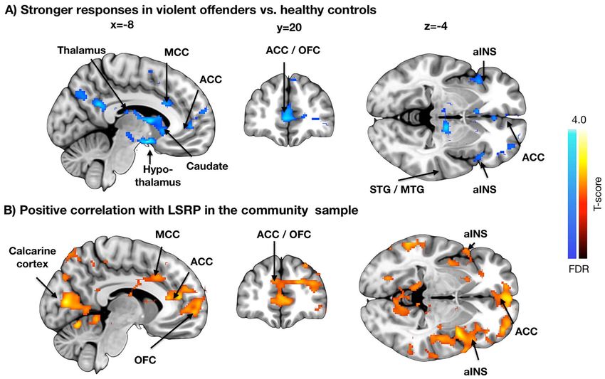
2     Cerebral Cortex, 2021, Vol. 00, No. 00

Introduction                                                           sample sizes and typically lack of direct comparison with crim-
                                                                       inal psychopathic offenders.
Psychopathy is a personality disorder characterized by persis-
tent antisocial behavior, impaired empathy and remorse, as well
as bold, disinhibited, and egotistical traits (Cooke and Michie        The Current Study
2001). In clinical and forensic settings, psychopathy predicts
                                                                       Forensic imaging studies on psychopathy are complicated
criminal behavior and violence (Salekin et al. 1997). For example,
                                                                       by clinical confounding factors and substance use history,
whereas prevalence of psychopathy is ∼1% in normal popula-
                                                                       even though the large psychological differences between
tion, it is ∼20% in incarcerated offenders (Hare 1991; in Fin-
                                                                       psychopaths and healthy controls makes them well powered
land 16.4%, Juriloo et al. 2014). The pervasive nature of both
                                                                       (Koenigs et al. 2011). Studies focusing on variation in psycho-

                                                                                                                                             Downloaded from https://academic.oup.com/cercor/advance-article/doi/10.1093/cercor/bhab072/6218172 by guest on 22 April 2021
behavioral and emotional symptoms suggest that psychopathy
                                                                       pathic traits in the general population have the advantage of
might have organic basis, and multiple studies have found that
                                                                       better control over comorbid psychiatric illnesses and substance
psychopathic offenders have atrophy in frontal cortex and in
                                                                       use, yet the restricted variation in psychopathic traits and cor-
limbic regions including insula and amygdala (Muller et al. 2008;
                                                                       responding self-reported trait measures compromises the sta-
Tiihonen et al. 2008; Yang et al. 2009; Ermer et al. 2012). These
                                                                       tistical power, which has been identified as a major problem in
structural impairments are coupled with abnormal function of
                                                                       neuroimaging studies (Cremers et al. 2017; Poldrack et al. 2017).
the limbic system, and psychopathic subjects show less affect-
                                                                       Finally, functional neuroimaging experiments on psychopathy
related activity in amygdala and hippocampus, striatum and
                                                                       have measured brain responses to isolated features such as
cingulate cortices while viewing emotional facial expressions,
                                                                       static faces or words that are not representative of the dynamic
and stronger activation of frontal cortical regions (Kiehl et al.
                                                                       and ever-changing real world, and there is prima facie doubt
2001; Dolan and Fullam 2009).
                                                                       regarding the generalizability of this approach to real-world
    When viewing empathy-eliciting scenes, psychopathic indi-
                                                                       social behavior (Adolphs et al. 2016). Here, we tested whether
viduals show significantly reduced frontocortical brain activity
                                                                       psychopathic traits in a large well-functioning community sam-
compared with healthy controls, consistent with their lowered
                                                                       ple (n = 100) are associated with 1) cortical density and 2) brain
care motivation (Decety et al. 2013; Meffert et al. 2013). The
                                                                       activity when viewing highly naturalistic violent episodes, and 3)
weakened limbic outputs combined with dysfunction in exec-
                                                                       whether these effects match with corresponding structural and
utive frontal cortical and social decision-making systems could
                                                                       functional alterations in convicted violent offenders (n = 19) with
thus predispose psychopaths to violent and antisocial behavior.
                                                                       psychopathic traits versus matched healthy controls (n = 19). We
Finally, conduct disorder (CD) is predictive of adult psychopathy
                                                                       show that in the community sample, the degree of psychopathic
(Burke et al. 2007). Similarly to adult psychopaths, adolescents
                                                                       traits is associated with lowered fronto-limbic structural
with CD have reduced gray matter volume in amygdala and
                                                                       integrity and elevated frontal and insular hemodynamic brain
insula (Fairchild et al. 2011). Some studies have found that par-
                                                                       activity while seeing violence, and that these alterations are
ticipants with CD show increased amygdala response to neutral
                                                                       similar with those observed in the forensic sample.
but not angry faces (Passamonti et al. 2010), whereas another
study has found that adolescents with conduct problems and
low callous-unemotional traits show heightened amygdala
                                                                       Materials and Methods
responses to briefly presented fearful faces (Viding et al. 2012).
    Most people never commit a crime or any sort of violent            All subjects gave an informed, written consent and were com-
attack, yet there is considerable variation in everyday antisocial     pensated for their participation. The ethics board of the Hospital
and mildly delinquent behavior. For example, ∼60% of people            District of Southwest Finland approved the protocol and the
lie during casual conversations (Feldman et al. 2002), in EU 40–       study was conducted in accordance with the Declaration of
60% of drivers regularly exceed speeding limits (EuropeanCom-          Helsinki. A total of 100 non-convicted volunteers drawn from
mission 2020), and ∼10% of the US population have used illicit         the community volunteer pool participated in the study (51
drugs (NIDA 2013). It has thus been proposed that psychopathy          females, mean age 31 years, range 20–57 years; see Table 1).
is not a categorical designation similarly as, for example, clinical   The exclusion criteria included a history of neurological or
diagnoses. Instead, it may be better to view psychopathy as a          psychiatric disorders, alcohol and substance abuse, current use
constellation of continuous, lower-order antisocial, and aversive      of medication affecting the central nervous system, medical
personality dimensions that vary in the non-incarcerated popu-         conditions precluding participation, and the standard magnetic
lation with normal range of social functioning (Levenson et al.        resonance imaging (MRI) exclusion criteria. Four additional sub-
1995; Miller et al. 2008). This hypothesis is supported by data        jects were scanned but 2 were excluded from analyses because
from community samples where high scores on psychopathic               their MRIs revealed gross brain abnormalities, and 2 others due
traits are associated with antisocial behavior such as aggression      to malfunctioning gradient coil resulting in unusable data. In
and racism (Hodson et al. 2009; Jones and Paulhus 2010). This          a separate forensic imaging experiment, 19 convicted violent
variation is also reflected in cortical structure: psychopathic        male offenders with high psychopathic traits and 19 age and
traits in general populations are associated with decreased gray       sex-matched control subjects (different from those described
matter density (GMD) in striatum and amygdala (Vieira et al.           previously) were studied (Table 1). See Supplementary Figure
2015), whereas some studies have found regional increased              S1 for the distributions of the primary and secondary psy-
cortical density in frontal cortical regions (Lasko et al. 2019).      chopathy scores. All offenders were inmates from the Turku
In healthy subjects, high levels of psychopathic traits are asso-      Prison currently serving a sentence for either murder (n = 5),
ciated with heightened brain responses to facial expressions           manslaughter (n = 5), attempted manslaughter (n = 3) or grievous
in amygdala and frontal cortex (Gordon et al. 2004). However,          bodily harm (n = 6). Mean recidivism rate was 2.4 times after
interpretation of these studies is difficult due to compromised        first conviction. All were screened for illicit drug use both in
Psychopathy in Criminal Offenders and General Population                       Nummenmaa et al.                3

Table 1 Subject characteristics with frequencies and means with standard deviations in parenthesis

                                                     Community sample                            Convicted offenders                         Matched controls

n (Males)                                            49                                          19                                          19
n (Females)                                          51                                          0                                           0
Age                                                  31.14 (9.31)                                31.16 (6.49)                                28.53 (7.69)
BMI                                                  23.86 (2.61)                                28.05 (3.90)                                25.10 (2.09)
PCL-R                                                —                                           26.47 (6.24)                                —
LSRP primary psychopathy                             23.42 (4.07)                                —                                           21.94 (3.26)
LSRP secondary psychopathy                           15.99 (3.18)                                —                                           13.56 (3.00)

                                                                                                                                                                Downloaded from https://academic.oup.com/cercor/advance-article/doi/10.1093/cercor/bhab072/6218172 by guest on 22 April 2021
Figure 1. Experimental design and sample stimuli. The subjects viewed a compilation of 137 movie clips with variable violent and nonviolent content.

the screening and on the day of the MRI scan. Information on                        a cinema or at home, that is, no specific task was assigned.
medication and psychiatric diagnoses of the forensic subjects is                    Visual stimuli were presented with NordicNeuroLab VisualSys-
presented in Supplementary Tables S1 and S2. Forensic subjects                      tem binocular display. Sound was conveyed with Sensimetrics
were escorted to the hospital imaging site by 2 prison guards                       S14 insert earphones. Stimulation was controlled with Presen-
who monitored the whole study protocol.                                             tation software. Before the functional run, sound intensity was
   Psychopathy in the convicted offenders was assessed with                         adjusted for each subject so that it could be heard over the
the Hare Psychopathy Checklist-Revised (PCL; Hare 1991), based                      gradient noise. Six independent raters evaluated the occurrence
on semi-structured interview by an experienced forensic psy-                        of violence in the movie clips with 4-s temporal accuracy. These
chologist/psychiatrist and review of collateral information. The                    ratings were subsequently averaged and used as regressors in
PCL-R measures 2 dimensions of psychopathic traits: primary                         the functional magnetic resonance imaging (fMRI) data analysis
psychopathy involving inclination to lie, lack of remorse, and                      (see below). Due to time constraints (the offenders also partici-
callousness, and secondary psychopathy involving impulsivity,                       pated in another study on the same visit), the task used for the
short temper, and low tolerance for frustration. Psychopathic                       offender sample was slightly trimmed in duration (12 min). Our
traits in the non-convicted population were measured with the                       previous work however shows that the task is robust to this kind
Levenson Self-Report Psychopathy Scale (LSRP; Levenson et al.                       of variations in the stimulus material selection (Lahnakoski et al.
1995). This self-report instrument is based on the 2-factor (pri-                   2012; Karjalainen et al. 2017, 2019).
mary/secondary) conceptualization of the PCL. One subject had
incomplete items in the secondary psychopathy scale items and
was left out from the corresponding analyses.                                       MRI Data Acquisition and Preprocessing
                                                                                    The MRI data were acquired using a Phillips Ingenuity TF PET/MR
                                                                                    3T whole-body scanner. High-resolution (1 mm3 ) structural
Experimental Design for functional magnetic
                                                                                    images were obtained with a T1-weighted sequence (time
resonance imaging
                                                                                    repetition [TR] 9.8 ms, time echo [TE] 4.6 ms, flip angle 7◦ , 250-
To map brain responses to seeing violence, we used our previ-                       mm FOV, and 256 × 256 reconstruction matrix). A radiologist
ously validated socioemotional “localizer” paradigm that allows                     screened the T1 images for structural abnormalities. A total
reliable mapping of various social and emotional functions (Lah-                    of 407 functional volumes were acquired with a T2∗ -weighted
nakoski et al. 2012; Karjalainen et al. 2017, 2019). Briefly, the sub-              echo-planar imaging sequence (TR = 2600 ms, TE = 30 ms, 75◦
jects viewed a medley of 137 movie clips (mean duration 11.5 s;                     flip angle, 240 mm FOV, 80 × 80 reconstruction matrix, 62.5 kHz
total duration 26.2 min) that have been curated to contain large                    bandwidth, 3.0-mm slice thickness, and 45 interleaved slices
variability of social and emotional content (Fig. 1). All videos                    acquired in ascending order without gaps). One functional run
were extracted from mainstream Hollywood movies with audio                          with 476 functional images was acquired (275 volumes for the
track in English. Because this task is designed to map neural                       forensic sample and their controls). MRI data were preprocessed
processing of naturalistic socioemotional events, the clips are                     using fMRIPprep 1.3.0.2 (Esteban et al. 2019). The following
not deliberately matched with respect to, for example, human                        preprocessing steps were performed on the anatomical T1-
motion or optic flow. The videos were presented in fixed order                      weighted (T1w) reference image: correction for intensity non-
across the subjects without breaks. Subjects were instructed to                     uniformity, skull-stripping, brain surface reconstruction, and
view the movies similarly as if they were viewing a movie at                        spatial normalization to the ICBM 152 Nonlinear Asymmetrical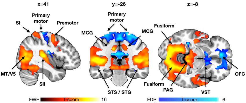
4      Cerebral Cortex, 2021, Vol. 00, No. 00

                                                                                                                                                                        Downloaded from https://academic.oup.com/cercor/advance-article/doi/10.1093/cercor/bhab072/6218172 by guest on 22 April 2021
Figure 2. Results from the voxel-based morphometry analyses. (A) Brain regions where convicted offenders had significant atrophy in comparison with control subjects.
(B) Brain regions where primary psychopathy scores were associated with decreased GMD, P < 0.01 FDR corrected. aINS = anterior insula, OFC = orbitofrontal cortex, SI
= primary somatosensory cortex, SII = secondary somatosensory cortex. .

template version 2009c using nonlinear registration with                             as nuisance covariates. Primary statistical threshold was set at
antsRegistration (ANTs 2.2.0) and brain tissue segmentation.                         P < 0.01, FDR corrected at cluster level.
The following preprocessing steps were performed on the func-
tional data: co-registration to the T1 reference image, slice-time
correction, spatial smoothing with a 6-mm Gaussian kernel,                           fMRI Data Analysis
automatic removal of motion artifacts using ICA-AROMA (Pruim                         The fMRI data were analyzed in SPM12. To reveal brain regions
et al. 2015), and resampling to the MNI152NLin2009cAsym                              activated by violence, a subjectwise (first-level) GLM was fitted
standard space.                                                                      to the data, where the blood oxygen level–dependent (BOLD)
                                                                                     signals were predicted with the violence regressors convolved
                                                                                     with the canonical hemodynamic response function. To identify
Voxel-Based Morphometry                                                              the brain regions showing enhanced brain activity consistently
Voxel-based morphometry (VBM) was done with SPM12 (Well-                             across the subjects, the individual beta images for the violence
come Trust Center for Imaging, London, United Kingdom, http://                       regressor were the entered to a second-level analysis. The effects
www.fil.ion.ucl.ac.uk/spm), which enables automated spatial                          of primary and secondary psychopathy were finally assessed by
normalization, tissue classification and radio-frequency bias                        adding them as covariates into the second-level models. Primary
correction to be combined with the segmentation step. Cutoff of                      statistical threshold was set at P < 0.05, False Discovery Rate
spatial normalization was 25 mm and medium affine regulariza-                        (FDR) corrected at cluster level.
tion 0.01 was used. Following normalization and segmentation
into grey and white matter, a modulation step was incorpo-
                                                                                     Connectivity Analyses
rated to take into account volume changes caused by spatial
normalization. Importantly, the modulation step corrects for the                     To test whether interregional coupling during movie viewing
differences in total brain size across subjects. Finally, the seg-                   varies across the psychopathic offenders and healthy controls
mented, normalized, and modulated GMD/WMD images were                                (and as a function of psychopathic traits in the community
smoothed using a Gaussian kernel of 8-mm full width at half                          sample), we performed connectivity analyses. We first extracted
maximum. Normalized and smoothed images were analyzed                                regional time courses from the detrended BOLD time series
with general linear model (GLM) where they were predicted                            from a priori regions of interest involved in emotional pro-
with primary and secondary psychopathy scores, while age and                         cessing including amygdala, insula, frontal pole, and thalamus
sex (for the sample of 100 non-convicted volunteers only; the                        (Saarimäki et al. 2016, 2018). These subjectwise time series were
forensic sample and their controls were all males) were entered                      then used as regressors in first level models, and the resulting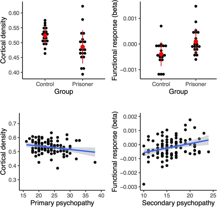
Psychopathy in Criminal Offenders and General Population Nummenmaa et al. 5
Downloaded from https://academic.oup.com/cercor/advance-article/doi/10.1093/cercor/bhab072/6218172 by guest on 22 April 2021
Figure 3. Brain regions showing increased (hot colors, 0.05 familywise error (FWE) corrected for visualization) and decreased (hot colors, P < 0.05 FDR corrected
for visualization) activity as a function of violence seen in the movies. Data for the community sample are shown for reference. MCG = middle cingulate gyrys,
STG = superior temporal gyrus, STS = superior temporal sulcus, and VST = ventral striatum.
contrast images were analyzed using GLM for population-level and sulcus. Deactivations were significantly weaker than acti-
inference similarly as in the VBM and BOLD-GLM analyses. vations, and were localized to primary motor cortex, OFC, and
ventral striatum.
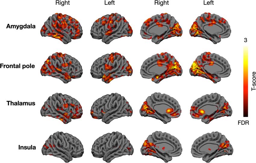
We then compared how the brain responses to seeing
Results violence in the psychopathic offenders differ from those of
healthy controls (Fig. 4). In the psychopathic offenders the
VBM
violent scenes provoked heightened activation in OFC, bilateral
Voxel-based morphometry (Fig. 2) revealed that offenders with insula, anterior and middle cingulate cortices, thalamus,
psychopathic traits had significantly lowered GMD in anterior and superior and middle temporal polysensory regions.
insula, orbitofrontal cortex (OFC) and secondary somatosensory Next, we assessed whether the degree of psychopathic traits
cortex (SII), indicating atrophy in these regions. No opposite associates with brain responses to violent scenes in the well-
effects were found. In the community sample, negative associa- functioning community sample, and found that the effects were
tion with primary LSRP scores (i.e., lower cortical density with comparable with those observed in the convicted offenders.
higher psychopathy scores scores) was observed in the same The strongest positive associations were observed for the
regions. Additional effects were observed in primary somatosen- secondary psychopathy scores, which were positively associated
sory cortex (SI), paracentral lobule and midcingulate cortex. No with violence-dependent BOLD responses in orbitofrontal and
regions showed significant positive associations with primary anterior insular cortices and along the whole anterior–posterior
psychopathy even with a more lenient threshold (P < 0.05 FDR axis of the cingulate cortex, and SI, and also in the visual
corrected), and neither positive nor negative associations were cortices. Effects for primary psychopathy were weaker but
found with secondary psychopathy. Analysis of the WM seg- overlapped with those of secondary psychopathy in the orbital
ments revealed only lowered density in the cerebellum and and frontal areas.
around the lingual gyrus in the psychopathic group. No oppo- Finally, to confirm the consistency of psychopathy-dependent
site effects were found. In the community sample, there were orbitofrontal alterations (that were consistently observed in all
no positive or negative associations with neither primary nor the analyses) and to address the direction of the associations
secondary psychopathy scores. between psychopathy and brain structure and function, we
generated a 6-mm spherical region of interest (ROI) in OFC.
The ROI was centered around a peak voxel (x = 1, y = 46, and z = 2)
fMRI
showing strong positive association between psychopathic traits
Viewing violent scenes elicited widespread and strong brain and violence-elicited brain responses in the community sample.
activation (Fig. 3). The effects spanned limbic and paralimbic Next, we extracted regional cortical density (for VBM data)
emotion circuits (thalamus, periaqueductal gray [PAG], and mid- and beta (for fMRI) values in this ROI for both the community
dle cingulate cortex) as well as the somatosensory cortices sample and the prisoners and their respective controls. This
(SI–SII) and components of the frontoparietal network (inferior analysis (Fig. 5) showed that in OFC, psychopathic offenders
parietal and lateral premotor cortices). Significant activation had increased atrophy (i.e., lowered cortical density; t = 2.59 and
clusters were also observed in visual (V1 and the MT–V5 com- P = 0.015) and increased responses to seen violence comparison
plex) and auditory cortices, as well as in regions involved in with healthy controls (i.e., betas, t = 3.08 and P = 0.003). The
face and social perception, such as the lateral fusiform gyrus results were comparable in the community sample—in the OFC
and posterior polysensory areas of the superior temporal gyrus primary psychopathy was negatively associated with cortical
6      Cerebral Cortex, 2021, Vol. 00, No. 00

                                                                                                                                                                         Downloaded from https://academic.oup.com/cercor/advance-article/doi/10.1093/cercor/bhab072/6218172 by guest on 22 April 2021
Figure 4. Brain regions whose responses to seen violence were stronger on convicted offenders versus controls (A) and positively associated with secondary psychopathy
scores in the community sample (B). The data are thresholded at P < 0.05, FDR corrected. MCC = middle cingulate cortex.

density (r = 0.22 and P = 0.028), while secondary psychopathy                         thalamus and insula, whose connectivity with medial temporal,
was positively associated with the functional responses to seen                       cortical midline, and lateral and medial frontal cortices were
violence (r = 0.30 and P = 0.002). Note that this analysis could be                   associated positively with the primary psychopathy scores.
considered “circular” (Kriegeskorte et al. 2009) for the functional
responses in the community sample that was used for ROI
definition, but not in the 3 other conditions (structural effects                     Discussion
in community sample, functional and structural effects in the
                                                                                      Our main findings were that 1) psychopathic traits among
prisoners, and controls). This analysis thus suggests that there
                                                                                      healthy individuals are associated with compromised cerebral
are consistent psychopathy-related structural and functional
                                                                                      structural integrity and amplified functional responses to
alterations in the orbitofrontal region.
                                                                                      seeing highly naturalistic violence and that 2) these alterations
                                                                                      resemble the differences observed between incarcerated violent
                                                                                      psychopathic offenders and healthy controls. Primary (affec-
Connectivity Analyses                                                                 tive) psychopathic traits were associated with compromised
The connectivity analyses revealed consistent hypoconnectivity                        gray matter integrity in the medial frontal, insular, and
in the psychopathic offenders (Fig. 6). All the tested regions                        somatosensory cortices, whereas secondary (impulse control
showed lowered connectivity with cuneus and calcarine cortex.                         related) psychopathic traits were associated with increased
The largest differences were observed for amygdala and frontal                        functional responses in these areas when viewing violent
pole, whose hypoconnectivity spanned inferior temporal cortex                         videos. Together these data provide structural and functional
and supramarginal gyrus, S1 and S2, cingulate and lateral and                         neurobiological evidence for the similar neural basis of criminal
medial frontal cortex as well as caudate and putamen. For tha-                        psychopathy and antisocial yet psychologically well-functioning
lamus, significant hypoconnectivity was observed with insula,                         personalities.
caudate and lateral frontal cortex. Altered connectivity from
insula was primarily restricted to cuneus and calcarine cortex.
                                                                                      Psychopathic Traits Are Associated
For the community sample, the results did not clearly parallel
                                                                                      with Cerebral Integrity
with those obtained in the forensic sample. Only connectivity
from amygdala, thalamus and frontal to calcarine cortex was                           The violent offenders had lowered GMD in OFC and anterior
negatively associated with primary (frontal pole) and secondary                       insulae. In the community sample, primary psychopathy scores
(amygdala and thalamus) psychopathy scores, thus resembling                           were associated with lowered cortical integrity in the same
the convicted offenders. Largest discrepancies were observed for                      areas. This effect was also specific to grey matter, and we found
Psychopathy in Criminal Offenders and General Population Nummenmaa et al. 7
Downloaded from https://academic.oup.com/cercor/advance-article/doi/10.1093/cercor/bhab072/6218172 by guest on 22 April 2021
Figure 5. Orbitofrontal cortical density (left; expressed as probability of a voxel belonging to gray matter) and functional responses (right) in the prisoners and controls
(top) and in the community sample (bottom). Error bars in the dotplots show ±2 standard deviation, the scatterplots show LS-regression lines with 95% confidence
interval.
no evidence of aberrant white matter density as a function of lobes in violent psychiatric patients and murderers (Raine
psychopathy except in the cerebellum of the convicted sample. et al. 1994, 1997, 1998; Volkow et al. 1995; George et al. 2004),
These results bear striking resemblance to prior VBM studies and the present data align well with these observations by
on criminal psychopaths. These studies have found significant demonstrating psychopathy-related alterations in the same
psychopathy-dependent atrophy in OFC/ACC (anterior cingulate brain regions in a healthy sample. Previous studies have
cortex) and insula (Tiihonen et al. 2008; Ermer et al. 2012), also found hippocampal and amygdala density reductions in
suggesting that similar neural alterations underlie criminal psy- psychopathic offenders (Ermer et al. 2012). Although these
chopathy and variation in everyday antisocial behavior in psy- effects were not manifested in our data with the a priori
chologically well-functioning individuals. statistical threshold, it is noteworthy that with slightly more
Frontal lobe dysfunction and poor executive functions are liberal (yet multiple comparison corrected) threshold, primary
hallmarks of disorders involving impulsivity (Morgan and psychopathy scores were also negatively associated with GMD
Lilienfeld 2000; Yang and Raine 2009). Numerous studies have in amygdala and hippocampus (P < 0.05, FDR corrected, data not
reported lowered glucose metabolism in frontal and temporal shown).8 Cerebral Cortex, 2021, Vol. 00, No. 00
Downloaded from https://academic.oup.com/cercor/advance-article/doi/10.1093/cercor/bhab072/6218172 by guest on 22 April 2021
Figure 6. Brain regions whose connectivity with amygdala, frontal pole, thalamus, and insula was lowered in the convicted offenders versus controls. The data are
thresholded at P < 0.05, FDR corrected at cluster level.
Prior structural imaging studies have yielded mixed results threatening events (Nummenmaa et al. 2008). Seeing violence
concerning the cerebral alterations associated with psychopa- also engaged the dorsal frontoparietal network involved in goal-
thy: Whereas many studies have found decreased frontocortical directed attention shifts and sustained attention (Corbetta and
and limbic densities in psychopathy (Tiihonen et al. 2008; Ermer Shulman 2002; Nummenmaa et al. 2017), likely resulting from
et al. 2012), some have found positive associations between focused attention on the survival-salient events (Vuilleumier
frontal/OFC densities and secondary (factor 2) psychopathy 2005). Consistent activations were found in the lateral fusiform
(Korponay et al. 2017). These discrepancies could be attributed gyrus centrally involved in facial expression recognition
to a multitude of factors, such as definition of and the (Calder and Young 2005), as well as posterior polysensory
criteria used for psychopathy, whether incarcerated or typical areas of the superior temporal sulci and gyri that integrate
populations with variation in psychopathy scores are studies, multitude of social signals and act as the centralized “hub”
and subjects’ substance use history (Koenigs et al. 2011). The of social perception in humans (Nummenmaa and Calder
present study in turn clearly replicates the association between 2009; Lahnakoski et al. 2012). Finally, seeing violence elicited
psychopathic traits and fronto-insular atrophy in both healthy significant activity in the motion-sensitive MT/V5 complex. This
volunteers (self-reports of psychopathy) and forensic sample likely reflects the increased motion kinematics in the violent
(objective measures of psychopathy), providing strong evidence episodes, and post hoc analyses indeed revealed that violence
for this regionally specific cerebral alteration associated with in the movies was associated with increased perceived human
psychopathy. motion (r = 0.17 and P < 0.05).
Altogether the coordinated activity of this network encodes
the threat value of the environment, focuses attentional
Brain Responses to Seeing Violence
resources to the critical features and prepares the fight-
Viewing violence elicited consistent responses in widespread or-flight response when needed. Remarkably, the increased
brain regions, including PAG that is a key part of the pain- activation of the somatosensory and premotor cortices was
inhibiting circuitry (Tracey and Mantyh 2007). PAG is also accompanied with significant deactivation in primary motor
centrally involved in generating freezing responses to distal cortex and rostral ACC. Although speculative, it is possible
threats and reactive aggression during threats (Panksepp 1998; that this distinction reflects the engagement of the violence
Gregg and Siegel 2001). Human fMRI studies have accordingly inhibition mechanism, whereby seeing others’ distress leads
demonstrated that proximity of threats activates the PAG in to somatosensory (SI–SII) and premotor mirroring of others
humans (Mobbs et al. 2007). Significant activations were also distress, leading to inhibitory activation at the motor cortex
found in thalamus, which likely modulates arousal levels during subsequently preventing engagement in violence. Previous fMRIPsychopathy in Criminal Offenders and General Population Nummenmaa et al. 9
studies on first-person simulated fighting have found that of the aversive stimulation (complex and naturalistic versus
dorsal ACC responses are increased when executing violent well-controlled).
actions, reflecting this region’s role in emotion regulation Criminal psychopathy was also associated with lowered con-
(Mathiak and Weber 2006). nectivity of the key nodes of the social and emotional brain net-
works, including amygdala, insula, thalamus, and frontal pole.
This effect was however specific to the incarcerated subjects and
Psychopathic Traits Mediate Neural Responses
was not consistently replicated in the community sample as a
to Violence
function of psychopathic traits. Thus, even though there are par-
Violent psychopathic offenders showed increased hemody- allels in the regional responsiveness of the brain’s affective cir-
namic responses to seeing violence in orbitofrontal and insular cuit in the convicted psychopaths and well-functioning subjects
Downloaded from https://academic.oup.com/cercor/advance-article/doi/10.1093/cercor/bhab072/6218172 by guest on 22 April 2021
cortices, and also throughout the cingulate cortex. In the with psychopathic traits, it is likely that the disrupted functional
healthy community sample, secondary psychopathy scores connectivity of this network is specific to criminal psychopathy.
were associated with increased hemodynamic responses in
comparable brain regions, suggesting similarities between brain
Limitations and Future Directions
characteristics of criminal psychopathy and more benign forms
of antisociality. It is also noteworthy that while structural The psychopathic traits of the community sample were assessed
alterations in the healthy sample were linked with primary using self-reports (LSRP) rather than PCL-R, as the latter is based
psychopathic traits, the functional responses were associated on interviews and extensive review of collateral information
with secondary psychopathic traits. It is likely that the latter (Hare 1991). The nature of psychopathic personality per se (e.g.,
reflects the contents of the naturalistic video stimulus which tendency to lie) makes self-report scales difficult to construct.
contained numerous episodes of impulsive violence, whose Numerous studies have however confirmed that self-reports on
processing may not be modulated by callousness-related traits. psychopathic traits predict real-life antisocial behavior (Furn-
Prior functional imaging studies have found that psy- ham et al. 2013); the brain imaging results from the forensic
chopaths typically have increased rather than decreased sample (evaluated with PCL-R) and community sample (evalu-
functional responses to emotional stimuli, typically facial ated with self-reports) were concordant. We aimed at recruiting
expression of fear (Kiehl et al. 2001; Dolan and Fullam 2009), prisoner subjects not using antipsychotics, antidepressants or
yet to our knowledge our study is the first one to measure anxiolytics; however, it was not possible to recruit a completely
psychopathy-related neural responses to naturalistic violence medication-free sample. The convicted offenders and healthy
scenes. One possible explanation for the amplified brain controls also differ from each other regarding the available
responses to violent scenes is that psychopathy involves quality and quantity of social interaction, leisure time activities
impaired inhibitory control during affective provocation, and so forth. Ideally, this kind of study should thus also involve a
which may lead to aggression during real-life encounters. It forensic but non-psychopathic sample. Our data are also cross-
is noteworthy that OFC and insula–regions whose structural sectional in nature and cannot resolve the potential causal link
integrity was negatively associated with psychopathy–showed between the cerebral structural and functional alterations and
positive association between secondary psychopathy and BOLD psychopathic traits. Because conduct problems in adolescence
responses to violence (Fig. 5). This might reflect a compensatory are predictive of adult psychopathy (Burke et al. 2007), future
functional activity due to compromised cerebral integrity in longitudinal studies are needed to delineate the developmental
these regions, and suggest a general role of the OFC in mediating neural pathways of psychopathy and assess the contribution of
antisocial behavior. early social environment to the observed neural alterations.
Both insula and SI are important for generating the bodily Our naturalistic movie viewing paradigm yields a high-
component of emotions (Damasio 1999; Nummenmaa et al. dimensional stimulus space and it is difficult to tease apart
2014, 2018). Furthermore, damage to the primary somatosensory the specific contribution of different aspects of violence (such
cortex (Adolphs et al. 2000) or its inhibition with transcranial as facial expressions and vocalizations). Yet, such isolated
magnetic stimulation (Pourtois et al. 2004) impairs facial expres- features are not representative of the real world either (Adolphs
sion recognition. Psychopathic individuals show affective disen- et al. 2016). Because evolution has prepared the brain to
gagement and dampened autonomic nervous system reactivity process a continuous sensory stream rather than “snapshots”
to a variety of emotional stimuli (Patrick et al. 1993). Combined, such as pictures and sound bursts, dynamic natural stimuli
our results suggest that this may be due to the compromised trigger more consistent and stronger neural responses than
integrity and atypical function of the somatosensory and insular the conventionally used well-controlled yet reduced stimuli
loops of the emotion circuit that serve the generation of the (Yao et al. 2007; Fox et al. 2009; Schultz et al. 2013). Violent
bodily component of emotion. The aversive somatic markers episodes span numerous overlapping and hierarchic time scales
generated by SI–SII and insula when seeing aggressive cues necessitating parallel processing of multiple sensory features.
could inhibit aggressive behavior (Blair 2001). Thus their com- Consequently, they cannot be adequately explored with fully
promised function and structure may lead to disinhibited affect controlled classic experimental designs.
due to lacking somatic feedback from evoked emotions during
affective provocation, potentially predisposing to aggression.
Yet, it must be noted that some prior studies have found that
Conclusions
psychopathic offenders show lower rather than greater neural Normally functioning individuals with high psychopathic traits
responses during passive viewing of empathy-evoking scenarios have structural and functional brain characteristics that are
(e.g., Meffert et al. 2013). We have no clear explanation for this, similar to violent offender with high psychopathic traits. These
but it is possible that it pertains to the degree of aggressive characteristics include both fronto-limbic cortical atrophy and
violence present in the scenes (e.g., fights and assaults versus enhanced brain activity in affective circuits while seeing vio-
controlled minor pain inflicted on hands) or the general context lence. Altogether these data show that integrity and function of10 Cerebral Cortex, 2021, Vol. 00, No. 00
the frontal and insular cortex associate with both extreme and European Commission. 2020. The frequency of speed limit
benign variations of antisocial behavior, providing neurobiolog- violations. In: safety MaTR, editor. https://ec.europa.eu/
ical support for a common neural basis of antisocial behaviors transport/road_safety/specialist/knowledge/speed/many_
with different severity. drivers_exceed_the_speed_limit/the_frequency_of_speed_li
mit_violations_en
Fairchild G, Passamonti L, Hurford G, Hagan CC, von dem Hagen
Supplementary Material EAH, van Goozen SHM, Goodyer IM, Calder AJ. 2011. Brain
Supplementary material can be found at Cerebral Cortex online. structure abnormalities in early-onset and adolescent-onset
conduct disorder. Am J Psychiat. 168:624–633.
Feldman RS, Forrest JA, Happ BR. 2002. Self-presentation and
Downloaded from https://academic.oup.com/cercor/advance-article/doi/10.1093/cercor/bhab072/6218172 by guest on 22 April 2021
Notes
verbal deception: do self-presenters lie more? Basic Appl Soc
We acknowledge director Juhani Järvi and other staff members Psychol. 24:163–170.
of Turku Prison who made it possible to safely transport and Fox CJ, Iaria G, Barton JJS. 2009. Defining the face processing
guard the inmates during the neuroimaging. Conflict of Interest: network: optimization of the functional localizer in fMRI.
The authors declare no conflict of interest. Hum Brain Mapp. 30:1637–1651.
Furnham A, Richards SC, Paulhus DL. 2013. The dark triad of
personality: a 10 year review. Soc Personal Psychol Compass.
Funding 7:199–216.
This work was supported by the Academy of Finland (grant no. George DT, Rawlings RR, Williams WA, Phillips MJ, Fong G, Kerich
294897), and European Research Council starting (grant 313000 M, Momenan R, Umhau JC, Hommer D. 2004. A select group
to L.N.), Valon Vuoksi Foundation (grants to L.L. and L.S.). of perpetrators of domestic violence: evidence of decreased
metabolism in the right hypothalamus and reduced rela-
tionships between cortical/subcortical brain structures in
References position emission tomography. Psychiatry Res Neuroimaging.
Adolphs R, Damasio H, Tranel D, Cooper G, Damasio AR. 2000. A 130:11–25.
role for somatosensory cortices in the visual recognition of Gordon HL, Baird AA, End A. 2004. Functional differences among
emotion as revealed by three-dimensional lesion mapping. J those high and low on a trait measure of psychopathy. Biol
Neurosci. 20:2683–2690. Psychiatry. 56:516–521.
Adolphs R, Nummenmaa L, Todorov A, Haxby JV. 2016. Data- Gregg TR, Siegel A. 2001. Brain structures and neurotransmitters
driven approaches in the investigation of social perception. regulating aggression in cats: implications for human aggres-
Phil Trans B. 371. sion. Progr Neuro-Psychopharmacol Biol Psychiatry. 25:91–140.
Blair RJR. 2001. Neurocognitive models of aggression, the antiso- Hare RD. 1991. Manual for the Hare Psychopathy Checklist-Revised.
cial personality disorders, and psychopathy. J Neurol. Neurosur Toronto, Canada: Multi-Health Systems.
Psychiatry. 71:727. Hodson G, Hogg SM, MacInnis CC. 2009. The role of "dark per-
Burke JD, Loeber R, Lahey BB. 2007. Adolescent conduct disorder sonalities" (narcissism, Machiavellianism, psychopathy), big
and interpersonal callousness as predictors of psychopathy five personality factors, and ideology in explaining prejudice.
in young adults. J Clin Child Adolesc Psychol. 36:334–346. J Res Personality. 43:686–690.
Calder AJ, Young AW. 2005. Understanding the recognition Jones DN, Paulhus DL. 2010. Different provocations trigger
of facial identity and facial expression. Nat Rev Neurosci. aggression in narcissists and psychopaths. Soc Psychol Per-
6:641–651. sonal Sci. 1:12–18.
Cooke DJ, Michie C. 2001. Refining the construct of psychopathy: Juriloo A, Lauerma H, Holmalahti T, Tyni S, Aarnio J, Viitanen P,
towards a hierarchical model. Psychol Assess. 13:171–188. Wuolijoki T, Mattila A, Lintonen T, Joukamaa M, et al. 2014.
Corbetta M, Shulman GL. 2002. Control of goal-directed and Psychopathic traits in a representative sample of Finnish
stimulus-driven attention in the brain. Nat Rev Neurosci. male prisoners. Nord J Psychiatr. 68:117–122.
3:201–215. Karjalainen T, Karlsson HK, Lahnakoski JM, Glerean E, Nuutila
Cremers HR, Wager TD, Yarkoni T. 2017. The relation between P, Jaaskelainen IP, Hari R, Sams M, Nummenmaa L. 2017.
statistical power and inference in fMRI. PLoS One. 12:e0184923. Dissociable roles of cerebral mu-opioid and type 2 dopamine
Damasio A. 1999. The feeling of what happens: body and emotion in receptors in vicarious pain: a combined PET-fMRI study. Cereb
the making of consciousness. New York: Harcourt Brace. Cortex. 27:4257–4266.
Decety J, Skelly LR, Kiehl KA. 2013. Brain response to empathy- Karjalainen T, Seppala K, Glerean E, Karlsson HK, Lahnakoski
eliciting scenarios involving pain in incarcerated individuals JM, Nuutila P, Jaaskelainen IP, Hari R, Sams M, Nummen-
with psychopathy. JAMA Psychiatry. 70:638–645. maa L. 2019. Opioidergic regulation of emotional arousal: a
Dolan MC, Fullam RS. 2009. Psychopathy and functional mag- combined PET-fMRI study. Cereb Cortex. 29:4006–4016.
netic resonance imaging blood oxygenation level-dependent Kiehl KA, Smith AM, Hare RD, Mendrek A, Forster BB, Brink J,
responses to emotional faces in violent patients with Liddle PF. 2001. Limbic abnormalities in affective processing
schizophrenia. Biol Psychiatry. 66:570–577. by criminal psychopaths as revealed by functional magnetic
Ermer E, Cope LM, Nyalakanti PK, Calhoun VD, Kiehl KA. 2012. resonance imaging. Biol Psychiatry. 50:677–684.
Aberrant paralimbic gray matter in criminal psychopathy. J Koenigs M, Baskin-Sommers A, Zeier J, Newman JP. 2011. Investi-
Abnorm Psychol. 121:649–658. gating the neural correlates of psychopathy: a critical review.
Esteban O, Markiewicz CJ, Blair RW, Moodie CA, Isik AI, Erra- Mol Psychiatry. 16:792–799.
muzpe A, Kent JD, Goncalves M, DuPre E, Snyder M, et al. 2019. Korponay C, Pujara M, Deming P, Philippi C, Decety J, Kosson
fMRIPrep: a robust preprocessing pipeline for functional MRI. DS, Kiehl KA, Koenigs M. 2017. Impulsive-antisocial psy-
Nat Methods. 16:111–116. chopathic traits linked to increased volume and functionalPsychopathy in Criminal Offenders and General Population Nummenmaa et al. 11
connectivity within prefrontal cortex. Soc Cogn Affect Neurosci. Poldrack RA, Baker CI, Durnez J, Gorgolewski KJ, Matthews PM,
12:1169–1178. Munafò MR, Nichols TE, Poline J-B, Vul E, Yarkoni T. 2017.
Kriegeskorte N, Simmons WK, Bellgowan PSF, Baker CI. 2009. Scanning the horizon: towards transparent and reproducible
Circular analysis in systems neuroscience: the dangers of neuroimaging research. Nat Rev Neurosci. 18:115–126.
double dipping. Nat Neurosci. 12:535–540. Pourtois G, Sander D, Andres M, Grandjean D, Reveret L, Olivier
Lahnakoski JM, Glerean E, Salmi J, Jaaskelainen I, Sams M, Hari E, Vuilleumier P. 2004. Dissociable roles of the human
R, Nummenmaa L. 2012. Naturalistic fMRI mapping reveals somatosensory and superior temporal cortices for processing
superior temporal sulcus as the hub for the distributed brain social face signals. Eur J Neurosci. 20:3507–3515.
network for social perception. Front Hum Neurosci. 6:14. Pruim RHR, Mennes M, Buitelaar JK, Beckmann CF. 2015. Evalua-
Lasko EN, Chester DS, Martelli AM, West SJ, DeWall SN. 2019. tion of ICA-AROMA and alternative strategies for motion arti-
Downloaded from https://academic.oup.com/cercor/advance-article/doi/10.1093/cercor/bhab072/6218172 by guest on 22 April 2021
An investigation of the relationship between psychopathy fact removal in resting state fMRI. Neuroimage. 112:278–287.
and greater gray matter density in lateral prefrontal cortex. Raine A, Buchsbaum M, LaCasse L. 1997. Brain abnormalities in
Personality Neurosci. 2:e7. murderers indicated by positron emission tomography. Biol
Levenson MR, Kiehl KA, Fitzpatrick CM. 1995. Assessing psycho- Psychiatry. 42:495–508.
pathic attributes in a noninstitutionalized population. J Pers Raine A, Buchsbaum MS, Stanley J, Lottenberg S, Abel L, Stod-
Soc Psychol. 68:151–158. dard J. 1994. Selective reductions in prefrontal glucose-
Mathiak K, Weber R. 2006. Toward brain correlates of natu- metabolism in murderers. Biol Psychiatry. 36:365–373.
ral behavior: fMRI during violent video games. Hum Brain Raine A, Meloy JR, Bihrle S, Stoddard J, LaCasse L, Buchsbaum
Mapping. 27:948–956. MS. 1998. Reduced prefrontal and increased subcortical brain
Meffert H, Gazzola V, den Boer JA, Bartels AAJ, Keysers C. 2013. functioning assessed using positron emission tomography in
Reduced spontaneous but relatively normal deliberate vicar- predatory and affective murderers. Behav Sci Law. 16:319–332.
ious representations in psychopathy. Brain. 136:2550–2562. Saarimäki H, Ejtehadian LF, Glerean E, Jaaskelainen IP, Vuilleu-
Miller JD, Gaughan ET, Pryor LR. 2008. The Levenson self-report mier P, Sams M, Nummenmaa L. 2018. Distributed affec-
psychopathy scale: an examination of the personality traits tive space represents multiple emotion categories across the
and disorders associated with the LSRP factors. Assessment. human brain. Soc Cogn Affect Neurosci. 13:471–482.
15:450–463. Saarimäki H, Gotsopoulos A, Jääskeläinen IP, Lampinen J,
Mobbs D, Petrovic P, Marchant JL, Hassabis D, Weiskopf N, Sey- Vuilleumier P, Hari R, Sams M, Nummenmaa L. 2016. Dis-
mour B, Dolan RJ, Frith CD. 2007. When fear is near: threat crete neural signatures of basic emotions. Cereb Cortex.
imminence elicits prefrontal-periaqueductal gray shifts in 6:2563–2573.
humans. Science. 317:1079–1083. Salekin RT, Rogers R, Sewell KW. 1997. Construct validity of
Morgan AB, Lilienfeld SO. 2000. A meta-analytic review of psychopathy in a female offender sample: a multitrait-
the relation between antisocial behavior and neuropsycho- multimethod evaluation. J Abnorm Psychol. 106:576–585.
logical measures of executive function. Clin Psychol Rev. Schultz J, Brockhaus M, Bulthoff HH, Pilz KS. 2013. What
20:113–136. the human brain likes about facial motion. Cereb Cortex.
Muller JL, Ganssbauer S, Sommer M, Dohnel K, Weber T, 23:1167–1178.
Schmidt-Wilcke T, Hajak G. 2008. Gray matter changes in Tiihonen J, Rossi R, Laakso MP, Hodgins S, Testa C, Perez J, Repo-
right superior temporal gyrus in criminal psychopaths. Evi- Tiihonen E, Vaurio O, Soininen H, Aronen HJ, et al. 2008. Brain
dence from voxel-based morphometry. Psychiatry Res Neu- anatomy of persistent violent offenders: more rather than
roimaging. 163:213–222. less. Psychiatry Res Neuroimaging. 163:201–212.
NIDA. 2013. Results from the 2013 National Survey on Drug Use and Tracey I, Mantyh PW. 2007. The cerebral signature for pain
Health: summary of national findings. Rockville, MD: Adminis- perception and its modulation. Neuron. 55:377–391.
tration SAaMHS. Viding E, Sebastian CL, Dadds MR, Lockwood PL, Cecil CAM, De
Nummenmaa L, Calder AJ. 2009. Neural mechanisms of social Brito SA, McCrory EJ. 2012. Amygdala response to Preattentive
attention. Trends Cognit Sci. 13:135–143. masked fear in children with conduct problems: the role of
Nummenmaa L, Glerean E, Hari R, Hietanen JK. 2014. Bodily maps callous-unemotional traits. Am J Psychiat. 169:1109–1116.
of emotions. Proc Natl Acad Sci U S A. 111:646–651. Vieira JB, Ferreira-Santos F, Almeida PR, Barbosa F, Marques-
Nummenmaa L, Hari R, Hietanen JK, Glerean E. 2018. Maps of Teixeira J, Marsh AA. 2015. Psychopathic traits are associated
subjective feelings. Proc Natl Acad Sci U S A. 115:9198–9203. with cortical and subcortical volume alterations in healthy
Nummenmaa L, Hirvonen J, Parkkola R, Hietanen JK. 2008. individuals. Soc Cogn Affect Neurosci. 10:1693–1704.
Is emotional contagion special? An fMRI study on neural Volkow ND, Tancredib LR, Grant C, Gillespie H, Valentine A, Mul-
systems for affective and cognitive empathy. Neuroimage. lani N, Wang GJ, Hollister L. 1995. Brain glucose metabolism
43:571–580. in violent psychiatric patients: a preliminary study. Psychiatry
Nummenmaa L, Oksama L, Glerean E, Hyona J. 2017. Corti- Res: Neuroimaging. 61:243–253.
cal circuit for binding object identity and location during Vuilleumier P. 2005. How brains beware: neural mechanisms of
multiple-object tracking. Cereb Cortex. 27:162–172. emotional attention. Trends Cogn Sci. 9:585–594.
Panksepp J. 1998. Affective neuroscience: the foundations of human Yang YL, Raine A. 2009. Prefrontal structural and functional
and animal emotions. Oxford: Oxford University Press. brain imaging findings in antisocial, violent, and psycho-
Passamonti L, Fairchild G, Goodyer IM, Hurford G, Hagan CC, pathic individuals: a meta-analysis. Psychiatry Res Neuroimag-
Rowe JB, Calder AJ. 2010. Neural abnormalities in early-onset ing. 174:81–88.
and adolescence-onset conduct disorder. Arch Gen Psychiatry. Yang YL, Raine A, Narr KL, Colletti P, Toga AW. 2009. Localization
67:729–738. of deformations within the amygdala in individuals with
Patrick CJ, Bradley MM, Lang PJ. 1993. Emotion in the criminal psychopathy. Arch Gen Psychiatry. 66:986–994.
psychopath - startle reflex modulation. J Abnorm Psychol. Yao HS, Shi L, Han F, Gao HF, Dan Y. 2007. Rapid learning in
102:82–92. cortical coding of visual scenes. Nature Neurosci. 10:772–778.You can also read