Cortical thickness distinguishes between major depression and schizophrenia in adolescents
←
→
Page content transcription
If your browser does not render page correctly, please read the page content below
Zhou et al. BMC Psychiatry     (2021) 21:361
https://doi.org/10.1186/s12888-021-03373-1

 RESEARCH                                                                                                                                           Open Access

Cortical thickness distinguishes between
major depression and schizophrenia in
adolescents
Zheyi Zhou1,2†, Kangcheng Wang3†, Jinxiang Tang4,5, Dongtao Wei1,2, Li Song1,2, Yadong Peng4,6, Yixiao Fu4* and
Jiang Qiu1,2,7*

  Abstract
  Background: Early diagnosis of adolescent psychiatric disorder is crucial for early intervention. However, there is
  extensive comorbidity between affective and psychotic disorders, which increases the difficulty of precise diagnoses
  among adolescents.
  Methods: We obtained structural magnetic resonance imaging scans from 150 adolescents, including 67 and 47
  patients with major depressive disorder (MDD) and schizophrenia (SCZ), as well as 34 healthy controls (HC) to
  explore whether psychiatric disorders could be identified using a machine learning technique. Specifically, we used
  the support vector machine and the leave-one-out cross-validation method to distinguish among adolescents with
  MDD and SCZ and healthy controls.
  Results: We found that cortical thickness was a classification feature of a) MDD and HC with 79.21% accuracy
  where the temporal pole had the highest weight; b) SCZ and HC with 69.88% accuracy where the left superior
  temporal sulcus had the highest weight. Notably, adolescents with MDD and SCZ could be classified with 62.93%
  accuracy where the right pars triangularis had the highest weight.
  Conclusions: Our findings suggest that cortical thickness may be a critical biological feature in the diagnosis of
  adolescent psychiatric disorders. These findings might be helpful to establish an early prediction model for
  adolescents to better diagnose psychiatric disorders.
  Keywords: Depression, Schizophrenia, Adolescence, Cortical thickness, Machine learning

Background                                                                             psychiatric disorders, have extensive comorbidities with
Psychiatric disorder is among the most important causes                                each other [4, 5]. Approximately 80% of patients with
of mortality in humans, which affects the quality of life                              SCZ experience depressive episode in the early disorder
and increases the social burden [1–3]. Psychotic (such as                              stages [6]. The depression prevalence among patients
schizophrenia [SCZ]) and affective disorders (such as                                  with SCZ can be as high as 40% [7, 8]. Moreover, pa-
major depressive disorder [MDD]), as two typical                                       tients with MDD have been shown to have a higher risk
                                                                                       of developing a psychotic disorder. In addition, depres-
* Correspondence: fuyixiao2016@126.com; qiuj318@swu.edu.cn                             sion often precedes psychotic symptoms in people with
†
4
 Zheyi Zhou and Kangcheng Wang contributed equally to this work.                       a high risk of SCZ [9, 10]. The presence of psychotic
 Department of Psychiatry, The First Affiliated Hospital of Chongqing Medical
                                                                                       symptoms in patients with depression is considered a
University, No.1, Yixueyuan Road, Yuzhong District, Chongqing 400016, China
1
 Key Laboratory of Cognition and Personality (SWU), Ministry of Education,             clinical depression subtype known as psychotic depres-
Chongqing 400715, China                                                                sion, which is associated with increased depressive
Full list of author information is available at the end of the article

                                        © The Author(s). 2021 Open Access This article is licensed under a Creative Commons Attribution 4.0 International License,
                                        which permits use, sharing, adaptation, distribution and reproduction in any medium or format, as long as you give
                                        appropriate credit to the original author(s) and the source, provide a link to the Creative Commons licence, and indicate if
                                        changes were made. The images or other third party material in this article are included in the article's Creative Commons
                                        licence, unless indicated otherwise in a credit line to the material. If material is not included in the article's Creative Commons
                                        licence and your intended use is not permitted by statutory regulation or exceeds the permitted use, you will need to obtain
                                        permission directly from the copyright holder. To view a copy of this licence, visit http://creativecommons.org/licenses/by/4.0/.
                                        The Creative Commons Public Domain Dedication waiver (http://creativecommons.org/publicdomain/zero/1.0/) applies to the
                                        data made available in this article, unless otherwise stated in a credit line to the data.
Zhou et al. BMC Psychiatry (2021) 21:361 Page 2 of 9 symptom severity [11, 12]. Further, both MDD and SCZ obtained from a group of individuals to determine significantly impair working memory, planning, shifting whether the tissue is more likely from a patient or a and so on [13, 14]. In addition, MDD and SCZ have sig- healthy individual [34]. Over the past decade, different nificant genetic similarities [15, 16]. This complex rela- machine learning methods using various brain features tionship between MDD and SCZ sometimes can impede have been developed to distinguish between psychotic diagnoses by psychiatrists. and affective disorders with a good accuracy ranging Adolescence is the critical period during psychological from 60 to 90% [35–38]. However, these studies on dis- development in which most psychiatric disorders are ini- ease identification have mostly focused on adults with- tially detected [17, 18]. Similarly, there is an overlap in out accounting for pathophysiology differences at the clinical characteristics of adolescents with MDD and different stages, especially in adolescence, which is a crit- SCZ [19]. At this stage, although there are not wide ef- ical development period. It remains unclear whether fects on behaviors influenced by psychiatric disorders, adolescent patients with MDD and SCZ can be distin- they can have significant negative effects later in life and guished via structural brain MRI scans. are potential health threat for future generations [20, Consequently, we used a support vector machine 21]. Prior to severe symptoms during adulthood, SCZ (SVM) to determine whether it could be used to accur- often begins developing during early adolescence [18, 22, ately identify adolescent patients with SCZ and MDD at 23]. Compared to patients with adult-onset SCZ, those the individual level based on anatomic brain parameters, with early-onset often exhibit more severe psychotic as well as to determine their key brain characteristics symptoms, poorer therapeutic outcomes, and greater [39]. We hypothesized that SVM could accurately distin- disability [24, 25]. In structural neuroimaging studies, al- guish among MDD, SCZ, and healthy controls. To our though a meta-analysis by Van et al. found a thinner knowledge, this is the first study to examine psychiatric cortex in adult patients with SCZ (especially in the disorders (MDD and SCZ) in adolescents using a ma- frontal and temporal lobe regions), Thormodsen et al. chine learning technique. This study provides the insight reported no significant difference in the cortical thick- of MDD and SCZ. Moreover, this study may contribute ness between adolescent patients with SCZ and healthy toward the identification of adolescent psychiatric disor- adolescents [26, 27]. These findings indicate changes in ders based on MRI scans and a scientific basis for early cerebral cortex of the adolescent patients with SCZ may clinical diagnosis of psychiatric disorder. take time to develop. The first episodes of affective disorder, including Methods MDD, appear at adolescence and cause serious distress Participants to the patients and their guardians [28, 29]. In the symp- The patients were diagnosed using a Structured Clinical toms of MDD, appetite and weight changes, energy loss, Interview for Diagnostic and Statistical Manual of Men- and insomnia can be seen among adolescents while con- tal Disorders (SCID-I/P, Chinese version) by two psychi- centration problems and anhedonia/loss of interest are atrists in the Department of Psychiatry, The First more frequent among adults [30]. Structural neuroimag- Affiliated Hospital of Chongqing Medical University be- ing studies have reported reduced cortical thickness in tween July 2015 and October 2017 [40]. All patients the dorsal lateral prefrontal cortex, lingual gyrus, and were screened for comorbidities of depression and pre- and postcentral gyrus in patients with early-onset schizophrenia to ensure that every patient had only one depression [31]. Reynolds et al. reported a thicker bilat- of the disorders at the time of diagnosis. The initial sam- eral dorsal-lateral prefrontal cortex and left caudal anter- ple comprised 175 participants, including 80 patients ior cingulate cortex in MDD adolescents [32]. with MDD, 61 patients with SCZ, and 34 age- and Contrastingly, previous studies have reported no signifi- gender-matched HC. We excluded participants aged < cant differences in the cortical thickness at the whole- 10 years (N = 1) and > 20 years (N = 2). Moreover, we ex- brain level between adult patients with MDD and cluded 22 participants due to identified head motion ar- healthy controls [31, 33]. Consistent with findings on tifacts after two specialists visually inspected the original SCZ, these findings indicate there are a lot of differences and segmentation images. Finally, we included 150 par- in symptoms and brain morphology between adolescents ticipants, including 67 patients with MDD, 49 patients and adults with depression. Therefore, pathophysio- with SCZ, and 34 HC. We assessed the history of dis- logical mechanisms might differ between adolescents eases for all participants to exclude any existing systemic and adults with MDD. diseases, including neurologic diseases and morphologic Machine learning is an emerging technology in recent anomalies in the brain. years that can help us better understand the patho- This study was approved by the Local Medical physiological mechanisms of the brain. It involves asses- Ethics Committee of the First Affiliated Hospital of sing the similarity of a brain MRI scan with images Chongqing Medical University. All methods were
Zhou et al. BMC Psychiatry   (2021) 21:361                                                                      Page 3 of 9

performed in accordance with the relevant guidelines          Statistical analysis
and regulations. All the study participants provided          SVM is a type of multivariate classification algorithm
written assent and their legal guardians provided             automatically identifying the hyperplane that differenti-
written informed consent.                                     ates two labeled classes in a training data feature set.
                                                              Subsequently, the individual (test data set) is automatic-
                                                              ally classified or predicted by the hyperplane. This
Magnetic resonance imaging data acquisition                   method is suitable for high-dimension imaging data set
All the participants were scanned on a 3 Tesla GE Signa       analysis [39]. As a diagnostic tool, SVM has been applied
Medical Systems (Milwaukee, Wisconsin, USA) with a            to MRI data to predict various pathologies, including
12-channel head coil at The First Affiliated Hospital of      MDD, SCZ, bipolar depression, etc. [35, 36, 38, 51, 52].
Chongqing Medical University. We acquired high-               We performed statistical analysis using the Library for
resolution anatomical T1-weighted spoiled gradient-           Support Vector Machines (LIBSVM) software package
recalled images covering the whole brain (TR = 8348 ms,       and Matlab 2017b (www.mathworks.com) [53]. The cor-
TE = 3272 ms, 156 axial slices, flip angle = 12°, field of    tical gray matter thickness of 68 brain regions was se-
view = 240.128 × 240.128 × 156 mm, matrix = 512 × 512,        lected as the model features without a priori regions of
voxel size = 0.469 × 0.469 × 1 mm3).                          interest. To remove the influence of sex, age and intra-
                                                              cranial volumes (ICV) while retaining disease-associated
                                                              neuroanatomical variations, we regressed the original
Magnetic resonance imaging data preprocessing                 data to correct for sex, age and ICV effects [54]. Subse-
The T1-weighted structural scans were processed using         quently, to avoid the effect of differences in the magni-
FreeSurfer (version 5.3.0, http://surfer.nmr.harvard.edu)     tude of cortical thickness across brain regions on the
image analysis suite to produce measures of gray matter       weight values, we standardized the regressed data
thickness [41, 42]. Using an automated brain segmenta-        through Z-transformation. To better explore the effect
tion process, the command “recon-all” was executed to         of different brain regions on classification for future clin-
estimate the brain region volume based on the Desikan-        ical application, we build three models: (1) separating
Killiany atlas [43]. Both original and processed images       MDD from HC; (2) separating SCZ from HC; (3) separ-
were visually inspected by two specialists to identify ex-    ating MDD from SCZ. Each model was generated by C-
cessive motion artifacts. According to the proposal of        SVC with a linear kernel due to the high dimensionality
Klapwijk et al., the criteria for visual quality control in   of the data [52]. Participants in each model were classi-
this study include: (1) whether the reconstructed image       fied using two nested leave-one-out cross-validations
is affected by movement; (2) whether the temporal pole        (LOO-CV). Here, one participant is excluded as the test-
is missing in the reconstruction; (3) whether the non-        ing set while the remaining participants are used as the
brain tissue is included in the reconstruction of the pial    training set within each iteration. To identify the best
surface; (4) whether parts of the cortex are missing in       classifier parameter, we performed a search over param-
the reconstruction [44]. A 4-point score was used. If ei-     eter C, a cost parameter of SVM classifier, whose values
ther of the two specialists thought the result of any         were in the set [C = 2−3, 2−2, 2−1, …, 23]. For each value of
above item was bad (score 1), this participant would be       C, the accuracy rate was measured using another leave-
excluded. The Euler number of all images in this study        one-out cross-validation within the training set. The C
are 2, which indicates the high data quality for cortical     parameter that produced the greatest classification effect
reconstruction. Moreover, no manual corrections were          in the training set was computed by the model. After
applied. After the execution of the command “recon-all”       identifying the best parameter, the left-out testing set
which contains a series of automatic preprocessing steps      was classified to determine the classification rate of the
such as Talairach transform computation and spherical         model. To further enrich the study, we also computed
registration [45, 46], the entire cortical surface was par-   balanced accuracy (BA) with the posterior probability
cellated into 34 regions per hemisphere [47]. Given the       interval (PI) of our model because of the unbalanced
reported cortical thickness abnormalities of MDD and          number between groups [55]. Finally, the accuracy of all
SCZ and the evidence that there are less individual vari-     three models was confirmed through permutation tests
ations in cortical thickness than in cortical gray matter     separately [56]. We randomized the labels (i.e., group
volume, we used cortical thickness as the main index          membership as MDD or SCZ) with a held constant ratio
[48–50]. However, other brain structure aspects (e.g.,        of 3000 times and calculated the classification accuracy
cortical and subcortical volume, cortical surface area)       within each iteration. P-values, the number of times that
provide information toward a broader understanding of         the accuracy was higher than our original classification
these disorders; therefore, we investigated these parame-     accuracy divided by the iteration times, reflected the sig-
ters as secondary indices.                                    nificance of our classification models.
Zhou et al. BMC Psychiatry (2021) 21:361 Page 4 of 9
Results bank superior temporal sulcus, left superior parietal
Sample characteristics gyrus, and right caudal anterior cingulate cortex were
No evidence of a group difference was found in the vari- the most important brain regions for SCZ-HC classifica-
ables of gender (MDD: 42% male; SCZ: 49% male; and tion. Regarding MDD-SCZ classification, the heavy-
HC: 44% male) and age (MDD: mean age = 16.22 ± 2.02 weighted regions were distributed across different brain
years; SCZ: 16.02 ± 1.80 years; HC: 16.32 ± 2.99 years). regions. The right pars triangularis, right postcentral
moreover, there was no significant among-group differ- gyrus, and caudal middle frontal gyrus were the most
ence in the intracranial volume. There was no significant important for MDD-SCZ classification.
difference in the current episode duration, age at onset Further, we explored the effect of the cerebellar-
between the MDD and SCZ groups. Compared to the subcortical volume, gray matter volume, and gray matter
number of patients with MDD, More patients with SCZ area on classification, respectively. An additional table
undergo medication and physical therapy, including presents the key model information and classification re-
electric shock and transcranial magnetic stimulation, sults [see Additional file 1]. Compared to the results
which is consistent with their pathology [57]. Table 1 using cortical thickness as the feature set, all the results
presents the clinical and demographic characteristics, as using other brain index as feature set were worse in all
well as their between-group comparisons. classification models, except using cerebellar-subcortical
volume as the feature set alone to distinguish MDD and
SVM classification SCZ, whose accuracy (62.93%) was the same with it
In case-classification, distinguishing patients with MDD using cortical thickness.
(positive class) and SCZ (positive class) from HC using
cortical gray matter thickness resulted in an accuracy of Discussion
79.21% (p = .002, 95% CIs of permutation test(per_CIs): To our knowledge, this is the first MRI study to distin-
39.60–71.29%, sensitivity: 83.58%, specificity: 70.59%, guish between adolescent psychiatric disorders (MDD
BA: 76.20% (95% PI: 66.72–83.88%)) and 69.88% (p = and SCZ) using machine learning techniques. We
.008, 95% per_CIs: 38.55–66.27%, sensitivity: 73.47%, employed a linear kernel nested SVM to create data-
specificity: 64.71%, BA: 68.45% (95% PI: 58.02–77.52%)), driven models for classifying patients with MDD,
respectively. The model of MDD-SCZ (MDD as the patients with SCZ, and HC based on whole-brain neuro-
positive class) resulted in an accuracy of 62.93% (p = anatomical features in MRI scans. The models using cor-
.045, 95% per_CIs: 37.07–64.66%, sensitivity: 64.18%, tical thickness could distinguish adolescents with MDD
specificity: 61.22%, BA: 62.25% (95% PI: 53.38–70.48%)), and SCZ from healthy adolescents with an accuracy of
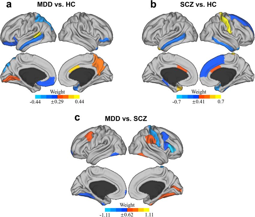
which was lower than the case-classification. Figure 1 79.21, and 69.88%, respectively. The classification be-
present the top 10 averaged weights of the brain regions tween adolescents with MDD and those with SCZ had a
in each classification model. Specifically, the right post- lower accuracy of 62.93%. Our findings indicate that ma-
central gyrus, the left temporal pole, and the right tem- chine learning using cortical thickness as the features
poral pole were the most important brain regions for can allow effective classification of psychiatric disorders
MDD-HC classification. On the other hand, the left among adolescents at an individual level.
Table 1 Clinical and demographic characteristics
Measures MDD (N = 67) SCZ (N = 49) HC (N = 34) p-value
Age (year) 16.22 ± 2.02 16.02 ± 1.80 16.32 ± 2.99 0.81a
Intracranial Volume 1459.73 ± 127.77 1448.33 ± 141.57 1484.68 ± 108.24 0.44a
Length of Current Episode (months) 7.88 ± 9.19 6.24 ± 12.29 – 0.41b
Age at Onset (year) 15.13 ± 2.17 15.39 ± 2.10 – 0.53b
Male (%) 28 (41.79) 24 (48.98) 15 (44.12) 0.56c
Prior Exposure to Medicine (%) 34 (50.75) 42 (85.71) – < 0.0001d
First Episode (%) 48 (71.64) 42 (85.71) – 0.11d
Family History of Mental Disorders (%) 7 (10.45) 8 (16.33) – 0.41d
Physical Intervention (%) 12 (17.91) 27 (55.10) – < 0.0001d
Values indicate the mean ± SD
Abbreviations: MDD Major Depressive Disorder, SCZ schizophrenia, HC healthy controls
a
Statistic computed using F-test
b
Statistic computed using two-sample t-tests
c
Statistic computed using χ2 test
d
Statistic computed using Fisher’s exact testZhou et al. BMC Psychiatry (2021) 21:361 Page 5 of 9 Fig. 1 The top 10 thickness brain regions contributing to classification accuracy in the SVM. a Brain regions with their thickness having the highest weight to distinguish patients with major depression and healthy controls. b Brain regions with their thickness having the highest weight to distinguish patients with schizophrenia and healthy controls. c Brain regions with their thickness having the highest weight to distinguish patients with major depression and schizophrenia Our findings indicate that structural brain MRI im- consistent with previous findings that patients with aging can be used to effectively identify MDD and SCZ childhood-onset schizophrenia presented with bilateral in adolescents. Previous studies widely used different deficits in the temporal, prefrontal, and parietal cortices structural indexes, including subcortical volume, gray [62]. Moreover, using the machine learning technique, matter density, gray matter volume, cortical thickness, cortical thickness has been reported to predict future- and cortical area, and all of them could distinguish be- onset of depression in adolescents with an accuracy of tween adults with psychiatric disorders and healthy 70% [63]. Besides, with volumes of both subcortical and adults [38, 58–60]. In this study, we focused on adoles- cerebellar regions as the feature set, the classification cent psychiatric patients and collected structural MRI model of MDD-SCZ resulted in a significant accuracy of data from patients with MDD and SCZ. Different struc- 62.93% (p = .044). This is because some subcortical nu- tural indexes were used as SVM model features respect- clei are also linked to MDD and SCZ, such as amygdala, ively for case- classification and MDD-SCZ which is associated with emotion [64, 65]. In addition, classification. Unlike previous findings on adults, only there is an interesting finding that we succeed to distin- cortical thickness could provide the best accuracy in guish adolescents with SCZ and HC but Thormodsen three adolescent classification models in our study. This et al. not [26]. In their study., there are no significant is consistent with the findings by Qiu et al., who used an evidence of cortical thickness difference between adoles- SVM based on various brain morphometric features to cent with SCZ and HC based on univariate analysis. In distinguish between 32 adult patients with first-episode our study, to further explore the brain morphology of MDD and 32 HC [61]. They reported that multiple cor- adolescents with SCZ, we succeed to distinguish them tical features could discriminate them with cortical using multivariate analysis. Although no significant evi- thickness providing the highest accuracy. Our findings dence is found in cortical thickness of each brain region indicate that the cortical thickness is already altered in between the two groups, there may be a particular adolescent patients with MDD and SCZ. This is spatial pattern of abnormal changes in cortical thickness
Zhou et al. BMC Psychiatry   (2021) 21:361                                                                       Page 6 of 9

across brain regions in adolescents with SCZ. That may         with higher depression levels were found to have poorer
be why we are successful. In a word, cortical thickness is     response inhibition and to perform worse on the stop-
a crucial structural brain index for identifying adolescent    signal task [89]. Neuroimaging studies have reported
patients with psychiatric disorders.                           that patients with MDD have increased functional con-
   For distinguishing adolescent patients with MDD from        nectivity in the right pars triangularis of the inferior
HC, the most important brain region was the temporal           frontal gyrus [90, 91]. This indicates a strong correlation
pole. The temporal pole, which is a node of the paralim-       of the right pars triangularis with depression in adoles-
bic system, plays an important role in socioemotional          cents. Patients with SCZ also present with reduced gray
and cognitive processing [66]. Defects in these processes      matter volume in the right inferior frontal gyrus [92].
are associated with depression [67, 68]. Gray matter           However, this is attributed to the generalized neuro-
abnormities in the temporal pole have been reported in         psychological impairment associated with SCZ rather
medication-naive patients with first-episode MDD [69,          than impaired inhibitory behavioral control, which is a
70]. Compared to healthy controls, individuals with de-        specific cognitive impairment [93]. Taken together, these
pression present with greater activation of the right an-      findings indicated that the right pars triangularis is asso-
terior temporal pole [71]. Previous studies also reported      ciated with response inhibition in adolescents with
abnormal functional connections between the right tem-         MDD and could be used to distinguish between adoles-
poral pole and other brain regions in patients with MDD        cent patients with MDD and SCZ.
[72–74]. Given the emotional instability in adolescents           This study has several limitations. First, the accuracies
and the abnormal emotional response to external stim-          of our models were all < 80%. To improve accuracy, we
uli, abnormal changes are more likely to occur in the          combined other indexes (cortical volume, cortical area,
temporal pole [75]. Therefore, the structure of this re-       and cerebellar-subcortical volume) with cortical thick-
gion could be used as a crucial biomarker for adolescent       ness as the feature set. An additional table presents these
depression.                                                    results [see Additional file 2]. After adding additional in-
   The left banks of the superior temporal sulcus, which       dexes into the feature set, no improved prediction accur-
is a crucial association area for biological motion percep-    acy was found. To prevent model overfitting and to
tion, was the most significant brain region for distin-        improve accuracy, we applied the least absolute shrink-
guishing between adolescents with SCZ and HC [76].             age and selection operator for feature selection and di-
The superior temporal sulcus is part of a neural circuit       mensionality reduction [94]. However, this did not
involved in perceiving intention from action and reac-         reduce dimensions, which could be attributed to the
tions to social and emotional events [77, 78]. Many stud-      complexity of brain structures and small sample size. In
ies have reported a reduced ability to extract social          addition, it is a limitation that we exclude those patients
information from bodily cues in patients with SCZ [79–         with psychiatric comorbidities to maximize the group
82]. Neuroimaging studies have reported that patients          difference to train the classifier. The patients with co-
with SCZ present with an aberrant pattern of superior          morbidities are valuable cases to investigate for diagnos-
temporal sulcus activity during basic biological motion        tic purposes. In the future, we will apply the models
tasks [83, 84]. Matsumoto et al. reported a negative cor-      here to these groups. Moreover, we obtained our sample
relation of the behavioral performance on basic bio-           from a single center. It is not clear whether our results
logical motion perception tasks and the gray matter            are reproducible and generalizable. In future studies, we
volume of the superior temporal sulcus in patients with        will obtain multi-center samples to validate these find-
SCZ [85]. Similarly, we observed adolescents with SCZ          ings and continue to focus on early psychiatric
had thinning cortical thickness of the left banks of the       disorders.
superior temporal sulcus than HC (p = 0.005, FDR cor-
rected). Our findings indicate that the superior temporal      Conclusions
sulcus could be associated with impaired extraction of         In summary, using a machine learning technique, we
social information in adolescents with SCZ.                    found that cortical thickness contributed toward distin-
   In our study, the most important brain region that dis-     guishing adolescent patients with MDD and SCZ. This
tinguishing between MDD and SCZ was the right pars             indicates that there are early-life structural brain abnor-
triangularis. The pars triangular is located in the inferior   malities in patients with MDD and SCZ. These findings
frontal gyrus, which is a crucial brain region for emo-        contribute toward biomarker-based clinical diagnosis
tional and cognitive control circuits [86]. Deng et al. re-    and demonstrate the utility of pattern recognition in ex-
ported that the right inferior frontal gyrus is highly         ploring the neurological basis of psychiatric disorders.
activated in a stop-signal task involving motor inhibitory     Further, this study provides an evidence regarding the
responses [87]. Damage to this area impairs the perform-       correct identification of adolescent psychiatric disorders
ance of the stop-signal task [88]. Moreover, individuals       based on neuroimaging. Future studies will focus on
Zhou et al. BMC Psychiatry          (2021) 21:361                                                                                                     Page 7 of 9

identifying other psychiatric disorders to improve the                            Competing interests
identification accuracy of specific diseases to contribute                        The authors declare that there are no conflicts of interest.

to early diagnosis and treatment of psychiatric diseases.                         Author details
                                                                                  1
                                                                                   Key Laboratory of Cognition and Personality (SWU), Ministry of Education,
Abbreviations                                                                     Chongqing 400715, China. 2Faculty of Psychology, Southwest University, No.2
MDD: Major depressive disorder; SCZ: Schizophrenia; HC: Healthy controls;         Tiansheng Road, Beibei District, Chongqing 400715, China. 3Faculty of
MRI: Magnetic resonance imaging; SVM: Support vector machine;                     Psychology, Shandong Normal University, Jinan 250014, Shandong, China.
                                                                                  4
BA: Balanced accuracy; FDR: False discovery rate; PI: Posterior probability        Department of Psychiatry, The First Affiliated Hospital of Chongqing Medical
interval; CI: Confidence interval; per_CIs: Confidence intervals of permutation   University, No.1, Yixueyuan Road, Yuzhong District, Chongqing 400016,
test                                                                              China. 5Sleep and Psychology Center, The Bishan Hospital of Chongqing,
                                                                                  Chongqing 402760, China. 6Department of Psychology, Chongqing Health
                                                                                  Center for Women and Children, Chongqing 401147, China. 7Collaborative
Supplementary Information                                                         Innovation Center of Assessment Toward Basic Education Quality, Southwest
The online version contains supplementary material available at https://doi.      University Branch, Beijing Normal University, Beijing 100875, China.
org/10.1186/s12888-021-03373-1.
                                                                                  Received: 14 October 2020 Accepted: 8 July 2021
 Additional file 1: Supplementary Table 1. Comparison of the model
 classification accuracy of the different brain indexes.
 Additional file 2: Supplementary Table 2. Comparison of the model                References
 classification accuracy of gray matter thickness with the different brain        1. Gore FM, Bloem PJ, Patton GC, Ferguson J, Joseph V, Coffey C, et al. Global
 indexes.                                                                             burden of disease in young people aged 10-24 years: a systematic analysis.
                                                                                      Lancet. 2011;377(9783):2093–102. https://doi.org/10.1016/S0140-6736(11
 Additional file 3: Supplementary Table 3. The processed data
                                                                                      )60512-6.
 supporting the conclusions of this article (cortical thickness).
                                                                                  2. Whiteford HA, Degenhardt L, Rehm J, Baxter AJ, Ferrari AJ, Erskine HE, et al.
 Additional file 4: Supplementary Table 4. The mean feature weight                    Global burden of disease attributable to mental and substance use
 and cross-validation ratio of all brain regions.                                     disorders: findings from the global burden of disease study 2010. Lancet.
 Additional file 5: Supplementary Table 5. The t-value and p-value be-                2013;382(9904):1575–86. https://doi.org/10.1016/S0140-6736(13)61611-6.
 tween different groups using t-test in univariate analysis (FDR corrected).      3. Lee FS, Heimer H, Giedd JN, Lein ES, Šestan N, Weinberger DR, et al. Mental
                                                                                      health. Adolescent mental health--opportunity and obligation. Science (80-
                                                                                      ). 2014;346:547–9.
Acknowledgements                                                                  4. Samsom JN, Wong AHC. Schizophrenia and depression co-morbidity: what
Not applicable.                                                                       we have learned from animal models. Front Psychiatry. 2015;6:13. https://
                                                                                      doi.org/10.3389/fpsyt.2015.00013.
                                                                                  5. Chunhua Z, Dezhi K, Xiaodong Z, Wei W, Rong X, Gongying L, et al.
Authors’ contributions
                                                                                      Rethinking schizophrenia and depression comorbidity as one psychiatric
Z.Z., J.Q. and D.W. designed the study. J.T., Y.P. and Y.F. collected the data.
                                                                                      disorder entity: evidence from mouse model. Front Neurosci. 2020;14:115.
Z.Z. and K.W. analyzed the data. Z.Z., K.W and L.S. drafted the manuscript. All
                                                                                  6. Rachel U, Birchwood M, Ross K, Brunett K, McCollum R, Jones L. The
authors contributed to revisions and approved the final version of the
                                                                                      evolution of depression and suicidality in first episode psychosis. Acta
manuscript for submission.
                                                                                      Psychiatr Scand. 2010;122(3):211-8.
                                                                                  7. Conley RR, Ascher-Svanum H, Zhu B, Faries DE, Kinon BJ. The burden of
Funding                                                                               depressive symptoms in the long-term treatment of patients with
This work was supported by the National Natural Science Foundation of                 schizophrenia. Schizophr Res. 2006;90(1-3):186-97.
China [31771231, 32071070, 32000760], Chongqing Science and Technology            8. Nasrettin S, Lie RK, Andreasssen OA, Ingrid M, Ivar RJ. Depressive symptoms
Commission [cstc2018jcyjAX0252, cstc2016shmszx130051], Chongqing                      in first episode psychosis: a one-year follow-up study. BMC Psychiatry. 2013;
Municipal Education Commission [2017SKG017], Chongqing Health                         13:106.
Commission and Chongqing Science and Technology Commission                        9. Häfner H, Maurer K, Trendler G, an der Heiden W, Schmidt M, Könnecke R.
[2019MSXM045], Natural Science Foundation of Chongqing [cstc2019jcyj-                 Schizophrenia and depression: challenging the paradigm of two separate
msxmX0520, cstc2020jcyj-msxmX0299], the planned project of Chongqing                  diseases—A controlled study of schizophrenia, depression and healthy
humanities and Social Sciences [2018PY80, 2019PY51], China Postdoctoral               controls. Schizophr Res. 2005;77(1):11-24.
Science Foundation Funded Project [2019 M662433], Fundamental Research            10. Schothorst PF, Emck C, van Engeland H. Characteristics of early psychosis.
Funds for the Central Universities [SWU119007], Postdoctoral Innovation Pro-          Compr Psychiatry. 2006;47(6):438–42. https://doi.org/10.1016/j.comppsych.2
ject in Shandong Province, Chang Jiang Scholars Program, National Out-                006.03.003.
standing Young People Plan, Chongqing Talent Program.                             11. Gournellis R, Oulis P, Howard R. Psychotic major depression in older people:
                                                                                      a systematic review. Int J Geriatr Psychiatry. 2014;29(8):784–96. https://doi.
Availability of data and materials                                                    org/10.1002/gps.4065.
The datasets analysed during the current study are available in                   12. Seon-Cheol P, Hwa-Young L, Jeong-Kyu S, Tae-Youn J, Min-Soo L, Jae-Min K,
Additional file 3. The code analysed during the current study is available in         et al. Distinctive clinical correlates of psychotic major depression: the CRES
https://github.com/zhouzheyi/adolescent_psychiatry_svm.                               CEND study. Psychiatry Investig. 2014;11(3):281-9.
                                                                                  13. Barch D, Sheline Y, Csernansky J, Snyder A. Working memory and prefrontal
                                                                                      cortex dysfunction: specificity to schizophrenia compared with major
Declarations                                                                          depression. Biol Psychiatry. 2003;53(5):376–84. https://doi.org/10.1016/
                                                                                      S0006-3223(02)01674-8.
Ethics approval and consent to participate                                        14. Snyder HR. Major depressive disorder is associated with broad impairments
This study was approved by the Local Medical Ethics Committee of the First            on neuropsychological measures of executive function: a meta-analysis and
Affiliated Hospital of Chongqing Medical University. All the study participants       review. Psychol Bull. 2013;139(1):81–132. https://doi.org/10.1037/a0028727.
provided written assent and their legal guardians provided written informed       15. Pain O, Dudbridge F, Cardno AG, Freeman D, Lu Y, Lundstrom S, et al.
consent.                                                                              Genome-wide analysis of adolescent psychotic-like experiences shows
                                                                                      genetic overlap with psychiatric disorders. Am J Med Genet B
Consent for publication                                                               Neuropsychiatr Genet. 2018;177(4):416–25. https://doi.org/10.1002/ajmg.
Not applicable.                                                                       b.32630.
Zhou et al. BMC Psychiatry (2021) 21:361 Page 8 of 9
16. Paksarian D, Trabjerg B, Merikangas K, Mors O, Børglum A, Hougaard D, et al. 37. Bhaumik R, Jenkins LM, Gowins JR, Jacobs RH, Barba A, Bhaumik DK, et al.
Adolescent residential mobility, genetic liability and risk of schizophrenia, Multivariate pattern analysis strategies in detection of remitted major
bipolar disorder and major depression. Br J Psychiatry. 2020;217(1):390-6. depressive disorder using resting state functional connectivity. Neuroimage
17. Patel V, Flisher AJ, Hetrick S, McGorry P. Mental health of young people: a Clin. 2017;16:390–8. https://doi.org/10.1016/j.nicl.2016.02.018.
global public-health challenge. Lancet. 2007;369(9569):1302–13. https://doi. 38. Koutsouleris N, Meisenzahl EM, Borgwardt S, Riecher-Rossler A, Frodl T,
org/10.1016/S0140-6736(07)60368-7. Kambeitz J, et al. Individualized differential diagnosis of schizophrenia and
18. Paus T, Keshavan M, Giedd JN. Why do many psychiatric disorders emerge mood disorders using neuroanatomical biomarkers. Brain. 2015;138(Pt 7):
during adolescence? Nat Rev Neurosci. 2008;9(12):947–57. https://doi.org/1 2059–73. https://doi.org/10.1093/brain/awv111.
0.1038/nrn2513. 39. Cortes C, Vapnik V. Support-vector networks. Mach Learn. 1995;20(3):273–97.
19. Wei S, Womer F, Geng H, Jiang X, Zhou Q, Chang M, et al. Similarities and https://doi.org/10.1007/BF00994018.
differences of functional connectivity in drug-naïve, first-episode adolescent 40. First M, Spitzer RL, Gibbon ML, Williams J. Structured clinical interview for
and young adult with major depressive disorder and schizophrenia. Sci Rep. DSM-IV-TR Axis I disorders. research version, non-patient edition. New York
2017;7(1):44316. https://doi.org/10.1038/srep44316. State Psychiatric Institute; 2002.
20. World Health Organization. Global health risks : mortality and burden of 41. Salat DH, Buckner RL, Snyder AZ, Greve DN, Desikan RS, Busa E, et al.
disease attributable to selected major risks. 2009. https://extranet.who.int/ Thinning of the cerebral cortex in aging. Cereb Cortex. 2004;14(7):721–30.
iris/restricted/handle/10665/44203. https://doi.org/10.1093/cercor/bhh032.
21. Patton GC, Viner RM, Linh le C, Ameratunga S, Fatusi AO, Ferguson BJ, et al. 42. Fischl B, Dale AM. Measuring the thickness of the human cerebral cortex
Mapping a global agenda for adolescent health. J Adolesc Health. 2010; from magnetic resonance images. Proc Natl Acad Sci U S A. 2000;97(20):
47(5):427–32. https://doi.org/10.1016/j.jadohealth.2010.08.019. 11050–5. https://doi.org/10.1073/pnas.200033797.
22. Fraguas D, Diaz-Caneja CM, Pina-Camacho L, Janssen J, Arango C. 43. Fischl B, van der Kouwe A, Destrieux C, Halgren E, Segonne F, Salat DH,
Progressive brain changes in children and adolescents with early-onset et al. Automatically parcellating the human cerebral cortex. Cereb Cortex.
psychosis: a meta-analysis of longitudinal MRI studies. Schizophr Res. 2016; 2004;14(1):11–22. https://doi.org/10.1093/cercor/bhg087.
173(3):132–9. https://doi.org/10.1016/j.schres.2014.12.022. 44. Klapwijk ET, van de Kamp F, van der Meulen M, Peters S, Wierenga LM.
23. Insel TR. Rethinking schizophrenia. Nature. 2010;468(7321):187–93. https:// Qoala-T: a supervised-learning tool for quality control of FreeSurfer
doi.org/10.1038/nature09552. segmented MRI data. Neuroimage. 2019;189:116–29. https://doi.org/10.1016/
24. Driver DI, Gogtay N, Rapoport JL. Childhood onset schizophrenia and early j.neuroimage.2019.01.014.
onset schizophrenia spectrum disorders. Child Adolesc Psychiatr Clin N Am. 45. Fischl B, Sereno MI, Dale AM. Cortical surface-based analysis. II: inflation,
2013;22(4):539–55. https://doi.org/10.1016/j.chc.2013.04.001. flattening, and a surface-based coordinate system. Neuroimage. 1999;9(2):
25. Grover S, Sahoo S, Nehra R. A comparative study of childhood/adolescent 195–207. https://doi.org/10.1006/nimg.1998.0396.
and adult onset schizophrenia: does the neurocognitive and psychosocial 46. Dale AM, Fischl B, Sereno MI. Cortical surface-based analysis. I Segmentation
outcome differ? Asian J Psychiatr. 2019;43:160–9. https://doi.org/10.1016/j.a and surface reconstruction. Neuroimage. 1999;9(2):179–94. https://doi.org/1
jp.2019.05.031. 0.1006/nimg.1998.0395.
26. Thormodsen R, Rimol LM, Tamnes CK, Juuhl-Langseth M, Holmen A, Emblem 47. Desikan RS, Segonne F, Fischl B, Quinn BT, Dickerson BC, Blacker D, et al. An
KE, et al. Age-related cortical thickness differences in adolescents with early- automated labeling system for subdividing the human cerebral cortex on
onset schizophrenia compared with healthy adolescents. Psychiatry Res. 2013; MRI scans into gyral based regions of interest. Neuroimage. 2006;31(3):968–
214(3):190–6. https://doi.org/10.1016/j.pscychresns.2013.07.003. 80. https://doi.org/10.1016/j.neuroimage.2006.01.021.
27. van Erp TGM, Walton E, Hibar DP, Schmaal L, Jiang W, Glahn DC, et al. 48. Winkler AM, Kochunov P, Blangero J, Almasy L, Zilles K, Fox PT, et al. Cortical
Cortical brain abnormalities in 4474 individuals with schizophrenia and 5098 thickness or grey matter volume? The importance of selecting the
control subjects via the enhancing neuro imaging genetics through Meta phenotype for imaging genetics studies. Neuroimage. 2010;53(3):1135–46.
analysis (ENIGMA) consortium. Biol Psychiatry. 2018;84(9):644–54. https://doi. https://doi.org/10.1016/j.neuroimage.2009.12.028.
org/10.1016/j.biopsych.2018.04.023. 49. Schmaal L, Hibar DP, Samann PG, Hall GB, Baune BT, Jahanshad N, et al.
28. Marin O. Developmental timing and critical windows for the treatment of Cortical abnormalities in adults and adolescents with major depression
psychiatric disorders. Nat Med. 2016;22(11):1229–38. https://doi.org/10.1038/ based on brain scans from 20 cohorts worldwide in the ENIGMA major
nm.4225. depressive disorder working group. Mol Psychiatry. 2017;22(6):900–9. https://
29. Bevan Jones R, Thapar A, Stone Z, Thapar A, Jones I, Smith D, et al. doi.org/10.1038/mp.2016.60.
Psychoeducational interventions in adolescent depression: a systematic 50. Lin Y, Li M, Zhou Y, Deng W, Ma X, Wang Q, et al. Age-related reduction in
review. Patient Educ Couns. 2018;101(5):804–16. https://doi.org/10.1016/j. cortical thickness in First-episode treatment-naive patients with
pec.2017.10.015. schizophrenia. Neurosci Bull. 2019;35(4):688–96. https://doi.org/10.1007/s122
30. Rice F, Riglin L, Lomax T, Souter E, Potter R, Smith DJ, et al. Adolescent and 64-019-00348-x.
adult differences in major depression symptom profiles. J Affect Disord. 51. Orru G, Pettersson-Yeo W, Marquand AF, Sartori G, Mechelli A. Using
2019;243:175–81. https://doi.org/10.1016/j.jad.2018.09.015. support vector machine to identify imaging biomarkers of neurological and
31. Truong W, Minuzzi L, Soares CN, Frey BN, Evans AC, MacQueen GM, et al. psychiatric disease: a critical review. Neurosci Biobehav Rev. 2012;36(4):
Changes in cortical thickness across the lifespan in major depressive 1140–52. https://doi.org/10.1016/j.neubiorev.2012.01.004.
disorder. Psychiatry Res. 2013;214(3):204–11. https://doi.org/10.1016/j. 52. Rubin-Falcone H, Zanderigo F, Thapa-Chhetry B, Lan M, Miller JM, Sublette
pscychresns.2013.09.003. ME, et al. Pattern recognition of magnetic resonance imaging-based gray
32. Reynolds S, Carrey N, Jaworska N, Langevin LM, Yang XR, Macmaster FP. matter volume measurements classifies bipolar disorder and major
Cortical thickness in youth with major depressive disorder. BMC Psychiatry. depressive disorder. J Affect Disord. 2018;227:498–505. https://doi.org/10.101
2014;14(1):83. https://doi.org/10.1186/1471-244X-14-83. 6/j.jad.2017.11.043.
33. Han KM, Choi S, Jung J, Na KS, Yoon HK, Lee MS, et al. Cortical thickness, 53. Chang C-C, Lin C-J. LIBSVM: a library for support vector machines. ACM
cortical and subcortical volume, and white matter integrity in patients with Trans Intell Syst Technol. 2011;2(3):1-27.
their first episode of major depression. J Affect Disord. 2014;155:42–8. 54. Koutsouleris N, Borgwardt S, Meisenzahl EM, Bottlender R, Möller H-J,
https://doi.org/10.1016/j.jad.2013.10.021. Riecher-Rössler A. Disease prediction in the at-risk mental state for psychosis
34. Mitelman SA. Transdiagnostic neuroimaging in psychiatry: a review. Psychiatry using neuroanatomical biomarkers: results from the FePsy study. Schizophr
Res. 2019;277:23–38. https://doi.org/10.1016/j.psychres.2019.01.026. Bull. 2012;38(6):1234–46. https://doi.org/10.1093/schbul/sbr145.
35. De Filippis R, Carbone EA, Gaetano R, Bruni A, Pugliese V, Segura-Garcia C, 55. Brodersen KH, Ong CS, Stephan KE, Buhmann JM. The balanced accuracy
et al. Machine learning techniques in a structural and functional MRI and its posterior distribution. Proc Int Conf Pattern Recognit. 2010;0:3121–4.
diagnostic approach in schizophrenia: a systematic review. Neuropsychiatr https://doi.org/10.1109/ICPR.2010.764.
Dis Treat. 2019;15:1605–27. https://doi.org/10.2147/NDT.S202418. 56. Ojala M, Garriga G. Permutation tests for studying classifier performance; 2009.
36. Gao S, Calhoun VD, Sui J. Machine learning in major depression: from 57. Kellner CH, Obbels J, Sienaert P. When to consider electroconvulsive
classification to treatment outcome prediction. CNS Neurosci Ther. 2018; therapy (ECT). Acta Psychiatr Scand. 2020;141(4):304–15. https://doi.org/1
24(11):1037–52. https://doi.org/10.1111/cns.13048. 0.1111/acps.13134.Zhou et al. BMC Psychiatry (2021) 21:361 Page 9 of 9
58. Gong Q, Wu Q, Scarpazza C, Lui S, Jia Z, Marquand A, et al. Prognostic 78. Blake R, Shiffrar M. Perception of human motion. Annu Rev Psychol. 2007;
prediction of therapeutic response in depression using high-field MR 58(1):47–73. https://doi.org/10.1146/annurev.psych.57.102904.190152.
imaging. Neuroimage. 2011;55(4):1497–503. https://doi.org/10.1016/j. 79. Kern RS, Penn DL, Lee J, Horan WP, Reise SP, Ochsner KN, et al. Adapting
neuroimage.2010.11.079. social neuroscience measures for schizophrenia clinical trials, part 2: trolling
59. Sacchet MD, Livermore EE, Iglesias JE, Glover GH, Gotlib IH. Subcortical the depths of psychometric properties. Schizophr Bull. 2013;39(6):1201–10.
volumes differentiate major depressive disorder, bipolar disorder, and https://doi.org/10.1093/schbul/sbt127.
remitted major depressive disorder. J Psychiatr Res. 2015;68:91–8. https:// 80. Peterman JS, Christensen A, Giese MA, Park S. Extraction of social
doi.org/10.1016/j.jpsychires.2015.06.002. information from gait in schizophrenia. Psychol Med. 2014;44(5):987–96.
60. Chin R, You AX, Meng F, Zhou J, Sim K. Recognition of schizophrenia with https://doi.org/10.1017/S003329171300144X.
regularized support vector machine and sequential region of interest 81. Vaskinn A, Sundet K, Ostefjells T, Nymo K, Melle I, Ueland T. Reading
selection using structural magnetic resonance imaging. Sci Rep. 2018;8(1): emotions from body movement: a generalized impairment in
13858. https://doi.org/10.1038/s41598-018-32290-9. schizophrenia. Front Psychol. 2015;6:2058.
61. Qiu L, Huang X, Zhang J, Wang Y, Kuang W, Li J, et al. Characterization of 82. Okruszek L, Pilecka I. Biological motion processing in schizophrenia -
major depressive disorder using a multiparametric classification approach systematic review and meta-analysis. Schizophr Res. 2017;190:3–10. https://
based on high resolution structural images. J Psychiatry Neurosci. 2014; doi.org/10.1016/j.schres.2017.03.013.
39(2):78–86. https://doi.org/10.1503/jpn.130034. 83. Kim J, Park S, Blake R. Perception of biological motion in schizophrenia and
62. Gogtay N, Weisinger B, Bakalar JL, Stidd R, de la Fernandez Vega O, Miller R, healthy individuals: a behavioral and FMRI study. PLoS One. 2011;6(5):
et al. Psychotic symptoms and gray matter deficits in clinical pediatric e19971. https://doi.org/10.1371/journal.pone.0019971.
populations. Schizophr Res. 2012;140(1-3):149–54. https://doi.org/10.1016/j. 84. Jimenez AM, Lee J, Reavis EA, Wynn JK, Green MF. Aberrant patterns of
schres.2012.07.006. neural activity when perceiving emotion from biological motion in
63. Foland-Ross LC, Sacchet MD, Prasad G, Gilbert B, Thompson PM, Gotlib IH. schizophrenia. Neuroimage Clin. 2018;20:380–7. https://doi.org/10.1016/j.
Cortical thickness predicts the first onset of major depression in adolescence. Int nicl.2018.08.014.
J Dev Neurosci. 2015;46(1):125–31. https://doi.org/10.1016/j.ijdevneu.2015.07.007. 85. Matsumoto Y, Takahashi H, Miyata J, Sugihara G, Murai T, Takahashi H.
64. Ferri J, Eisendrath SJ, Fryer SL, Gillung E, Roach BJ, Mathalon DH. Blunted Neural basis of altered earlier attention and higher order biological motion
amygdala activity is associated with depression severity in treatment- processing in schizophrenia. Soc Neurosci. 2018;13(5):594–601. https://doi.
resistant depression. Cogn Affect Behav Neurosci. 2017;17(6):1221–31. org/10.1080/17470919.2017.1366363.
https://doi.org/10.3758/s13415-017-0544-6. 86. Roberts G, Lord A, Frankland A, Wright A, Lau P, Levy F, et al. Functional
65. Lawrie SM, Whalley HC, Job DE, Johnstone EC. Structural and functional Dysconnection of the inferior frontal gyrus in Young people with bipolar
abnormalities of the amygdala in schizophrenia. Ann N Y Acad Sci. 2003; disorder or at genetic high risk. Biol Psychiatry. 2017;81(8):718–27. https://
985:445–60. https://doi.org/10.1111/j.1749-6632.2003.tb07099.x. doi.org/10.1016/j.biopsych.2016.08.018.
66. Olson IR, Plotzker A, Ezzyat Y. The enigmatic temporal pole: a review of 87. Deng W, Rolls ET, Ji X, Robbins TW, Banaschewski T, Bokde ALW, et al.
findings on social and emotional processing. Brain. 2007;130(Pt 7):1718–31. Separate neural systems for behavioral change and for emotional responses
https://doi.org/10.1093/brain/awm052. to failure during behavioral inhibition. Hum Brain Mapp. 2017;38(7):3527–37.
67. Zobel I, Werden D, Linster H, Dykierek P, Drieling T, Berger M, et al. Theory https://doi.org/10.1002/hbm.23607.
of mind deficits in chronically depressed patients. Depress Anxiety. 2010; 88. Aron AR, Robbins TW, Poldrack RA. Inhibition and the right inferior frontal
27(9):821–8. https://doi.org/10.1002/da.20713. cortex: one decade on. Trends Cogn Sci. 2014;18(4):177–85. https://doi.org/1
68. Bistricky SL, Ingram RE, Atchley RA. Facial affect processing and depression 0.1016/j.tics.2013.12.003.
susceptibility: cognitive biases and cognitive neuroscience. Psychol Bull. 89. Legrand A, Price M. Emotionally Valenced stimuli impact response inhibition
2011;137(6):998–1028. https://doi.org/10.1037/a0025348. in those with substance use disorder and co-occurring anxiety and
69. Peng J, Liu J, Nie B, Li Y, Shan B, Wang G, et al. Cerebral and cerebellar gray depression symptoms. J Affect Disord. 2020;266:639–45. https://doi.org/10.1
matter reduction in first-episode patients with major depressive disorder: a 016/j.jad.2020.02.008.
voxel-based morphometry study. Eur J Radiol. 2011;80(2):395–9. https://doi. 90. Cai Y, Liu J, Zhang L, Liao M, Zhang Y, Wang L, et al. Grey matter volume
org/10.1016/j.ejrad.2010.04.006. abnormalities in patients with bipolar I depressive disorder and unipolar
70. Lener MS, Kundu P, Wong E, Dewilde KE, Tang CY, Balchandani P, et al. depressive disorder: a voxel-based morphometry study. Neurosci Bull. 2015;
Cortical abnormalities and association with symptom dimensions across the 31(1):4–12. https://doi.org/10.1007/s12264-014-1485-5.
depressive spectrum. J Affect Disord. 2016;190:529–36. https://doi.org/10.101 91. Rolls ET, Cheng W, Du J, Wei D, Qiu J, Dai D, et al. Functional connectivity of
6/j.jad.2015.10.027. the right inferior frontal gyrus and orbitofrontal cortex in depression. Soc Cogn
Affect Neurosci. 2020;15(1):75–86. https://doi.org/10.1093/scan/nsaa014.
71. Beauregard M, Paquette V, Levesque J. Dysfunction in the neural circuitry of
92. Guo F, Zhu YQ, Li C, Wang XR, Wang HN, Liu WM, et al. Gray matter volume
emotional self-regulation in major depressive disorder. Neuroreport. 2006;
changes following antipsychotic therapy in first-episode schizophrenia
17(8):843–6. https://doi.org/10.1097/01.wnr.0000220132.32091.9f.
patients: a longitudinal voxel-based morphometric study. J Psychiatr Res.
72. Cheng W, Rolls ET, Qiu J, Liu W, Tang Y, Huang CC, et al. Medial reward and
2019;116:126–32. https://doi.org/10.1016/j.jpsychires.2019.06.009.
lateral non-reward orbitofrontal cortex circuits change in opposite
93. Ethridge LE, Soilleux M, Nakonezny PA, Reilly JL, Hill SK, Keefe RS, et al.
directions in depression. Brain. 2016;139(Pt 12):3296–309. https://doi.org/10.1
Behavioral response inhibition in psychotic disorders: diagnostic specificity,
093/brain/aww255.
familiality and relation to generalized cognitive deficit. Schizophr Res. 2014;
73. Bai T, Zu M, Chen Y, Xie W, Cai C, Wei Q, et al. Decreased connection
159(2-3):491–8. https://doi.org/10.1016/j.schres.2014.08.025.
between reward systems and Paralimbic cortex in depressive patients. Front
94. Cui LB, Liu L, Wang HN, Wang LX, Guo F, Xi YB, et al. Disease definition for
Neurosci. 2018;12:462. https://doi.org/10.3389/fnins.2018.00462.
schizophrenia by functional connectivity using Radiomics strategy.
74. Pang Y, Chen H, Wang Y, Long Z, He Z, Zhang H, et al. Transdiagnostic and
Schizophr Bull. 2018;44(5):1053–9. https://doi.org/10.1093/schbul/sby007.
diagnosis-specific dynamic functional connectivity anchored in the right
anterior insula in major depressive disorder and bipolar depression. Prog
Neuro-Psychopharmacol Biol Psychiatry. 2018;85:7–15. https://doi.org/10.101 Publisher’s Note
6/j.pnpbp.2018.03.020. Springer Nature remains neutral with regard to jurisdictional claims in
75. Bailen N, Green L, Thompson R. Understanding Emotion in Adolescents: A published maps and institutional affiliations.
Review of Emotional Frequency, Intensity, Instability, and Clarity. Emot Rev.
2019;11(1):63-73.
76. Allison T, Puce A, McCarthy G. Social perception from visual cues: role of the
STS region. Trends Cogn Sci. 2000;4(7):267–78. https://doi.org/10.1016/S13
64-6613(00)01501-1.
77. Zacks JM, Braver TS, Sheridan MA, Donaldson DI, Snyder AZ, Ollinger JM,
et al. Human brain activity time-locked to perceptual event boundaries. Nat
Neurosci. 2001;4(6):651–5. https://doi.org/10.1038/88486.You can also read


