Dyad of CD40/CD40 Ligand Fosters Neuroinflammation at the Blood-Brain Barrier and Is Regulated via JNK Signaling: Implications for HIV-1 Encephalitis
←
→
Page content transcription
If your browser does not render page correctly, please read the page content below
9454 • The Journal of Neuroscience, July 14, 2010 • 30(28):9454 –9464

Neurobiology of Disease

Dyad of CD40/CD40 Ligand Fosters Neuroinflammation at
the Blood–Brain Barrier and Is Regulated via JNK Signaling:
Implications for HIV-1 Encephalitis
Servio H. Ramirez,1 Shongshan Fan,1 Holly Dykstra,1 Nancy Reichenbach,1 Luis Del Valle,1,2 Raghava Potula,1
Richard P. Phipps,3,4 Sanjay B. Maggirwar,3 and Yuri Persidsky1
Departments of 1Pathology and Laboratory Medicine, and 2Neuroscience, Temple University School of Medicine, Philadelphia, Pennsylvania 19140,
and Departments of 3Microbiology & Immunology, and 4Environmental Medicine, University of Rochester School of Medicine, Rochester, New York 14642

Human immunodeficiency virus 1 (HIV-1) infection may result in activation of peripheral monocytes followed by their infiltration into
the CNS, where the release of proinflammatory mediators causes neurologic disease. Previously, we detected high levels of soluble CD40
ligand (CD40L) in CSF and plasma of HIV-infected patients with cognitive impairment. We now show that CD40, a receptor for CD40L, is
highly expressed in brain endothelial cells of patients affected by HIV-1 encephalitis (HIVE), suggesting an important role for the
CD40/CD40L dyad in regulating blood– brain barrier (BBB) functions. This concept was further supported by in vitro experiments.
Exposure of primary human brain microvascular endothelial cells (BMVECs) to CD40L upregulated the expression of adhesion molecules
intracellular adhesion molecule-1 and vascular cell adhesion molecule-1, which caused a fourfold increase in monocyte adhesion to
BMVECs and stimulated migration across an in vitro BBB model. Investigations into the intracellular signaling pathways that govern
these events revealed that cJUN-N-terminal kinase (JNK) is critical to CD40 activation in the BMVECs. CD40L induced activation of
mixed-lineage-kinase-3 and JNK, leading to the subsequent activation of cJUN/AP-1 (activating-protein-1). JNK inhibition in the BMVECs
prevented CD40L-mediated induction of adhesion molecules, monocyte adhesion, and transendothelial migration. These new findings support
the concept that the CD40/CD40L dyad plays an important role in HIVE neuroinflammation.

Introduction                                                                                                       matory mediators (Mondal et al., 2004; Ramirez et al., 2008)
Despite the introduction of highly active antiretroviral therapy                                                   control leukocyte trafficking into the CNS. Increased expres-
(HAART), which efficiently suppresses viral replication and nor-                                                   sion of adhesion molecules and BBB permeability has been
malizes immunologic parameters, a significant number of hu-                                                        demonstrated in HAND patients (Eugenin et al., 2006). A
man immunodeficiency virus 1 (HIV-1)-infected patients show                                                        disrupted BBB allows accumulation of toxic serum proteins
progressive loss of cognitive abilities. These cognitive deficits are                                              and increased infiltration of monocytes and lymphocytes,
collectively termed HIV-associated neurocognitive disorder                                                         thereby accelerating inflammation and viral entry into the
(HAND) (Boissé et al., 2008; Minagar et al., 2008). The patho-                                                    CNS. HAART fails to control BBB leakage and inflammation
genesis of HAND involves activation of monocytes and their sub-                                                    in HAND patients (Avison et al., 2004b; Eilers et al., 2008), in
sequent recruitment into the CNS, altering blood– brain barrier                                                    part because it does not reduce the high levels of CD40 ligand
(BBB) function and resulting in HIV-1 encephalitis (HIVE)                                                          (CD40L) found in the plasma and CSF of HIV-1-infected pa-
(Persidsky et al., 2006a). The effector molecules and mechanisms                                                   tients (Sipsas et al., 2002; Sui et al., 2007). As demonstrated in
that regulate monocyte migration across the BBB remain poorly                                                      other systems (Piguet et al., 2001; Ishikawa et al., 2005; Sitati et
defined. Enhanced expression of adhesion molecules on brain                                                        al., 2007), high levels of soluble CD40L (sCD40L) can regulate
microvascular endothelial cells (BMVECs) triggered by inflam-                                                      CNS inflammation at the level of the BBB.
                                                                                                                       CD40L (CD154) is a 33 kDa type II membrane glycoprotein
                                                                                                                   from the tumor necrosis factor ␣ (TNF␣) family. CD40L is ex-
Received Nov. 22, 2009; revised May 21, 2010; accepted May 30, 2010.                                               pressed predominantly by activated leukocytes and platelets (Li,
   This study was supported (in part) by research funding from National Institutes of Health (NIH) Grants          2008). In addition to the membrane-bound form of the protein,
RO1MH65151, R01AA17398, and RO1AA015913 (to Y.P.); RO1NS054578 (to S.B.M. and R.P.P.); PO1MH64570,
                                                                                                                   31 kDa and/or 18 kDa versions of CD40L can be secreted or shed
RO1MH56838, and RO1NS066801 (to S.B.M.); RO1DE011390 (to R.P.P. and S.B.M.); and RO1HL078603 and
RO1ES01247 (to R.P.P.). Human BMVECs were provided by Drs. Marlys H. Witte and Michael Bernas through contract     from activated cells. Either form of CD40L retains the ability to
with the University of Arizona Medical Center (Tucson, AZ). The NIH National NeuroAIDS Consortium is acknowl-      activate CD40, a 45 to 50 kDa type I membrane glycoprotein
edged for brain tissue specimens used in this study.                                                               expressed at a low level in resting cells of myeloid and vascular
   Correspondence should be addressed to Yuri Persidsky, Department of Pathology and Laboratory Medicine,          origin (Sui et al., 2007; Mancino et al., 2008; Pluvinet et al., 2008).
Temple University School of Medicine, 3401 N. Broad Street, Philadelphia, PA 19140. E-mail: yuri.persidsky@tuhs.
temple.edu.
                                                                                                                   CD40 expression is rapidly upregulated in these cells after expo-
   DOI:10.1523/JNEUROSCI.5796-09.2010                                                                              sure to proinflammatory mediators (Sui et al., 2007; Pluvinet et
Copyright © 2010 the authors 0270-6474/10/309454-11$15.00/0                                                        al., 2008).
Ramirez et al. • CD40/CD40L Fosters Neuroinflammation at the BBB J. Neurosci., July 14, 2010 • 30(28):9454 –9464 • 9455
Table 1. Clinical data for human brain tissue samples
Case Age Viral load
number HIV-1 infection (year) Gender (copies/ml) Other systemic diseases
a
1 HIVE severe 37 M 750,000 Pulmonary aspergillosis
2 HIVE severe 44 M 389,120 Cachexia, Kaposi sarcoma of lymph nodes, chronic hepatitis
3 HIVE severe 32 M 1,098,947 Bacterial bronchopneumonia
4 HIVE severe 42 M 6,940 Bacterial bronchopneumonia
5 HIV-1b 51 M 65 Pulmonary embolus, pulmonary aspergilloma
6 HIV-1 43 M 252,604 Pneumocystis carinii pneumonia, disseminated cytomegalovirus, liver steatosis
7 HIV-1 47 M 3,861 Hepatitis C, micronodular cirrhosis, focal segmental glomerulosclerosis
8 Seronegative control 33 F NA Cystic fibrosis, septicemia
9 Seronegative control 55 M NA Bronchopneumonia
10 Seronegative control 46 M NA Hepatitis C, cirrhosis, atherosclerosis
11 Seronegative control 52 M NA Status post aortic valve repair, myocardial infarction, bronchopneumonia
M, Male; F, female; NA, not applicable.
a
Severe HIVE is defined by the expression of human leukocyte antigen HLA-DR on 80 to 90% of microglia; HIV-1 infection of ramified microglia; formation of 1 to 2 microglial nodules and 25 to 50 infiltrating macrophages per 5 ⫻ 10-power
fields; and abundant multinucleated cells, as described by Mathur et al. (2004).
b
No significant neuropathologic changes were found.
Elevated levels of sCD40L are found in a variety of diseases in ing the above supplements but lacking ECGS and heparin. Under these
which sCD40L is thought to initiate or potentiate inflammation conditions, the BMVEC cultures were routinely evaluated for the pres-
(Tsakiris et al., 2000; Heeschen et al., 2003; Devaraj et al., 2006). ence of endothelial markers and barrier formation.
Inflammatory conditions increase the expression of the CD40 Primary human monocytes were supplied by the Human Immunol-
ogy Core at the University of Pennsylvania (Philadelphia, PA). The cells
receptor on the surface of endothelial cells and the shedding of
were isolated by countercurrent centrifugal elutriation and maintained
the ligand (Chai et al., 2006). In HIV-1 neuropathogenesis, a in DMEM containing heat-inactivated 10% FBS, penicillin (100 U/ml),
connection between CD40 and microglia has been established. streptomycin (100 U/ml), and L-glutamine (2 mM) (Ramirez et al., 2009),
Upregulation of CD40 expression has been detected on microglia and were used within 24 h of isolation.
of HIV-1-infected brain tissues (D’Aversa et al., 2005). CD40L Immunofluorescence staining and image analysis. Evaluation of CD40
was also shown to potentiate the ability of HIV-1 protein (Tat) to expression was performed on frozen brain tissue from seven HIV-1-
activate monocytes and microglia leading to the secretion of neu- infected patients [four with severe encephalitis (Persidsky et al., 2006b)
rotoxic inflammatory mediators (Sui et al., 2007). and three without evidence of encephalitis], and four seronegative age-
A role for the CD40/CD40L dyad in brain endothelium re- matched controls were provided by the National NeuroAIDS Consor-
mains largely unknown. Herein, we detected high levels of CD40 tium (Washington, DC) (Table 1). Indirect immunofluorescence was
on brain endothelium in patients with HIVE. We demonstrated performed on serial sections cut at a thickness of 5 m. Monoclonal
antibodies to human CD68 (diluted 1:200, Abcam), CD163 (1:200, Santa
that engagement of endothelial CD40 promotes adhesion and
Cruz Biotechnology), intercellular adhesion molecule-1 (ICAM-1), and
migration of leukocytes across an in vitro BBB model. Our studies vascular cell adhesion molecule-1 (VCAM-1) (diluted 1:200, R&D Sys-
also show that CD40 signaling converges to the cJUN-N-terminal tems) or polyclonal antibodies to human CD40 (diluted 1:50, Abcam),
kinase (JNK) signaling pathway, which was found to mediate the together with rhodamine-conjugated Ulex europeus agglutinin 1 lectin
effects of CD40L on the endothelial regulation of leukocyte ad- (UEA-1) (2 g/ml, Vector Laboratories), were placed on sections over-
hesion and migration. night at 4°C. Tissue sections were rinsed, and secondary antibodies con-
jugated to Alexa-488 (diluted 1:250, Invitrogen) were then added for 1 h.
Materials and Methods The slides were mounted with Prolong antifade reagent containing DAPI
Reagents. Recombinant human sCD40L was purchased from ProSpec. (Invitrogen).
Recombinant membrane-bound CD40L [CD40L(M)] and corresponding We performed semiquantitative image analysis to determine the up-
control membranes that lack CD40L were generated in a Baculovirus-based regulation of CD40 in HIVE. For each case, images from five fields were
expression system (Ray et al., 2005; Sui et al., 2007). Neutralizing anti- acquired with equal acquisition parameters and ⬍20⫻ objective magni-
bodies to human CD40, recombinant human TNF␣, and CC chemokine fication for both CD40 and UEA-1 using a Coolsnap EZ CCD camera
ligand 2 (CCL2)/monocyte chemoattractant protein 1 (MCP-1) were (Photometrics) coupled to a Nikon i80 Eclipse (Nikon). Up to 20 vessels
purchased from R&D Systems. The following inhibitors were obtained (with diameters between 30 and 60 m) per field were identified using
from Calbiochem: Janus kinase (JAK) inhibitor P6; JAK-3 inhibitor-VI the UEA-1 label and then were matched to the corresponding CD40
(3⬘-pyridyl oxindole derivative); IB-␣ kinase (IKK) inhibitor-X [N-(6- staining. The integrated intensity (average optical densities from all the
chloro-9H-b-carbolin-8-yl) nicotinamide)]; p38 inhibitor SB202190; pixels measured) was determined by placing a defined rectangular box
extracellular signal-regulated kinase (ERK) inhibitor FR180204; JNK (10 ⫻ 100 m) region of interest within the CD400-labeled vessels using
inhibitor I [(L)-HIV-TAT48 –57-PP-JBD20]; and JNK inhibitor II Axiovision imaging software (Zeiss). For display purposes, Axiovision
(SP600125). Unless specified, all other reagents were purchased from was also used to pseudo-color the acquired fluorescence images.
Sigma-Aldrich. Flow cytometry. Surface expression of adhesion molecules was mea-
Cell culture. Primary BMVECs were supplied by Dr. Michael Bernas sured in BMVECs by flow cytometry. Briefly, after various treatments
and Dr. Marlys Witte (University of Arizona, Tucson, AZ). BMVECs 2 ⫻ 10 6 cells were placed in staining solution (2% BSA in PBS with 0.5%
were isolated from vessels of normal tissue derived from brain resections NaN3) containing fluorophore-conjugated antibodies to CD40 (anti-
performed for the treatment of intractable epilepsy. The BMVEC cul- CD40-Phycoerythrin, R&D Systems), CD54 (anti-ICAM-1-allophy-
tures were used until passage 5 and were expanded in DMEM/F-12 media cocyanin, BD Biosciences), and CD106 (anti-VCAM-1 FITC, BD
supplemented with 10% heat-inactivated fetal bovine serum (FBS), en- Biosciences) for 30 min on ice. Cells were then washed and fixed in 2%
dothelial cell growth supplement (ECGS) (BD Bioscience), heparin (1 methanol-free formaldehyde (Thermo Scientific) in 1⫻ PBS. Acquisi-
mg/ml, Sigma), amphotericin B (2.5 g/ml, Invitrogen), penicillin (100 tion and analysis of the labeled cells were then performed using a FACS-
U/ml, Invitrogen), and streptomycin (100 g/ml, Invitrogen). Before Calibur flow cytometer (BD Biosciences). Acquisition parameters and
experimentation, BMVEC monolayers were placed with media contain- gating were controlled by CellQuest software (BD Biosciences). Data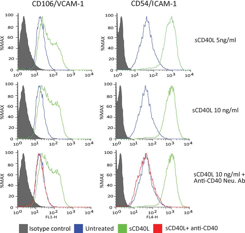
9456 • J. Neurosci., July 14, 2010 • 30(28):9454 –9464 Ramirez et al. • CD40/CD40L Fosters Neuroinflammation at the BBB Figure 1. Upregulation of CD40 expression in microvessels in HIV-1-infected brain tissue. Frontal cortex brain tissue specimens were obtained from seven cases of HIVE of different severity (moderate to severe according to previously described criteria) (Avison et al., 2004a) along with HIVE cases and seronegative age-matched controls (n ⫽ 4). Serial frozen sections (5 m thick) were cut and stained for CD68 (monocyte–macrophage marker, cyan). On serial sections, cerebral vessels were identified by double staining with UEA-1 (red) and antibodies to CD40 (green). A, Infiltration of monocytes (a feature of HIVE) was confirmed by staining of CD68 (V, Vessel; PS, perivascular space). B, Although no staining for CD40 was seen in controls, there was a substantial increase in CD40 levels in HIVE (arrowheads). C, Higher magnification demonstrated minimal-to-no staining for CD40 in controls and upregulation of CD40 in HIVE. In addition, glial cells were also CD40 positive in HIVE (arrowheads) but not in controls. Stained sections were observed by immunofluorescent microscopy (objective 10⫻ and 40⫻), and digital images were acquired by a cooled CCD camera. D, Semiquantitative assessments of CD40 levels were performed as described in Materials and Methods. CD40 was upregulated 2.5-fold and 5-fold in HIV-positive and severe HIVE cases, respectively, compared with controls. Data are shown as mean ⫾ SEM. *p ⬍ 0.01. Original magnification: A, B, 100⫻; C, 400⫻. analysis was performed with FlowJo software (Tree Star). The data rep- Transcription factor ELISA. Evaluation of DNA binding by activated resent the mean fluorescence intensity of gated populations (as deter- cJun was performed using the TransAM cJun transcription factor assay mined by isotype-matched controls) (supplemental Fig. S1 A, B, available (Active Motif). BMVECs (2 ⫻ 10 6) were exposed to increasing concen- at www.jneurosci.org as supplemental material) of at least 10,000 events trations of sCD40L or anisomysin (control) for 30 min followed by lysis recorded in a single experiment, which was repeated at least three times. and extraction of the nuclear fraction. Nuclear proteins (10 g) were Western blot. BMVEC monolayers were treated and lysed for either allowed to bind to oligonucleotide-coated 96-well plates. The bound total protein or nuclear protein. Cell lysates were prepared using the proteins were quantitated using the antibodies described above and mea- CelLytic-M cell lysis reagent (Sigma) containing protease inhibitor mix- suring absorbance at 450 nm in a Spectramax M5 (Molecular Devices). ture (Sigma) and phosphatase inhibitor mixture set I (Calbiochem). Pro- Adhesion and migration assays. Quantitative adhesion and migration teins from the nuclear fraction were isolated using the CelLytic nuclear assays were performed as previously described (Ramirez et al., 2008). For extraction kit (Sigma). The protein content in the lysate/fraction was adhesion and migration assays, BMVECs were seeded on collagen type then determined using the BCA protein assay (Thermo Scientific). Pro- I-coated 96-well plates (adhesion) or on 3 m 24-well tissue culture tein lysate/fraction containing 20 g of protein was mixed with 2⫻ Lae- inserts (FluoroBlok, BD Bioscience) at a density of 2.5 ⫻ 10 4 cells/well. mmli (sample loading) buffer containing -mercaptoethanol and then Confluent monolayers were then exposed to the indicated experimental boiled for 5 min. The proteins were then resolved by SDS-PAGE (4 –20% treatments. Freshly isolated human monocytes at 5 ⫻ 10 6 cells/ml were precast gels) (Thermo Scientific), followed by electrophoretic transfer to labeled with 5 M the fluorescent tracer, calcein-AM (Invitrogen). All nitrocellulose membranes. The primary antibodies were diluted in 1⫻ treatments were removed from the endothelial cells before adding mono- TBS/0.1% Tween 20 and used to detect the following proteins: anti- cytes at 2.5 ⫻ 10 5 cells/well or insert. CD40 (diluted 1:500); anti--tubulin (diluted 1:2000); anti-lamin For adhesion assays, the monocytes and endothelial cells were incu- A/C (diluted 1:2000) from Abcam; anti-ERK1/2 (diluted 1:1000); anti- bated together for 15 min and then rinsed three times with 1⫻ PBS to phospho (p)-ERK1/2 thr202/tyr204 (diluted 1:1000); anti-JNK eliminate nonadherent monocytes. For migration assays, the monocytes (1:500); anti-p-JNK thr183/tyr185 (diluted 1:250); anti-MLK3 (diluted were placed for 2 h in the upper chamber of the tissue culture insert 1:500); anti-p-MLK3 thr277/ser281 (diluted 1:250); anti-cJun (diluted system, while chemoattractant CCL2/MCP-1 (50 ng/ml) was added to 1:1000); and anti-p-cJun ser63 (diluted 1:500) (all purchased from Cell the lower chamber to stimulate migration. The fluorescence of adherent Signaling Technology). All antibodies (except for -tubulin, 1 h at room monocytes or migrated monocytes was measured using a Spectramax M5 temperature) were incubated with the membranes overnight at 4°C un- fluorescence plate reader (Molecular Devices). The number of migrated der gentle shaking. Bound primary antibodies were exposed to the cor- monocytes was determined from external standards of known numbers responding species-specific peroxidase-conjugated secondary antibody of labeled monocytes. The results for adhesion are represented as the (diluted 1:5000) (Thermo Scientific) for 1 h at room temperature and mean ⫾ SEM fold adhesion (number of adherent monocytes for each detected using the supersignal West-Femto chemluminescent substrate experimental condition divided by the basal adhesion of the untreated (Thermo Scientific), acquired on a G:Box Chemi HR16 (Syngene) gel control). The results for migration are shown as the average fold migra- documentation system. tion ⫾ SEM; the fold migration is derived from the number of migrated
Ramirez et al. • CD40/CD40L Fosters Neuroinflammation at the BBB                                                              J. Neurosci., July 14, 2010 • 30(28):9454 –9464 • 9457

                                                                                                                                 culature. Control brains demonstrated a
                                                                                                                                 minimal level of CD40 expression (Fig.
                                                                                                                                 1 B, case 8); however, CD40 expression
                                                                                                                                 was readily detectable in endothelial cells
                                                                                                                                 (Fig. 1 B, arrowheads) in brain tissues af-
                                                                                                                                 fected by severe HIVE (Fig. 1 B, cases 1, 2,
                                                                                                                                 3, and 4) and to a lesser extent in tissue
                                                                                                                                 from HIV-1-infected patients without
                                                                                                                                 HIVE (data not shown). Consistent with
                                                                                                                                 previous observations (D’Aversa et al.,
                                                                                                                                 2002), astrocytes and microglia in severe
                                                                                                                                 HIVE cases were positive for CD40 (Fig.
                                                                                                                                 1C, cases 1 and 4), and no immunostain-
                                                                                                                                 ing was detected in controls (Fig. 1C, case
                                                                                                                                 8). The increase in CD40 immunostaining
                                                                                                                                 seen in HIVE cases paralleled the upregu-
                                                                                                                                 lation of adhesion molecules in brain en-
                                                                                                                                 dothelium, VCAM-1 (supplemental Fig.
                                                                                                                                 S2 B, available at www.jneurosci.org as
Figure 2. Brain endothelial cells exposed to CD40L increase monocyte adhesion and transendothelial migration. A, Basal supplemental material), and to a lesser de-
expression of CD40 was found in BMVECs, but was undetectable in resting HeLa cells. Analysis of adhesion was performed with a gree ICAM-1 (data not shown) compared
fluorescence-based assay using primary human BMVECs and monocytes as described in Materials and Methods. After the forma- with controls. This new finding suggested
tion of monolayers, the BMVECs were treated with the indicated test conditions or with TNF␣ (20 ng/ml) for 24 h. Treatments were that CD40 plays a role in promoting leu-
removed and fluorescent-labeled monocytes were added to the BMVECs. B, Adhesion assay demonstrating endothelial activation
                                                                                                                                 kocyte infiltration across the BBB.
and subsequent monocyte adhesion induced by increasing concentrations of membrane-bound CD40L [shown as CD40L(M)].
Control membrane [CTR(M)] preps lacking CD40L had no appreciable induction in adhesion. Coincubation of CD40L(M) with                Next, we assessed CD40 expression in
neutralizing antibodies (Neut. AbCD40) to CD40 blocked the effects of CD40L. C, Adhesion assays with sCD40L also shows a dose brain endothelium. For each case, up to
response in adhesion that is blocked by the presence of CD40-neutralizing antibodies. Transendothelial migration assays were five fields were acquired at ⬍20⫻ objec-
performed as describe in Materials and Methods. The indicated treatments were added for 24 h and then removed, followed by tive magnification for both CD40 and
monocyte addition and chemotaxis toward the relevant chemokine CCL2/MCP-1 (50 ng/ml). D, E, A dose-dependent increase in UEA-1. Up to 20 vessels per field were
monocyte migration was found after addition of CD40L(M) (D) and sCD40L (E) to BMVEC monolayers. The effects of either form of identified using UEA-1 staining and then
CD40L were specific since the degree of migration induced by CD40L was eliminated by antibody neutralization. Of note, non- matched to the corresponding CD40 la-
chemoattractant-driven migration also showed an increase in basal migration when BMVECs were stimulated with either beling. The integrated intensity was mea-
membrane-bound or soluble CD40L. Data shown as mean ⫾ SEM. *p ⬍ 0.01.                                                            sured by placing a defined rectangular
                                                                                                                                 region of interest on the vessel. We found
monocytes for each experimental condition divided by the number of                                 that CD40 expression was upregulated over twofold in HIV-1
migrated monocytes in the untreated, no chemoattractant control.                                   patients (without HIVE) and over fivefold in cases of HIVE when
   Statistical analysis. The values shown in all figures and those men-
                                                                                                   compared with the seronegative controls (Fig. 1 D, p ⬍ 0.01).
tioned in the text represents the average ⫾ SEM of experiments that were
conducted multiple times (as indicated). Statistical significance ( p ⬍
                                                                                                   These data suggest that CD40 expression is indeed increased in
0.05) was determined by performing unpaired, two-tailed Student’s t test                           brain endothelium during HIVE, and levels appear elevated even
or ANOVA using Prism v5 software (GraphPad Software).                                              in HIV-1 infection without evidence of encephalitis.

Results                                                                                  CD40L promotes monocyte adhesion to and migration across
CD40 is upregulated in brain endothelium from patients                                   human BMVECs
with HIVE                                                                                Increased expression of CD40 can potentially promote leukocyte
To establish clinical relevance of CD40 expression in BMVECs in                          adhesion to and migration across endothelium. Indeed, aug-
the context of HIV-1 infection, we evaluated the CD40 levels in                          mented CD40L levels have been found in inflammatory condi-
the frontal cortex derived from seven HIV-1-infected patients, of                        tions including HIV-1 CNS infection (Sui et al., 2007; Rizvi et al.,
which four cases were determined to be representative of severe                          2008). However, the ability of CD40L to mediate monocyte ad-
HIVE (Persidsky et al., 1999; Persidsky et al., 2006b) and no                            hesion to and/or migration across brain endothelium has not
neuropathologic changes were detected in three other cases (Ta-                          been studied. Therefore, we sought to determine whether CD40L
ble 1). As a negative control, the CD40 levels were also examined                        could affect monocyte adhesion to BMVEC monolayers. We con-
in frontal cortex obtained from seronegative patients. Since the                         ducted immunoblot analyses and found detectable amounts of
degree of neurologic deficit in HIV-infected individuals is                              CD40 in the lysates of BMVECs, but not in negative control cells
strongly correlated with the number of activated macrophages                             (HeLa) (Fig. 2A).
and microglia within the basal ganglia and frontal lobes (Persidsky                         Next, we performed monocyte adhesion assays in which
et al., 1999; Avison et al., 2004b), we first verified the severity of                   monolayers of BMVECs were exposed to two forms of CD40Ls,
HIVE by detecting CD68-positive cells in the perivascular spaces.                        membrane-bound CD40L and CD40L(M); this form of CD40L
Although very few CD68 cells were found in control cases, HIVE                           mimics CD40L (that is expressed on the surface of activated cells)
brains featured mononuclear cell infiltration (Fig. 1 A) (see addi-                      and sCD40L (similar to CD40L that is shed or released by acti-
tional histopathology in supplemental Fig. S2 A, available at                            vated cells). Doses of CD40L(M) were similar to those previously
www.jneurosci.org as supplemental material). Next, we exam-                              validated in cell culture (Ray et al., 2005; Ryan et al., 2005). Sol-
ined CD40 expression by staining with anti-CD40 antibodies and                           uble CD40L was applied in concentrations used by other investi-
double-labeling with the lectin UEA-1 to identify the microvas-                          gators (Kehry and Castle, 1994). Adhesion assays revealed that
9458 • J. Neurosci., July 14, 2010 • 30(28):9454 –9464 Ramirez et al. • CD40/CD40L Fosters Neuroinflammation at the BBB
exposure to CD40L(M) induced a fivefold
increase in the number of monocytes at-
taching to the BMVEC monolayers ( p ⬍
0.01) (Fig. 2 B). This effect of CD40L(M)
was specific to CD40L, at all doses. Expo-
sure to equivalent amounts of control
membranes that lack CD40L resulted in
monocyte attachment comparable to un-
treated BMVECs. Similarly, sCD40L pro-
duced ⬃3.5-fold increase in monocyte
adhesion over untreated BMVECs (Fig.
2C). Increases in monocyte adhesion were
completely blocked after coadministra-
tion of CD40 neutralizing antibodies, but
not by addition of isotype-matched non-
immune serum (Fig. 2 B, C).
Analogous to sCD40L (Sipsas et al.,
2002; Sui et al., 2007), high levels of TNF␣
are also present in the blood of HIV-1-
infected patients especially in those with
greater BBB impairment (Sharief et al.,
1992). We sought to determine whether
TNF␣ augments the ability of CD40L to
induce monocyte adhesion. Pretreatment
of BMVECs with TNF␣ induced mono-
cyte adhesion by almost fourfold; this was
further enhanced after coadministration
of sCD40L, suggesting an additive effect of
these two proinflammatory mediators on
monocyte adhesion (Fig. 2C). Figure 3. Increased surface expression of ICAM-1 and VCAM-1 on brain endothelial cells after sCD40L exposure. FACS analyses
Subsequently, we tested whether of adhesion molecules ICAM-1 and VCAM-1 were performed in BMVECs exposed to increasing concentrations of sCD40L. Induction
CD40 engagement in BMVECs would of adhesion molecules ICAM-1 and VCAM-1 was observed after sCD40L exposure for 24 h. The upregulation of adhesion molecules
promote monocyte passage (migration) was also dose dependent and was prevented by introduction of neutralizing anti-CD40 antibodies.
across BMVEC monolayers. BMVECs
were treated with both CD40L(M) and sCD40L, after which fluo- nearly a fourfold to fivefold increase in ICAM-1 expression was
rescently labeled human monocytes were placed in the upper observed in BMVECs under these conditions. These effects of
chamber and allowed to migrate across the BMVEC monolayer. sCD40L were completely reversed by preincubation of the cells
Addition of CD40L caused a dose-dependent increase in mono- with neutralizing antibodies to CD40. These data indicate that
cyte passage across BMVEC monolayers (up to twofold, p ⬍ 0.01) CD40 stimulation by CD40L promoted adhesion molecule
(Fig. 2 D, E). We also used CCL2/MCP-1 as a chemoattractant expression.
implicated in HIVE pathogenesis (Persidsky et al., 1999). Applica-
tion of CCL2/MCP-1 to the lower chamber of BBB construct CD40L stimulates expression of adhesion molecules via
increased monocyte migration by 2.5-fold compared with mod- activation of JNK-dependent signaling events
els without chemokine addition. This effect of CCL2/MCP-1 was To understand the molecular basis for the observed induction of
more intense (over fourfold increase for the highest concentra- ICAM-1 and VCAM-1, we determined whether sCD40L might
tion tested) in BMVEC monolayers pretreated with either CD40L activate specific signaling pathways, and thereby promote
configuration (Fig. 2 D, E). As in the case of the adhesion, sCD40L-mediated effects on adhesion molecule production by
CD40L-mediated transendothelial migration was also signifi- BMVECs. We explored putative signaling molecules previously
cantly blocked by CD40 neutralization. Our data suggest that the identified as targets for stimulation by CD40L including JAK
CD40/CD40L interaction in BMVECs may play an important (Klein et al., 2003), IKK (Schwabe et al., 2001), JNK, and
role in controlling trans-BBB migration of monocytes during mitogen-activated protein kinase (MAPK) family members
HIVE. ERK1/2 and p38 (Yu et al., 2004; Chen et al., 2006; Li and Nord,
2009) that determine activation of transcription factors such as
CD40L promotes expression of adhesion molecules signal transducers and activators of transcription (STAT), nu-
in BMVECs clear factor B, and cJUN, respectively. We used specific cell-
To better understand how CD40 engagement regulates monocyte permeable pharmacological and biological inhibitors of relevant
migration across the BBB, we determined whether CD40L treatment signaling molecules to dissect CD40L-induced increased expres-
caused increased expression of adhesion molecules on BMVECs. sion of adhesion molecules. BMVECs were pretreated with the
BMVECs were exposed to sCD40L for 24 h, either alone or together inhibitors for 30 min then coincubated with sCD40L for 24 h,
with CD40 neutralizing antibodies. The cells were stained using anti- after which ICAM-1 and VCAM-1 expression was detected by
bodies to VCAM-1, ICAM-1, or isotype-matched nonimmune anti- FACS analysis. The JAK/STAT inhibitor (pyridone 6, 10 M) and
bodies. Treatment of BMVECs with sCD40L resulted in a twofold JAK3/STAT3 inhibitor VI (3⬘-pyridyl oxindole-based com-
increase in cell surface expression of VCAM-1 (Fig. 3), whereas pound, 1 M) produced no decrease in expression of eitherRamirez et al. • CD40/CD40L Fosters Neuroinflammation at the BBB J. Neurosci., July 14, 2010 • 30(28):9454 –9464 • 9459
Figure 4. sCD40L induction of ICAM-1 and VCAM-1 in BMVECs is JNK dependent. FACS analyses were performed to determine which pathway was responsible for the upregulation of adhesion
molecules during CD40 activation. Endothelial cells were coincubated with sCD40L and cell-permeable inhibitors to JAK1/3, IKK␣, ERK1/2, and JNK1. A–F, The following inhibitors were introduced
for 30 min after coincubation with sCD40L (10 ng/ml): JAK/STAT inhibitor pyridone 6 (10 M) (A); JAK3/STAT3 inhibitor VI (1 M), a 3⬘-pyridyl oxindole-based compound (B); IKK␣ inhibitor
N-(6-chloro-9H-b-carbolin-8-yl)nicotinamide (1 M) (C); p38 inhibitor FHPI (5 M) (D); ERK1/2 inhibitor FR180204 (10 M) (E); and the cell-permeable JNK-binding domain peptide inhibitor (F ).
Histograms on the left side indicate VCAM-1 expression. Histograms on the right indicate ICAM-1 expression. The histograms are a single representation of at least three independently performed
experiments showing similar results. The filled area under the curve shows the IgG isotype control, the green line indicates the expression after sCD40L exposure, and the orange line indicates
expression after sCD40L plus respective inhibitor.
ICAM-1 or VCAM-1 (Fig. 4). Inhibition of the IKK/NFB path- tected (Fig. 5D), suggesting this kinase does not mediate CD40L
way with the potent IKK ATP-competitor N-(6-chloro-9H-b- signaling in BMVECs.
carbolin-8-yl)nicotinamide (1 M) only marginally diminished To verify whether CD40L activates JNK, additional experi-
VCAM-1 surface levels without effect on ICAM-1 expression. ments were performed in which BMVECs were exposed to
Inhibition of p38 (by FHPI, 5 M) and ERK1/2 (by FR180204, 10 sCD40L and then examined for the phosphorylation of cJUN at
M) failed to prevent CD40L-induced upregulation of adhesion sites commonly targeted by JNK. Since phosphorylation of cJUN
molecules (Fig. 4). These results were in stark contrast to the at Ser-63 rapidly triggers its translocation, we prepared nuclear
effects of a JNK inhibitor (cell-permeable JNK-binding domain extracts and subjected them to immunoblot analyses. Prompt
peptide) that caused a nearly complete inhibition of ICAM-1 and accumulation of cJUN was observed in BMVECs treated with
VCAM-1. These data indicate that enhanced expression of adhe- sCD40L (Fig. 5E). Moreover, cJUN protein detected in the nu-
sion molecules by signaling events triggered by the CD40L–CD40 cleus was found predominantly phosphorylated at Ser-63, fur-
dyad is sensitive to inhibition of JNK. ther suggesting activation of the JNK.
Finally, we tested whether the increased phosphorylation of
CD40L activates JNK and ERK1/2 but not p38 cJUN in sCD40L-treated BMVECs resulted in higher DNA-
To test whether JNK-dependent signaling was activated by binding activity of cJUN containing complexes of transcription
CD40L in BMVECs, we examined the ability of sCD40L to pro- factors (largely referred to as the AP-1 family). Nuclear extracts
mote the phosphorylation and thus activation of mixed-lineage were prepared, and the DNA-binding activity of AP-1 was mea-
kinase-3 (MLK3), a molecule that targets JNK. As determined by sured. AP-1 DNA-binding activity was rapidly stimulated in
immunoblot analyses, rapid phosphorylation of MLK3 at resi- sCD40L-treated cells (Fig. 5F ), indicating activation of AP-1.
dues Thr-277 and Ser-281 occurred in sCD40L-exposed BMVECs
(Fig. 5A). This effect indicates activation of MLK3 because auto- Inhibition of JNK signaling blocks monocyte adhesion and
phosphorylation at Thr-277 and Ser-281 is an obligatory inter- transendothelial migration stimulated by sCD40L
mediate step of MLK3 activation, after its dimerization in To test whether activation of JNK signaling mechanisms by
response to specific stimuli (Du et al., 2005). sCD40L in BMVECs is biologically significant, we determined
To test whether downstream kinases known to be activated by whether JNK inhibition could decrease the monocyte migration in-
MLK3 were also involved in sCD40L-induced expression of ad- duced by sCD40L. BMVEC exposure to sCD40L led to a 3.3-fold
hesion molecules, cell lysates were analyzed for activation of JNK increase in the number of monocytes adhering to the BMVEC
and other MAPKs, namely p38 and ERK1/2. As shown in Figure monolayers ( p ⬍ 0.01) (Fig. 6A). Increased adhesion was re-
5, B and C, immunoblots revealed enhanced phosphorylation of duced by 2-fold and 1.6-fold in the cells exposed to sCD40L
JNK (including both JNK1 and JNK2 isoforms of JNK) and together with a JNK biological inhibitor (JBD peptide) or a phar-
ERK1/2, indicating activation of these kinases. However, under macological inhibitor (SP600125), respectively. To verify
these conditions, phosphorylation of p38 MAPK was not de- whether JNK signaling indeed plays a crucial role in regulation of9460 • J. Neurosci., July 14, 2010 • 30(28):9454 –9464 Ramirez et al. • CD40/CD40L Fosters Neuroinflammation at the BBB
monocyte adhesion, we also used a posi-
tive control, TNF␣. Treatment of BMVEC
monolayers with TNF␣ increased mono-
cyte adhesion more than fivefold, which
was reduced by 60% after treatment with
JNK inhibitors and is consistent with pre-
viously published data (Ramirez et al.,
2008).
Next, we tested whether JNK inhibi-
tion in endothelial cells could prevent
monocyte passage (migration) across
BMVEC monolayers activated with
sCD40L. sCD40L application increased
migration by 2.2-fold, which was reduced
by 30% after addition of JNK inhibitors
(Fig. 6 B). We used CCL2/MCP-1 as a rel-
evant chemoattractant (applied to the
lower chamber of BBB models). In the
presence of CCL2/MCP-1, sCD40L treat-
ment increased migration nearly fivefold.
Remarkably, as seen without chemoat-
tractant, JNK inhibitors reduced transen-
dothelial migration by almost 70% ( p ⬍
0.01). Thus, JNK inhibitors significantly
attenuated monocyte adhesion and trans-
endothelial migration in CD40-activated
endothelial monolayers. Additionally,
JNK inhibitors were also effective at
blocking the synergistic effect seen in ad-
hesion/migration when sCD40L was
added in the presence of a second inflam-
matory factor, TNF␣, or a chemoattrac-
tant, CCL2/MCP-1.
To ensure that the effects by JNK in-
hibitors seen in the functional assays are Figure 5. Activation of CD40 in BMVECs leads to ERK1/2 and JNK-1 activation but not p38 MAPK activation. Western blots are
due to JNK inhibition and not that of shown for the phosphorylated/activated status of ERK1/2, JNK1 and p38 MAPK after CD40 activation. A, Western blots for MLK3,
broad inhibition on other kinases (like upstream activators of JNK1, show increases in p-MLK3 after 5 min stimulation of sCD40L. BMVECs were exposed to sCD40L for 5,
ERK); lysates from cells treated with 15, and 30 min, lysed, and Western blots performed. B, Phosphorylation of JNK1 (thr183/tyr185) is shown along with densitometry
values calculated from the ratio of phospho JNK1/total JNK; the values are expressed as a fold change relative to untreated control.
sCD40L or in combination with JNK in- C, Phosphorylation status of ERK1/2 (thr202/tyr204) is shown. D, The unchanged phosphorylation status of p38 MAPK (thr180/
hibitor were evaluated. The blots in Figure tyr182) is shown. Corresponding blots for total ERK1/2, JNK1, and p38 MAPK protein are shown below the blots probed with the
5G show the nuclear accumulation of phospho-specific antibody. -tubulin was used as the loading control (A–D). E, Activation of c-JUN was determined by assessing
phosphorylated c-jun in the CD40L the increase in c-JUN nuclear translocation and phosphorylation of ser63. F, The DNA-binding activity of activated c-JUN complexes
treated along with its significant reduc- was determined using an ELISA-based transactivation assay. Nuclear extracts from sCD40L-treated BMVECs were prepared for the
tion in cells that were coincubated with indicated time points. The results are shown as the average ⫾ SEM (n ⫽ 3) optical density measurements taken at 450 nm.
CD40L and JNK inhibitor. In contrast, *Statistical significance ( p ⬍ 0.05). G, Activation and nuclear translocation of c-JUN after stimulation by sCD40L is inhibited by the
ERK activation by CD40L was unaffected presence of JNK inhibitor II (2 M; SP600125). The unchanging levels of lamin A/C serve as a control for nuclear fraction loading. H,
by the presence of JNK inhibitors (Fig. In contrast to c-JUN, JNK inhibitor II (2 M; SP600125) does not block the activation of ERK after CD40L activation (cytosolic fraction
5H ). Therefore, adhesion and transendo- shown).
thelial migration stimulated by CD40/
CD40L are mediated by JNK and not by other kinases (like ERK). sCD40L led to a fourfold increase in monocyte adhesion to BM-
VEC monolayers and paralleled the upregulation of adhesion
Discussion molecules. Using BBB models, we demonstrated that sCD40L
CD40/CD40L interactions in endothelial cells have been impli- pretreatment of BMVECs produced a fourfold increase in
cated in several pathologic conditions including atherosclerosis, monocyte migration across endothelial monolayers in re-
allograft rejection, Alzheimer’s disease (AD), and chronic inflam- sponse to a relevant chemoattractant, CCL2/MCP-1. Next, we
mation (for review, see Town et al., 2001; Phipps, 2008). Using defined sCD40L/CD40 signaling in BMVECs, demonstrating
human brain tissues affected by HIVE, we found that prominent fast activation of ERK1/2 and JNK1 in response to CD40L.
upregulation of CD40 in brain endothelium. CD40 expression Interestingly, only inhibition of JNK1 reversed the induction
levels paralleled the severity of HIVE, expression of adhesion of ICAM-1 and VCAM-1. Activation of the JNK1 pathway by
molecules, and immune cell infiltration into the neuropile. Since sCD40L led to an increase in phosphorylation, nuclear trans-
CD40L levels are increased in the blood of HIV-1-infected pa- location, and DNA-binding activity of the transcription factor
tients, we investigated whether CD40 –CD40L interactions at the c-Jun. Suppression of JNK1 prevented the enhanced mono-
BBB promoted neuroinflammation. Exposure of BMVECs to cyte adhesion to and transendothelial migration acrossRamirez et al. • CD40/CD40L Fosters Neuroinflammation at the BBB J. Neurosci., July 14, 2010 • 30(28):9454 –9464 • 9461
MCP-1. Costimulatory molecules (includ-
ing its own receptor CD40) and adhesion
molecules (ICAM-1, VCAM-1, E-selectin)
are induced by the CD40/CD40L dyad
(Elgueta et al., 2009). In terms of the endo-
thelium, activation of CD40 is a critical
component of inflammatory response and a
strong contributor to chronic inflammation
in cardiovascular disease (Hassan et al.,
2009).
In the CNS, a number of studies focus-
ing on neurons and glial cells point to the
importance of CD40 in the pathobiology
of chronic neuroinflammation (Tan et al.,
1999a; Town et al., 2001; Calingasan,
2002). For example, in studies related to
AD, exposure of -amyloid (A) peptides
to cultured microglia induces the expres-
sion of CD40, which is further elevated
with the coaddition of interferon-␥ (Tan
et al., 1999b). More importantly, the up-
regulation of microglial CD40 by A1– 42,
and in combination with soluble CD40L,
results in significant neuronal loss (Tan et
al., 1999b). In vivo studies have also shown
that the -amyloid plaque burden and
microgliosis are markedly reduced
in TgAPPsw (AD mouse model) mice
crossed with CD40L knock-out mice (Tan
et al., 2002b). Therefore, interruption of
the CD40/CD40L dyad directly attenuates
the neuroinflammation associated with
the pathology of AD (for comprehensive
review, see Tan et al., 2002a).
Figure 6. Inhibition of JNK in BMVECs attenuates CD40-mediated monocyte migration. The effect on adhesion and transendo- Thus far, the inflammatory contribu-
thelial migration of monocytes was evaluated in BMVECs exposed to JNK inhibitors coincubated with sCD40L (100 ng/ml). A, tion of CD40/CD40L in the brain has cen-
Adhesion assays of untreated, sCD40L-treated, and TNF␣-treated endothelial cells exposed to JNK1 inhibitor, JNK binding domain
tered primarily on neurons, astrocytes
peptide (10 M), and pharmacological JNK1 inhibitor SP600125 (2 M) for 24 h. Before assay, all treatments were removed and
calcein–AM-labeled monocytes were added to the BMVECs and allowed to adhere for 15 min, unattached cells were rinsed, and the
and microglial cells, while the effects of
fluorescence was measured. The data are represented as mean ⫾ SEM fold difference, which is the adhesion value from treated CD40 on brain endothelial cells remain
BMVECs divided by the basal adhesion value from untreated cells. B, Transendothelial migration of monocytes was performed largely unexplored. The involvement of
across sCD40L-stimulated BMVEC monolayers in the presence of JNK inhibitors. All treatments (as above; except no TNF␣) were CD40 in the brain vasculature is under-
removed before monocyte introduction, and, where indicated, chemotaxis was performed toward CCL2/MCP-1 (50 ng/ml). The scored in a study by Togo et al. (2000)
data are represented as the fold difference (mean ⫾ SEM) of migration, which is the value from the migration of treated cells where strong CD40-positive immunola-
divided by the basal migration of untreated cells without chemoattractant. All data collected were from at least three independent beling of cerebral vessels is observed in
experiments performed in triplicate; *p ⬍ 0.01, stastical significance. postmortem tissue from AD cases and
other neurological diseases. The studies
reported here provide the first evidence
BMVEC monolayers exposed to sCD40L. Together, upregula- of CD40 upregulation in the brain vasculature in HIVE
tion of CD40 in HIVE brain tissue, and increased monocyte adhe- neuroinflammation.
sion and transmigration across BMVECs exposed to CD40L, During HIVE, viral infection of blood-borne or resident CNS
accompanied by an increase in adhesion molecules, point to an im- macrophages induces reactive astrocytosis, microglial nodules,
portant role for the CD40/CD40L dyad in BBB disruption during BBB dysfunction, and infiltration of monocytes into the perivas-
neuroinflammation. cular spaces (Boissé et al., 2008). Chronic inflammation in HIVE
CD40 belongs to the TNF receptor family and is expressed in causes disruption in neuronal networks leading to cognitive def-
immune cells (Chen et al., 2006). CD40 is also detected in non- icits (Kaul et al., 2001). The findings presented here demonstrate
immune cells like those comprising the vascular wall, in particu- the abundant upregulation of CD40 in the vasculature of brain
lar, in endothelial cells (Urbich and Dimmeler, 2004). Activation tissues affected by HIVE. CD40 levels detected on brain endothe-
of CD40 results from engagement to its natural ligand CD40L. lium parallel overexpression of VCAM-1 and ICAM-1 (Persidsky
sCD40L has similar induction characteristics to the transmem- et al., 1997). Our findings complement early studies where a marked
branous form. Triggering of CD40 –CD40L interaction initiates increase of CD40 was found in activated microglia in HIVE brain
multiple signaling cascades that lead to the release of key proin- (D’Aversa et al., 2005). It is possible that shed or secreted sCD40L
flammatory mediators including interleukin-1 (IL-1), IL-6, IL-8, cells by HIV-1-infected cells in the brain exacerbates and perpetuates
IL-10, TNF␣, macrophage inflammatory protein-1␣, and CCL2/ inflammatory responses by acting on and upregulating CD40 in the9462 • J. Neurosci., July 14, 2010 • 30(28):9454 –9464 Ramirez et al. • CD40/CD40L Fosters Neuroinflammation at the BBB
brain endothelium. Cognitive impairment is clearly associated with effectors. JNK1 contributes to the inflammatory response via
BBB impairment (Avison et al., 2004a) and increased levels of AP-1 transcription factors. Our analysis demonstrates that the
CD40L in CSF and serum (Sui et al., 2007). AP1 transcription factor c-JUN had increased phosphorylation,
Activation of the endothelium is a central component of nuclear translocation, and DNA-binding activity after CD40 ac-
immune-mediated inflammatory diseases of the CNS. In the case tivation. Using human umbilical vein endothelial cells, Xia et al.
of HIVE and multiple sclerosis, immune access to the CNS is a (2009) reported that the sCD40L-induced shedding of soluble
required step in the induction of chronic neuroinflammation ICAM-1 and VCAM-1 was a JNK-1- and p38 MAPK-mediated
(Minagar et al., 2002; Trebst et al., 2003). To evaluate whether phenomenon. Our data also suggest that signal integration from
CD40L activates brain endothelial cells to allow for increased CD40 in the BMVECs may occur via MLK3. Thus, sCD40L en-
monocyte– endothelial cell interaction, we performed adhesion gagement of CD40 generates signaling that activates MLK3 3
assays using primary human BMVECs. We showed that CD40L JNK1 3 c-JUN, leading to the induction of ICAM-1 and
greatly increased adhesion of monocytes to endothelial cells. VCAM-1 in the BMVECs. If JNK1 inhibition blocks adhesion
Similarly, transendothelial migration was also enhanced in a molecule expression by sCD40L, then monocyte adhesion and
dose-dependent manner with CD40L. In the presence of a sec- transendothelial migration would also be affected. The results
ondary inflammatory mediator such as TNF␣ or in CCL2/MCP- presented here demonstrate that inhibition of JNK drastically
1-driven chemotaxis, sCD40L produces synergistic effects reduces monocyte adhesion to and migration of monocytes
leading to greater adhesion and transendothelial migration. across CD40-activated brain endothelial cells. Overall, our find-
Higher adhesion of resting and activated T-cells to primary ings support the key concept that sCD40L is an important regu-
BMVECs exposed to sCD40L alone or in combination with lator of brain endothelial activation that could potentiate the
TNF␣ has been shown previously (Omari and Dorovini-Zis, progression of HIV-1-driven neuroinflammation and HAND.
2003). In addition to increases in cytokine production, activation Uncovering the signaling mechanism has significant implications
of the CD40/CD40L promoted expression of adhesion molecules beyond HIV-1 CNS infection, given the observed upregulation of
ICAM-1 and VCAM-1 (Elgueta et al., 2009). Our analysis of CD40L in Alzheimer’s disease, multiple sclerosis, stroke, and nu-
ICAM-1 and VCAM-1 indicates sCD40L dose-dependent induc- merous inflammatory conditions outside of the brain (Ishikawa
tion of both adhesion molecules. These results differ from an et al., 2005; Desideri et al., 2008; Elgueta et al., 2009; Rezai-Zadeh
early report using BMVECs that showed a marginal increase in et al., 2009).
VCAM-1 after CD40 activation with anti-CD40 antibodies and a The BBB is responsible for maintaining the delicate neuronal
high VCAM-1 expression after BMVECs had been exposed to environment optimal for synaptic communication. Therefore, it
both HIV-1 and CD40 activation (Moses et al., 1997). Perhaps may also be important to understand whether sCD40L breaches
the manner in which CD40 was activated may account for some barrier function by effects on the tight junction complexes. Sup-
of the differences since our studies used the CD40L to activate the porting this idea is the ability of sCD40L to decrease transendo-
receptor. thelial electrical resistance, a measure of barrier “tightness”
Intracellular signaling events triggered by the CD40/CD40L (unpublished results). Thus, in conjunction with its proinflam-
system have been characterized in immune cells (Elgueta et al., matory inducing ability, CD40 may also acts as a BBB modulator.
2009). In the brain, however, the focus on the CD40 pathway
activation has centered on microglia, with little known about
CD40 signaling in the brain endothelium (Chen et al., 2006). We References
explored the pathways that are activated by CD40/CD40L and the Avison MJ, Nath A, Greene-Avison R, Schmitt FA, Greenberg RN, Berger JR
(2004a) Neuroimaging correlates of HIV-associated BBB compromise.
potential role in neuroinflammation via enhanced expression of
J Neuroimmunol 157:140 –146.
ICAM-1 and VCAM-1. Although CD40 lacks intrinsic enzymatic Avison MJ, Nath A, Greene-Avison R, Schmitt FA, Bales RA, Ethisham A,
activity, its trimerization with CD40L promotes interaction Greenberg RN, Berger JR (2004b) Inflammatory changes and break-
mainly with the TNF receptor-associated factors (TRAFs). Di- down of microvascular integrity in early human immunodeficiency virus
rectly and via the TRAFs, CD40 initiates multiple and diverse dementia. J Neurovirol 10:223–232.
signaling cascades involved in inflammation including: JAK/ Boissé L, Gill MJ, Power C (2008) HIV infection of the central nervous
STAT, IKK/NFB, p38 MAPK, ERK1/2, and JNK1/AP1 (Elgueta system: clinical features and neuropathogenesis. Neurol Clin 26:799 –
819, x.
et al., 2009). In the current report, we have systematically inhib-
Borda JT, Alvarez X, Mohan M, Hasegawa A, Bernardino A, Jean S, Aye P,
ited the known pathways that participate in CD40-mediated in- Lackner AA (2008) CD163, a marker of perivascular macrophages, is
flammatory response in BMVECs activated by sCD40L and up-regulated by microglia in simian immunodeficiency virus encephalitis
detected a nearly complete inhibition of the two adhesion mole- after haptoglobin-hemoglobin complex stimulation and is suggestive of
cules with JNK-1 inhibitors. breakdown of the blood-brain barrier. Am J Pathol 172:725–737.
Analysis of phosphorylation status showed that not only JNK1 Calingasan NY, Erdely HA, Altar CA (2002) Identification of CD40 ligand in
but also ERK1/2 was rapidly activated by sCD40L. Interestingly, Alzheimer’s disease and in animal models of Alzheimer’s disease and
brain injury. Neurobiol Aging 23:31–39.
the p38 MAPK appeared active in the BMVECs under resting Chai H, Yan S, Wang H, Zhang R, Lin PH, Yao Q, Chen C (2006) CD40
conditions; however, it was not further activated/phosphorylated ligand increases expression of its receptor CD40 in human coronary ar-
by sCD40L. This observation contrasts to a previous study impli- tery endothelial cells. Surgery 140:236 –242.
cating that p38 MAPK activation was involved in cytokine pro- Chen C, Chai H, Wang X, Jiang J, Jamaluddin MS, Liao D, Zhang Y, Wang H,
duction after CD40 stimulation (Mathur et al., 2004). Our data Bharadwaj U, Zhang S, Li M, Lin P, Yao Q (2008) Soluble CD40 ligand
are different from results obtained using coronary endothelial induces endothelial dysfunction in human and porcine coronary artery
cells where CD40L increased phosphorylation of MAPK p38 and endothelial cells. Blood 112:3205–3216.
Chen K, Huang J, Gong W, Zhang L, Yu P, Wang JM (2006) CD40/CD40L
ERK1/2 supporting important tissue differences between brain dyad in the inflammatory and immune responses in the central nervous
and endothelial cells from other tissues (Chen et al., 2008). Since system. Cell Mol Immunol 3:163–169.
ERK1/2 inhibition did not prevent adhesion molecule upregula- D’Aversa TG, Weidenheim KM, Berman JW (2002) CD40-CD40L interac-
tion by CD40, we investigated JNK1 activation of its downstream tions induce chemokine expression by human microglia: implications forRamirez et al. • CD40/CD40L Fosters Neuroinflammation at the BBB J. Neurosci., July 14, 2010 • 30(28):9454 –9464 • 9463
human immunodeficiency virus encephalitis and multiple sclerosis. Am J thelial cells regulates CD4⫹ T cell adhesion to endothelium. J Neuroim-
Pathol 160:559 –567. munol 134:166 –178.
D’Aversa TG, Eugenin EA, Berman JW (2005) NeuroAIDS: contributions Persidsky Y, Stins M, Way D, Witte MH, Weinand M, Kim KS, Bock P,
of the human immunodeficiency virus-1 proteins Tat and gp120 as well as Gendelman HE, Fiala M (1997) A model for monocyte migration
CD40 to microglial activation. J Neurosci Res 81:436 – 446. through the blood-brain barrier during HIV-1 encephalitis. J Immunol
Desideri G, Cipollone F, Necozione S, Marini C, Lechiara MC, Taglieri G, 158:3499 –3510.
Zuliani G, Fellin R, Mezzetti A, di Orio F, Ferri C (2008) Enhanced Persidsky Y, Ghorpade A, Rasmussen J, Limoges J, Liu XJ, Stins M, Fiala M,
soluble CD40 ligand and Alzheimer’s disease: evidence of a possible Way D, Kim KS, Witte MH, Weinand M, Carhart L, Gendelman HE
pathogenetic role. Neurobiol Aging 29:348 –356. (1999) Microglial and astrocyte chemokines regulate monocyte migra-
Devaraj S, Glaser N, Griffen S, Wang-Polagruto J, Miguelino E, Jialal I (2006) tion through the blood-brain barrier in human immunodeficiency
Increased monocytic activity and biomarkers of inflammation in patients virus-1 encephalitis. Am J Pathol 155:1599 –1611.
with type 1 diabetes. Diabetes 55:774 –779. Persidsky Y, Ramirez SH, Haorah J, Kanmogne GD (2006a) Blood-brain
Du Y, Böck BC, Schachter KA, Chao M, Gallo KA (2005) Cdc42 induces barrier: structural components and function under physiologic and
activation loop phosphorylation and membrane targeting of mixed lin- pathologic conditions. J Neuroimmune Pharmacol 1:223–236.
eage kinase 3. J Biol Chem 280:42984 – 42993. Persidsky Y, Heilman D, Haorah J, Zelivyanskaya M, Persidsky R, Weber GA,
Eilers M, Roy U, Mondal D (2008) MRP (ABCC) transporters-mediated Shimokawa H, Kaibuchi K, Ikezu T (2006b) Rho-mediated regulation
efflux of anti-HIV drugs, saquinavir and zidovudine, from human endo- of tight junctions during monocyte migration across the blood-brain bar-
thelial cells. Exp Biol Med (Maywood) 233:1149 –1160. rier in HIV-1 encephalitis (HIVE). Blood 107:4770 – 4780.
Elgueta R, Benson MJ, de Vries VC, Wasiuk A, Guo Y, Noelle RJ (2009) Phipps RP (2008) CD40: lord of the endothelial cell. Blood 112:3531–3532.
Molecular mechanism and function of CD40/CD40L engagement in the Piguet PF, Kan CD, Vesin C, Rochat A, Donati Y, Barazzone C (2001) Role
immune system. Immunol Rev 229:152–172. of CD40-CVD40L in mouse severe malaria. Am J Pathol 159:733–742.
Eugenin EA, Gamss R, Buckner C, Buono D, Klein RS, Schoenbaum EE, Pluvinet R, Olivar R, Krupinski J, Herrero-Fresneda I, Luque A, Torras J,
Calderon TM, Berman JW (2006) Shedding of PECAM-1 during HIV Cruzado JM, Grinyó JM, Sumoy L, Aran JM (2008) CD40: an upstream
infection: a potential role for soluble PECAM-1 in the pathogenesis of master switch for endothelial cell activation uncovered by RNAi-coupled
NeuroAIDS. J Leukoc Biol 79:444 – 452. transcriptional profiling. Blood 112:3624 –3637.
Hassan GS, Merhi Y, Mourad WM (2009) CD154 and its receptors in in- Ramirez SH, Heilman D, Morsey B, Potula R, Haorah J, Persidsky Y (2008)
flammatory vascular pathologies. Trends Immunol 30:165–172. Activation of peroxisome proliferator-activated receptor gamma (PPAR-
Heeschen C, Dimmeler S, Hamm CW, van den Brand MJ, Boersma E, Zeiher gamma) suppresses Rho GTPases in human brain microvascular endo-
AM, Simoons ML (2003) Soluble CD40 ligand in acute coronary syn- thelial cells and inhibits adhesion and transendothelial migration of
dromes. N Engl J Med 348:1104 –1111.
HIV-1 infected monocytes. J Immunol 180:1854 –1865.
Ishikawa M, Vowinkel T, Stokes KY, Arumugam TV, Yilmaz G, Nanda A,
Ramirez SH, Potula R, Fan S, Eidem T, Papugani A, Reichenbach N, Dykstra
Granger DN (2005) CD40/CD40 ligand signaling in mouse cerebral mi-
H, Weksler BB, Romero IA, Couraud PO, Persidsky Y (2009) Metham-
crovasculature after focal ischemia/reperfusion. Circulation
phetamine disrupts blood-brain barrier function by induction of oxida-
111:1690 –1696.
tive stress in brain endothelial cells. J Cereb Blood Flow Metab
Kaul M, Garden GA, Lipton SA (2001) Pathways to neuronal injury and
29:1933–1945
apoptosis in HIV-1-associated dementia. Nature 410:988 –994.
Ray DM, Akbiyik F, Bernstein SH, Phipps RP (2005) CD40 engagement
Kehry MR, Castle BE (1994) Regulation of CD40 ligand expression and use
prevents peroxisome proliferator-activated receptor gamma agonist-
of recombinant CD40 ligand for studying B cell growth and differentia-
induced apoptosis of B lymphocytes and B lymphoma cells by an NF-
tion. Semin Immunol 6:287–294.
kappaB-dependent mechanism. J Immunol 174:4060 – 4069.
Kim WK, Alvarez X, Fisher J, Bronfin B, Westmoreland S, McLaurin J,
Rezai-Zadeh K, Gate D, Town T (2009) CNS infiltration of peripheral im-
Williams K (2006) CD163 identifies perivascular macrophages in nor-
mune cells: D-Day for neurodegenerative disease? J Neuroimmune Phar-
mal and viral encephalitic brains and potential precursors to perivascular
macrophages in blood. Am J Pathol 168:822– 834. macol 4:462– 475.
Klein B, Tarte K, Jourdan M, Mathouk K, Moreaux J, Jourdan E, Legouffe E, Rizvi M, Pathak D, Freedman JE, Chakrabarti S (2008) CD40-CD40 ligand
De Vos J, Rossi JF (2003) Survival and proliferation factors of normal interactions in oxidative stress, inflammation and vascular disease.
and malignant plasma cells. Int J Hematol 78:106 –113. Trends Mol Med 14:530 –538.
Li H, Nord EP (2009) IL-8 amplifies CD40/CD154-mediated ICAM-1 pro- Ryan EP, Pollock SJ, Murant TI, Bernstein SH, Felgar RE, Phipps RP (2005)
duction via the CXCR-1 receptor and p38-MAPK pathway in human Activated human B lymphocytes express cyclooxygenase-2 and cycloox-
renal proximal tubule cells. Am J Physiol Renal Physiol 296:F438 – 445. ygenase inhibitors attenuate antibody production. J Immunol
Li N (2008) Platelet-lymphocyte cross-talk. J Leukoc Biol 83:1069 –1078. 174:2619 –2626.
Mancino A, Schioppa T, Larghi P, Pasqualini F, Nebuloni M, Chen IH, Schwabe RF, Schnabl B, Kweon YO, Brenner DA (2001) CD40 activates
Sozzani S, Austyn JM, Mantovani A, Sica A (2008) Divergent effects of NF-kappa B and c-Jun N-terminal kinase and enhances chemokine secre-
hypoxia on dendritic cell functions. Blood 112:3723–3734. tion on activated human hepatic stellate cells. J Immunol 166:6812– 6819.
Mathur RK, Awasthi A, Wadhone P, Ramanamurthy B, Saha B (2004) Re- Sharief MK, Ciardi M, Thompson EJ, Sorice F, Rossi F, Vullo V, Cirelli A
ciprocal CD40 signals through p38MAPK and ERK-1/2 induce counter- (1992) Tumour necrosis factor-alpha mediates blood-brain barrier dam-
acting immune responses. Nat Med 10:540 –544. age in HIV-1 infection of the central nervous system. Mediators Inflamm
Minagar A, Shapshak P, Fujimura R, Ownby R, Heyes M, Eisdorfer C (2002) 1:191–196.
The role of macrophage/microglia and astrocytes in the pathogenesis of Sipsas NV, Sfikakis PP, Kontos A, Kordossis T (2002) Levels of soluble
three neurologic disorders: HIV-associated dementia, Alzheimer disease, CD40 ligand (CD154) in serum are increased in human immunodefi-
and multiple sclerosis. J Neurol Sci 202:13–23. ciency virus type 1-infected patients and correlate with CD4(⫹) T-cell
Minagar A, Commins D, Alexander JS, Hoque R, Chiappelli F, Singer EJ, counts. Clin Diagn Lab Immunol 9:558 –561.
Nikbin B, Shapshak P (2008) NeuroAIDS: characteristics and diagnosis Sitati E, McCandless EE, Klein RS, Diamond MS (2007) CD40-CD40 ligand
of the neurological complications of AIDS. Mol Diagn Ther 12:25– 43. interactions promote trafficking of CD8⫹ T cells into the brain and pro-
Mondal D, Pradhan L, Ali M, Agrawal KC (2004) HAART drugs induce tection against West Nile virus encephalitis. J Virol 81:9801–9811.
oxidative stress in human endothelial cells and increase endothelial re- Sui Z, Sniderhan LF, Schifitto G, Phipps RP, Gelbard HA, Dewhurst S, Mag-
cruitment of mononuclear cells: exacerbation by inflammatory cytokines girwar SB (2007) Functional synergy between CD40 ligand and HIV-1
and amelioration by antioxidants. Cardiovasc Toxicol 4:287–302. Tat contributes to inflammation: implications in HIV type 1 dementia.
Moses AV, Williams SE, Strussenberg JG, Heneveld ML, Ruhl RA, Bakke AC, J Immunol 178:3226 –3236.
Bagby GC, Nelson JA (1997) HIV-1 induction of CD40 on endothelial Tan J, Town T, Paris D, Placzek A, Parker T, Crawford F, Yu H, Humphrey J,
cells promotes the outgrowth of AIDS-associated B-cell lymphomas. Nat Mullan M (1999a) Activation of microglial cells by the CD40 pathway:
Med 3:1242–1249. relevance to multiple sclerosis. J Neuroimmunol 97:77– 85.
Omari KM, Dorovini-Zis K (2003) CD40 expressed by human brain endo- Tan J, Town T, Paris D, Mori T, Suo Z, Crawford F, Mattson MP, Flavell RA,You can also read