Chronic within-hive video recordings detect altered nursing behaviour and retarded larval development of neonicotinoid treated honey bees - Nature
←
→
Page content transcription
If your browser does not render page correctly, please read the page content below
www.nature.com/scientificreports

            OPEN           Chronic within-hive video
                           recordings detect altered nursing
                           behaviour and retarded larval
                           development of neonicotinoid
                           treated honey bees
                           Paul Siefert1 ✉, Rudra Hota2, Visvanathan Ramesh2 & Bernd Grünewald1

                           Risk evaluations for agricultural chemicals are necessary to preserve healthy populations of honey bee
                           colonies. Field studies on whole colonies are limited in behavioural research, while results from lab
                           studies allow only restricted conclusions on whole colony impacts. Methods for automated long-term
                           investigations of behaviours within comb cells, such as brood care, were hitherto missing. In the present
                           study, we demonstrate an innovative video method that enables within-cell analysis in honey bee (Apis
                           mellifera) observation hives to detect chronic sublethal neonicotinoid effects of clothianidin (1 and
                           10 ppb) and thiacloprid (200 ppb) on worker behaviour and development. In May and June, colonies
                           which were fed 10 ppb clothianidin and 200 ppb thiacloprid in syrup over three weeks showed reduced
                           feeding visits and duration throughout various larval development days (LDDs). On LDD 6 (capping
                           day) total feeding duration did not differ between treatments. Behavioural adaptation was exhibited
                           by nurses in the treatment groups in response to retarded larval development by increasing the overall
                           feeding timespan. Using our machine learning algorithm, we demonstrate a novel method for detecting
                           behaviours in an intact hive that can be applied in a versatile manner to conduct impact analyses of
                           chemicals, pests and other stressors.

                           Analyses of agrochemical effects, such as neonicotinoids, are necessary to preserve healthy populations of honey
                           bee colonies. According to the latest risk assessment of the European Food Safety Authority (EFSA 2018 1),
                           screening 735 documents that monitored the effects of the neonicotinoids clothianidin, imidacloprid and thia-
                           methoxam, such experiments are most commonly based on the behaviour or vitality of caged bees studied in a
                           laboratory (314 experiments; cf.2; e.g.3). Other approaches comprise colony, field, or semi-field experiments (74,
                           101 and 27 experiments, respectively). While field studies have limitations in analysing individual behaviour,
                           laboratory studies typically allow only restricted conclusions on colony conditions (cf.4).
                               Various effects of neonicotinoids on honey bee behaviour have been elucidated in the past, including impaired
                           learning and memory5,6, influence on motor function7, and impaired navigation and foraging behaviour8–11.
                           Furthermore, effects on immune system12,13 and brood viability14 have been also demonstrated, though the field
                           relevance of these impairments remains unclear15. On the other hand, field studies involving the placement of bee
                           hives close to treated crop fields or feeding full hives with neonicotinoids in the field failed to show homogenous
                           systematic effects on colony fitness16–21 (reviews22,23).
                               Notably, the effects of neonicotinoids on brood development are controversial. Retarded brood development
                           has been demonstrated in various in vitro rearing experiments (for review see24), but some authors summarize
                           no delaying effect of clothianidin on honey bee larvae25 or on the solitary bee Osmia bicornis26 under such con-
                           ditions. While some authors have reported the relatively high larval toxicity of neonicotinoids through in vitro
                           experiments (cf.14), others describe a relatively high tolerance when using combs in functioning hives27. This
                           suggests that hives with many individuals may compensate adverse effects or present in vitro diets and rearing

                           1
                             Institut für Bienenkunde, Polytechnische Gesellschaft Frankfurt am Main, Goethe-Universität, Frankfurt am Main,
                           Germany. 2Center for Cognition and Computation, Institut für Informatik, Goethe-Universität, Frankfurt am Main,
                           Germany. ✉e-mail: siefert@bio.uni-frankfurt.de

Scientific Reports |   (2020) 10:8727 | https://doi.org/10.1038/s41598-020-65425-y                                                          1
www.nature.com/scientificreports/ www.nature.com/scientificreports
experiments requiring improvement since a universal consensus on a specific methodology for testing pesticides
on honey bee brood has yet to be achieved14.
Evidence shows that neonicotinoids impact the size of food producing glands28,29 and their non-neuronal
acetylcholine secretion30, and reduce the amount of protein in workers31. Since a relatively high concentration of
non-neuronal acetylcholine in brood food suggests its crucial role in development32–34, neonicotinoids, that act
agonistically on the acetylcholine receptor, are likely to interfere with developmental processes (for review see24).
It was recently demonstrated that in vitro larval development is impaired upon blockage of acetylcholine recep-
tors and that acetylcholine is synthesized in the canal cells of hypopharyngeal glands30. However, the target within
developing larvae and mode of action of neonicotinoids within larvae remain to be determined.
To understand the social organisation of insect colonies, including those of honey bees, cooperative brood
care is a common research focus35. However, cooperative brood care remains rather difficult to observe in a living
colony because visual inspections must be performed deep within the colonies. There is a current lack of methods
available to monitor individual behaviours involved in cooperative brood care, while changes in larval nutrition,
which depend on age and caste, make this a complex field in honey bee research. In this report, we describe a
novel video-based technique for recording and analysing brood care within honey bee colonies and studying the
effects of insecticides on brood care behaviour in living hives. Our approach overcomes the existing gap between
laboratory and field effects by providing non-invasive long-term quantifications of within-hive brood care. We
monitored larval development and larvae-worker interactions in longitudinal truncated brood cells in undis-
turbed small observation hives and developed an analysis method to determine the cause, number and duration
of brood cell visits over several weeks of continuous recording time. For the very first time, we report that field
realistic concentrations (cf.17,31,36–38) of neonicotinoids alter nursing behaviour within honey bee colonies.
Results
Colony Development. During the first few days after experiment initiation workers used existing wax of
the provided combs to fix them to the glass of the brood area (experimental design in Fig. 1). This wax was
primarily taken from the visible (first) cell layer so that these cells appeared shrinking during this period (see
Supplementary Video S1). First layer cells were gradually extended in the following days (see Supplementary
Videos S1 and S2). Eggs were visible in the first layer shortly after experiment initiation but nurses did not con-
tinue feeding hatched larvae and cannibalized them (as described for other larvae39) until the inner cells were
occupied to a certain degree (most of inner cells were invisible). Since cell walls were very thin developing lar-
vae were visible in the second cell layer as well. Rearing in the second layer preceded rearing larvae in the first,
showing that nurses prioritised the inner cells for brood rearing (see Supplementary Video S2). Successful brood
rearing (development until capping) in the first layer did not start before five to seven days into the experiment so
we delayed recordings likewise. Therefore, all colony members were exposed to treatments for at least this period
of time prior to experiment analysis. In the first cell layer, offspring development experienced a peak in May and
June. Thereafter, the number of occupied brood cells decreased and displayed scattered, if any, capped cells during
August. Therefore, the possible time window for successful experiments was constrained to May to July.
Characterization of behaviours. Our experiment equipment allowed us to gather an extensive amount of
behavioural data of workers within the cells. We manually distinguished behaviours to detect feedings within the
cells and present characteristics of the most commonly seen behaviours: building, feeding and a motionless state
with indications for heating behaviour (see below). During building activity workers went back and forth within
the cell several times with frequent antennal movement, head movement, and longitudinal turns.
Feeding events were separated into two distinct behaviours since feedings were always preceded by an inspec-
tion. Inspections prior to food supply were discernible by workers entering the cell with strong antennal move-
ments and mandibles and antennal tips pointing towards the larva, sometimes accompanied by longitudinal
turns. The turns occurred more often in cells containing young larvae, reflecting the workers attempting to locate
the correct position for food provision. Inspection duration varied according to larval age. In cells of young
larvae inspections took relatively long compared to the feeding duration. Inspections of five-day old larvae were
sometimes reduced to an undetectable minimum at the given frame rate. Food provision was initiated by clatter-
ing mandibles (seen in high frame rate macro-recordings) during which the worker gradually approached until
secreting food next to the larva. During food provision nurses remained motionless within the cell. The gradual
approach followed by an entire stop of body motion was an important characteristic in analysis. Throughout
inspections and feedings head alignment of the worker (pointing towards the larva) with consistent antennal
movement were other well detectable characteristics. Secreted jelly was only visible if the feeding location was
at or near the glass. Furthermore, worker jelly could not be distinguished from modified worker jelly (see40; dis-
tinction to royal jelly see41). However, based on the described worker appearance and frequent larval movement
during food provision, feedings were very easily detected.
Presumable heating events were discriminated by a head alignment (not towards the larva) and little to no
antennal movement. We observed up to 1.5 hours of continuous long-term cell occupations displaying almost
solely abdominal movements, if any. Although we could not measure emitted heat, we found supporting evidence
that the events represent heating. In a fifth experimental arrangement used for general macro-recordings that was
located on top of a heat-emitting radiator, those continuous long-term cell occupations were not present; instead,
workers supplied a clear fluid inside the cell that was soon taken up again, which we interpreted as cooling (see
Supplementary Video S3). Vice versa, cooling events were absent from the 16 analysed experiments of this study,
which were not located directly over a heat-emitting radiator. Primarily during this heating activity, workers
pushed eggs down, thus displaying the main reason for egg descent. This is supported by missing egg descent
observations for hives placed above heat-emitting radiators, where heating events were not present (cf. abruptly
moving eggs in Supplementary Video S2 and missing decent in Supplementary Video S3).
Scientific Reports | (2020) 10:8727 | https://doi.org/10.1038/s41598-020-65425-y 2www.nature.com/scientificreports/ www.nature.com/scientificreports
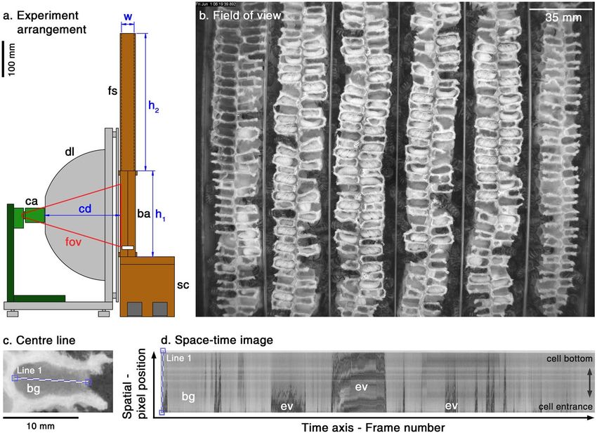
Figure 1. Recording and analysis methods to detect nursing parameters within cells. In our experiment
arrangement (a) we recorded longitudinal truncated cells in the brood area (ba; h1 = 235 mm) of the
observation hive using a digital video camera (ca) at focal distance (cd = 210 mm) and red light-emitting
(λpeak = 660 nm) dome-lighting (dl). The observation hive was 35 mm in width (w) and fitted with two
MiniPlus-frames in the food super (fs, h2 = 380 mm) and a feeder on top (not shown). To make the brood area
level with the lighting and camera, the hive was fixed on a sub-construction (sc, height: 165 mm). The field of
view (fov; b) covered 220 × 175 mm and approximately 420 truncated cells (13.5% of total brood area). Cell
visits were visualised by concatenating the temporal change of each pixel of a line (Line 1; c) along the x-axis,
thereby generating a space-time image (d) with the cell entrance at the bottom. Since bees appeared as dark
structural changes (‘events’; ev) in front of the background (bg in c and d), event parameters were automatically
determined after applying a user defined grey-threshold for event detection. Thereafter, feeding events were
either manually classified, or – later, after development – automatically classified using a convolutional neural
network algorithm analysing the event structure (e.g. feeding event structure looked similar to the one shown in
the centre of the image).
Nursing parameters. To test whether neonicotinoids influenced feeding behaviour in the colonies, we
determined the duration of each feeding visit (feeding visit duration; Fig. 2) and subsequently calculated the
following nursing parameters for each larval development day (LDD): the daily and cumulative feeding visits
(feeding visits/cumulative feeding visits; Fig. 3a–e) and duration (feeding duration/cumulative feeding duration;
Fig. 3f–j). We also analysed the feeding timespan of the nurses, indicating the larval development time (Fig. 4a).
Furthermore, we show the effects on LDD 5 for cumulative feeding visits (Fig. 4b) and duration (Fig. 4c), and
feeding visit duration (Fig. 4d). Finally, we tested if feeding timespan was depending on the duration of exposi-
tion to the substances (Fig. 5), and tested the influence on development after high neonicotinoid administration
(Fig. 6). By statistically analysing the results for each experiment individually, in addition to the mean, we allow a
transparent view of significant differences within each colony.
We analysed the time spent in the cell during a feedings visit shown in Fig. 2. This includes inspection times
prior to feedings. On LDDs 1 and 2 feeding visits lasted 122.0 ± 10.4 (mean ± SEM; n = 4) and 118.1 ± 3.3 sec-
onds, respectively, in control colonies. We observed relatively high variations on LDD 1 due to varying time for
proper inspection of the location of the newly hatched larva. Nurses in all treatments fed the longest on LDD 3
(133.0 ± 4.8 seconds in control) followed by LDD 4 (122.4 ± 2.8 seconds in control). On LDD 5, a 25% decrease
in feeding visit duration was present in all treatments (89.3 ± 2.4 seconds in control). This possibly represents
the change in nutrition from worker jelly to modified worker jelly, when increasingly honey stomach contents
are fed. On LDD 6 feeding visit duration was the shortest (79.8 ± 2.8 seconds in control). As already graspable
on LDD 4, we observed a dose-dependent appearance on LDD 5 and 6 showing that treated bees visited the cells
longer compared to the control. Neonicotinoid treatment showed significant differences for feeding visit duration
on LDD 5 (Fig. 4d) in May 2016 (F(3, 3284) = 6.22, p < 0.001), May 2018 (F(3, 5037) = 7.74, p < 0.001), June
2016 (F(3, 1186) = 26.98, p < 0.001) and July 2016 (F(3, 1015) = 2.96, p = 0.031), but not the mean (F(3, 12) =
1.25, p = 0.336). Feeding visit duration was significantly increased in 10 ppb clothianidin treated colonies in May
2016 (t = −3.412, p = 0.001, n = 714) and June 2016 (t = −6.720, p < 0.001, n = 452), and 200 ppb thiacloprid
Scientific Reports | (2020) 10:8727 | https://doi.org/10.1038/s41598-020-65425-y 3www.nature.com/scientificreports/ www.nature.com/scientificreports
140 Control
Clothianidin: 1 ppb
Clothianidin: 10 ppb
130 Thiacloprid: 200 ppb
Feeding visit duration
(mean sec ± SEM)
120
110
100
90
80
70
1 2 3 4 5 6
Larval development day
Figure 2. Duration of feeding visits on larval development days. During the first two days mean feeding
visits lasted about 2 minutes in control colonies, and took the longest on the third day. On day five feeding
visit duration majorly decreased to 1.5 minutes. See text for details. Treatments induced a reoccurring dose-
dependent effect on days 4 to 6 of development, which was not significant due to differing results in one of the
four experiments (n = 4; cf. Figs. 2, 3). The feeding visit duration includes inspection times prior to feedings.
SEM = standard error of mean.
treatment increased feeding visit duration in May 2016 (t = −3.763, p < 0.001, n = 856), May 2018 (t = −4.840,
p < 0.001, n = 1272) and June 2016 (t = −5.439, p < 0.001, n = 142). 1 ppb clothianidin significantly increased
feeding visit duration in July 2016 (t = −2.495, p = 0.039, n = 233). From a total of 16 post-hoc comparisons of
feeding visit duration on LDD 5, 10 were not significant.
The number of feeding visits per day increased from LDDs 1 to 5 in control colonies. We counted 4.4 ± 0.3
feeding visits (mean ± SEM, n = 4) on the first and 7.1 ± 0.6 feedings on the second day, followed by 11.2 ± 0.6,
23.1 ± 1.3 and 47.4 ± 1.7 average feedings on the third, fourth and fifth day, respectively (Fig. 3e; numbers in
coloured boxes; overview in Supplementary Table S1). On LDD 6, larvae received an average of 19.3 ± 4.7 feed-
ings until the cell was capped. Control colonies roughly doubled their feeding effort with each LDD except the
last. The mean cumulative feeding visits (Fig. 3e; upper ends of coloured boxes, y-axis) were 4.4 ± 0.3, 11.5 ± 0.9,
22.7 ± 1.4, 45.8 ± 2.1, 93.2 ± 3.5 and 112.5 ± 2.4 on LDDs 1 to 6, respectively. As shown in Fig. 3j, feeding visit
duration was strongly correlated with feeding visits. Controls fed for a mean of 8.9 ± 0.6, 13.8 ± 1.5, 24.7 ± 2.1,
47.8 ± 2.3, 71.4 ± 2.4 and 26.0 ± 6.5 minutes on LDDs 1 to 6, respectively. Cumulative feeding duration was
8.9 ± 0.6, 22.7 ± 2.1, 47.4 ± 4.1, 95.2 ± 5.4 and 166.6 ± 6.4 minutes on LDDs 1 to 5, respectively, and the total
feeding duration was 192.6 ± 4.0 minutes at cell capping on LDD 6. These values include the varying inspection
times preceding food provision. In the mean of treated colonies cumulative feeding visits were lower than the
control starting with the second development day (Fig. 3e). This tendency was increasing with every LDD but
was levelled throughout LDD 6. Concomitant results were present for feeding duration (Fig. 3j). Treated colonies
showed reoccurring patterns between May and June (Figs. 3a–c and f–h) but not in July (Fig. 3d,i). Up to LDD
5, 200 ppb thiacloprid treatment showed less daily and cumulative feeding visits and durations than the control.
This was also the case for 10 ppb clothianidin in May and June 2016, and for 1 ppb clothianidin treated colonies
up to LDD 4 in all experiments except in May 2018. On LDD 6 however, more daily feedings were counted in
the treatments in 10 of 12 cases, hereby decreasing the differences to the control in total feedings until capping.
In July 2016 the thiacloprid treatment was at level with the control and exceeded its feeding visits (Fig. 3d) and
durations (Fig. 3i) on LDDs 4 and 5. In this experiment, treatment with 1 ppb clothianidin reduced nursing
parameters more than 10 ppb clothianidin. Compared to the other experiments, the nursing parameters were
highest on LDDs 1–5 and lowest on LDD 6 in all colonies of July. For all parameters in Fig. 3, out of 195 post-hoc
comparisons to the control, 69 remained significant and 13 were above the threshold after p-value adjustment (see
Supplementary Table S1).
Figure 4b,c illustrate mean cumulative feeding visits and durations, respectively, as a function of neonicotinoid
treatment five days after the first feeding (same as upper ends of LDD 5 boxes in Fig. 3). The one-way ANOVA
showed that the number of feeding visits were significantly different for the treatments in May 2016 (F(3, 67) =
5.01, p = 0.003), May 2018 (F(3, 110) = 7.18, p < 0.001), June 2016 (F(3, 25) = 6.72, p = 0.002), and July (F(3, 16)
= 4.22, p = 0.022) but not the mean (F(3, 12) = 1.43, p = 0.282). While in July 2016 no statistical differences were
present in pairwise comparisons, 10 ppb clothianidin significantly reduced feeding visits in May 2016 (t = 2.813,
p = 0.024, n = 17) and June 2016 (t = 3.114, p = 0.018, n = 14), and 200 ppb thiacloprid in May 2016 (t = 2.820,
p = 0.023, n = 21) and May 2018 (t = 4.002, p < 0.001, n = 29). Similar significances were present for feeding dura-
tion in May 2016 (F(3, 67) = 3.98, p = 0.011), May 2018 (F(3, 110) = 7.64, p < 0.001), June 2016 (F(3, 25) = 5.06,
p = 0.007), and July (F(3, 16) = 4.68, p = 0.016) but not the mean (F(3, 12) = 1.15, p = 0.370). Again, no statistical
differences were present in pairwise comparisons in July. 10 ppb clothianidin reduced feeding duration in June
2016 (t = 3.006, p = 0.023, n = 14) but not in May 2016 (t = 2.126, p = 0.082, n = 17), and 200 ppb thiacloprid in
May 2016 (t = 2.586, p = 0.041, n = 21) and May 2018 (t = 3.932, p < 0.001, n = 29). In June 2016 only 6 larvae
in the control and 4 larvae in the thiacloprid treatment were available for analysis. Corresponding to the small
number of larvae, tested differences were not significant.
Scientific Reports | (2020) 10:8727 | https://doi.org/10.1038/s41598-020-65425-y 4www.nature.com/scientificreports/ www.nature.com/scientificreports
May 2016 May
May 2018
2018 June 2016 July 2016 Mean
a b c d e
120
28.6 3.8
23.2 6.2
19.9 28.5 7.6 19.3
100 51.8 27.8
27.8 32.1
32.1
22.4
18.3
18.3 14.2 30.9 33.0
Feeding visits
32.2 51.6
41.0
41.0 56.6
80 35.5
49.4
49.7 51.2
48.4 47.4
42.3 37.5
37.5 59.6 48.6
50.6 42.3 38.2 46.6
60 38.2
40.2 42.0
39.0 40.8
35.2
35.2 32.3 35.5
40 20.6
20.6
25.2 29.4
23.3 19.5
19.5 20.0
20.0 24.5 25.4 23.1
18.6 20.6 20.5 19.7
19.6 15.9 22.2 18.8
20 12.2
12.2 14.7
14.7 13.6 15.3 12.4
9.7 11.3
11.3 10.7
10.7 11.5 11.4 12.2 11.2
8.5 9.8 9.5 9.5 8.9
6.3 8.4 8.7 6.8 7.7
7.7 6.6 7.5
7.5
6.6 3.0 7.6 7.4 7.3 8.8 5.6 7.0 7.1
4.6 6.8 6.3 3.0 3.9 4.2 4.9 4.8 4.4
4.2 3.8 3.12.6 4.12.7 4.6
4.6 6.5
6.5 5.1
5.1 3.0
3.5
3.5 3.7 3.2 3.3 3.5 2.0 5.2 4.4 5.0 4.8 4.4 4.5 4.0 3.3
0
f g h i j
Control
Clothianidin: 1 ppb
Clothianidin: 10 ppb
Thiacloprid: 200 ppb
200 5.6
Feeding visit duration (min)
29.6
28.8 40.2 38.3 7.6 12.3 26.0
75.0 31.0
48.3 38.1 46.4 20.0 43.6 49.9
25.6
67.2 55.3 80.8
150 63.6
70.5
72.4 75.1
77.1 71.4
85.7 58.7 86.4 74.7 76.4
65.5 62.0 66.4
68.3 69.7
71.3
100 60.2 66.4
60.3 48.7 59.8
43.5 52.1 50.8 47.8
49.0 41.2 41.3 44.7
40.1 43.4 42.7
46.1 34.4 45.5 40.2
50 32.6 35.9 29.9
23.9 30.7 28.7
20.4 22.3 21.9 26.3 26.1 24.7
18.7 22.6 20.5 21.0 19.8
19.0 19.6 13.7 16.6 17.0 15.7 18.1
12.2 9.7 6.6 8.2 11.9 11.4 15.2 13.0 8.6 8.6 10.4 14.9 13.8 9.6 9.3 8.8
8.2 7.4 4.5 5.6 7.8 11.9 8.4 4.85.4 9.1 6.65.9 7.3 7.2 4.5 10.5 7.5 9.7 9.9 8.9 8.2 7.5 6.4
LDD 0
Control
CLO: 1 ppb
CLO: 10 ppb n = 17
THI: 200 ppb n = 21
Control
CLO: 1 ppb
CLO: 10 ppb n = 30
THI: 200 ppb n = 29
Control
CLO: 1 ppb
CLO: 10 ppb n = 14
THI: 200 ppb n = 4
Control
CLO: 1 ppb
CLO: 10 ppb n = 5
THI: 200 ppb n = 5
Control
CLO: 1 ppb
CLO: 10 ppb n = 4
THI: 200 ppb n = 4
6
5
4
3
2
n = 19
n = 14
n = 27
n = 28
n=6
n=5
n=5
n=5
n=4
n=4
1
Figure 3. Feeding visits and durations in all experiments. Numbers in boxes represent the mean value on
each larval development day (LDD; colour-coded) for feeding visits (a–b) and feeding duration (f–j). Bold
italic numbers in boxes represent statistical differences to the control on this LDD (Bonferroni-Holm type I
error-corrected adjusted p of Student’s t-test after significant one-way ANOVA; number of tests used for p-
value adjustment = 3). The upper ends of the coloured boxes show the mean cumulative value on the y-axis,
equivalent to the sum of all numbers below. If dotted, significant statistical differences (same test) of cumulative
values to the control were present. Graphs (e,j) show the mean of (a–d and f–i), respectively. Treatments were in
most cases below the controls daily and cumulative and value, but levelled throughout LDD 6 (red boxes). The
upper ends of LDD 6 represent the total value until cell capping. Feeding duration was strongly correlated to the
number of feeding visits. Parameter means of all experiments (Mean) were not significant due to the results of
July 2016. Abbreviations: CLO = clothianidin; THI = thiacloprid; ppb = parts per billion; SEM = standard error
of mean. Samples sizes (n) are number of analysed cells in the experiments (a,f: May 2016, b,g: May 2018, c,h:
June 2016, d,i: July 2016) and the number of colonies (e,j: Mean).
Development time. In 2016 and 2018 we measured the feeding timespan as an indicator for development
time, since timestamps of the first and last feedings were available in all analysed cells. To calculate the real devel-
opment times, oviposition, larval hatch and cell capping timestamps were required. Since oviposition required
between 10 to 20 seconds, its detection on our images was principally possible, and easily determined by a sudden
appearance of the egg in the cell. However, this was not the case in all cells that we used for analysis. Sometimes
the egg and subsequent larval hatch were hidden from sight due to the viewing angle, and some recordings started
with eggs already present in the cells. In numerous cases, eggs were pushed down during the first three days of
development and disappeared from the field of view after a worker bee entered and appeared motionless the cell
(suspected as heating events; in some cases larval hatch occurred during this period). Therefore, we measured the
feeding timespan.
One-way ANOVA displayed a statistically significant difference in feeding timespans for the treatments
(Fig. 4a) in May 2016 (F(3, 67) = 6.18, p < 0.001), May 2018 (F(3, 110) = 4.82, p = 0.003) and June 2016 (F(3, 25)
= 6.04, p = 0.003). We observed that means were negatively correlated to feeding visits and duration (Fig. 4b,c,
respectively) and positively correlated with nursing activity on LDD 6 (Fig. 3). Thiacloprid treatment increased
the feeding timespan in May 2016 from 5.33 ± 0.09 to 5.74 ± 0.07 days (mean ± SEM, t = −3.656, p = 0.002,
n = 21), and in May 2018 from 5.31 ± 0.05 to 5.54 ± 0.07 days (t = −2.571, p = 0.039, n = 29). 10 ppb clothia-
nidin increased the feeding timespan in June 2016 from 5.45 ± 0.14 to 5.89 ± 0.07 days (t = −3.053, p = 0.021,
n = 14). The remaining 13 of the 16 post-hoc comparisons were not significant. We found no significant differ-
ence between the means of all experiments (Fig. 4a, Mean) (F(3, 12) = 0.72, p = 0.560), possibly due to the low
amount of tested colonies.
Scientific Reports | (2020) 10:8727 | https://doi.org/10.1038/s41598-020-65425-y 5www.nature.com/scientificreports/ www.nature.com/scientificreports
6.0 *
a b
Cumulative feeding visits 5 days
Control
** Clothianidin: 1 ppb
110
after first feeding (± SEM)
5.8 Clothianidin: 10 ppb
Thiacloprid: 200 ppb
Feeding timespan
*
5.6
(days ± SEM)
90
5.4
* *
5.2 ***
70 *
5.0
4.8 50
19
14
17
21
27
28
30
29
14
19
14
17
21
27
28
30
29
14
6
5
4
5
5
5
5
4
4
4
4
6
5
4
5
5
5
5
4
4
4
4
220 120
Cumulative feeding duration 5 days
d ***
c ***
Feeding visit duration 5 days
after first feeding (min ± SEM)
after first feeding (s ± SEM)
200
110 ***
***
180
100 *** *
160
*
140 *
*** 90
120
1276
1346
1272
1147
80
953
765
714
856
298
298
452
142
247
233
256
283
100
19
14
17
21
27
28
30
29
14
4
4
4
4
6
5
4
5
5
5
5
4
4
4
4
May 2016 May 2018 June 2016 July 2016 Mean May 2016 May 2018 June 2016 July 2016 Mean
Experiment Experiment
Figure 4. Feeding timespan and nursing parameters on larval development day 5. Mean feeding timespan (a)
from the first to the last feeding was significantly increased up to 0.44 days (10.56 hours) after chronic treatment
with 10 ppb clothianidin or 200 ppb thiacloprid in all experiments except July 2016. Five days after first feeding,
neonicotinoid treatment significantly reduced mean cumulative feeding visits (b) and mean cumulative feeding
duration (c), whereas mean feeding visit duration was prolonged (d). This shows an interdependence of the
affected nursing parameters and feeding timespan (or development time of the larvae). Asterisks indicate
significant differences to control (Bonferroni-Holm adjusted p-value of Student’s t-test: *www.nature.com/scientificreports/ www.nature.com/scientificreports
26
Egg Uncapped larva
24 Capped larva Pupa
22 *** 20.84 ± .068
**
20
Mean development time (d)
18
16 *** *
14
12
10
8
6
*** ***
4
2
0
*** **
Control Clothianidin: Thiacloprid:
100 ppb 8800 ppb
Figure 6. Worker development times (mean ± SEM) of various observable stages in high neonicotinoid
treatments. The embryonic (egg) stage was significantly delayed in colonies treated with 100 ppb clothianidin
and 8800 ppb thiacloprid. Greatest relative delay was present in the uncapped larval stage (17.3% and 7.2%
increase in clothianidin and thiacloprid treatments, respectively). No significant differences were observed
during larval development in the capped cell. While clothianidin increased pupal development time, thiacloprid
surprisingly decreased it. The control needed 20.50 ± 0.07 days for complete worker ontogenesis (numbers for
complete ontogenesis are not the sum of means). In the 100 ppb clothianidin treatment complete ontogenesis
was delayed by 1.19 days or 28.56 hours and in the 8800 ppb thiacloprid treatment by 0.34 days or 8.16 hours.
Statistical test: ANOVA, Bonferroni-Holm adjusted p-values of Student’s t-test; number of tests used for p-value
adjustment = 2. Asterisks indicate significance levels: *www.nature.com/scientificreports/ www.nature.com/scientificreports
treatments. In pairwise comparison to the control, statistically significant differences were present in the 100 ppb
clothianidin treatment for egg (t = −8.396, p < 0.001, n = 13), uncapped larva (t = −6.518, p < 0.001, n = 14)
and pupa (t = −4.755, p < 0.001, n = 14) stages and complete ontogenesis (t = −4.217, p < 0.001, n = 14), and
likewise in the 8800 ppb thiacloprid treatment (t = −3.984, p = 0.002; t = −4.925, p < 0.001; t = 2.915, p = 0.010;
t = −3.390, p = 0.004, respectively, n = 10). All of the 8 post-hoc comparisons were significant, while the ANOVA
of the capped larva stage showed no statistical differences. Development times were increased in all cases, except
for pupal development in the thiacloprid colony, which was surprisingly shortened. The shown delay in embryo
development suggests a mechanism independent from feeding behaviour of nurses. These results support our
previous observations of retarded brood development in our low concentration experiments. However, one
should keep in mind that data is from only one colony each and that larval development time could have been
influenced by the obvious differences in brood area development (3 to 75 capped cells after 17 days as described
above; cf. Fig. 5).
Unrepeated weighting experiments in the low concentration experiment in May 2018 also showed delayed
development. Upon correlating weight and development duration in 22 uncapped cells per treatment we deter-
mined that larvae in the 200 ppb thiacloprid treated hive required 10.8 hours longer than the control to gain
150 mg of weight (regression in control: y = 7E−7 × 9.2365, R2 = 0.89; thiacloprid: y = 2E−5 × 7.4259, R2 = 0.92). Larvae
in the 10 ppb clothianidin treatment required 5.4 hours longer (y = 2E−5 × 7.5218, R2 = 0.93). In general, larvae
weight between 137.6 and 158.3 mg at cell capping42.
Food uptake. We tested whether the neonicotinoid-spiked sugar solution was taken up by colonies by regular
weighting of the feeder. Control hives collected between 2,101 and 2,766 g (mean ± SEM: 2,452 ± 184 g, n = 4)
of sugar solution during experiments. Colonies treated with 1 ppb clothianidin, 10 ppb clothianidin and 200
ppb thiacloprid collected 2,299 ± 229 g, 2,320 ± 245 g, and 2,094 ± 251 g, respectively (four colonies per treat-
ment). One-way ANOVA showed no significant differences in food uptake between treatments (F(3,12) = 0.42,
p = 0.743).
Discussion
Our novel video recording and analysing methods have proven successful to process a comprehensive amount of
honey bee brood care behaviour continuously over several weeks. For the first time, we were able to demonstrate
significant differences of nursing behaviour and larval development time from within a functioning hive that was
chronically fed with neonicotinoids. Notably, in May (Fig. 3a,b and f,g) statistical differences in nursing amount
and duration were present already during the initial days of development. Furthermore, treatment with 200 ppb
of the partial nicotinergic agonist thiacloprid did not induce opposing results compared to 10 ppb of the more
effective agonist clothianidin. In June (Fig. 3c,h) only few cells were analysable in the control and the thiacloprid
treatments and statistical differences in daily and cumulative nursing parameters were missing. We observed high
nursing activity in the initial LDDs of July (Fig. 3d,i) and found no significant differences between the means of all
experiments (Fig. 3e,j; Mean). In conclusion, we can neither clearly confirm nor refute a neonicotinoid effect on
nursing behaviour and larval development throughout all experiments, although we observed various indications
of such effects. This includes experiments using high neonicotinoid dosages in which brood area development
was obviously retarded and developmental stages were delayed (Fig. 6). A further factor influencing development
time was the duration between experiment initiation and the first feeding of the larva. We chose all larvae for
analysis without bias towards this factor as shown in the distributions in Fig. 5. The effect was treatment inde-
pendent and could be related to the amount of capped brood within the colony. We conclude that there was no
accumulating effect by the neonicotinoids from 7 to 20 days that further retarded larval development. It should
be further investigated if the results of July were due to seasonal circumstances and if hives rear brood differently,
as shown in the relatively high daily feeding visits and feeding duration compared to the other experiments
conducted in May and June. We want to emphasize at this point that our hive architecture was optimized for
observation and video recordings, with a relatively small amount of colony members. Although the validity of the
observed effects within managed full colonies remains to be proven, we, nevertheless, were able to quantify social
behaviour in the brood area within a hive that was stable throughout the whole study period of up to six weeks.
Therefore, we assume that our conclusions can be assigned to full-sized colonies with the given caution based on
the different hive dimensions.
To date, investigations on neonicotinoid effects have focused on neuronal impairments (see introduction for
references, review22,23) because acetylcholine is the most abundant neurotransmitter in the insect brain43 and is
used in various brain regions of the honey bee, such as the antennal lobes and mushroom body44. Therefore, alter-
ations in behaviour are expected. Publications noting high non-neuronal acetylcholine concentration in brood
food30,32–34 suggest neonicotinoid impairments of development as a further possibility. The cholinergic system is
suggested to be important in insect development45 but the non-neuronal cholinergic system of insects is largely
understudied. Various delaying effects by neonicotinoids and other insecticides targeting the cholinergic system,
such as dimethoate, monocrotophos, methylparathion or quinalphos, have been demonstrated elsewhere (for
review see24). Currently, the targets of non-neuronal acetylcholine, how it affects honey bee larvae and its modes
of action remain to be discovered. However, it should be noted that other nest-mates and the queen are also fed by
nurses46,47, thus broadening the range of possible targets of non-neuronal acetylcholine within the hive.
In this study we have shown indications of delayed development using various neonicotinoid concentrations
within our hives, as shown in Figs. 4a and 6. However, because larval development and nursing behaviour are
closely linked to each other, we do not know whether the tested neonicotinoids primarily affected nurses, larvae
or both at the same time and had no possibility to disentangle these possibilities in the shown experiments of
this study. Furthermore, we do not know whether alterations in A) behaviour, B) development, or both are the
underlying causes.
Scientific Reports | (2020) 10:8727 | https://doi.org/10.1038/s41598-020-65425-y 8www.nature.com/scientificreports/ www.nature.com/scientificreports
Regarding A), behavioural alterations, the reduced number of feeding visits and durations within the treat-
ments, presented in Fig. 3, could have been the reason for the prolonged feeding timespan, and thus, brood devel-
opment observed. In this case, neonicotinoids would have had a negative effect on the nervous systems of nurses
to such a significant degree that they affected feeding behaviour and/or induced incorrect decisions. It is expected
that nurses adjusted food composition40,48–50 and nursing activity51,52 according to the condition and developmen-
tal stage of the larvae. Previous studies have shown a correlation between nursing duration and the development
time of larvae42,53,54. On the other hand, queen larvae that are fed more often than worker larvae have a lower
mean weight between hours 6 and 90 of development55. This suggests that food amount between castes is not the
exclusive factor for affecting growth, but that growth rate may instead be linked to food composition. Although
we were unable to measure the amount of food provided during each visit, there were no differences observed in
feeding durations over the first three days (Fig. 2). Assuming that food amount is positively correlated with the
time spent in a cell during feeding, we presume an effect on development duration based on alterations in the
amount of food provided was not being present. The mean total of 112.5 ± 2.4 nursing visits in the control colo-
nies is below observations of previous studies in this field49,56. However, we postulate that our values are the most
precise thus far due to a greater sample size with automated analyses.
Other brood rearing relevant behavioural impairments, such as irregular inspections or the inability of tem-
perature preservation, could also have induced the observed delay in development and further analysis of these
behaviours using our video data is intended. Temperature has been shown to have an influence on development
time57. The surrounding room temperature was 26.9 ± 1.4 °C, 26.9 ± 1.7 °C, 27.6 ± 1.4 °C and 29.5 ± 1.3 °C in May
2016, June 2016, July 2016 and May 2018, respectively (mean ± SD, start to end of the experiments, 30 minutes
reading interval). We also derived the standard deviations of temperature measurements across each individual
day. These standard deviations ranged from 1.5 (May 2018) to 4.3 (May 2016), with a mean standard deviation
of 3.0 for all experiments in 2016 and 3.1 for May 2018. Hives were approximately 4 °C above room temperature
(within-hive sensor were placed on the floor in the brood area, level with the entrance). We were unable to accu-
rately determine whether primarily motionless bees displayed heating events, although these events were not
observed in a hive with over-heated surroundings (this hive was at a different location). In an unpublished study,
we delayed the development of larvae reared under constant temperature in vitro using high concentrations of
imidacloprid in our lab; therefore, temperature is unlikely to be the only reason for retarded larval development
in our experiments. Alternative possibilities include chemical miscommunication, for example by impaired per-
ception of nurses or by impaired emittance of larval pheromones. Since the mean of the experiments had similar
values in cumulative amount and duration on LDD 6 (Fig. 3) the nurses presumably perceived the state of the
larva at the time point of capping correctly.
Regarding B), nurses could have adapted their behaviour because of impairments in larval development,
if sensory perception was well functioning. Since the direct transmission of neonicotinoids from nurse to
larva seems to be very low58, a further possibility is that development within the nurses was affected, and that
food secreting organs were impaired. Neonicotinoids have been shown to influence the size28 and activity30 of
food-producing glands (for review see24) and underlying endocrine controls within age polyethism59. For exam-
ple, the development of hypopharyngeal glands is retarded in caged bees using 2 and 3 ppb imidacloprid in a
sugar solution and pollen pastry, respectively28. Unpublished results from our lab using colonies systematically
treated with high concentrations of clothianidin (100 ppb) and thiacloprid (8800 ppb) suggest similar effects.
Hypopharyngeal gland volume of seven-day-old workers was significantly reduced, glands appearing white
and undeveloped as of newly emerged bees. Gland impairments could affect food quality, for example, affect-
ing non-neuronal acetylcholine concentrations found in the jelly30,32–34. However, retarded development may be
independent of gland secretions and feeding behaviour of nurses, since eggs in colonies treated with high neon-
icotinoid concentrations showed a developmental delay (Fig. 6). Yet, queens previously received feedings by the
workers as well. Because development is mainly mediated by hormones, we should also envisage an impairment
of endocrine organs within larvae and/or nurses, as a couple of studies have suggested up to date60,61. This should
be investigated in further analyses.
We anticipate that our versatile method can be used for a variety of behavioural monitoring studies that
require precise time determination under various stressor influences. Apart from general effects from other bee-
keeping chemicals, this includes bees’ reactions to parasites such as Varroa destructor under various conditions,
and, for example, after the extraction and insertion of food or chosen substances into individual cells.
Materials and Methods
Animals and colonies. We used small observation hives (outer dimensions H × W × D: 617 × 271 × 42 mm;
Fig. 7) that were each filled with 300 g Apis mellifera carnica (approximately 3,000 individuals) taken from bee
hives of the Institut für Bienenkunde in Oberursel as well as one queen. Sister queens were taken from our on-site
queen breeding programme. Hives were divided by a queen grid into an upper food super (inner dimensions
H × W × D: 359 × 229 × 35 mm) and a lower brood area (inner dimensions H × W × D: 235 × 211 × 35 mm) by
preventing the queen from entering the food super. At the start of the experiments it contained two wooden
MiniPlus-sized frames with mounted comb foundations, while visual inspection was granted by acrylic glass.
The brood area had six “comb stripes” (35 mm stripes of an empty well-constructed Zander frame comb) that
were turned 90° to allow video observation of the truncated cells through a 4 mm layer of anti-reflective glass
(Energy Vision 1, Hecker, Dortmund). This enabled the continuous recording of nursing behaviour and devel-
oping worker bees from oviposition until adult eclosion within individual cells (Fig. 1b). We further developed
this concept of looking sideways into the cells from a previous publication56. All wax that was set into the hives
was previously treated with a toxin against wax moths (B401, Vita Europe Ltd, Basingstoke) to prevent their
development during the experiments. One comb stripe had a maximum of 40 vertical cells per side. Bees had six
or seven horizontal cells for brood rearing, alternating due to offset comb structure. Therefore, a comb stripe had
Scientific Reports | (2020) 10:8727 | https://doi.org/10.1038/s41598-020-65425-y 9www.nature.com/scientificreports/ www.nature.com/scientificreports
fov
Figure 7. Technical drawing of the observation hive. The recorded brood area (fov = field of view) contained
six glass stripes (light grey / red overlay) to mount combs (not shown), which were pressed against the glass
using metal bars (not shown) across top and bottom wood blocks (deep red). The upper food super contained
two MiniPlus-sized frames (yellow) and was separated from the brood area by a queen grid above the glass
stripes. The food super was visible though embedded acrylic glass. The hive was fixed at the bottom using angle
connectors (dark grey).
a maximum of 520 cells and a total of 3120 cells within the brood area (six comb stripes). As shown in Fig. 1b,
the camera observed approximately 70 vertical cells in each comb stripe and a total of 420 in the field of view.
Therefore we observed approximately 13.5% of all available cells in the brood area. A comb stripe was laterally
confined by two glasses and vertically by two wood blocks. Serving as spacer, the wood blocks allowed firm
attachment of the glass to the comb stripe. To create passages to the food super the upper blocks were narrower
(H × W × D: 18 × 25 × 35 mm) than the bottom one (H × W × D: 18 × 35 × 35 mm).
Treatments. We simultaneously recorded four hives with various treatments: control, 1 ppb clothianidin, 10
ppb clothianidin and 200 ppb thiacloprid (Sigma-Aldrich Chemie GmbH, Munich). The hives were positioned
in a dark and temperate (~27 °C) room whose temperature was continuously monitored. With a feeder on top of
the food super, we applied control sugar solution (ApiInvert, Südzucker AG, Germany + 0.0002% acetone) or in
acetone dissolved neonicotinoids in sugar solution. Since feeding started with experiment initiation, all bees were
Scientific Reports | (2020) 10:8727 | https://doi.org/10.1038/s41598-020-65425-y 10www.nature.com/scientificreports/ www.nature.com/scientificreports
Application Requirements/
Tasks Constraints Design Choice
Sample video at 1 fps/JPEG real-time
Long term recording Limited hard disk storage space
compression
Good contrast of background
Honey bee detection Use of bright empty honey combs & red light
foreground required
Sufficient video resolution & Analysis requires acceptable Balanced camera distance/5.3 MP
number of observable cells image quality camera/4.8 µm pixel spacing
Record without shadows
Reflections occur on glass Usage of dome lighting
through glass
Bees might get disturbed by
Keep hive naturally dark Usage of 660 nm red light/monochrome cameras
visible light
Feedings occur within cells; Compress 2D + time data to 1D + time data by
Observe nursing within cells constrained cell-entering projections along a line interval orthogonal to
possibility the cell entry direction.
Development time is linked to
Analyse larval development time Control surrounding room temperature
temperature
Table 1. Tasks and application requirements to record nursing within a hive.
potentially exposed to the treatments from the first experiment day. The weight of the feeder was measured before
refilling. Hives were connected to the outside using a tunnel in the brood area. After a maximum of three weeks
there were sufficient capped cells for further analysis. We then stopped the recordings and fully disassembled the
hives, sweeping all colony members into an artificial swarm box. Queens were not in the swarm. We extensively
cleaned the hives with a brush and warm water to remove remaining food. Feeders were cleaned with warm
water as well. We then used a new set of six well-constructed comb stipes and two MiniPlus-sized frames with
mounted comb foundations for the next repetition. We refilled the colonies with new bees as described above
on the following day to the end of a three week experiment. We restarted the experiments as long as the brood
season (February – October in Germany, coordinates: N50°13′4.296″ O 8°32′54.024″) permitted or until too little
cells at the glass were occupied by developing larvae with decreasing offspring (typically between August and
September). Sister queens were available from May. Due to this effectively to three months limited experiment
time per year we did not keep track of adult emergence, since this would have required another 12 days. However,
we followed the full worker ontogenesis using high neonicotinoid concentrations (100 ppb clothianidin and 8800
ppb thiacloprid) in 2017 on cost of the number of possible repetitions.
Video registration and data analyses. We evaluated a total of 26,483 feeding visits in 234 brood cells
of 16 hives (number of cells in control: 57; 1 ppb clothianidin: 52; 10 ppb clothianidin: 66; 200 ppb thiacloprid:
59), recorded in four repetitions and two experimental years. In 2016 experiments were from May 17th to June 6th
(May 2016), from June 7th to June 27th (June 2016) and June 28th to July 18th (July 2016). In 2018 one experiment
from May 14th to June 1st (May 2018) was available. A total of 60 cells were analysed using manual classification
(6,539 feeding events) and 174 were analysed with a convolutional neural network algorithm (19,944 feeding
events). Nursing behaviour within honey bee colonies and its analysis within our experiment arrangement was
evaluated by the following assumptions: Firstly, nursing always takes place within the comb cells; secondly, all
bees within the colony are of approximately the same size and appearance (i.e. patterns and colours); and thirdly,
workers require a certain amount of time within the cell for nursing. Given these strong constraints, we had
strong expectations regarding which video pixels were going to change over which timeframe within the fixed
comb structure upon a nurse entering a cell to feed. Therefore, we were able to compress our video data from
three dimensions (width, height and time) into two (width and time) without losing important information,
and were further able to record at one frame per second. For 2D compression we followed a systems engineering
methodology for the design of the video analysis system62.
Key factors that influence the design of a vision system include sensor choices, application contexts, as well as
tasks and other requirements such as hardware constraints and costs (among others; Table 1). Some important
parameters for the contextual setup of data collection for hive observations include (from and Fig. 1a): cd – cam-
era distance from brood area, dl – dome lighting, h1 – brood area height and sc – height of sub-construction.
The recording arrangement was constructed by setting these parameters as follows: To increase the likelihood
of developing larvae within the visible cell layer, we balanced the arrangement to record a large area of cells and
preserve sufficient video details for later analysis (for video resolution see Supplementary Video S2). Brood area
height was adapted from the Zander-frame height (205 mm) plus additional 30 mm space to place wooden spac-
ers at the top and bottom for comb fixation (see red rectangles in Fig. 7). Brood area width was set to create the
most approximate square by multiplying the 35 mm glass-stripe width (space to include combs and inter-comb
spacing for bees to pass) by 6, resulting in 210 mm. For optimal illumination, we constructed a dome-light size
that encircled our observation area and set the height of the sub-construction to align the centres of the observa-
tion area and the dome lighting.
We recorded the brood area using a 5.3 MP monochrome USB3 camera (PL-D725MU-T, PixeLINK, Ottawa),
a C-mount lens with 12.5 mm focal length (LM12HC, Kowa Optical Products Co., Ltd., Tokyo) and a red
light-emitting dome-light beyond the range of honey bee colour vision (λpeak = 660 nm). One comb stipe was
recorded with 2048 × 420 pixels (H × W). The 1″ camera sensor fulfilled photon detection requirements under
Scientific Reports | (2020) 10:8727 | https://doi.org/10.1038/s41598-020-65425-y 11www.nature.com/scientificreports/ www.nature.com/scientificreports
User input: Scene Sensor
line start/end points:
(x1,y1), (x2,y2)
Bresenham line
algorithm 3D image (grayscale) Crop feature space
(S-T Image) using
event start/stop
information from
Spatio-temporal Event list
Pixel coords 1D + time (S-T Image)
projection
Event-sized
User input:
S-T Image
Level of cell entrance Cropping, Binary
to crop STI to current Classification & S-T Image (binary)
cell size & threshold Filling
Prune/pad and
for binarization
concatenate to
227x227x3 to fit
Event detection Event list (all) in network
User classification Auto classification
Network-sized
User input:
Event list image
Minimum duration
Filter events
and depth over time (possible feedings)
of visit
Neural network
User input: classification
Feeding verification (vgg16)
Event list
(feedings)
Figure 8. Object-process diagram for our application: Boxes with rounded corners represent transformations,
while regular boxes represent data objects. We used a manual classification (user classification) and used the
resulting and further classified image data to develop an automated STI classification using a neural network
algorithm (for more information on the Object-process Methodology, see65).
monochrome lighting conditions. Recording at one frame per second was reasonable to sufficiently observe nurs-
ing behaviours and minimized the amount of required hard-disk space, thus allowing continuous recording of
the four experiment arrangements simultaneously throughout the season. However, we acquired high temporal
resolution cameras to ensure the flexibility of experimental arrangements. StreamPix (version 6.3.0.155 and 7.4,
NorPix Inc., Montreal) was used as recording software. For analysis, we used a manual or automated classification
process shown in the block diagram of Fig. 8. While one could process video frames at the original resolution,
the application context, the analysis goals and computational requirements motivated us to perform projections
of videos (2D + time) into images (1D + time). The space-time image (STI) constructed is presented in Fig. 1d.
For STI generation the brightness information of the cells centre pixels from the bottom to the entrance (using
Bresenham’s line algorithm; Fig. 1c) was concatenated over time. Entering bees appeared as dark structural
change on the STI (from now: ‘events’). STI representation has been used in a number of real-world video moni-
toring tasks (e.g63,64). In this work, we simply concatenated the pixel values selected at the observation lines. The
temporal properties of the filtered signal retain relevant information regarding the dynamics in the video due to
illumination variation, shadows, reflections, occlusions and background motion. However, the STI representation
clearly shows features that facilitate the classification of these effects based on visual patterns.
Because the three-week recordings had over one million frames, STIs were divided into groups of 65,499
frames to enable common display possibilities. In a programmed graphical user interface (MATLAB 2014a –
2018a by MathWorks, Natick) running on Windows 7 we stored events if brightness was below a threshold (user
adjusted for best detection, bees appeared darker than the background wax which was variable in brightness). To
narrow down possible feeding events during manual classification, events were filtered by duration (10–300 sec-
onds) and depth over time (bees use at least 65% of available space in a cell over a user-defined amount of time).
These filters were applied prior to consecutive verification by hand. In case of doubt, comparisons were made with
the corresponding video material.
We observed that events on the STIs had a similar pattern appearance according to the behaviour performed
by bees. Therefore, feedings could usually be characterised quickly without consideration of the video footage
during manual classification. We used this circumstance to train a convolutional neural network (Vgg16) with
63,411 truncated event images we had classified for this purpose. We trained the algorithm to discriminate
between “feeding” (19,472 manually classified events used for training; including 6,539 of the 2016 experiments),
“heating” (10,691 events), “building” (28,149 events) and “unknown” (5,099 events) by analysing patterns and
Scientific Reports | (2020) 10:8727 | https://doi.org/10.1038/s41598-020-65425-y 12www.nature.com/scientificreports/ www.nature.com/scientificreports
structures within the event STIs (for behaviour characterisation see results). The images were normalised in
brightness and contrast and then cropped or padded black and concatenated to fit a specific size (227 × 227 × 3
pixels; height, width, colour channels) in the neural network input layer. Although this shortened some events
to 227 seconds (pixels), the important information was sufficiently preserved. Behavioural changes within events
were extremely rare. For network training all image labels were split into two random batches, one for algorithm
improvement and the other one for testing accuracy. Although our two-class neural network (feeding/unknown)
was able to detect feedings with 99.4% accuracy, we chose an analysis with four classes (95.9% accuracy) in order
to also analyse the effects on other behaviours in the future. During neural network classification we bypassed the
maximum visit length filter (set from 300 to 10,000 seconds) and renounced depth over time filtering to include
long lasting heating events in the list (“Event list (all)” in Fig. 8). Afterwards, we excluded incorrectly classi-
fied feeding events above 300 seconds from the final event list. Code has been made available at: https://github.
com/paulsiefert/honey-bee-behaviour.
Statistics. Statistics were calculated using IBM SPSS Statistics, version 23.0.0.2, with α = 0.05. We applied
decimal logarithm transformations on dependent variables “Feeding visits”, “Cumulative feeding visits”, “Feeding
duration” and “Cumulative feeding duration” to normalize positive skewness. Dependent variables “Feeding visit
duration”, “Feeding timespan” and “Development time” were not transformed. Because we expected altering
nursing parameters throughout larval development data were split into six larval development days (LDDs).
We ran statistics on each experiment individually and the means of all experiments (Mean) to allow a differen-
tiated assessment of the statistical results by each reader. For each dependent variable we performed a one-way
ANOVA. If significant, we used the Student’s t-test to compare each of the three neonicotinoid treatments to the
control. If the t-test significance was below 0.05 we adjusted significance levels using Bonferroni-Holm type I
error-correction. The lowest significant p-value was multiplied by three, the second lowest by two and the third by
one. We did not compare between neonicotinoid treatments, therefore, the number of tests used for significance
level adjustment was three. The same tests and corrections were used for development time in 100 ppb clothiani-
din and 8800 ppb thiacloprid treated colonies in 2017, here the number of tests used for significance level adjust-
ment was two. Statistical results in the text are given in the following format: F(dfnumerator, dfdenominator) = F-value,
p-value; df = degrees of freedom.
Data availability
The datasets generated during and/or analysed during the current study are available from the corresponding
author on reasonable request.
Received: 7 July 2019; Accepted: 29 April 2020;
Published: xx xx xxxx
References
1. Evaluation of the data on clothianidin, imidacloprid and thiamethoxam for the updated risk assessment to bees for seed treatments
and granules in the EU. EFSA Support. Publ. 15, 1–31 (2018).
2. Lundin, O., Rundlöf, M., Smith, H. G., Fries, I. & Bommarco, R. Neonicotinoid insecticides and their impacts on bees: A systematic
review of research approaches and identification of knowledge gaps. PLoS One 10, e0136928 (2015).
3. Shpigler, H. Y. & Robinson, G. E. Laboratory assay of brood care for quantitative analyses of individual differences in honey bee (Apis
mellifera) affiliative behavior. PLoS One 10, 1–14 (2015).
4. Heimbach, F. et al. The Challenge: Assessment of risks posed by systemic insecticides to hymenopteran pollinators: New perception
when we move from laboratory via (semi-)field to landscape scale testing? Environ. Toxicol. Chem. 36, 17–24 (2017).
5. Wright, G. A., Softley, S. & Earnshaw, H. Low doses of neonicotinoid pesticides in food rewards impair short-term olfactory memory
in foraging-age honeybees. Sci. Rep. 5, 15322 (2015).
6. Decourtye, A. et al. Imidacloprid impairs memory and brain metabolism in the honeybee (Apis mellifera L.). Pestic. Biochem.
Physiol. 78, 83–92 (2004).
7. Williamson, S. M., Willis, S. J. & Wright, G. A. Exposure to neonicotinoids influences the motor function of adult worker honeybees.
Ecotoxicology 23, 1409–1418 (2014).
8. Fischer, J. et al. Neonicotinoids interfere with specific components of navigation in honeybees. PLoS One 9, e91364 (2014).
9. Schneider, C. W., Tautz, J., Grünewald, B. & Fuchs, S. RFID tracking of sublethal effects of two neonicotinoid insecticides on the
foraging behavior of Apis mellifera. PLoS One 7, e30023 (2012).
10. Henry, M. et al. A common pesticide decreases foraging success and survival in honey bees. Science 336, 348–350 (2012).
11. Tison, L. et al. Honey bees’ behavior is impaired by chronic exposure to the neonicotinoid thiacloprid in the field. Environ. Sci.
Technol. 50, 7218–7227 (2016).
12. Brandt, A., Gorenflo, A., Siede, R., Meixner, M. & Büchler, R. The neonicotinoids thiacloprid, imidacloprid, and clothianidin affect
the immunocompetence of honey bees (Apis mellifera L.). J. Insect Physiol. 86, 40–47 (2016).
13. Brandt, A. et al. Immunosuppression in honeybee queens by the neonicotinoids thiacloprid and clothianidin. Sci. Rep. 7, 4673 (2017).
14. Grillone, G., Laurino, D., Manino, A. & Porporato, M. Toxicity of thiametoxam on in vitro reared honey bee brood. Apidologie 48,
635–643 (2017).
15. Henry, M. et al. Reconciling laboratory and field assessments of neonicotinoid toxicity to honeybees. Proc. R. Soc. B Biol. Sci. 282,
20152110 (2015).
16. Pilling, E., Campbell, P., Coulson, M., Ruddle, N. & Tornier, I. A four-year field program investigating long-term effects of repeated
exposure of honey bee colonies to flowering crops treated with thiamethoxam. PLoS One 8, e77193 (2013).
17. Rundlöf, M. et al. Seed coating with a neonicotinoid insecticide negatively affects wild bees. Nature 521, 77–80 (2015).
18. Siede, R. et al. Performance of honey bee colonies under a long-lasting dietary exposure to sublethal concentrations of the
neonicotinoid insecticide thiacloprid. Pest Manag. Sci. 73, 1334–1344 (2017).
19. Woodcock, B. A. et al. Country-specific effects of neonicotinoid pesticides on honey bees and wild bees. Science 356, 1393–1395
(2017).
20. Rolke, D., Persigehl, M., Peters, B., Sterk, G. & Blenau, W. Large-scale monitoring of effects of clothianidin-dressed oilseed rape
seeds on pollinating insects in northern Germany: residues of clothianidin in pollen, nectar and honey. Ecotoxicology 25, 1691–1701
(2016).
Scientific Reports | (2020) 10:8727 | https://doi.org/10.1038/s41598-020-65425-y 13You can also read