Engineered ribosomes with tethered subunits for expanding biological function
←
→
Page content transcription
If your browser does not render page correctly, please read the page content below
ARTICLE
                  https://doi.org/10.1038/s41467-019-11427-y                 OPEN

                  Engineered ribosomes with tethered subunits
                  for expanding biological function
                  Erik D. Carlson1,2,3,6,8, Anne E. d’Aquino1,2,3,4,8, Do Soon Kim1,2,3, Emily M. Fulk1,2,3, Kim Hoang1,7, Teresa Szal5,
                  Alexander S. Mankin 5 & Michael C. Jewett 1,2,3,4
1234567890():,;

                  Ribo-T is a ribosome with covalently tethered subunits where core 16S and 23S ribosomal
                  RNAs form a single chimeric molecule. Ribo-T makes possible a functionally orthogonal
                  ribosome–mRNA system in cells. Unfortunately, use of Ribo-T has been limited because of
                  low activity of its original version. Here, to overcome this limitation, we use an evolutionary
                  approach to select new tether designs that are capable of supporting faster cell growth and
                  increased protein expression. Further, we evolve new orthogonal Ribo-T/mRNA pairs that
                  function in parallel with, but independent of, natural ribosomes and mRNAs, increasing the
                  efficiency of orthogonal protein expression. The Ribo-T with optimized designs is able to
                  synthesize a diverse set of proteins, and can also incorporate multiple non-canonical amino
                  acids into synthesized polypeptides. The enhanced Ribo-T designs should be useful for
                  exploring poorly understood functions of the ribosome and engineering ribosomes with
                  altered catalytic properties.

                  1 Department of Chemical and Biological Engineering, Northwestern University, 2145 Sheridan Road, Tech E-136, Evanston, IL 60208, USA. 2 Chemistry of Life

                  Processes Institute, Northwestern University, 2170 Campus Drive, Evanston, IL 60208, USA. 3 Center for Synthetic Biology, Northwestern University, 2145
                  Sheridan Road, Evanston, IL 60208, USA. 4 Interdisciplinary Biological Sciences Graduate Program, Northwestern University, Hogan 2-100, 2205 Tech Drive,
                  Evanston, IL 60208, USA. 5 Center for Pharmaceutical Biotechnology, University of Illinois at Chicago, Chicago, IL 60607, USA. 6Present address:
                  Department of Chemical Engineering, Stanford University, Stanford, CA 94305, USA. 7Present address: Department of Biology, Johnson and Wales
                  University, Providence, RI 02903, USA. 8These authors contributed equally: Erik D. Carlson, Anne E. d’Aquino. Correspondence and requests for materials
                  should be addressed to M.C.J. (email: m-jewett@northwestern.edu)

                  NATURE COMMUNICATIONS | (2019)10:3920 | https://doi.org/10.1038/s41467-019-11427-y | www.nature.com/naturecommunications                                1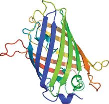
ARTICLE                                                                      NATURE COMMUNICATIONS | https://doi.org/10.1038/s41467-019-11427-y

T
       he ribosome is a molecular machine responsible for the                    ribosome (termed Ribo-T), whereby the small and large subunits
       polymerization of α-amino acids into proteins1,2. In all                  tethered together via helix h44 of the 16S rRNA and helix H101
       kingdoms of life, the ribosome is made up of two sub-                     of the 23S rRNA (Fig. 1a, c). Not only could this hybrid rRNA be
units3–5. In bacteria, these correspond to the small (30S) subunit               assembled into a functional ribosome in a cell, but Ribo-T could
and the large (50S) subunit. The 30S subunit contains the 16S                    support bacterial growth in the absence of wild-type ribosomes
ribosomal RNA (rRNA) and 21 ribosomal proteins (r-proteins),                     (Fig. 1b). We also used Ribo-T to create the first functionally
and is involved in translation initiation and decoding the mRNA                  orthogonal ribosome–mRNA system (Fig. 1d, e), and demon-
message6. The 50S subunit contains the 5S and 23S rRNAs and 33                   strated that Ribo-T could be evolved to synthesize protein
r-proteins, and is responsible for accommodation of amino acid                   sequences that the natural ribosome cannot easily translate by
substrates, catalysis of peptide bond formation, and protein                     selecting otherwise dominantly lethal rRNA mutations in the 50S
excretion7,8.                                                                    subunit. This provided the first example of engineering new
   The extraordinarily versatile catalytic capacity of the ribosome              function in the large subunit of an orthogonal ribosome that was
has driven extensive efforts to harness it for novel functions, such             previously inaccessible22. Similar results were obtained more
as reprogramming the genetic code9–13. For example, the ability to               recently with an analogously-designed ribosome with conjoined
modify the ribosome’s active site to work with substrates beyond                 subunits23,24. It should be noted that while remaining function-
those found in nature such as mirror-image (D-α-) and backbone-                  ally independent, orthogonal tethered ribosomes still share many
extended (β- and γ-) amino acids14,15, could enable the synthesis                components with native translation machinery (e.g., r-proteins,
of new classes of sequence-defined polymers to meet many goals                    elongation factors and initiation factors)12.
of biotechnology and medicine11,16. Unfortunately, cell viability                    Although the functional independence of Ribo-T conceptually
constraints limit the alterations that can be made to the ribosome.              enables new opportunities for exploring poorly understood
   To bypass this limitation, recent developments have focused on                functions of the ribosome, facilitating orthogonal genetic systems,
the engineering of specialized ribosome systems. The concept is                  and engineering ribosomes with altered chemical properties,
to create an independent, or orthogonal, translation system                      Ribo-T possesses limitations that could hinder its broad appli-
within the cell dedicated to production of one or a few target                   cations25. For example, cells with only Ribo-T exhibit a slower
proteins while wild-type ribosomes continue to synthesize                        growth rate than cells with natural wild type ribosomes (doubling
genome-encoded proteins to ensure cell viability. Pioneering                     time τ = 70 ± 2 min as opposed to τ = 35 ± 1 min for wild-type),
efforts by Hui and DeBoer17, and subsequent improvements by                      noting that part of this growth rate defect may arise from the
Chin and colleagues18–21, first created a specialized small ribo-                 circular permutation of the large subunit alone and not the
somal subunit. By modifying the Shine-Dalgarno (SD) sequence                     tethering26. In addition, the rate of protein synthesis in the Ribo-
of an mRNA and the corresponding anti-Shine Dalgarno (ASD)                       T cells is ~45% of that of the wild-type22 possibly due to slow
sequence in 16S rRNA, they generated orthogonal 30S subunits                     assembly and the resulting reduced number of functionally-active
capable of primarily translating a specific kind of engineered                    translating Ribo-T ribosomes25. Furthermore, the implemented
mRNA, while largely excluding them from translating endogen-                     orthogonal system was simply a modified version of previous
ous cellular mRNAs. These advances enabled the selection of                      works18,27, evolved in the context of untethered ribosomes using
mutant 30S ribosomal subunits capable of re-programming cel-                     different plasmid backbones and promoters. Finally, it is not clear
lular logic19 and enabling new decoding properties20.                            if the Ribo-T system is compatible with orthogonal non-canonical
   Unfortunately, such techniques have been restricted to the                    amino acid (ncAA) incorporation machinery for applications that
small subunit because the large subunits freely exchange between                 could expand the range of genetically encoded chemistry. Taken
pools of native and orthogonal 30S. This limits the engineering                  together, these features of the original Ribo-T system lim-
potential of the large subunit, which contains the peptidyl                      ited some applications.
transferase center (PTC) active site and the nascent peptide exit                    Here, we address these limitations through the development of
tunnel. We addressed this limitation with a fully orthogonal                     an improved Ribo-T design. Specifically, we used evolutionary

                      a                                             b                                                           c        3′   5′
                            The Ribo-T System                            Cell growth with Ribo-T
                                                                                                                  Proteome
                                                                        SQ171fg Δ7rrn
                                                                                                    50 S                        23S
                                                                                                                    Ribo-T      H101
                                                                                                                  ribosome
                                                                                                    30 S
                                                                                 mRNA                                            2858         2857
                                                                                                                    Ribo-T
                                         50 S
                                                                                                                                    T1        T2
                                                                    d    Orthogonal function

                                                                        Proteome                           50 S    Orthogonal   1453
                                                                                        50 S                                                  1454
                                                                                                                    function
                                                                         Wild-type   30 S             o30 S
                                                                        ribosomes
                                         30 S                                                   X                               16 S
                                                                                        mRNA         o-mRNA                      h44
                                                                    e                                 o-SD
                                                                         o-mRNA 5′
                                                                         o-16S                 3′
                                                                                                       o-ASD                             5′   3′

Fig. 1 Ribo-T system improvement strategies. a Schematic of Ribo-T showing tether (red) and orthogonal ribosome binding site (yellow). b The tether is
optimized in cells growing exclusively from the Ribo-T plasmid. c Previously published Ribo-T tether sequence. d Orthogonal function evolved for Ribo-T. e
Previously published orthogonal mRNA (o-mRNA) Shine-Dalgarno (SD) sequence and orthogonal 16S rRNA anti-SD (o-ASD) sequence shown

2                   NATURE COMMUNICATIONS | (2019)10:3920 | https://doi.org/10.1038/s41467-019-11427-y | www.nature.com/naturecommunications
NATURE COMMUNICATIONS | https://doi.org/10.1038/s41467-019-11427-y ARTICLE
a WT Ribo-T v1 b Library 1 Library 2 Library 3 Library 4 d Ribosome
plasmid n Tether 1: 5′–3′ Tether 2 : 5′–3′ Growth rate (h–1)
3′ 5′ 3′ 5′ 3′ 5′ 3′ 5′ 3′ 5′ 3′ 5′
WT 3 0.76 ± 0.02
L4-1 1 0.65
L4-2 1 0.64
L4-3 1 0.63
23S H101 L4-4 1 0.63
L4-5 1 0.63
L4-6 1 0.63
2858 2861 2856 2861 2856 2861 2856 2861 2856 L4-7 12 0.63 ± 0.04
2857 2857
L4-8 1 0.62
L4-9 1 0.62
2858
T1 T2 7A-20A 7U-20U 7A-20A 7A-20A 8N 9N 15N 10N L3-1 1 0.60
1453 L3-2 1 0.59
1453 L4-10 1 0.58
1454 1454 1449 1454 1449 1454 1449 1454 1449 1454
L4-11 1 0.57
L4-12 2 0.57 ± 0.06
L4-13 1 0.56
16S h44 L4-14 1 0.55
L4-15 1 0.54
L4-16 1 ACACATGTAGGAGAA GTGGGTATAT 0.54
pRibo-T v1 3 0.53 ± 0.06
L4-17 7 0.53 ± 0.03
5′ 3′ 5′ 3′ 5′ 3′ 5′ 3′ 5′ 3′ 5′ 3′ L3-3 1 0.52
L4-18 1 0.52
c SQ171fg strain
L4-19
L1-1
1
1 16 9
0.49
0.49
L3-4 1 0.49
Proteome L4-20 1 0.48
50 S L4-21 1 0.48
L4-22 1 0.48
rRNA & tRNA L4-23 1 0.47
30 S L4-24 1 0.47
mRNA
Tether library L4-25 1 0.47
pCSacB L4-26 1 0.47
L3-5 1 0.45
wt ptRNA Genome Δ7rrn Select for growth L4-27 1 0.45
L3-6 1 0.44
L4-28 1 0.44
L3-7 1 0.44
Transform plasmid library, L4-29 1 0.40
L1-2 1 12 9 0.39
select against wt plasmid Proteome L2-1 1 12 10 0.39
Ribo-T L3-8 1 0.38
rRNA & tRNA L4-30 1 0.38
L1-3 1 14 9 0.37
wt L1-4 2 9 10 0.36 ± 0.05
mRNA L2-2 1 8 11 0.34
pRibo-T L3-9 1 0.33
ptRNA Genome Δ7rrn L2-3 8 13 12 0.33 ± 0.01
L1-5 1 9 12 0.32
L1-6 1 10 19 0.28
Fig. 2 Optimizing tether sequence improves performance. a Wild-type 23S rRNA helix 101 and 16S rRNA helix 44 are connected to create Ribo-T with 9A
for 5′ tether, T1, and 8A for 3′ tether, T2. b Library 1: paired 5′ tether T1 poly A from 7–20 nucleotides, with 3′ tether T2 poly T from 7–20 nucleotides.
Library 2: unpaired polyA on both T1 and T2, ranging in 7–20 nucleotides long. Library 3: randomized T1 (8N) and T2 (9N) keeping residues of opened H101
and h44 apex loops. Library 4: randomized apex-to-apex T1 (15N) and T2 (10N) of tether. c Selection scheme for improved tethers. Strains lacking genomic
copies of rRNA operons (Δ7rrn) are transformed with plasmid-based Ribo-T tether libraries, and the wild-type pCSacB plasmid (wt) is removed. d Tether
sequences and growth rates of analyzed colonies. Error bars = 1SD of noted independent colonies, n. The top 15 Ribo-T design winners (L4-1 through L4-
13) were co-cultured and passaged for 3 days. Between each passage, the bulk culture populations were sequenced and analyzed. Source data for d can be
found in the Source Data file
approaches to select new RNA tethers that connect the 16S and length and sequence composition (Fig. 2a, b). The original Ribo-
23S rRNA by sampling an extended pool of tether variants dif- T’s (Ribo-T v1) 9-adenine tether T1 connects the 3′ 16S rRNA
ferring in their composition and length. By testing libraries residue G1453 of helix 44 (h44) to the 5′ 23S rRNA C2858 of
amounting to more than 1015 members, we isolated Ribo-T helix 101 (H101), and a 8-adenine tether T2 links G2857 of H101
variants with improved properties. Specifically, cells carrying the to G1454 of h44 (Fig. 2a). Our initial choice of these oligo(A)
improved variant, which we term Ribo-T version 2 (Ribo-T v2) tethers for Ribo-T was based on the simplicity of the linker
has a 24% increase in growth rate (0.75 h−1, in SQ171fg strain) as sequence and its presumed resistance to the action of cellular
compared to the original Ribo-T (Ribo-T v1; T1: 9A, T2: 8A) and nucleases22. We wondered if replacing unpaired linkers with
a 12% increase in final OD600 at 37 °C as compared to Ribo-T v1 sequences capable of base pairing with each other and forming a
(final Ribo-T v2 OD600 = 0.9, in SQ171fg strain). In minimal double stranded RNA stem would be beneficial for Ribo-T sta-
media, these advantages are even more striking, with Ribo-T v2 bility and functionality. To test this, we designed four libraries of
possessing a 79% improvement in final OD600 at 37 °C relative to T1 and T2 tethers at the H101/h44 subunit connection point
Ribo-T v1. We then used directed evolution to improve the (Fig. 2b). Libraries 1 and 2 explore tether length in a paired and
orthogonal function of Ribo-T. The optimized orthogonal (o) unpaired format, respectively, without the apex loop remnants
Ribo-T v2 (mRNA Shine-Dalgarno (SD): 5′-CAACCAC-3′, 16S present in our original library design22. Specifically, library 1
anti-SD (ASD): 5′-CUGUGG-3′) has a 208% increase in overall explores tether length with potential base pairing using a 7A-20A
expression of the target protein, and possessed a 16% increase in T1 tether paired with a 7U-20U T2 tether (for a total library size
orthogonality (with an orthogonal cat reporter) as compared to of 196 members). Library 2 explores a dual poly(A) tether ranging
oRibo-T v1. To demonstrate the utility of the oRibo-T v2, we from 7A-20A (196 members). Libraries 3 and 4 explore tether
expressed a diverse set of proteins ranging from small (25 kDA) sequence with fixed length of the published pRibo-T tether22.
to large (116 kDa). Lastly, oRibo-T v2 was leveraged to synthesize Library 3 keeps the apex loop remnants of the original Ribo-T
superfolder green fluorescent protein (sfGFP) possessing multi- sequence for an 8N/9 N randomized library of 1.7 × 10¹⁰ mem-
ple, identical ncAAs. Our improvements expand Ribo-T’s appli- bers, while library 4 fully randomizes the h44-tether-H101
cations and make the Ribo-T system better suited for studying structure for a 15N/10N randomized library of 1.1 × 10¹⁵ mem-
and leveraging orthogonal translation in vivo. bers, although the entire sequence space was not accessed
experimentally because of transformation limitations.
Results Following library construction (Supplementary Fig. 1), the
Tether optimization improves growth of Ribo-T cells. We first resulting libraries were individually transformed into
sought to improve Ribo-T function by optimizing the tether for the Escherichia coli SQ171fg strain22, which was evolved from
NATURE COMMUNICATIONS | (2019)10:3920 | https://doi.org/10.1038/s41467-019-11427-y | www.nature.com/naturecommunications 3ARTICLE NATURE COMMUNICATIONS | https://doi.org/10.1038/s41467-019-11427-y
a b SQ171 “sg” SQ171fg
Ribo-T v1 Ribo-T v2 1.5
1.15 1.10 p < 0.05
3′ 5′ 3′ 5′
rate (hr–1)
1
Growth
0.75
0.60
0.38
0.5 0.20
23S
H101 0
p < 0.05
1.0 1.0
1 0.9
0.8
max OD600
Relative
0.8
0.6 0.3 0.4
2861 2856
2858 2857 0.4
0.2
0
rRNA wt Ribo-T v1 Ribo-T v2
T1 T2 T1 T2
c sg fg sg fg sg fg
1453
1454 1449 1454
Dilution
16S
h44
d
16S/23S
23S
5′ 3′ 5′ 3′ 16S
Fig. 3 Optimizing tether sequence improves performance. a Ribo-T v1: previously published tether sequence. Ribo-T v2: fastest growing and most frequent
selected tether sequence. b Growth rate and max OD600 of SQ171 slow growing (sg) and SQ171 and fast growing (fg) cells growing with pAM552 (wild-
type rrnb operon), pRibo-T v1 and pRibo-T v2 (n = 6; paired t-test [two-sided], p < 0.05). Error bars = 1SD. c Spot plated SQ171 and SQ171fg cells growing
with pAM552, pRibo-T v1 and pRibo-T v2 imaged after 48 h at 37 °C. d Total RNA extraction from SQ171 and SQ171fg cells growing with pAM552, pRibo-T
v1 and pRibo-T v2. Source data for b–d can be found in the Source Data file
the SQ171 strain28 that lacks chromosomal rRNA alleles and individually grown in separate liquid cultures, combined at equal
survives on the pCSacB plasmid that carries the wt rrnB operon OD600 in co-culture, in triplicate, and passaged for three days.
and the tRNA67 plasmid that carries missing tRNA genes. The Between each passage, both the bulk populations and individual
pCSacB plasmid also contains a counter selectable marker sacB resultant colonies from plated cultures were sequenced and
gene, that confers sensitivity to sucrose. Distinct from the analyzed (Supplementary Fig. 3). After 3 passaging days, all three
SQ171 strain, the SQ171fg strain contains mutations that were cultures converged to sequence L4-7, which we term Ribo-T v2
previously shown to improve the growth of the Ribo-T cells22. (Fig. 3a).
The Ribo-T 23S rRNA in each library contains an A2058G In both liquid culture growth (Fig. 3b) and plate growth assays
mutation, conferring resistance to erythromycin that facilitates (Fig. 3c), cells supported exclusively by pRibo-T v2 outperform
the selection of cells expressing functional Ribo-T. Colonies grew pRibo-T v1 in both SQ171 and SQ171fg strains. Specifically, in
from all libraries in the presence of sucrose (indicating the loss of the SQ171fg strain, the pRibo-T v2 plasmid improves growth rate
the pCSacB plasmid) and erythromycin, demonstrating full by 24% and the maximum OD600 in LB media by 12% as
support of the cellular protein synthesis by tethered Ribo-T compared to the pRibo-T plasmid (n = 6, paired t-test [two-
expressed from the plasmid (Fig. 2c). Agarose gel electrophoresis sided], p < 0.05). The benefits are more pronounced in the
of total RNA of a sampling of colonies from each library show the original SQ171 strain, where growth rate improves by 86%, and
expected dominant Ribo-T size RNA corresponding to the max OD600 by 70% as compared to pRibo-T (n = 6, paired t-test
16S–23S chimera instead of the individual 16S and 23S bands, [two-sided], p < 0.05). The growth curves also highlight a
confirming no significant wild-type ribosome contamination significantly reduced lag time in cell growth for Ribo-T v2 cells
or tether cleavage (Supplementary Fig. 2). Individual colonies versus Ribo-T v1 cells in both SQ171 and SQ171fg strains
(~50–100) were picked from each library (biasing towards bigger (Supplementary Fig. 4). Agarose gel electrophoresis of total RNA
colonies), tethers were sequenced, and growth rates were extracted from cells supported by pRibo-T v2 plasmids show the
determined (Fig. 2d). While viable clones supported by intact expected 16S–23S sized RNA, and the loss of individual 16S and
tethered ribosomes were isolated from each library (Supplemen- 23S rRNA bands (Fig. 3d).
tary Fig. 2), Library 4 was most successful in yielding clones with We next tested if the Ribo-T v2 growth improvement
improved growth rates compared to pRibo-T v1 (Fig. 2d). properties were robust, by comparing growth relative to Ribo-T
We next carried out additional evolutionary experiments to let v1 in different strains (i.e., SQ171, SQ171fg, POP2136), at various
the cells with the top 15 most improved tether sequences that growth temperatures (30, 37, and 42 °C) and different media
emerged from this selection compete in liquid culture. Specifi- (Supplementary Fig. 5). We observed appreciable improvements
cally, the top 15 strains (Fig. 2d, L4–1 through L4-13) were in each case. The advantage of Ribo-T v2 was especially
4 NATURE COMMUNICATIONS | (2019)10:3920 | https://doi.org/10.1038/s41467-019-11427-y | www.nature.com/naturecommunicationsNATURE COMMUNICATIONS | https://doi.org/10.1038/s41467-019-11427-y ARTICLE
pronounced at 30 °C in M9-casamino acids (M9CA) minimal Fig. 7a) enables both a positive and a negative selection from a
media with a 78% and 69% improvement, respectively, in final single gene product: chloramphenicol acetyltransferase encoded
max OD and average doubling time over Ribo-T v1 (Supple- in the cat gene confers resistance to chloramphenicol (Cm),
mentary Fig. 5e, f). Since the Ribo-T v2 design showed superior whereas the fused upp gene codes for uracil phosphoribosyl-
growth characteristics, remained uncleaved, and outperformed tranferase causing cell death in the presence of 5-fluorouracil (5-
other tether sequences in a liquid culture competition, this FU) (Supplementary Fig. 8).
construct was selected for future experiments. For the negative selection step, the wild-type SD sequence (5′-
While we do not have a simple explanation for why the newly AAGGAGG-3′) for the cat-upp gene on plasmid plpp5-catupp-
selected tethers improve the growth rate of Ribo-T v2 cells p15A (Fig. 4, Supplementary Fig. 7a) was entirely randomized.
relative to Ribo-T v1, it may be attributed to the possible partial We then transformed the plasmid library into BL21(DE3)Δupp
pairing of the new tethers. Specifically, chemical probing and cells and plated on M9 minimal media agar plates supplemented
modeling of the secondary structure29 suggest that a segment of with 10 µg ml−1 5-FU. Surviving cells produce mRNA that is not
the tethers may form a base-paired duplex (Supplementary efficiently translated by endogenous ribosomes (desired out-
Fig. 6). Conceivably, the structure of the improved tethers may come), or have non-functional plasmids. In the initial attempts of
either facilitate the Ribo-T v2 assembly, which as we know is one the subsequent positive selection, we had difficulty selecting
of the main limiting properties of the original Ribo-T design25 or robust orthogonal SD/ASD pairs from this o-SD mRNA pool
may better facilitate the relative movement of the tethered with a randomized ASD-Ribo-T library directly. Therefore, we
subunits during initiation, elongation of termination steps of performed a first round of positive selection using untethered
translation. ribosomes with the small subunit carrying randomized ASD in
order to limit the o-mRNA sequence space to just orthogonal and
sufficiently active o-mRNA sequences. Specifically, the 16S rRNA
Improvement of Ribo-T orthogonal function. After selecting ASD sequence of plasmid-based untethered ribosomes (Supple-
optimized tethers, we sought to improve the orthogonality of the mentary Fig. 7b) was randomized, the plasmids were transformed
tethered ribosome system. Orthogonal function of Ribo-T is into the surviving cells from our negative selection, and then
achieved by altering the mRNA SD sequence and the corre- plated on LB-agar plates in the presence of 100 µg ml−1 Cm.
sponding ASD sequence of the 16S rRNA. In this way, a spe- Surviving colonies were picked, and plasmids were isolated and
cialized pool of orthogonal Ribo-T (oRibo-T) is created that sequenced (Round 1, Supplementary Fig. 9b).
exclusively translates the cognate mRNA and in principle, should To identify top performing o-mRNAs, we evaluated the round
be functionally isolated from the pool of wild-type mRNA and 1 selected SD/ASD pairs for overall reporter expression levels and
ribosomes. Our oRibo-T system22 utilized a modified version of a assessed the extent of cross-talk with wild-type. This initial
previously developed orthogonal 30S subunit system18, not one characterization of orthogonal SD/ASD pair activity was
developed in the Ribo-T context. We hypothesized that because performed using a Cm-resistance assay and the cat-upp reporter
initiation with Ribo-T is limiting22,25, optimizing the SD/ASD plasmids. To test overall activity, each set of cognate o-mRNA
pairing could improve orthogonal system functionality. and o-16S rRNA plasmids was added to the same cells and
The goal of this effort was to improve orthogonal protein resistance to Cm assessed. Additionally, to measure orthogonality
expression by oRibo-T v2, while minimizing cross-talk of the of the corresponding mRNAs with the wild-type ribosome pool
orthogonal mRNA with wild-type ribosomes. To this end, we (i.e., how much cross-talk exists between wild-type ribosomes and
used a robust directed evolution approach18 to select highly our selected orthogonal mRNAs), each orthogonal mRNA
functional and orthogonal Ribo-T v2/mRNA pairs (Fig. 4). construct was independently co-transformed into fresh BL21
Specifically, a fusion of the cat and upp genes (Supplementary (DE3)Δupp cells with plasmid coding for wild-type ribosomes
BL21(DE3)Δupp Postive selection (+Chloramphenicol)
Proteome
50 S 50 S
Wild-type 50 S
Round 2 pairs,
ribosome 30 S
Ribo-T v2 rRNA 30 S
mRNA o-30S
Genome
rDNA
SD
ASD
3′
Cat-upp rRNA Ribo-T v2
Reporter DNA
plasmid library
library
Clone into
Negative selection (+ 5-Fluorouracil) pRibo-T v2
50 S
Reporter DNA
50 S Round 1 pairs, 30 S Active orthogonal
untethered rRNA mRNA from round 1
30 S
Cell death when
CAT-UPRT expressed Transform isolated reporters
Postive selection (+Chloramphenicol)
Reporter DNA Untethered rRNA Reporter DNA
plasmid library
50 S 50 S
ASD
3′
30 S No CAT-UPRT, cell lives 30 S
o-30S
rDNA
Fig. 4 Improving orthogonal pairs. Selection scheme to optimize orthogonal Shine-Dalgarno (SD) and anti-Shine-Dalgarno (ASD) pairs in untethered and
tethered context
NATURE COMMUNICATIONS | (2019)10:3920 | https://doi.org/10.1038/s41467-019-11427-y | www.nature.com/naturecommunications 5ARTICLE NATURE COMMUNICATIONS | https://doi.org/10.1038/s41467-019-11427-y
a b + Pair – Pair d
5′ mRNA
El er
n
2000
io
dd
ut
wt BL21(DE3)Δupp sf-gfp
3′ kDa
La
16S
Fluorescence/OD600
1500
A 5′ 198
1 3′ 1000
98
B 5′ n 500 62
2 3′ Round 1 9 49
0 PDB: 1EMA PDB: 3Q3E
85% 85% 91% 93% 90% 92% 77% 82% 89% 91% 76% 89%
3 3′ Round 2 1 Name: Superfolder green 38 Name: N-glycosyltransferase of
v1 1.A 2.B 3.B 2.C 3.C 4.D 5.D 6.D 7.D 8.E 9.E
fluorescent protein (sfGFP) A. pleuropneumoniae (ApNGT)
% Orthogonality Mass: 26,886 Da 28 Mass: 71,561 Da
C 5′ Orthogonal pair 17
Helical: 9% Helical: 47%
14
2 3′ Round 1 6 Beta sheet: 49% Beta sheet: 10%
3 3′ Round 2 1
c + Pair – Pair e
El er
n
io
dd
120
ut
D 5′ kDa
La
BL21(DE3)Δupp Cat
100
4 3′ Round 1 1
IC50 (μg ml–1)
3′ 80 198
5 Round 1 2
60 98
6 3′ Round 2 4
40
7 3′ Round 2 1 62
20
PDB: 3CLA 49 PDB: 1JYX
E 5′ 0 Name: Chloramphenicol Name: Beta-galactosidase (LacZ)
75% 72% 85% 88% 66% 65% 62% 64% 68% 69% 67% 85% Acetyltransferase (CAT) 38 Mass: 116,483Da
8 3′ Round 1 13 v1 1.A 2.B 3.B 2.C 3.C 4.D 5.D 6.D 7.D 8.E 9.E Mass: 25,663 Da Helical: 13%
9 3′ Round 2 1 % Orthogonality 28
Helical: 28% Beta sheet: 40%
Orthogonal pair Beta sheet: 30%
17
14
Fig. 5 Selected orthogonal pair sequences and function in Ribo-T v2. a Top evolved orthogonal mRNA and 16S with predicted pairing. Selection round is
noted by round 1 or round 2 to the right of each pair. n denotes number of isolated members with that sequence from the selection. b–e Orthogonal pair
notation: Original orthogonal Ribo-T system denoted by v1, and x.y where x is o16S number and y is o-mRNA letter (pORTx.y plasmid name format).
b Orthogonal expression of super folder green fluorescent protein (sf-gfp) in BL21(DE3)Δupp.+ pair: both o-rRNA and o-mRNA expressed, − pair: just o-
mRNA expressed without cognate o-rRNA. Percent orthogonality is shown below column labels. A higher percentage value is desired, indicating a
lower background expression of o-mRNA as compared to the expression with the cognate orthogonal rRNA. Error bars = 1SD of n = 3 independent
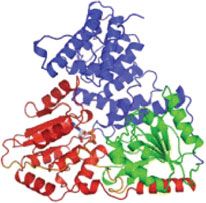
experiments. The protein’s structure and details are listed to the right of the graph. c Orthogonal expression of Cm acetyltransferase (cat) in BL21(DE3)
Δupp. Error bars = 1 standard error in IC50 curve fitting. The protein’s structure and details are listed to the right of the graph. d Orthogonal expression of
N-glycosyltransferase of A. pleuropneumoniae (ApNGT) in BL21(DE3). The protein’s structure and details are listed to the right of the graph. e Orthogonal
expression of Beta-galactosidase (LacZ) in BL21(DE3). The protein’s structure and details are listed to the right of the graph. Source data for b, c can be
found in the Source Data file
(pAM552, Supplementary Fig. 7b). Round 1 strains were plated were measured with two protein expression assays: fluorescent
on a range of Cm concentrations (0, 0.5, 1, 2.5, 5, 10, 20, 40, 60, protein expression and antibiotic resistance. These two assays
80, 100, 200, 300, 400, and 500 μg ml−1), and maximum growth were chosen to validate and demonstrate that these orthogonal
concentrations noted (Supplementary Fig. 9a). Evolved pairs had Ribo-T v2 exhibit comparable relative orthogonal expression
increased cognate pair activity (black bars) well above the regardless of the protein they express. Importantly, a metric for
background expression of the o-mRNA by wild-type ribosomes quantifying orthogonality is critical, because it segregates the
(white bars). Furthermore, orthogonal pair activity was signifi- activity of oRibo-T v2 from that of wild-type ribosomes, and
cantly increased over the previous orthogonal system22 normalizes orthogonality across the two different assays. Percent
(pAM552o/A, Supplementary Fig. 9). orthogonality is calculated as:
We used a set of 14 best-performing orthogonal mRNAs for a
second round of positive selection with a library of Ribo-T v2 Apair AmRNA
% orthogonality ¼ ´ 100 ð1Þ
with the ASD sequence randomized. First, the active and Apair
orthogonal mRNA (o-mRNA B-P, Supplementary Fig. 9b) were
isolated, pooled and transformed into the BL21(DE3)Δupp strain. Where Apair is the activity of the orthogonal pair (sfGFP fluor-
Then, the ASD sequence on pRibo-T v2 plasmid was randomized, escence divided by OD600 for the fluorescent protein expression
transformed into BL21(DE3)Δupp containing the top performing assay, or half maximal inhibitory concentration (IC50) for the
orthogonal mRNAs, and plated on LB-agar plates supplemented CAT assay), and AmRNA is the activity of just the orthogonal
with 100 μg ml−1 Cm (Fig. 4). Surviving colonies were picked, mRNA expressed without the cognate orthogonal ribosome (i.e.,
and plasmids were isolated and sequenced. Top performing pairs, the crosstalk with wild-type ribosomes). The extent of ortho-
aligned using the ribosome binding site (RBS) calculator30,31, are gonality (%) is shown below each pair in the activity plots in
shown in Fig. 5a. The alignments show that while the selected Fig. 5. With this metric, a higher percentage value indicates a
orthogonal SD/ASD pairs are different from wild-type sequences, lower background expression of o-mRNA in the absence of
they have high complementarity between themselves. Our cognate oRibo-T v2 as compared to the expression when the
orthogonal Ribo-T constructs with improved v2 tethers are cognate oRibo-T v2 is present.
named pORTx.y, where x is a number indicating the orthogonal For evaluation of selected orthogonal pairs, SD variants were
ASD sequence (1–9), and y is a letter indicating the correspond- cloned into vectors containing the sf-gfp and cat genes,
ing cognate SD sequence (A–E). Corresponding rRNA plasmids respectively. ASD variants were freshly cloned into the pRibo-T
with untethered ribosomes are named pOx.y. v2 plasmid. Plasmid pairs were transformed into a naïve BL21
(DE3)Δupp strain for testing. Expression of sfGFP was measured
as final fluorescence normalized by the final OD600 reading
Evaluation of evolved orthogonal pairs. With improved ortho- (Fig. 5b) and activity of CAT was evaluated as IC50 (Fig. 5c). Of
gonal Ribo-T v2/mRNA pairs in hand, we assessed performance note, pair activity is improved in both sfGFP and CAT assays over
with two key metrics: (i) the overall activity and (ii) the ortho- the original published oRibo-T system22 (noted as v1), as well as
gonality to wild-type ribosomes. Pair activity and orthogonality the published v1 orthogonal pair cloned with the optimized v2
6 NATURE COMMUNICATIONS | (2019)10:3920 | https://doi.org/10.1038/s41467-019-11427-y | www.nature.com/naturecommunicationsNATURE COMMUNICATIONS | https://doi.org/10.1038/s41467-019-11427-y ARTICLE
tether sequences (noted as 1.A). We observed that some pairs combined with every orthogonal pair. Specifically, v2 tethers and
achieved high sfGFP expression (e.g., pORT3.C, Fig. 5b), other improved orthogonal pairs worked synergistically to improve
pairs conferred particularly strong resistance to Cm (e.g., pORT7. orthogonal function over the v1 tethers by up to 55% (Supple-
D, Fig. 5c), some pairs achieved high orthogonality (e.g., pORT3. mentary Fig. 12a). The CAT assay did not show significant dif-
B, Fig. 5b, c), some pairs had moderate activity but poor ference between v1 and v2 tethers (Supplementary Fig. 12b),
orthogonality (e.g., pORT4.D, pORT8.E, Fig. 5b, c), and some presumably because of the less sensitive assay range compared to
pairs achieved a balance of high activity and orthogonality e.g., the sfGFP fluorescence assay.
pORT2.B, pORT3.B, Fig. 5b, c). When considering both To further demonstrate the utility of the oRibo-T v2 system, we
assays, and metrics of pair activity and orthogonality, we selected expressed additional recombinant proteins aiming to represent a
o-mRNA B (oSD: 5′-CAACCAC) paired with o-ASD #2 diverse range of protein sizes, structures, and functions.
(5′-UGUGGU) (selected in Round 1 in untethered context), Specifically, we cloned E. coli β-galactosidase (LacZ) and N-
and o-ASD #3 (5′-CUGUGG) (selected in Round 2 in v2 tether glycosyltransferase of A. pleuropneumoniae (ApNGT) into our
context). in vivo orthogonal reporter construct (plpp5.B). We then purified
To directly compare performance of the newly selected the encoded proteins, and compared their expression across
orthogonal pairs against our original orthogonal pair22, we oRibo-T v2 and oRibo-T v1 (Fig. 5d, e and Supplementary
cloned the previous o-ASD sequence into the Ribo-T v2 plasmid Fig. 10b–d). Importantly, cells carrying oRibo-T v2 had a 37%
to generate pORT1, and the cognate orthogonal SD sequence into higher expression of LacZ and a 22% higher expression of
the sf-gfp and cat reporter plasmids to generate plpp5.A.gfp and ApNGT over oRibo-T v1 (n = 3, paired t-test [two-sided], p <
plpp5.A.cat (Fig. 5a). For plasmids pORT2 and pORT3 paired 0.05). These results demonstrate Ribo-T v2’s utility in producing
with orthogonal GFP reporter B (plpp5.B.gfp), we observed a variety of proteins of various sizes (25–116kDa), structural
activity increases of 154% and 208%, respectively, compared to compositions (9–47% alpha helical and 10–49% beta sheets), and
pORT1. Percent orthogonality also increased by 6% and 8% (n = functions (fluorescence, antibiotic resistance, hydrolysis, and
6, paired t-test [two-sided], p < 0.05), respectively (Fig. 5b). For glycosylation).
plasmids pORT2 and pORT3 paired with orthogonal cat reporter
B (plpp5.B.cat), pair activity increased 77% and 121% over
pORT1, respectively. Percent orthogonality increased 13% (for 2. Incorporation of non-canonical amino acids by Ribo-T. Engi-
B) and 16% (for 3.B) over pORT1, respectively (Fig. 5c). While neering the translation apparatus is a key emerging opportunity
the orthogonal GFP reporter C (plpp5.C.gfp) had higher in synthetic biology38–40. One of the central reasons to develop an
functionality than the orthogonal GFP reporter B (plpp5.B.gfp) orthogonal Ribo-T system is the possibility of selecting otherwise
with pORT2 and pORT3, its orthogonality was lower than that of dominantly lethal rRNA mutations in the peptidyl transferase
the reporter B (Fig. 5b, c). The new mRNA/oRibo-T pairs (o- center that facilitate the translation of new abiological polymers
mRNA B: 5′-CAACCAC; o-ASD #3: 5′-CUGUGG) are poised to made with the use of an expanded genetic code9,39. Such efforts
expand the versatility of the fully orthogonal ribosome–mRNA require that the Ribo-T platform is compatible with orthogonal
system. ncAA incorporation machinery and, up to now, compatibility has
yet to be shown in the Ribo-T system, and multiple ncAA
incorporations with a tethered o-ribosome has yet to be achieved.
Orthogonal pair activity in other E. coli strains. To test system
We therefore tested whether oRiboT is compatible with
versatility in a wide range of strains, top performing plasmid pairs
multiple site-specific ncAA incorporation into proteins. Specifi-
for the sfGFP reporter set were next transformed into BL21 Star
cally, we assessed the ability of orthogonal Ribo-T v2 (pORT3) to
(DE3) (Invitrogen) and a variant of the fully recoded C321.ΔA
site-specifically incorporate p-azido-L-phenylalanine (pAzF) into
strain32,33, MCJ1217. These strains provide benefits for ncAA
sfGFP, using a previously reported orthogonal transfer RNA
incorporation using amber suppression and we recently showed
(tRNA) and aminoacyl-tRNA synthetase (aaRS) pair from
that C321.ΔA could be coupled with extensively engineered
Methanocaldococcus jannaschii41 (henceforth referred to as
synthetases for multi-site incorporation of up to 30 ncAAs into a
pAzFRS). Importantly, the idea was not to engineer oRibo-T to
single biopolymer in vivo34 and developed for cell-free protein
be better than a natural ribosome at incorporating pAzF, which is
synthesis applications as well33,35–37. Following transformation,
known to be incorporated efficiently, but rather to show that
we evaluated the ability of our top performing oRibo-T v2/o-
oRibo-T and the pAzF orthogonal translation system were able to
mRNA pairs to express sfGFP (Supplementary Fig. 10a). General
cooperate in producing protein(s) with multiple ncAAs.
trends observed in the BL21(DE3)Δupp strain hold for these
To minimize plasmid requirements for ncAA incorporation,
additional strains: pORT2.B, pORT3.B, pORT2.C and pORT3.C
we first combined the oRibo-T v2 rRNA and the reporter gene on
sets perform better than the original pair (>200% of pORT1
one plasmid. Since relative directional orientation of the two
expression under similar conditions), with maintained high
expression cassettes from a single plasmid can have a significant
orthogonality. The best-performing orthogonal pairs similarly
impact on system performance42–44, we built and tested
benefitted specialized 30S subunits in a non-tethered context
combined rRNA/mRNA plasmids in both the forward and
(Supplementary Fig. 11).
reverse directions (Supplementary Fig. 13a). While pORT3.B.gfp
forward and reverse constructs had similar overall expression, the
Synergistic effect of evolved tethers and orthogonal pairs. We growth characteristics of the reverse orientation was significantly
next set out to study the effects of improved tethers and ortho- better than the forward orientation (Supplementary Fig. 13b,
gonal pairs on the oRibo-T system performance. To do this, select graph inset), and so this orientation was selected for future
orthogonal ASD sequences were cloned into both our improved experiments.
oRibo-T v2 plasmid as well as our original published oRibo-T v1 We then tested ncAA incorporation. The genomically-recoded
(with tether sequences 9A/8A)22. Using both our orthogonal organism derived from C321.ΔA (MCJ.1217) lacking UAG stop
sfGFP and CAT assays, we measured the activity (fluorescence for codons was co-transformed with our combined reporter gene and
sfGFP, and IC50 for CAT) of our orthogonal pairs in the context an orthogonal translation system plasmid containing an aaRS:
of either Ribo-T v1 or v2 tethers. In our sfGFP assay, we observed tRNA pair previously engineered for incorporation of pAzF41.
improvements in activity and orthogonality for Ribo-T v2 when We quantitatively assessed the incorporation of pAzF into sfGFP
NATURE COMMUNICATIONS | (2019)10:3920 | https://doi.org/10.1038/s41467-019-11427-y | www.nature.com/naturecommunications 7ARTICLE NATURE COMMUNICATIONS | https://doi.org/10.1038/s41467-019-11427-y
a D36 E132
construct carrying T1 (CAATGAACAATTGGA) and T2
K101 E213 (GATAACTAGT) being the winning variant. The new Ribo-T
v2 system exhibits up to an 86% improvement in growth rate and
B.gfpTAG D190 70% improvement in maximum OD600 (in SQ171 strain), as
AmpR
ColE1 compared to the original Ribo-T v1. The improvement in tether
16S 23S 5S design was insufficient to bring the growth rate of the Ribo-T v2
pAM cells to that of wild type cells. We believe this reflects a funda-
o-anti-SD mental limitation of this Ribo-T architecture, which is based on
pORT3 insertion of a circularly permutated 23S rRNA into a 16S rRNA
helix at H101 and h44. The unusual structure and transcription
b order of the rRNA segments in Ribo-T causes notable assembly
2500
MCJ.1217 defects25. We are not sure whether it is the circular permutation
of the large subunit rRNA or disruption of the continuity of the
2000
small subunit rRNA that are the primary cause of the assembly
problems. However, in spite of assembly limitations, Ribo-T v2
Fluor./OD600
1500
has marked improvements over the original Ribo-T variant.
1000
Furthermore, after the selection of enhanced orthogonal Ribo-T
v2/mRNA pairs, orthogonal Ribo-T v2 (pORT3) exhibits a
500
~200% increase in activity for sfGFP expression and also
improved orthogonality compared to our original orthogonal
0 system22.
pAzF + – + – + – + – The improvements presented here to the Ribo-T platform
rRNA pAM pORT3 pAM pORT3
enhance the usefulness of the system for biochemical assays (e.g.,
faster growth for RNA extractions, and higher density cultures for
sf-gfp 1-TAG 5-TAG
increased preparation of Ribo-T v2 variants for in vitro utiliza-
Fig. 6 Incorporation of ncAA p-azido-L-phenylalanine (pAzF) by orthogonal tion), and applications. Specifically, these improvements allowed
Ribo-T. a Combined rRNA and sf-gfp plasmid with sf-gfp gene is replaced us to demonstrate the usefulness of the orthogonal mRNA-Ribo-
with a 1TAG or 5TAG version to create pORT3B.gfp1TAG and pORT3B. T v2 system for two different applications. We demonstrated that
gfp5TAG (orthogonal Ribo-T with ASD sequence 3 and orthogonal sfGFP orthogonal Ribo-T v2 is capable of synthesizing a range of diverse
message B containing 1 TAG or 5 TAG, respectively). Wild-type rrnb operon proteins of different sizes, structures, and functions with
was cloned as a negative control for background orthogonal expression enhanced efficiency over Ribo-T v1. Second, as a proof of con-
(pAM.B.gfp1TAG and pAM.B.gfp5TAG). b Expression of sf-gfp with 1TAG cept, we demonstrated that oRibo-T can be leveraged for the site-
or 5TAG in C321.ΔA derived strain MCJ.1217 (C321.ΔA.mutS+.Δλred. specific incorporation of multiple ncAAs into proteins. We
Δupp), in the presence of (+) or absence of (−) pAzF. Error bars = 1SD of showed successful Ribo-T mediated incorporation of up to five
n = 6 independent experiments. Source data for b can be found in the pAzF residues with >10-fold expression above background.
Source Data file Looking forward, the new Ribo-T v2 is expected to become a
versatile tool for many biotechnology, engineering, and basic
variants with 1 or 5 TAG codons at amino acid positions D190 (1 science applications. These applications and opportunities have
TAG) or D36, K101, E132, D190, and E213 (5 TAG) (Fig. 6a, sparked enthusiasm, resulting in parallel work featuring a con-
Supplementary Fig. 13c). Cells containing plasmids encoding for ceptually similar design of an orthogonal stapled ribosomes23,24.
orthogonal Ribo-T with ASD sequence 3 and orthogonal sfGFP Although the stapled ribosomes leveraged our same circular
message B containing 1 TAG (pORT3B.gfp1TAG) or 5 TAG permutation and helix connections found in the Ribo-T design
(pORT3B.gfp5TAG) were grown in LB media supplemented with (H101 and h44)22,47, recently reported improvements to the
pAzF. Upon analyzing fluorescence, we found oRibo-T v2 to be initial stapled system yielded a strain carrying tens of mutations
successful in translating the sf-gfp gene containing not only one within the evolved strain24, which leaves some uncertainty about
TAG but even five internal TAG codons with expression levels portability of that system. Our Ribo-T v2 construct was originally
>six-fold and >10-fold above background, respectively (Fig. 6b). developed in a widely used strain28, and is portable and func-
The expression levels are statistically significant (paired t-test tional in several other strains without extensive strain modifica-
[two-sided], p < 0.05) and in line with previously reported values tions. These attributes make our orthogonal Ribo-T v2 system
in the literature for this system configuration45. Similar expres- robust for a variety of applications and studies. This includes
sion was observed with the untethered orthogonal ribosome modifying the catalytic capacity of the ribosome for improved
system with plasmids pO2B.gfp1TAG and pO2B.gfp5TAG incorporation of ncAAs such as backbone-extended monomers
(Supplementary Fig. 13d). Our results highlight the effective (e.g., β-, D-, or γ- amino acids)48,49 into polypeptides and bio-
utility of our combined plasmid design for incorporation of polymers, probing single and multi-mutations in highly con-
ncAAs. Furthermore, our work demonstrates a key proof-of- served rRNA nucleotides, translation of problematic protein
concept result that confirms compatibility and utility of a Ribo-T sequences, and the creation of an orthogonal central dogma,
v2-based orthogonal system with widely used and standardized which may insulate genetic programs from host regulation and
orthogonal translation components33,45,46. allow expansion of the roles of these processes within the cell12.
Methods
Discussion Construction of the tether libraries. Plasmid construction and DNA manipula-
Here, we present improvements to the original Ribo-T platform. tions were performed following standard molecular biology techniques. The
This second-generation design was developed using tether libraries of tether sequences were introduced into the wild-type pRibo-T plasmid
by inverse PCR amplification with Phusion polymerase (NEB) with primers listed
libraries varying in both the length and composition of the tether in Supplementary Table 1. All primers were synthesized by Integrated DNA
sequence. We identified several sequences at the h44/H101 Technologies. Amplification was followed by re-circularization with the Gibson
junction capable of supporting robust cell growth with the assembly reaction50 (Supplementary Fig. 1). Specifically, Ribo-T backbone plasmid
8 NATURE COMMUNICATIONS | (2019)10:3920 | https://doi.org/10.1038/s41467-019-11427-y | www.nature.com/naturecommunicationsNATURE COMMUNICATIONS | https://doi.org/10.1038/s41467-019-11427-y ARTICLE
was prepared by PCR amplification with primers 5′-GGAGGGCGCTTACCAC 55 °C 30 s, 72 °C 2 min) × 25, and 72 °C final extension for 10 min. Plasmid pCP20
TTTG and 5′-GGTTAAGCTACCTACTTCTTTTG using pRibo-T22 as template. was transformed into a kanamycin-resistant colony to remove the KanR cassette by
Using Phusion polymerase, PCR was performed at 98 °C initial denaturing for the incorporated flippase sites51. Transformed cells were plated on LB agar sup-
3 min, (98 °C 30 sec, 55 °C 30 sec, 72 °C 70 sec)x25, and 72 °C final extension plemented with 50 μg ml−1 carbenicillin and grown overnight at 30 °C. Colonies
for 10 min. This amplifies the pRibo-T vector, excluding the tethers and 23S were picked, plated on LB agar plates, and grown overnight at 42 °C to select for
region of the plasmid. loss of pCP20 plasmid. Colonies were checked for kanamycin sensitivity, and
To generate the tether libraries (Fig. 2b), primer pools were first prepared from deletion was confirmed by sequencing of PCR product from colony PCR using
primers listed in Supplementary Table 1. For library 1, equimolar amounts of primers 5′-TGCCAGGGTAAAGGTTAG and 5′-GACGGTTGCACCAAAC, and
primers T1-A7-f through T1-A20-f were mixed to create the forward primer pool, Multiplex PCR mix (Qiagen), flanking the deletion site.
and equimolar amounts of primers T1-T7-r through T1-T20-r were mixed to For plasmid compatibility with the rRNA pAM552 plasmid backbone, the
create the reverse primer pool. For library 2, equimolar amounts of primers T1-A7- origin of replication on pLpp5oGFP22 was first switched from pMB1 to p15A.
f through T1-A20-f were mixed to create the forward primer pool, and equimolar Plasmid origin of replication p15A was synthesized by IDT as a gBlock
amounts of primers T1-A7-r through T1-A20-r were mixed to create the reverse (Supplementary Table 1), and amplified using primers 5′-GATGGCCTTTTTGC
primer pool. Library 3 is generated using primers T1-8N-f and T2-9N-r. Library 4 GTTTC and 5′-CTGAGAGTGCACCATACAG with Phusion polymerase (NEB)
is generated using primers T1-15N-f and T2-10N-r. In four separate PCRs under and 98 °C initial denaturing for 3 min, (98 °C 30 sec, 55 °C 30 s, 72 °C 30 s) × 25
the same reaction conditions just described, respective library primers were used cycles, and 72 °C final extension for 10 min. Plasmid pT7wtK22 was amplified with
with template pRibo-T to generate PCR products of tether libraries flanking CP23S primers 5′- GGATCTGTATGGTGCACTC and 5′- TGTAGAAACGCAAAAAGG
rRNA (Supplementary Fig. 1). Following gel extraction of the Ribo-T backbone and CCATC with 98 °C initial denaturing for 3 min, (98 °C 30 sec, 55 °C 30 sec, 72 °C
4 tether libraries from 0.7% agarose gels with E.Z.N.A. gel extraction kit (Omega), 2 min) × 25 cycles, and 72 °C final extension for 10 min. Following digestion
50 ng of Ribo-T backbone was re-circularlized in four separate Gibson assembly with DpnI (NEB), correct sized DNA was gel extracted from a 0.7% agarose gel
reactions with three-fold molar excess of respective libraries. Two microliters of with E.Z.N.A. gel purification kit (Omega). Using Gibson assembly50, 50 ng of
each library was transformed into POP2136 cells (F−glnV44 hsdR17 endA1 thi-1 backbone was recircularized with three-fold molar excess of p15A insert and
aroB mal−cI857 λ PR TetR) via electroporation and incubated at 30 °C to repress transformed into DH5α electrocompetent cells, plated on LB agar plates
expression of the pL promoter with POP2136 constitutively expressed cI repressor. supplemented with 30 µg ml−1 kanamycin and isolated for sequence confirmation.
In all, 40–80 colonies were selected from each library plate and library diversity was Next, cat-upp gene was prepared from pRepCM3 plasmid52, containing an
verified by DNA sequencing (Northwestern Sequencing Core). For each library, internal TAG codon for amber suppression. The TAG codon was mutated back to
transformations and plating was scaled until total number of colonies exceeded 3x CAA with inverse PCR using primers 5′- CACCCTTGTTACACCGTTTTCCAT
the theoretical library sizes. Plates were then washed and miniprepped with the E.Z. GAGCAAACTGAAACGTTTTCATCGCTC and 5′- CTCATGGAAAACGGTGT
N.A miniprep kit (Omega) to prepare the four plasmid libraries. AAC, pRepCM3 template, and Phusion polymerase (NEB) with 98 °C initial
denaturing for 3 min, (98 °C 30 s, 55 °C 30 s, 72 °C 105 s) × 25, and 72 °C final
extension for 10 min. PCR product was gel extracted from a 0.7% agarose gel with
Replacement of the wild-type ribosome by Ribo-T v2. SQ171 and SQ171fg cells
E.Z.N.A. gel extraction kit (Omega), and recircularized with Gibson assembly50.
harboring the pCSacB plasmid were transformed with the Ribo-T v2.0 library
Recircularized plasmid was transformed into DH5α electrocompetent cells and
preparations (Supplementary Fig. 1). In brief, 20 ng of plasmid was added to 50 μL
plated on LB agar plates supplemented with tetracycline at 20 µg ml−1.
of electrocompetent cells. Cells were resuspended in 800 μL of SOC and incubated
Ptrp promoter through the cat-upp was amplified from pRepCM-CAA with
for 1 h at 37 °C with shaking. A 250 μL aliquot of recovering cells was transferred to
primers 5′-GGTGGTAGATCTGTGCACTTCAAAAATCGATG and 5′-GGTGG
1.85 ml of SOC supplemented with 50 μg ml−1 of carbenicillin and 0.25% sucrose
TGCGGCCGCCAAGCTTCGAATTCTTTATTTCG, adding BglII and NotI sites
(final concentrations) and grown overnight at 37 °C with shaking. Cells were spun
respectively (underlined), with Phusion polymerase (NEB) with 98 °C initial
down and plated on LB agar plates containing 50 μg ml−1 carbenicillin, 5% sucrose
denaturing for 3 min, (98 °C 30 s, 55 °C 30 s, 72 °C 1 min) × 25, and 72 °C final
and 1 mg ml−1 erythromycin.
extension for 10 min. Plasmid pT7wtK-p15A and column purified PCR product (E.
Z.N.A. cycle pure kit from Omega) were digested with BglII and NotI (NEB) for 1 h
Selecting mutants and analyzing tethers. Colonies that appeared after 24–48 h at 37 °C, and gel extracted with E.Z.N.A. gel extraction kit (Omega). 50 ng of
incubation of the plates at 37 °C were inoculated in a Costar flat bottom 96-well pT7wtK-p15A backbone was ligated with three-fold molar excess Ptrp-cat-upp
plate containing 100 μL of LB supplemented with 50 μg ml−1 carbenicillin and 1 insert with T4 ligase (NEB) for 14 h at 16 °C. Product was transformed into DH5α
mg ml−1 erythromycin. Growth rates were monitored at 37 °C in a BioTek electrocompetent cells and plated on LB agar plates supplemented with kanamycin
microplate reader. Absorbance at 600 nm was read every 10 min (continuous linear at 30 μL ml−1. Plasmids were isolated with E.Z.N.A. miniprep kit (Omega) and
shaking with a 2-mm amplitude). Doubling times were calculated from the growth sequence confirmed. T7 promoter was then deleted using inverse PCR with
curve readings during logarithmic growth as determined by regression. phosphorylated primers 5′-GTGCACTTCAAAAATCGATG and 5′-GGATCCG
The fastest growing tether mutants were inoculated in 2 ml LB supplemented TCGACCTGCAG with Phusion polymerase (NEB) with 98 °C initial denaturing
with 50 μg ml−1 carbenicillin, 5% sucrose and 1 mg ml−1 erythromycin and grown for 3 min, (98 °C 30 sec, 55 °C 30 sec, 72 °C 3 min) × 25, and 72 °C final extension
for 24–48 h. Plasmids were isolated from clones and tethers were sequenced for 10 min. Following gel extraction with E.Z.N.A. gel extraction kit (NEB) product
(Northwestern Sequencing Core). Tether composition and library diversity were was ligated with T4 ligase (NEB) for 14 h at 16 °C, and transformed into DH5α
analyzed by sequencing with primers 5′- GCTGTCGTCAGCTCGTGTTG-3′ for electrocompetent cells and plated on LB agar plates supplemented with kanamycin
T1 site and 5′-CTGGAGAACTGAGGGG-3′ for T2 site. at 30 μL ml−1. Plasmids were isolated with E.Z.N.A. miniprep kit (Omega) and
sequence confirmed. This plasmid is named pPtrp-catupp-p15A.
Liquid culture competition assay. The top 15 Ribo-T v2 tether winners identified Plasmid pPtrp-p15A (Δcatupp) was prepared from pPtrp-catupp-p15A by PCR
in the initial library screen were transformed individually into SQ171fg cells. Each with primers 5′-AAGAATTCGAAGCTTGG (forward primer binding at the 3′ end
were grown individually in separate liquid cultures. The cultures were grown of cat-upp gene, including a NotI restriction site in PCR product) and 5′- GCATC
overnight at 37 °C, with shaking, in LB supplemented with 50 μg ml−1 carbenicillin AGCGGCCGCAACGCTGCGTAGCAACAGATCTCCTCCTTATGAAAGCGAC
and 1 mg ml−1 erythromycin. After ~18 h, the OD600 of each culture was mea- (reverse primer binding at 5′ end of gene), adding a BglII/NotI cloning site.
sured. Equal OD600 units of each culture were combined into a co-culture, in Following column purification (E.Z.N.A. cycle pure kit, Omega), product was
triplicate, and passaged for 3 days. Between each passage, both the bulk populations digested with NotI (NEB), gel extracted (E.Z.N.A gel extraction kit, Omega), and
and individual resultant colonies from plated culture were sequenced via sanger ligated with T4 ligase (NEB) for 14 h at 16 °C. Product was transformed into DH5α
sequencing and analyzed. electrocompetent cells and plated on LB agar plates supplemented with kanamycin
at 30 μL ml−1. Plasmids were isolated with E.Z.N.A. miniprep kit (Omega) and
sequence confirmed.
Total RNA analysis of tethered Ribo-T v2. Successful replacement of the wild Plasmid plpp5-catupp-p15A was prepared from plasmid pPtrp-catupp-p15A
type of pCSacB plasmid with the pRibo-T plasmids carrying Ribo-T v2 was con- and synthesized gBlock (IDT) lpp5-oRBS-BglII (Supplementary Table 1). First,
firmed via total RNA extraction. Total RNA was extracted from these clones using pPtrp-catupp-p15A was amplified with primers 5′-CACTGGATATACCACCG
RNeasy Mini Kit (Qiagen) and analyzed by agarose gel electrophoresis (Supple- TTG and 5′-GGAAAGCCACGTTGTGTCTC. The linear product is pPtrp-catupp-
mentary Fig. 2). p15A excluding the Ptrp promoter. Promoter lpp553 with orthogonal ribosome
binding site and BglII restriction site22 was amplified from gBlock lpp5-oRBS-BglII
Selection of new orthogonal pairs. Before selection could be carried out for a with primers 5′-GAGACACAACGTGGCTTTCC and 5′-CAACGGTGGTATATC
highly orthogonal and active 16S/mRNA pair, the BL21(DE3)Δupp strain was CAGTG. Both PCRs were run with Phusion polymerase (NEB) with 98 °C initial
prepared by deleting the genomic copy of upp from the BL21(DE3) strain using denaturing for 3 min, (98 °C 30 s, 55 °C 30 s, 72 °C 90 sec) × 25, and 72 °C final
Datsenko-Wanner recombination51 and replacement with a kanamycin resistance extension for 10 min. Following gel extraction from 0.7% agarose gel with E.Z.N.A.
(KanR) cassette. The deletion cassette was PCR amplified from pKD4 plasmid51 gel extraction kit (Omega), 50 ng of backbone was recircularized with three-fold
with primers 5′-AATCCGTCGATTTTTTTTGTGGCTGCCCCTCAAAGGAGAA molar excess of lpp5-oRBS-BglII insert using Gibson assembly50. Product was
AGAGTTGTGTAGGCTGGAGCTGCTTC and 5′-AAAAAAAAGCCGACTCTT transformed into DH5α electrocompetent cells, plated in LB plates supplemented
AAAGTCGGCTTTAATTATTTTTATTCTGTCCATATGAATATCCTCCTTAG, with 30 µg ml−1 kanamycin, incubated at 37 °C and plasmids isolated and
with Phusion polymerase (NEB) and 98 °C initial denaturing for 3 min, (98 °C 30 s, sequenced.
NATURE COMMUNICATIONS | (2019)10:3920 | https://doi.org/10.1038/s41467-019-11427-y | www.nature.com/naturecommunications 9You can also read