Association of condensin with chromosomes depends on DNA binding by its HEAT-repeat subunits

Page created by Virgil Jones
 
CONTINUE READING
Association of condensin with chromosomes depends on DNA binding by its HEAT-repeat subunits
articles

                                                   Association of condensin with chromosomes depends on
                                                   DNA binding by its HEAT-repeat subunits
                                                   Ilaria Piazza1–3, Anna Rutkowska1,2, Alessandro Ori2, Marta Walczak1,2,5, Jutta Metz1,2, Vicent Pelechano4,
                                                   Martin Beck2 & Christian H Haering1,2

                                                   Condensin complexes have central roles in the three-dimensional organization of chromosomes during cell divisions, but
                                                   how they interact with chromatin to promote chromosome segregation is largely unknown. Previous work has suggested that
                                                   condensin, in addition to encircling chromatin fibers topologically within the ring-shaped structure formed by its SMC and kleisin
© 2014 Nature America, Inc. All rights reserved.

                                                   subunits, contacts DNA directly. Here we describe the discovery of a binding domain for double-stranded DNA formed by the
                                                   two HEAT-repeat subunits of the Saccharomyces cerevisiae condensin complex. From detailed mapping data of the interfaces
                                                   between the HEAT-repeat and kleisin subunits, we generated condensin complexes that lack one of the HEAT-repeat subunits
                                                   and consequently fail to associate with chromosomes in yeast and human cells. The finding that DNA binding by condensin’s
                                                   HEAT-repeat subunits stimulates the SMC ATPase activity suggests a multistep mechanism for the loading of condensin
                                                   onto chromosomes.

                                                   The segregation of chromosomes during cell divisions depends on                     Condensin complexes isolated from Xenopus laevis egg extracts
                                                   the action of multisubunit protein complexes named condensins.                   are able to bind to and change the topological and supercoiling states
                                                   How condensins structure mitotic and meiotic chromosomes has,                    of circular plasmid DNAs in the presence of topoisomerases 10,11.
                                                   however, remained largely unknown 1–3. Similarly little is known                 Individual SMC2–SMC4 or non-SMC subcomplexes, in contrast,
                                                   about the molecular mechanisms behind the increasing number of                   bind to DNA only at very low salt conditions, fail to associate stably
                                                   roles for condensin complexes during interphase4. A major reason                 with chromatin in cell extracts and cannot promote changes in DNA
                                                   for the lack of understanding of condensin function is that the inter-           topology or support the transformation into rod-shaped chromo-
                                                   action of the complex with its chromosome substrates has not yet                 somes of chromatin added to meiotic frog egg extracts12. Budding-
                                                   been well defined.                                                               and fission-yeast Smc2–Smc4 dimers, on the contrary, have been
                                                      Condensin complexes are composed of two structural maintenance                reported to bind to DNA and, in the case of the former, to induce
                                                   of chromosomes (SMC) proteins (Smc2 and Smc4), which are charac-                 the formation of knotted structures into plasmid DNA13–15. These
npg

                                                   terized by ~45-nm-long antiparallel coiled coils, which separate ‘hinge’         findings are consistent with the suggestions that condensin’s major
                                                   dimerization domains from ATPase ‘head’ domains. Smc2 and Smc4                   DNA binding activity might be exerted by the Smc2–Smc4 subcom-
                                                   associate via their hinge domains and, after sandwiching two molecules           plex and that this activity is either enhanced12 or diminished15 by the
                                                   of ATP in between each other, via their ATPase head domains5. A third            non-SMC subcomplex.
                                                   subunit of the kleisin protein family6 binds to the Smc2 head via its               An alternative hypothesis for how condensin complexes bind to
                                                   N-terminal helix-turn-helix (HTH) motif and to the Smc4 head via its             DNA comes from the finding that DNA linearization or proteolytic
                                                   C-terminal winged-helix domain (WHD)7. In addition to connecting                 opening of the tripartite ring structure formed by the Smc2, Smc4
                                                   the Smc2 and Smc4 heads, the kleisin subunit recruits two subunits that          and kleisin subunits releases the association between yeast condensin
                                                   are predicted to be composed mostly of α-helical huntingtin, elongation          and circular minichromosomes in vitro and, in the case of the latter,
                                                   factor 3, protein phosphatase 2A, Tor1 kinase (HEAT)-repeat motifs               between condensin and chromosomes in vivo16. Condensin rings
                                                   (Fig. 1a)7,8. In the budding yeast S. cerevisiae, the kleisin subunit Brn1       might therefore encircle chromatin fibers topologically in a manner
                                                   forms a single non-SMC subcomplex with the HEAT-repeat subunits                  similar to that used by cohesin rings to entrap sister chromatids17.
                                                   Ycs4 and Ycg1. In human cells, the SMC2–SMC4 dimer associates with               Interestingly, efficient release of condensin from minichromosomes
                                                   two distinct non-SMC subcomplexes. Condensin I contains the γ-kleisin            required substantially higher salt concentrations than did release of
                                                   CAP-H and the HEAT-repeat subunits CAP-D2 and CAP-G, and                         cohesin16, thus suggesting that condensin might make additional
                                                   condensin II contains the β-kleisin CAP-H2 and the HEAT-repeat                   direct protein-chromatin contacts with DNA (as described above)
                                                   subunits CAP-D3 and CAP-G2 (ref. 9).                                             or with histones18,19.

                                                   1Cell Biology and Biophysics Unit, European Molecular Biology Laboratory (EMBL), Heidelberg, Germany. 2Structural and Computational Biology Unit, EMBL,

                                                   Heidelberg, Germany. 3International PhD Programme, EMBL, Heidelberg, Germany. 4Genome Biology Unit, EMBL, Heidelberg, Germany. 5Present address:
                                                   Max F. Perutz Laboratories, Vienna, Austria. Correspondence should be addressed to C.H.H. (christian.haering@embl.de).

                                                   Received 6 December 2013; accepted 24 April 2014; published online 18 May 2014; doi:10.1038/nsmb.2831

                                                   560                                                           VOLUME 21      NUMBER 6      JUNE 2014     nature structural & molecular biology
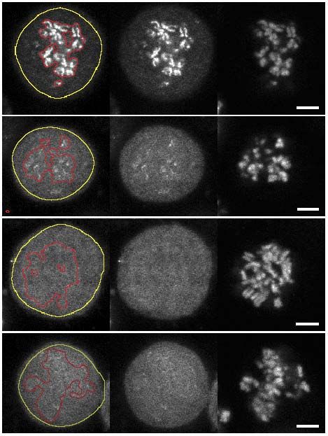
Association of condensin with chromosomes depends on DNA binding by its HEAT-repeat subunits
articles

                                                      Although existing models propose defined tasks in DNA bind-                                                 The finding that the condensin non-SMC subcomplex efficiently
                                                   ing for the condensin SMC and kleisin components, the role of the                                           binds to DNA was surprising, because the only predicted bona fide
                                                   two HEAT-repeat subunits has remained enigmatic. To gain insights                                           DNA-binding motifs present in the subcomplex are the HTH and
                                                   into their contribution to condensin function, we biochemically                                             WHD motifs at the N and C termini of the kleisin subunit20. In kleisin
                                                   and structurally characterized the two proteins as part of the con-                                         proteins, both motifs are, however, involved in binding to the SMC
                                                   densin non-SMC subcomplex. We discovered that DNA binds to the                                              head domains21–23. To test whether these motifs were responsible
                                                   S. cerevisiae non-SMC subcomplex and thereby activates the Smc2–                                            for the observed DNA gel shift, we purified a subcomplex that con-
                                                   Smc4 ATPases. Consistently with a role of the DNA-binding site in                                           tained a truncated version of Brn1 lacking both motifs (Brn1∆NC;
                                                   recruiting condensin to chromosomes, we found that condensin com-                                           Fig. 1e and Supplementary Fig. 1). This Brn1∆NC–Ycs4–Ycg1
                                                   plexes that cannot assemble one of the HEAT-repeat subunits fail to                                         complex bound the 30-bp dsDNA with an efficiency even slightly
                                                   associate with mitotic chromosomes in yeast and human cells. Our                                            higher than that for the Brn1–Ycs4–Ycg1 subcomplex (Fig. 1f
                                                   findings suggest that DNA binding by the non-SMC subcomplex and                                             and Supplementary Fig. 2). We conclude that the non-SMC sub-
                                                   activation of the SMC ATPase constitute the first steps in the topologi-                                    complex binds to dsDNA substrates, most probably via its HEAT-
                                                   cal loading of condensin rings onto chromosomes.                                                            repeat subunits.

                                                   RESULTS                                                                                                     Selective binding to double-stranded DNA helices
                                                   The condensin non-SMC subcomplex binds DNA                                                                  The Smc2–Smc4 dimer of condensin has previously been reported to
                                                   To investigate the properties of the condensin non-SMC subcom-                                              bind to DNA via its hinge domain24,25. We therefore compared binding
                                                   plex, we coexpressed and purified from insect cells the S. cerevisiae                                        of the S. cerevisiae Smc2–Smc4 hinge and the non-SMC subcomplex to
                                                   kleisin subunit Brn1 and the HEAT-repeat subunits Ycg1 and Ycs4                                              single-stranded (ssDNA) and dsDNA. The Brn1–Ycs4–Ycg1 subcom-
                                                   (Supplementary Fig. 1 and Supplementary Table 1). Coomassie                                                  plex bound with low-micromolar affinity (Kd ≈ 2.0 µM) to a 30-bp A-T
© 2014 Nature America, Inc. All rights reserved.

                                                   staining of the peak elution fractions indicated that the three subunits                                     dsDNA but not to a 30-base poly(T) ssDNA substrate (Fig. 2a). The
                                                   formed a stoichiometric Brn1–Ycs4–Ycg1 subcomplex (Fig. 1b).                                                 Smc2–Smc4 hinge, in contrast, bound only the ssDNA substrate with
                                                      Because previous evidence suggested that condensin makes direct                                           appreciable affinity (Kd ≈ 0.3 µM; Supplementary Fig. 3), consistently
                                                   contacts with chromatin, we tested whether the non-SMC subcomplex                                           with the reported Kd and substrate specificity for the mouse Smc2–Smc4
                                                   might provide a platform for DNA binding. Although addition of the                                          hinge25. This experiment also suggests that the non-SMC subcomplex
                                                   non-SMC complex did not notably affect the electrophoretic mobil-                                            binds dsDNA with little or no DNA sequence specificity, because the
                                                   ity of a 15-bp double-stranded DNA (dsDNA) substrate, it resulted                                            affinities for two different 30-bp dsDNA substrates were very similar.
                                                   in the formation of a slower-migrating species with 30-bp, 45-bp or                                             Because the kleisin subunits of fission-yeast and human condensin
                                                   6.5-kb dsDNA (Fig. 1c and Supplementary Fig. 2). Fluorescence                                                complexes have been reported to directly interact with tails of his-
                                                   anisotropy assays to estimate the binding affinities between the                                             tone H2A and H2A.Z18, we also tested binding of the S. ­cerevisiae
                                                   Brn1–Ycs4–Ycg1 subcomplex and DNA in solution (Fig. 1d)                                                     non-SMC subcomplex to in vitro–reconstituted yeast nucleo-
                                                   revealed low-micromolar affinities for binding of the non-SMC sub-                                          somes26 (Supplementary Fig. 3). Although we could not detect an
                                                   complex to the 30- and 45-bp dsDNA substrates (Kd ≈ 2 µM).                                                  ­electrophoretic mobility shift of the nucleosomal DNA, we noticed that

                                                   Figure 1 DNA binding by condensin non-SMC                             a                                              b             c          Molar ratio protein/DNA
                                                   subcomplexes. (a) Schematic representation
                                                                                                                                                                                      0.25
                                                                                                                                                                                      0.50
                                                                                                                                                                                      0.75
                                                                                                                                                                                      1.00
                                                                                                                                                                                      1.50
                                                                                                                                                                                      2.00
                                                                                                                                                                                      4.00

                                                                                                                                                                                                        0.25
                                                                                                                                                                                                        0.50
                                                                                                                                                                                                        0.75
                                                                                                                                                                                                        1.00
                                                                                                                                                                                                        1.50
                                                                                                                                                                                                        2.00
                                                                                                                                                                                                        4.00

                                                                                                                                                                                                                              0.25
                                                                                                                                                                                                                              0.50
                                                                                                                                                                                                                              0.75
                                                                                                                                                                                                                              1.00
                                                                                                                                                                                                                              1.50
                                                                                                                                                                                                                              2.00
                                                                                                                                                                                                                              4.00
                                                                                                                                                                                      0

                                                                                                                                                                                                        0

                                                                                                                                                                                                                              0
                                                   of the five condensin subunits labeled with the                    Smc2     Smc4                                      MW
                                                                                                                                                                        (kDa)
                                                   S. cerevisiae protein names. Amino acid residue                     1
npg

                                                                                                                                                                         140
                                                                                                                                                   HTH

                                                   numbers and positions of the helix-turn-helix            Brn1       111                                Ycs4
                                                   (HTH) motif, winged-helix domain (WHD) and                                             754             Ycg1           115                                                                     **
                                                   blocks of HEAT repeats are indicated. Small
                                                                                                                                    W
                                                                                                                                636 H D                   Brn1
                                                                                                            Ycg1
                                                   ovals indicate regions rich in α-helices.              1        HEAT                           1035
                                                                                                                                                                         80

                                                   (b) Analysis of Brn1–Ycs4–Ycg1 after gel                 Ycs4                                                         65
                                                   filtration by SDS-PAGE and Coomassie                   1                                          1176
                                                                                                                                                                              *
                                                                                                                    HEAT               HEAT
                                                   staining. MW, molecular weight.                                                                                              1 2 3 4 5 6 7 8    9 10 11 12 13 14 15 16 17 18 19 20 21 22 23 24
                                                                                                                                                                                  15-bp dsDNA         30-bp dsDNA              45-bp dsDNA
                                                   (c) Electrophoretic mobility shift assay
                                                   (EMSA) of 15- to 45-bp dsDNA substrates,                                                                                                                         +unlabeled

                                                                                                                       d                                                          e                 f              30-bp dsDNA +antibody
                                                                                                                                                                                                                                                     D A

                                                   with 0.2 µM 6-carboxyfluorescein (6-FAM)–
                                                                                                                                                                                                                                                ei DN

                                                                                                                                                                                                                                                       A
                                                                                                                                                                                                                                                      N
                                                                                                                                                                                                                                              ot A/

                                                                                                                                                                                                                  1 5 10 1 5 10
                                                                                                                                                                                                                                                  n/
                                                                                                                                                                                                                                            Pr DN

                                                   labeled dsDNA in the presence of 0.0–0.8 µM               1.0                                                                          MW
                                                                                                                                                                                                        0.25
                                                                                                                                                                                                        0.50
                                                                                                                                                                                                        1.00
                                                                                                                                                                                                        2.00
                                                                                                                                                                                                        4.00

                                                                                                                                                                                                                       2.00

                                                                                                                                                                                                                              4.00

                                                                                                                                                                                                                                     2.00

                                                   Brn1–Ycs4–Ycg1. Unbound (*) and slower-                                                                                               (kDa)
                                                                                                                      Normalized anisotropy (∆A)

                                                                                                                                                                                                        0

                                                                                                                                                                                                                                     0

                                                                                                                                                                                          170
                                                   migrating species (**) are indicated. (d) Binding                                                     45-bp dsDNA
                                                                                                                                                     Kd = 2.10 ± 0.16 µM      Ycs4
                                                   affinities of Brn1–Ycs4–Ycg1 to 15- to 45-bp                                   30-bp dsDNA
                                                                                                                                                                              Ycg1
                                                                                                                                                                                          130
                                                                                                                               Kd = 1.67 ± 0.18 µM
                                                   6-FAM–labeled dsDNA substrates, determined                                                                                                                                            ***
                                                                                                             0.5                                                                                                                         **
                                                   by measurement of fluorescence anisotropy                                                                                              100
                                                   changes (∆A) upon addition of protein at the
                                                   indicated concentrations. Dissociation constants                                                                                       70
                                                                                                                                                 15-bp dsDNA               Brn1∆NC
                                                   (Kd) were calculated by fitting mean ∆A values
                                                                                                               0                                                                               *
                                                   for each protein concentration, assuming a                                                                                                    1 2 3 4 5 6       7 8 9 10 11 12 13 14
                                                                                                                 0         5      10           15          20          25
                                                   single-site binding model. Points and error bars                          Protein concentration (µM)                                                      30-bp dsDNA
                                                   indicate mean and s.d., respectively, of n = 3
                                                   independent experiments. (e) Analysis of purified Brn1∆C–Ycs4–Ycg1 as in b. The central region of Brn1 stains only weakly with Coomassie, as shown.
                                                   (f) Electrophoretic mobility shift assay of a 30-bp dsDNA with Brn1 ∆NC–Ycs4–Ycg1 as in c (lanes 1–6) after addition of unlabeled 30-bp competitor
                                                   DNA (lanes 7–12) or an antibody against the hexahistidine (His 6) tag on Ycs4 (lanes 13–14). Unbound (*), slower-migrating (**) and antibody-
                                                   supershifted (***) species are indicated. One representative experiment of n = 3 independent replicates is shown in c and f.

                                                   nature structural & molecular biology                                    VOLUME 21                    NUMBER 6   JUNE 2014                                                                       561
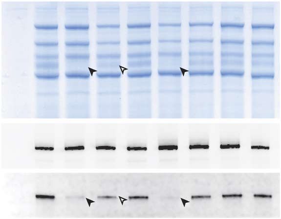
Association of condensin with chromosomes depends on DNA binding by its HEAT-repeat subunits
articles

                                                    a                              Brn1–Ycs4–Ycg1
                                                                                 Molar ratio protein/DNA
                                                                                                                                                                                                                                                                                                                              b                            Molar ratio          +30-bp
                                                                                                                                                                                                                                                                                                                                                          protein/DNA            DNA
                                                                              0 1 2 4 8         0 1 2 4 8
                                                                                                                                                                                                                                                                                                                                                            0 1 2 4              2 4
                                                                                                                                                                                                                                                                                                                                   10-bp
                                                                                                                                                                                                   1.0                                                                                                                             linker DNA

                                                                                                                                                                      Normalized anisotropy (∆A)
                                                                                                                                                                                                                                                                                                                                                                         *
                                                                                                                                                                                                                      30-bp dsDNA (poly (A-T))
                                                                                                                                                         **                                                             Kd = 1.77 ± 0.20 µM
                                                                                                                                                                                                   0.5

                                                                                                                                                                                                                                                                                                                                                Nuc-167                                   Nuc-167
                                                                                                                                                                                                                                   30-bp ssDNA (poly (T))
                                                                                                                                                                                                                                                                                                                                   10-bp                                                  167 bp
                                                                                                                                                                                                                                                                                                                                                 167 bp
                                                                                                                                                                                                                                                                                                                                   linker DNA
                                                                                                                                                                                                    0                                                                                                                                                                                     30 bp
                                                    *                                                                                                                                                    0      5         10       15       20       25
                                                                                                                                                                                                                    Protein concentration (µM)                                                                                                              1 2 3 4              5 6
                                                                              30-bp ssDNA                   30-bp dsDNA
                                                                                (poly (T))                   (poly (A-T))

                                                    c                                                                         d                                                                              tRNA genes
                                                                                                                                                                                                                                                     0.30
                                                                                                                                                                                                                                                                                                 e                          1.0
                                                                                                                                                                                                                                                                                                                                   RNA polymerase II–transcribed genes

                                                                              0.4                                                                       2.0

                                                                                                                                                                                                                                                                                                                                                                                                    (Htz1) nucleosome occupancy (RPM)
                                                                                                                                                                                                                                                                                                                                                                                   0.22

                                                                                                                                                                                                                                                            (Htz1) nucleosome occupancy (RPM)
                                                                                                                                                                                                                                                                                                                                                                                            0.50
                                                                                                                                                                                                                                             0.20
                                                                                                                                                        1.5
                                                                                                                             log2 (Brn1-PK6 IP/input)

                                                                                                                                                                                                                                                                                                log2 (Brn1-PK6 IP/input)
                                                                                                                                                                                                                                                     0.25                                                                   0.5
                                                   log2 (Brn1-PK6 IP/input)

                                                                              0.3             0.293                                                                                                                                                                                                                                                                                0.20
                                                                                                                                                                                                                                                                                                                                                                                            0.40
                                                                                                                                                        1.0
                                                                                                                                                                                                                                             0.15                                                                            0                                                     0.18
                                                                              0.2                                                                                                                                                                    0.20
                                                                                                                                                        0.5                                                                                                                                                                                                                                 0.30
© 2014 Nature America, Inc. All rights reserved.

                                                                                                                                                                                                                                                                                                                                                                                   0.16
                                                                                                         0.122
                                                                                                                                                                                                                                                     0.15                                                                  –0.5
                                                                              0.1                                                                        0
                                                                                                                                                                                                                                             0.10                                                                                                                                           0.20
                                                                                                                                                                                                                                                                                                                                                                                   0.14
                                                                                                                                               –0.5
                                                                                                                                                                                                                                                                                                                           –1.0
                                                                               0                                                                                                                                                                     0.10
                                                                                    Nucleosome Nucleosome                                                           −400    −200       0        200         400                                                                                                                   −400    −200       0        200         400
                                                                                     enriched   depleted                                                            Distance from transcription start site (bp)                                                                                                                   Distance from transcription start site (bp)

                                                   Figure 2 DNA and chromosome binding of the non-SMC subcomplex. (a) EMSA and fluorescence anisotropy binding assays of 6-FAM–labeled 30-bp
                                                   dsDNA and ssDNA substrates with increasing Brn1–Ycs4–Ycg1 concentrations. Unbound (*) and slower-migrating species (**) are indicated. Points and
                                                   error bars indicate mean and s.d., respectively, of n = 3 independent experiments. (b) EMSA of a 167-bp DNA assembled into a nucleosome (Nuc-167)
                                                   and a small fraction (~2%) of free 167-bp DNA with increasing Brn1–Ycs4–Ycg1 concentrations before and after addition of a ten-fold excess of 30-bp
                                                   competitor dsDNA. Upshifted free 167-bp DNA (*) is indicated. (c) Box plots showing assignment of Brn1 with six PK tags (Brn1-PK6) ChIP-seq reads
                                                   from yeast strain C3632 to nucleosome-enriched or nucleosome-depleted regions of the budding yeast genome; P < 4 × 10⁻⁹, Wilcoxon two-sided test.
                                                   Horizontal lines define the median and boxes the 25th and 75th percentiles; whiskers represent the maximum and minimum values. n = 16 chromosomes.
                                                   IP, immunoprecipitation. (d) Metagene analysis for the transcription start site (vertical dashed line) of all S. cerevisiae protein-coding genes. Condensin
                                                   (blue line) expressed as enrichment over the input and average nucleosome (black line) or histone H2A.Z nucleosome (dashed gray line) occupancy
                                                   expressed in reads per million (RPM), computed for each 10-bp bin and represented by smoothing splines. (e) Metagene analysis for all S. cerevisiae
                                                   tRNA genes as in d. One representative experiment of n = 3 independent replicates is shown in a and b.

                                                   a small fraction of 167-bp DNA that had not been incorporated into                                                                                                                            with that of nucleosomes in general27 or histone H2A.Z-containing
npg

                                                   nucleosomes was readily upshifted upon addition of the Brn1–Ycs4–                                                                                                                             nucleosomes in particular28 (Fig. 2c and Supplementary Fig. 3). The
                                                   Ycg1 subcomplex (Fig. 2b). The condensin non-SMC subcomplex                                                                                                                                   preference of condensin localization to nucleosome-free regions at
                                                   therefore has a preference to bind free DNA over nucleosomal DNA.                                                                                                                             transcription start sites was also evident at the promoters of tRNA genes
                                                      The tendency for condensin’s association with free rather than                                                                                                                             (Fig. 2d) but not of RNA polymerase II–transcribed genes (Fig. 2e).
                                                   nucleosome-bound DNA became more apparent when we compared,                                                                                                                                   Nevertheless, condensin binding patterns in the promoter-adjacent
                                                   by chromatin immunoprecipitation and massive parallel sequencing                                                                                                                              regions of both classes of genes appeared to correlate negatively with
                                                   (ChIP-seq), the genome-wide distribution of budding-yeast condensin                                                                                                                           nucleosome binding patterns. Thus, binding of the S. cerevisiae non-
                                                                                                                                                                                                                                                                            SMC complex to free DNA helices in vitro
                                                                                                                                                                                                                                                                            reflects the preferred positioning of con-
                                                          a                                                                                                           b                            Ct Brn1∆NC–Ycs4–
                                                                                                                                                                                                        Ycg1∆C               CtYcg1∆C              CtYcs4                   densin complexes in vivo.
                                                                                      Ycs4–Ycg1∆C
                                                                                      Ct Brn1∆NC–

                                                                                                                                                                                                                         128.00
                                                                                                                                                                                                                         16.00
                                                                                                                                                                                                                         32.00
                                                                                                                                                                                                                         64.00

                                                                                                                                                                                                                                             16.00
                                                                                                                                                                                                                                             32.00
                                                                                                                                                                                                                                             64.00

                                                                                                                                                                                                                                                                                                 Molar ratio
                                                                                                            CtYcg1∆C

                                                                                                                                                                                                   0.25
                                                                                                                                                                                                   0.50
                                                                                                                                                                                                   1.00
                                                                                                                                                                                                   2.00
                                                                                                                                                                                                   4.00
                                                                                                                                                                                                   8.00

                                                                                                                                                                                                                         4.00
                                                                                                                                                                                                                         8.00

                                                                                                                                                                                                                                             4.00
                                                                                                                                                                                                                                             8.00
                                                                                                                                                    CtYcs4

                                                                                                                                                                                                                                                                                                protein/DNA
                                                                                                                                                                                                                                                                                                                                     Figure 3 Both HEAT-repeat subunits are
                                                                                                                                                                                                   0

                                                                                                                                                                                                                                             0
                                                                                                                                                                                                                         0

                                                                                                     MW                 MW                                     MW                                                                                                                                                                    necessary for efficient dsDNA binding.
                                                                                                    (kDa)              (kDa)                                  (kDa)
                                                                                                                                                                                                                                                                                                    ***                              (a) Purified Ct Brn1∆NC–Ycs4–Ycg1∆C complex,
                                                                                                    170                130                                    170                                                                                                                                                                    CtYcg1∆C and CtYcs4, analyzed by SDS-
                                                                              Ycs4                                                                            130                                                                                                                               **                                   PAGE and Coomassie staining. (b) EMSA of
                                                                                                    130                100
                                                                 Ycg1∆C                                                                                                                                                                                                                                                              a 6-FAM–labeled 60-bp dsDNA at increasing
                                                                                                    100                70                                     100                                                                                                                                                                    concentrations of Ct Brn1∆NC–Ycs4–Ycg1∆C,
                                                                                                                                                              70                                                                                                                                                                     CtYcg1∆C or CtYcs4. Unbound (*), Ct
                                                                                                    70                 55
                                                           Brn1∆NC                                                                                                                                                                                                                                                                   Brn1∆NC–Ycs4–Ycg1∆C or CtYcg1∆C-bound
                                                                                                                                                              55      *                                                                                                                                                              (**) and CtYcs4-bound complexes (***) are
                                                                                                    55                 40
                                                                                                                                                                                                                                                                                                                                     indicated. One representative experiment of
                                                                                                                                                                                                   1
                                                                                                                                                                                                   2
                                                                                                                                                                                                   3
                                                                                                                                                                                                   4
                                                                                                                                                                                                   5
                                                                                                                                                                                                   6
                                                                                                                                                                                                   7
                                                                                                                                                                                                                          8
                                                                                                                                                                                                                          9
                                                                                                                                                                                                                         10
                                                                                                                                                                                                                         11
                                                                                                                                                                                                                         12
                                                                                                                                                                                                                         13
                                                                                                                                                                                                                         14

                                                                                                                                                                                                                                             15
                                                                                                                                                                                                                                             16
                                                                                                                                                                                                                                             17
                                                                                                                                                                                                                                             18
                                                                                                                                                                                                                                             19
                                                                                                                                                                                                                                             20

                                                                                                                                                                                                                          60-bp dsDNA                                                                                                n = 3 independent replicates is shown in b.

                                                   562                                                                                                                                                                  VOLUME 21         NUMBER 6                     JUNE 2014                                                  nature structural & molecular biology
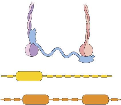
Association of condensin with chromosomes depends on DNA binding by its HEAT-repeat subunits
articles

                                                       a                            Ycs4
                                                                                                                                                                                                                                                      b
                                                                                K33         100            200               300            400             500              600              700            800         900           1035                                                1100
                                                                    Ycg1        6       7      2       2      0          3      4       2      0        4      4         1      3         5      1       1      7       3    2       3 3                                                   1000
                                                                                                                                                                                                                                                                                            900
                                                                                K33             K131                 K261                                                                                     K823      K895                                                                800
                                                                                    K64                                                                                                                                                                                                     700
                                                                                                                                                                                                                                                                                            600

                                                                                                                                                                                                                                                                         Ycg1
                                                                                                                                                                                                                                                                                            500
                                                                                                                                                K439 K491
                                                                                                                                                                                                                                                                                            400
                                                                                                                                            K421 K488                K573                                                                                                                   300
                                                                                K42       100              200             300          K409 K473                    K568                 K691 754                                                                                          200
                                                                                                                                                                                                                                                                                            100
                                                                    Brn1        2       3    2         4         4       1    1         1       6       12       4       4        5       5   2
                                                                                K42      K99                   K232          K316        K414                                K669                                                                                                           100
                                                                                                               K236                         K421                     K573                                                                                                                   200
                                                                                                                                             K439                                                                                                                                           300
                                                                                                                                                                                                                                                                                            400
                                                                                                                                              K445
                                                                                                                              K461                                                                                                                                                          500
                                                                                                                                                    K450                                                                                                                                    600
                                                                                                                             K432

                                                                                                                                                                                                                                                                         Ycs4
                                                                                                                                                                                                                                                                                            700
                                                                                                            K398 K428                                                                                                                                                                       800
                                                                                                           K391 K424                                         K693                                                                                                                           900
                                                       Ycs4                                                                                                                                                                                                                                1000
                                                                    K145                 K286          K377 K420 K498                                       K682                          K872           K987
                                                                                                                                                                                                                                                                                           1100
                                                           1   3    4       4       3       4      1       7         5       3      5       4       3        5       2       5        3       7      4       4      1     3      9   3                                                     1200
                                                       1       100          200              300            400               500            600             700 K773                         K883           1000         1100       1176
                                                                                                                                                                                                                                                                                                      100 200 300 400 500 600 700
                                                                                                                                                                 Ycg1                                                                                                                                            Brn1

                                                                                                   Brn1–Protein A                                                                                                                              MW
                                                       c                                                                                                                                                 d                                    (kDa)                                    115         Superose 6 10/300
© 2014 Nature America, Inc. All rights reserved.

                                                                                                                                                                                                                                                      Absorbance at 280 nm (a.u.)
                                                                   110–438                  439–531                      110–531                    1–754
                                                                                                                                                                                                                                               140
                                                                                                                                                                                 MW                                                            115                                         95
                                                               IN       U       B       IN         U        B IN             U          B IN            U        B                                                  CtYcg1∆C                                                                                                             17
                                                                                                                                                                                 (kDa)                                                         80
                                                                                                                                                                                 170                                                           70                                          75
                                                                                                                                                                                                                                                                                                                             158
                                                       Ycs4                                                                                                                                                                                    50                                                                                  44
                                                                                                                                                                             *130                                                                                                          55
                                                                                                                                                                                                                                               40
                                                                                                                                                                                                                                                                                                             670
                                                                                                                                                                                                                                              30                                           35
                                                                                                                                                                                 170                                                          25
                                                       Ycg1                                                                                                                                                                                                                                15
                                                                                                                                                                                                                                              15
                                                                                                                                                                                 130                                    Ct Brn1
                                                                                                                                                                                                                         562-633                                                                     10                 15                    20
                                                                                                                                                                                 130                                                                                                                        Elution volume (ml)

                                                                                                                                                                                 95
                                                                                                                                                                                                         e                                     MW                                                 Superdex 200
                                                                                                                                                                                                                                                             Absorbance at 280 nm (a.u.)

                                                                                                                                                                                 72                                                           (kDa)                                               26/600
                                                       Brn1                                                                                                                      55                                                                                                        40                                           17
                                                                                                                                                                                 43                                     CtYcs4                140
                                                                                                                                                                                                                                              115                                                           670
                                                                                                                                                                                 34                                                                                                        30
                                                                                                                                                                                                                                                                                                                       158
                                                                                                                                                                                 26                                                           80                                                                              44
                                                                                                                                                                                                                                                                                           20
                                                                                                                                                                                                                                              65

                                                                                                                                                                                                                        Ct Brn1                                                            10
                                                                                                                                                                                                                         225-583              50
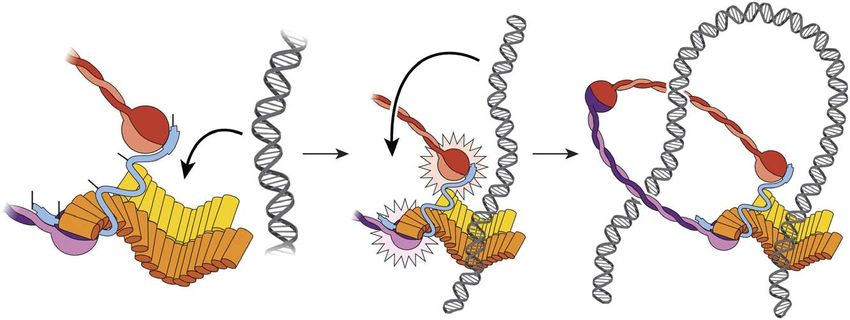
                                                   Figure 4 Subunit geometry of the non-SMC subcomplex. (a) Linkage
                                                   map showing the positions of all high-confidence intersubunit links identified                                         100       150      200   250
npg

                                                   in the S. cerevisiae non-SMC subcomplex by cross-linking and MS. The numbers of lysine residues present in                  Elution volume (ml)
                                                   windows of 50 residues are indicated in squared boxes; HEAT-repeat, HTH and WHD motifs are indicated in color.
                                                   Cross-links between the N or C termini of Ycg1 with Brn1 are indicated by dark- or light-blue lines, respectively; cross-links between the N or C
                                                   termini of Ycs4 with Brn1 are indicated by pink or red lines, respectively. (b) Scatter-plot graph of the cross-links described in a. Residue numbers are
                                                   indicated on the axes. (c) Western blotting against the PK9 tag on Ycs4 or the HA6 tag on Ycg1 in endogenous condensin subunits copurified with IgG
                                                   beads. Samples are from S. cerevisiae expressing Protein A–fused fragments of the indicated Brn1 residues. IN, input; U, unbound; B, bound (10×
                                                   concentrated compared to input) fractions. A band that results from binding of the anti-PK antibody by the full-length Brn1–Protein A is indicated by an
                                                   asterisk. (d) SDS-PAGE and Coomassie staining of the Ct Brn1562-633–Ycg1∆C complex after Ni-NTA chromatography, ion-exchange chromatography and
                                                   gel-filtration chromatography (graph). (e) Purification of a stable Ct Brn1225-583–Ycs4 complex as in d.

                                                   Efficient DNA binding requires both HEAT-repeat subunits                                                                                                               discrete band ratio or into a diffuse streak of slower-migrating species
                                                   We next tested whether either of the two condensin HEAT-repeat sub­                                                                                                    that accumulated in the wells of the gel, respectively (Fig. 3b). These
                                                   units is capable of binding DNA individually. Because we could not                                                                                                     results suggest that the isolated condensin HEAT-repeat subunits are
                                                   purify sufficient amounts of the individual S. cerevisiae HEAT-repeat                                                                                                  able to interact with DNA, albeit with considerably lower affinity than
                                                   subunits, we expressed and purified the Ycs4 and Ycg1 subunits from the                                                                                                when they are part of the non-SMC subcomplex.
                                                   thermophilic yeast Chaetomium thermophilum (denoted by Ct prefix)29
                                                   and compared their DNA binding activities to that of the Ct non-SMC                                                                                                    Subunit geometry of the condensin non-SMC subcomplex
                                                   subcomplex (Fig. 3a and Supplementary Fig. 4). In gel shift experi-                                                                                                    To gain insights into the three-dimensional organization of the
                                                   ments, addition of the Ct Brn1∆NC–Ycs4–Ycg1∆C subcomplex to a 60-bp                                                                                                    S. cerevisiae non-SMC subcomplex, we generated an interaction
                                                   dsDNA substrate produced a discrete slow-migrating band (Fig. 3b),                                                                                                     map of its subunits by using a cross-linking MS approach 30,31
                                                   similarly to the shift produced by the S. cerevisiae Brn1∆NC–Ycs4–Ycg1                                                                                                 (Supplementary Fig. 5). This identified 45 unique intersubunit
                                                   subcomplex (Fig. 1f). CtYcg1 or CtYcs4, in contrast, at high protein/                                                                                                  and 48 intrasubunit cross-links (Supplementary Tables 2 and 3).
                                                   DNA ratios, shifted either just a fraction of the dsDNA substrate into a                                                                                               Remarkably, out of the 45 intersubunit cross-links, only two connected

                                                   nature structural & molecular biology                                                                             VOLUME 21                       NUMBER 6                    JUNE 2014                                                                                                         563
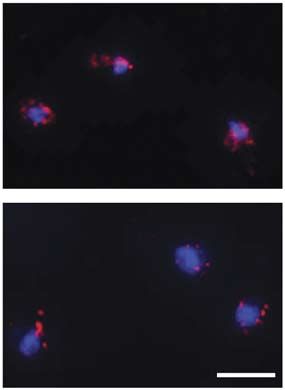
Association of condensin with chromosomes depends on DNA binding by its HEAT-repeat subunits
articles

                                                   a                                                               Patch 3                                   Patch 2                                       Patch 1
                                                                                                                                                       M4                                             M1              M2

                                                   Homo sapiens                            427
                                                   Mus musculus                            420
                                                   Xenopus tropicalis                      411
                                                   Drosophila melanogaster                 405
                                                   Arabidopsis thaliana                    394
                                                   Encephalitozoon cuniculi                227
                                                   Schizosaccharomyces pombe               418
                                                   Chaetomium thermophilum                 520
                                                   Saccharomyces cerevisiae                390
                                                                                                                                                                                                                            *
                                                                                                         M6              M7                            M4              M5                             M1              M2   M3
                                                                             Untagged

                                                                                        Wild type

                                                   b                                                M1   M2   M3   M4   M5    M6   M7    MW     c     Wild type             M1                M2                 M3
                                                                                                                                        (kDa)
                                                               Smc4
                                                           Smc2-HA6                                                                     170
                                                                Ycs4
                                                                Ycg1                                                                    130
                                                            Brn1-PK6
                                                                                                                                        100             M4                  M5                M6                 M7

                                                                                                                                        130
                                                            Brn1-PK6
© 2014 Nature America, Inc. All rights reserved.

                                                                                                                                        170
                                                                 Ycg1
                                                                                                                                        130

                                                   Figure 5 A conserved region within the Brn1 kleisin subunit for Ycg1 binding. (a) Multisequence alignment of the Ycg1-binding region of S. cerevisiae
                                                   Brn1 with homologous γ-kleisins, identifying three patches of conserved amino acid residues. Boxed residues were mutated to the indicated sequences
                                                   in mutants M1–M7. Two residues mutated in brn1-60 are indicated by a gray bar and asterisk. (b) Coimmunoprecipitation of condensin subunits with
                                                   the Brn1-PK6 proteins from yeast cell extracts (strains C3632, C3665, C3651, C3641, C3635, C3658, C3649 and C3634), analyzed by Coomassie
                                                   staining and western blotting with antibodies against Ycg1 or the PK epitope on Brn1. The Coomassie-stained bands were used in subsequent MS
                                                   analyses. Loss or reduction of Ycg1 binding in mutants M1, M4 and M2 is indicated by closed or open arrowheads, respectively. (c) Tetrad dissection
                                                   analysis of diploid cells from b at 30 °C on rich medium. Circles indicate kanamycin-resistant colonies linked to the wild-type or mutant BRN1-PK6
                                                   alleles. One representative experiment of n = 3 biological replicates is shown in b and c.

                                                   Ycs4 with Ycg1, whereas 27 or 16 links connected Brn1 with Ycs4 or                                  or in an additional patch just N terminal to it (patch 3; Fig. 5a)
                                                   Ycg1, respectively (Fig. 4a), thus suggesting that the HEAT-repeat                                  might prevent incorporation of Ycg1 into condensin complexes. We
                                                   subunits make little or no direct contact. This conclusion is consist-                              expressed mutant Brn1 versions from one of the two endogenous
                                                   ent with the observations that the two HEAT-repeat subunits do                                      alleles in diploid S. cerevisiae cells (Supplementary Fig. 7 and
                                                   not copurify (Supplementary Fig. 6), or do so only with very low                                    Supplementary Table 4) and assayed for copurification of Ycg1.
                                                   efficiency7, in the absence of the kleisin protein.                                                 Strikingly, Brn1 mutant M2 reduced binding and mutants M1 and
                                                      Notably, almost all cross-links with Ycg1 residues clustered within                              M4 completely abolished binding to Ycg1 without affecting the
npg

                                                   a small region of Brn1 (residues 409–573; Fig. 4a,b). To test whether                               association with the other three condensin subunits (Fig. 5b and
                                                   this region forms a distinct binding domain for Ycg1, we expressed in                               Supplementary Fig. 7).
                                                   budding yeast a series of Brn1 versions truncated either from the N or                                 As would be expected if incorporation of Ycg1 into condensin
                                                   the C terminus and assayed which constructs bound endogenous Ycg1                                   complexes were essential for function, tetrad analysis demonstrated
                                                   (Supplementary Fig. 6). Ycg1 copurified only with Brn1 constructs                                   that Brn1 mutants M1, M2 and M4 failed to support growth at 30 °C
                                                   that included the region between residues 439 and 531. Furthermore,                                 in cells that also expressed Smc2 with six hemagglutinin tags (HA 6)
                                                   this region of the kleisin subunit was sufficient for forming a stable                              (Fig. 5c). In the absence of a tag on Smc2, mutants M1 and M4 were
                                                   complex with Ycg1 of S. cerevisiae (Fig. 4c and Supplementary Fig. 6)                               able to sustain cell growth at 25 °C but not at 37 °C. We used these
                                                   and C. thermophilum (Fig. 4d). Most cross-links with Ycs4 were located                              two mutants to test whether the observed growth defects were due to
                                                   within a region N-terminal to the Ycg1-interacting domain (Fig. 4a,b).                              defects in chromosome segregation that result from condensin inacti-
                                                   Although Ycs4 copurified from yeast extracts with Brn1 fragments that                               vation32,33, by using live-cell microscopy to monitor partitioning of the
                                                   contained the region between residues 224 and 340, robust interaction                               fluorescently marked arm of chromosome V (ref. 16) in cells released
                                                   required residues 110–438 (Supplementary Fig. 6). This region was                                   from G1 phase at 37 °C (Supplementary Fig. 7). Remarkably, only
                                                   sufficient for forming complexes with Ycs4 of S. cerevisiae (Fig. 4c) and                           28% or 21% of Brn1-mutant M1 or M4 cells, respectively, successfully
                                                   C. thermophilum (Fig. 4e). We conclude that distinct binding domains                                segregated the marked sister chromatids into opposite daughter cells
                                                   exist within the central region of the condensin kleisin subunit for the                            within the time course of the experiment, compared to 75% of Brn1
                                                   recruitment of each HEAT-repeat subunit.                                                            wild-type cells. We conclude that yeast condensin complexes that are
                                                                                                                                                       deficient in recruiting Ycg1 are unable to support proper chromosome
                                                   Active condensin complexes require both HEAT-repeat subunits                                        segregation and cell division. Interestingly, mutations within patch 3
                                                   Because Ycg1 seems to bind only to a short region of Brn1, we rea-                                  abolished condensin function without notably affecting Ycg1 or Ycs4
                                                   soned that mutation of conserved hydrophobic residues clustered                                     binding (Fig. 5b,c), thus suggesting that this patch has an essential
                                                   in two conserved patches within this region (patch 1 and patch 2)                                   function other than recruiting the HEAT-repeat subunits.

                                                   564                                                                                   VOLUME 21   NUMBER 6      JUNE 2014   nature structural & molecular biology
articles

                                                   Figure 6 The Ycg1 subunit is essential for            a          Brn1-PK6 DAPI
                                                                                                                                                                                                                               b
                                                   condensin recruitment onto yeast chromosomes.                                           2.5                           CEN4                   rDNA

                                                                                                                                                                                                                               Percentage genomic DNA
                                                   (a) Chromosome spreads prepared from                                                        * **           0.20

                                                                                                                                                 Relative fluorescence
                                                                                                                                                    **

                                                                                                        Wild type
                                                                                                                                           2.0
                                                   asynchronous diploid yeast cells (C3632,
                                                                                                                                                              0.15
                                                   C3651, C3856 and C3857), probed with                                                    1.5
                                                   anti-PK antibody (red) and stained with 4′,6-                                                              0.10
                                                                                                                                           1.0
                                                   diamidino-2-phenylindole (DAPI; blue). Brn1-
                                                   PK6 signals were quantified. In box plot at right,

                                                                                                        M1 M2 M4
                                                                                                                                           0.5                0.05
                                                   horizontal lines define the median, boxes define
                                                                                                                                             0                   0
                                                   the 25th and 75th percentiles, and whiskers                                                  M2 M1 M1             M1 M2 M4 M1 M1         M1 M2 M4 M1 M1

                                                                                                                                                                           Wild type

                                                                                                                                                                                                                                                        Wild type

                                                                                                                                                                                                                                                                    Untagged

                                                                                                                                                                                                                                                                                           Untagged
                                                                                                                                                                                                                                                                               Wild type
                                                   define the 10th and 90th percentiles.                                                           M4 M2                      M4 M2                  M4 M2
                                                   *P = 8.1 × 10⁻¹⁰; **P < 2.2 × 10⁻¹⁶ by one-                                 5 µm                   M4                         M4                     M4

                                                   sided Wilcoxon-Mann-Whitney test. n = 201
                                                                                                                 c                                                                     d

                                                                                                                                                                                           Brn1-Pk6 IP Brn1(M1 M2 M4)-PK6 IP
                                                   (wild type), 211 (M2), 243 (M1 M4) or 217                                                             130
                                                   (M1 M2 M4) nuclei from two independent
                                                   experiments.(b) ChIP-qPCR in asynchronous

                                                                                                        log2 Brn1(M1 M2 M4)-PK6 IP (RPM)
                                                                                                            12

                                                                                                                                                                                                               (RPM)
                                                                                                                          Correlation 0.82
                                                   cells of diploid strains expressing wild-type            10
                                                   (C3632), single mutants (C3665, C3651 and
                                                   C3635) or mutant combinations (C3856 and                  8
                                                                                                                                                           0
                                                   C3857) of Brn1-PK6 at the centromere of                                                               130
                                                                                                             6
                                                   chromosome IV (CEN4) and the rDNA locus (5′
                                                                                                             4

                                                                                                                                                                                              (RPM)
                                                   untranslated region of RDN37-1 and RDN37-2).
                                                   Data represent mean values of n = 4 ChIP                  2
                                                   experiments and two technical replicates per              0
© 2014 Nature America, Inc. All rights reserved.

                                                   experiment ± s.d. (c) Scatter plot representing                                                          0
                                                                                                                                                           450 kb             460 kb             470 kb
                                                   sequence coverage of ChIP-seq reads from yeast                 0     2    4     6    8 10 12
                                                   expressing wild-type Brn1-PK6 (strain C3632,                      log2 Brn1-PK6 IP (RPM)                         RDN37-1 qPCR RDN37-2 qPCR
                                                   x axis) and the Brn1 (M1 M2 M4)-PK6 mutant
                                                   (strain C3857, y axis). Each point represents a 1,000-bp window; the nonparametric Spearman correlation index is indicated. (d) Sequence reads (reads
                                                   per million, RPM) of wild-type and mutant Brn1-PK6 cells from c at the rDNA locus on chromosome XII, displayed on a linear scale. The region used for
                                                   qPCR analysis in b is indicated. In c and d, sequence reads were normalized to the total number of reads for each sample individually to measure Brn1
                                                   positions independent of the absolute efficiency of Brn1 immunoprecipitation.

                                                   Recruitment to chromosomes by the HEAT-repeat subunits                                             CAP-G or CAP-G2 that coimmunoprecipitated with all three CAP-
                                                   To test whether the presence of both HEAT-repeat subunits in                                       H mutants or the double and triple CAP-H2 mutants, respec-
                                                   condensin complexes is required for condensin’s association with                                   tively, were greatly reduced (Fig. 7a and Supplementary Fig. 8).
                                                   chromosomes, we first measured the protein levels associated with                                  Notably, none of the mutations affected copurification of the other
                                                   mitotic chromosome spreads of wild-type and mutant versions of                                     three condensin subunits. We then expressed, at levels below that of
                                                   Brn1 that either reduced (mutant M2) or abolished (M1 M4 dou-                                      the endogenous protein, wild-type and mutant versions of CAP-H
                                                   ble or M1 M2 M4 triple mutants) binding to Ycg1. The amounts                                       fused to enhanced GFP (EGFP) in a HeLa cell line that also expresses
                                                   of condensin on chromosomes were markedly reduced in all three                                     histone H2B tagged with mCherry34 and measured the amounts of
                                                   mutants (Fig. 6a). Chromatin immunoprecipitation and quantita-                                     CAP-H associated with mitotic chromosomes in cells arrested with
                                                   tive PCR (ChIP-qPCR) at two different chromosomal binding sites                                    nocodazole (Fig. 7b). Condensin I enrichment on chromosomes,
npg

                                                   confirmed a substantial decrease in condensin association in single,                               which we define by the mean EGFP signal ratio of chromosomal to
                                                   double or triple Brn1 mutants (Fig. 6b). To distinguish whether the                                cytoplasmic regions, decreased from ~2.2 in cells expressing wild-
                                                   decrease of condensin levels at these sites was due to the reposition-                             type CAP-H to ~1.2–1.4 in cells expressing the CAP-H mutant
                                                   ing of condensin to other chromosomal locations or due to a global                                 versions (Fig. 7c; a value of 1.0 corresponds to no condensin enrich-
                                                   reduction in chromosome association, we mapped the genome-wide                                     ment on chromosomes). Similarly, condensin II enrichment on chro-
                                                   positions of the Brn1 triple mutant by ChIP-seq and compared them                                  mosomes decreased substantially in the CAP-H2 double and triple
                                                   to the positions of wild-type Brn1. Our analyses detected no obvi-                                 mutants but less so in the CAP-H2 single mutant that did not notably
                                                   ous differences between the distributions of wild-type and mutant                                  affect CAP-G2 binding (Supplementary Fig. 8). We conclude that
                                                   Brn1 (Fig. 6c,d and Supplementary Fig. 7). These findings do not                                   the HEAT-repeat subunits have an essential and conserved role for
                                                   suggest a role of the HEAT-repeat subunits in targeting to specific                                the recruitment of condensin complexes onto chromosomes in yeast
                                                   chromosome sites but instead suggest that the HEAT-repeat subunits                                 and human cells.
                                                   have a crucial function in the general recruitment of condensin onto
                                                   chromosomes.                                                                                       DNA binding by the non-SMC complex stimulates the SMC ATPase
                                                      To test whether this function is conserved in mammalian con-                                    Loading of the condensin-related cohesin complex onto chromosomes
                                                   densin complexes, we expressed in human embryonic kidney (HEK)                                     depends on ATP hydrolysis by its Smc1–Smc3 subunits, which has
                                                   293 cells wild-type or M1 single, M2 M4 double or M1 M2 M4 triple                                  been suggested to initiate transport of chromatin fibers into cohesin
                                                   mutants of the CAP-H kleisin subunit of condensin I or the CAP-H2                                  rings35,36. If condensin used a similar mechanism for chromosome
                                                   kleisin subunit of condensin II (Fig. 5a and Supplementary Fig. 8).                                loading, we reasoned that DNA binding by the HEAT-repeat sub­
                                                   We then immunoprecipitated CAP-H or CAP-H2 via their                                               units might function as a trigger to activate the Smc2–Smc4 ATPase.
                                                   N-terminal Flag tags and probed for copurification of the other four                               We found that, consistently with ­ previous reports13, the presence
                                                   condensin subunits. Remarkably, the amounts of the Ycg1 homologs                                   of DNA had no apparent effect on the low ATPase activity of the

                                                   nature structural & molecular biology                     VOLUME 21                     NUMBER 6                      JUNE 2014                                                                                                         565
articles

                                                   Figure 7 The CAP-G HEAT-repeat subunit is required for condensin                                           a                                       b                 Flag-
                                                                                                                                                                                                                       EGFP–
                                                                                                                                                                                                                                Histone
                                                                                                                                                                                                                                 H2B-      c

                                                                                                                                                                       Wild type
                                                   binding to human chromosomes. (a) Western blotting of coprecipitated                                                                  M1                            CAP-H    mCherry

                                                                                                                                                                                                                                           Flag-EGFP–CAP-H fluorescence
                                                                                                                                                                                      M2 M2                                                                               2.5
                                                   condensin subunits. The indicated Flag-EGFP–CAP-H proteins were

                                                                                                                                                                                                      Wild type
                                                                                                                                                                                   M2 M4 M4    MW
                                                   immunoprecipitated from lysates of transiently transfected HEK293 cells.

                                                                                                                                                                                                                                                chromosomes/cytosol
                                                                                                                                                                                              (kDa)
                                                   One representative experiment of n = 2 independent experiments is shown.                                    SMC4
                                                                                                                                                                                              150                                                                         2.0
                                                   (b) Mean EGFP intensities measured in chromosome and cytoplasmic                                           CAP-G                           120
                                                   regions. Samples are Flag-EGFP–CAP-H proteins transiently expressed in

                                                                                                                                                                                                      M2
                                                   nocodazole-arrested HeLa cells expressing histone H2B-mCherry. Cells                                                                                                                                                   1.5
                                                                                                                                                              CAP-D2                          150
                                                   (yellow lines) and chromosomes (red lines) are delineated according to total                                SMC2

                                                                                                                                                                                                      M1 M2 M4 M2 M4
                                                   EGFP or mCherry signals, respectively, and mean EGFP intensities were                                                                      120

                                                   measured in chromosome and cytoplasmic regions. (c) Ratios between                                                                         150                                                                         1.0
                                                                                                                                                              Flag-
                                                                                                                                                                                                                                                                                            M2 M2 M1

                                                                                                                                                                                                                                                                                Wild type
                                                   chromosomal and cytoplasmic EGFP mean intensities, calculated from one                                     EGFP–
                                                                                                                                                                                              120                                                                                              M4 M2
                                                   representative experiment of three biological replicates and plotted as mean                               CAP-H                                                                                                                               M4
                                                   ± s.d. n = 92 (wild type), 59 (M2), 39 (M2 M4) or 76 (M1 M2 M4) cells.                                                                     85
                                                                                                                                                                                                                                    5 µm

                                                   Smc2–Smc4 dimer (~0.5 molecules ATP hydrolyzed per Smc2–Smc4                                              DISCUSSION
                                                   per minute; Fig. 8a,b). Addition of the Brn1–Ycs4–Ycg1 non-SMC                                            A DNA-binding site formed by the HEAT-repeat subunits
                                                   subcomplex approximately doubled the Smc2–Smc4 ATPase activity                                            How condensin complexes interact with their chromatin substrates has
                                                   in the absence of DNA. Remarkably, simultaneous addition of the                                           remained poorly understood. The Smc2–Smc4 dimer has been previ-
                                                   Brn1–Ycs4–Ycg1 subcomplex and DNA enhanced the Smc2–Smc4                                                  ously reported to bind DNA, although binding was considerably less
                                                   ATPase activity more than ten-fold (Fig. 8b). The DNA-dependent                                           pronounced compared to that of condensin holocomplexes12 and was
                                                   stimulation of the Smc2–Smc4 ATPase activity must have been                                               disrupted even by medium salt concentrations13. Interaction with DNA
© 2014 Nature America, Inc. All rights reserved.

                                                   due to the binding of the non-SMC subcomplex to the Smc2–Smc4                                             was proposed to be mediated by the Smc2–Smc4 hinge domains, which,
                                                   head domains, because we observed no stimulation by DNA when                                              however, bound much more efficiently to ssDNA than to dsDNA25 and
                                                   we used a non-SMC subcomplex containing a version of Brn1 that                                            might therefore perform specialized functions of condensin, for example
                                                   lacks the Smc2–Smc4 interaction motifs (Brn1∆NC). To rule out that                                        during processes that generate unpaired DNA strands, such as DNA-
                                                   the increase in activity was due to the presence of a contaminating                                       damage repair or transcription38. In previous studies, no DNA binding
                                                   ATPase, we repeated the assay with an Smc2–Smc4 dimer that con-                                           could be detected for frog or fission-yeast non-SMC subcomplexes12,15.
                                                   tained point mutations in each of the two Walker B ATP-hydrolysis                                            We found that the budding-yeast non-SMC subcomplex, in con-
                                                   motifs. As expected, we measured no DNA-dependent ATPase activ-                                           trast, binds to dsDNA with high selectivity over ssDNA (Fig. 2a and
                                                   ity for the mutant Smc2–Smc4 dimer, even in the presence of the                                           Supplementary Fig. 3). Our finding that binding to different dsDNA
                                                   non-SMC subcomplex (Fig. 8b). Our findings are in full agreement                                          substrates occurred with low micromolar affinity, was reversible and
                                                   with the report that the ATPase activity of the Xenopus condensin                                         depended on a minimum DNA length (Fig. 1 and Supplementary Fig. 2)
                                                   holocomplex is higher than that of the SMC2–SMC4 subcomplex                                               does not support the possibility of mere nonspecific electrostatic
                                                   and increases in the presence of DNA12.                                                                   interactions. These would also seem unlikely for a protein complex
                                                                                                                                                             with a predicted negative surface charge (pI = 4.9). Furthermore, the
                                                                                                                                                                                      non-SMC subcomplex from an evolutionarily
                                                    a                                 b                                                                                               distant yeast species displayed very similar
                                                                         Q –
                                                                       52 Q)

                                                                                                               7
                                                                                                                                                                                      DNA binding properties (Fig. 3b). Because
                                                                     13 13

                                                                          )
                                                                   (E 11
                                                              Sm mc mc4

                                                                                                                                                                                      neither the HTH nor the WHD motifs of the
                                                                c4 2(E

                                                                                                               6
npg

                                                                                    Molecules ATP hydrolyzed
                                                                S –S

                                                                                     per Smc2–Smc4 (min–1)
                                                                 c2

                                                                                                                                                                                      kleisin subunit were required for DNA binding
                                                             Sm

                                                                                                               5
                                                                       MW
                                                                      (kDa)
                                                                                                               4
                                                    Smc4                170
                                                    Smc2                                                       3
                                                                        130                                                                                                                            Figure 8 DNA binding by the non-SMC
                                                                        100                                    2                                                                                       subcomplex activates the Smc2–Smc4
                                                                        70                                                                                                                             ATPases. (a) Wild-type and Walker B–mutant
                                                                                                               1
                                                                                                                                                                                                       Smc2–Smc4 complexes, analyzed by SDS-PAGE
                                                                        55
                                                                                                                                                                                                       and Coomassie staining. (b) Hydrolysis rates
                                                                                                                   –   DNA    –    DNA      –   DNA      –     DNA        –            DNA
                                                                                                                                                                                                       of ATP (1.25 mM) in S. cerevisiae wild-type or
                                                                                                                                Brn1–       Brn1∆NC–                     Brn1–
                                                                                                                              Ycs4–Ycg1    Ycs4–Ycg1                   Ycs4–Ycg1
                                                                                                                                                                                                       hydrolysis-defective Smc2–Smc4 dimers
                                                                                                                                                                                                       (0.5 µM), measured in the presence or absence
                                                                                                                             Smc2–Smc4                 Smc2(E1113Q)–Smc4(E1152Q)
                                                                                                                                                                                                       of the Brn1–Ycs4–Ycg1 subcomplex (1.5 µM),
                                                                                                                                                                                                       the Brn1∆NC–Ycs4–Ycg1 subcomplex (1.5 µM)
                                                    c                Brn1–Ycs4–Ycg1                                    Smc2–Smc4                                                                       lacking the Smc2–Smc4 interaction motifs
                                                                                                                       ATPase
                                                                                                                                                                                                       and/or 6.5-kb linearized plasmid DNA (10 nM).
                                                                                                                                                                                                       Columns and error bars indicate mean and s.d.,
                                                                              754                                                                                                                      respectively, of n = 3 technical replicates.
                                                                  531
                                                                                                                                                                                                       (c) Multistep model for the topological loading
                                                               438                                                                                                                                     of condensin onto chromosomes. Binding of
                                                         1                                                                                                                                             duplex DNA to the HEAT-repeat subunits (left)
                                                             111
                                                                                                                                                                                                       activates of the Smc2–Smc4 ATPase activity
                                                                                                                                                                                                       (middle) to trigger the transfer of DNA into the
                                                                                                                                                                                                       condensin ring (right). Numbers indicate
                                                                                                                                                                                                       the binding regions for Ycs4 and Ycg1 in the
                                                                                                                                                                                                       S. cerevisiae Brn1 kleisin subunit.

                                                   566                                                                                    VOLUME 21     NUMBER 6       JUNE 2014                   nature structural & molecular biology
articles

                                                   (Fig. 1e,f), we suggest that the non-SMC subcomplex binds DNA via             not support this possibility. Yet the identification of a direct DNA-
                                                   its HEAT-repeat subunits. Support for the notion that HEAT repeats            binding site in the condensin non-SMC subcomplex explains how
                                                   can serve as DNA-binding motifs comes from a recent crystal struc-            condensin can still bind to chromosomes even without encircling
                                                   ture of the AlkD glycosylase, which revealed the binding of six HEAT          them in an ATP hydrolysis–dependent manner. A direct protein-DNA
                                                   repeats to the phosphate backbone of a 12-bp DNA helix39 with similar         interaction could also be the reason for the requirement of high salt
                                                   affinities to those that we measured for the interaction of the condensin     conditions to efficiently release condensin (but not cohesin) from
                                                   non-SMC subcomplex with short dsDNA substrates (Fig. 1d).                     linearized minichromosomes in vitro16.
                                                      In addition to binding to DNA, condensin holocomplexes have                   Our data suggest that binding to DNA via the HEAT-repeat subunits
                                                   been reported to associate with nucleosomes12, either via the bind-           serves as the first step in the condensin-loading mechanism. This
                                                   ing of a phosphorylated N-terminal extension of the γ-kleisin sub­            inter­action consequently activates the Smc2–Smc4 ATPases (Fig. 8b).
                                                   units to histones H2A or H2A.Z18 or via binding of the HEAT-repeat            We hypothesize that activation of the ATPase cycle, in a second step,
                                                   subunits to methylated histone H4 tails19. In contrast, we could not          triggers the topological loading of condensin rings onto the tethered
                                                   detect evidence for an interaction between the non-SMC subcom-                chromatin fiber, potentially by inducing a temporary opening of the
                                                   plex and nucleosomes in vitro (Fig. 2b) or for the colocalization of          ring for the passage of DNA (Fig. 8c). For cohesin complexes, the role
                                                   condensin with nucleosomes on chromosomes in vivo (Fig. 2c–e                  of the HEAT-repeat subunits in the tethering step might have been
                                                   and Supplementary Fig. 3). This is consistent with the lack of the            taken over by a separate Scc2–Scc4 protein complex. Interestingly,
                                                   N-terminal extension in the γ-kleisin subunits of many yeast species,         Scc2 and Scc4 also contain α-helical repeats and were recently shown
                                                   including S. cerevisiae. It is therefore unlikely that the interaction with   to stimulate cohesin’s ATPase activity for efficient chromosome load-
                                                   nucleosomes serves as the universal basis for targeting condensin to          ing in vitro44. How the HEAT-repeat subunits precisely contact DNA,
                                                   chromosomes. We propose that the newly discovered DNA-binding                 how this leads to activation of the SMC ATPases and how these steps
                                                   domain fulfills this function instead.                                        are regulated by post-translational modifications are important ques-
© 2014 Nature America, Inc. All rights reserved.

                                                                                                                                 tions for future research.
                                                   Geometry of the non-SMC subcomplex
                                                   Cross-linking MS and copurification analyses showed that the two              Methods
                                                   HEAT-repeat subunits bind to distinct regions of the central domain of        Methods and any associated references are available in the online
                                                   the budding-yeast kleisin subunit. A short region within this domain,         version of the paper.
                                                   which contains two conserved patches of hydrophobic residues,
                                                   is necessary (Figs. 5b and 7a and Supplementary Fig. 8) and sufficient        Accession codes. ChIP-seq data have been deposited in the Gene
                                                   (Fig. 4) for mediating the interaction with the Ycg1-type HEAT-repeat         Expression Omnibus database under accession number GSE55948.
                                                   subunit. This is consistent with the finding that the temperature-
                                                                                                                                 Note: Any Supplementary Information and Source Data files are available in the online
                                                   sensitive phenotype of the brn1-60 mutant, which is caused by muta-
                                                                                                                                 version of the paper.
                                                   tions within patch 1, can be rescued by Ycg1 overexpression 40. The
                                                   kleisin region that is required for stable binding to the Ycs4-type           Acknowledgments
                                                   HEAT-repeat subunit is, in contrast, considerably larger (Fig. 4).            We are grateful to M. Cohen, V. Rybin, M. Saravanan and C. Tischer for assistance
                                                                                                                                 with yeast experiments, biophysical assays, nucleosome preparation and image
                                                      Linkage of the HEAT-repeat subunits by the kleisin notably
                                                                                                                                 segmentation, to Y. Frosi for suggesting the mutant analysis in human cells and to
                                                   increased DNA binding (Fig. 3b). This enhancement could be due                S. Amlacher and E. Hurt (University of Heidelberg) for providing C. thermophilum
                                                   to conformational changes and/or the formation of a combined DNA-             cDNA and condensin sequences. We thank I. Berger for extensive advice and
                                                   binding pocket when the two HEAT-repeat subunits bind to the klei-            training in the MultiBac technology and the EMBL Advanced Light Microscopy,
                                                   sin subunit. The cooperative action of both HEAT-repeat subunits is           Genomics and Proteomics Core Facilities for technical support. We thank V. Benes
npg

                                                                                                                                 and B. Baying for discussion, technical advice and help with the preparation of
                                                   presumably essential for condensin function, because association with         genomic libraries and sequencing. We thank F. Baudin, J. Ellenberg, D. Gilmour,
                                                   chromosomes of yeast or human condensin complexes that contain                F. Melchior, S. Milles, A. Musacchio, C. Müller and members of the Haering
                                                   only one HEAT-repeat subunit is strongly reduced (Figs. 6 and 7               laboratory for discussion and advice. This work was supported by funding from the
                                                   and Supplementary Fig. 8), and mutations in either HEAT-repeat                EMBL and the German Research Foundation (DFG) grant HA 5853/2-1 (C.H.H.).
                                                                                                                                 A.O. was supported by postdoctoral fellowships from the Alexander von Humboldt
                                                   protein reduce the levels of chromosome-associated Smc4 in yeast32.
                                                                                                                                 foundation and Marie Curie Actions.
                                                   Because the kleisin subunits of prokaryotic SMC complexes bind to
                                                   pairs of WHD subunits that have no resemblance to the eukaryo-                AUTHOR CONTRIBUTIONS
                                                   tic HEAT-repeat subunits21, the role of the latter in recruitment of          I.P., A.R., A.O., M.W., J.M. and C.H.H. designed and performed the experiments;
                                                   eukaryotic condensin complexes to chromosomes must have been                  I.P., A.O. and M.B. analyzed the cross-linking MS experiments; I.P. and V.P.
                                                                                                                                 performed bioinformatics analysis of generated and published ChIP-seq data; and
                                                   acquired after their evolutionary divergence from a common precur-            I.P. and C.H.H. conceived the project and wrote the manuscript with contributions
                                                   sor SMC complex.                                                              from all authors.

                                                   A multistep model for loading condensin onto chromosomes                      COMPETING FINANCIAL INTERESTS
                                                                                                                                 The authors declare no competing financial interests.
                                                   A central feature of SMC protein complexes is the entrapment of chro-
                                                   mosomal DNA within their large ring structures16,41. How chromatin            Reprints and permissions information is available online at http://www.nature.com/
                                                   fibers end up within these protein rings is, however, not understood.         reprints/index.html.
                                                   For cohesin, it has been suggested that ATP hydrolysis by the Smc1–
                                                   Smc3 head domains drives the temporary opening of the ring35,36,              1. Hirano, T. Condensins: universal organizers of chromosomes with diverse functions.
                                                   presumably at the Smc1–Smc3 hinge interface42, to allow the entry of             Genes Dev. 26, 1659–1678 (2012).
                                                   chromosomes. Whether condensin might use a similar mechanism is               2. Piazza, I., Haering, C.H. & Rutkowska, A. Condensin: crafting the chromosome
                                                                                                                                    landscape. Chromosoma 122, 175–190 (2013).
                                                   not known. The observation that Smc2 proteins that are defective in           3. Aragón, L., Martinez-Perez, E. & Merkenschlager, M. Condensin, cohesin and the
                                                   ATP hydrolysis can still associate with mitotic chromosomes 43 does              control of chromatin states. Curr. Opin. Genet. Dev. 23, 204–211 (2013).

                                                   nature structural & molecular biology                 VOLUME 21     NUMBER 6      JUNE 2014                                                                   567
articles

                                                   4. Wood, A.J., Severson, A.F. & Meyer, B.J. Condensin and cohesin complexity:                  25. Griese, J.J., Witte, G. & Hopfner, K.-P. Structure and DNA binding activity of the
                                                       the expanding repertoire of functions. Nat. Rev. Genet. 11, 391–404 (2010).                    mouse condensin hinge domain highlight common and diverse features of SMC
                                                   5. Anderson, D.E., Losada, A., Erickson, H.P. & Hirano, T. Condensin and cohesin                   proteins. Nucleic Acids Res. 38, 3454–3465 (2010).
                                                       display different arm conformations with characteristic hinge angles. J. Cell Biol.        26. Lowary, P.T. & Widom, J. New DNA sequence rules for high affinity binding to
                                                       156, 419–424 (2002).                                                                           histone octamer and sequence-directed nucleosome positioning. J. Mol. Biol. 276,
                                                   6. Schleiffer, A. et al. Kleisins: a superfamily of bacterial and eukaryotic SMC protein           19–42 (1998).
                                                       partners. Mol. Cell 11, 571–575 (2003).                                                    27. Mavrich, T.N. et al. A barrier nucleosome model for statistical positioning of
                                                   7. Onn, I., Aono, N., Hirano, M. & Hirano, T. Reconstitution and subunit geometry of               nucleosomes throughout the yeast genome. Genome Res. 18, 1073–1083
                                                       human condensin complexes. EMBO J. 26, 1024–1034 (2007).                                       (2008).
                                                   8. Neuwald, A.F. & Hirano, T. HEAT repeats associated with condensins, cohesins,               28. Albert, I. et al. Translational and rotational settings of H2A.Z nucleosomes across
                                                       and other complexes involved in chromosome-related functions. Genome Res. 10,                  the Saccharomyces cerevisiae genome. Nature 446, 572–576 (2007).
                                                       1445–1452 (2000).                                                                          29. Amlacher, S. et al. Insight into structure and assembly of the nuclear pore
                                                   9. Ono, T., Fang, Y., Spector, D.L. & Hirano, T. Spatial and temporal regulation of                complex by utilizing the genome of a eukaryotic thermophile. Cell 146, 277–289
                                                       condensins I and II in mitotic chromosome assembly in human cells. Mol. Biol.                  (2011).
                                                       Cell 15, 3296–3308 (2004).                                                                 30. Leitner, A. et al. Probing native protein structures by chemical cross-linking, mass
                                                   10. Kimura, K. & Hirano, T. ATP-dependent positive supercoiling of DNA by 13S                      spectrometry, and bioinformatics. Mol. Cell. Proteomics 9, 1634–1649 (2010).
                                                       condensin: a biochemical implication for chromosome condensation. Cell 90,                 31. Leitner, A. et al. Expanding the chemical cross-linking toolbox by the use of multiple
                                                       625–634 (1997).                                                                                proteases and enrichment by size exclusion chromatography. Mol. Cell Proteomics
                                                   11. Kimura, K., Rybenkov, V.V., Crisona, N.J., Hirano, T. & Cozzarelli, N.R. 13S                   11, M111.014126 (2012).
                                                       condensin actively reconfigures DNA by introducing global positive writhe:                 32. Lavoie, B.D., Hogan, E. & Koshland, D. In vivo dissection of the chromosome
                                                       implications for chromosome condensation. Cell 98, 239–248 (1999).                             condensation machinery: reversibility of condensation distinguishes contributions
                                                   12. Kimura, K. & Hirano, T. Dual roles of the 11S regulatory subcomplex in condensin               of condensin and cohesin. J. Cell Biol. 156, 805–815 (2002).
                                                       functions. Proc. Natl. Acad. Sci. USA 97, 11972–11977 (2000).                              33. Cuylen, S., Metz, J., Hruby, A. & Haering, C.H. Entrapment of chromosomes by
                                                   13. Stray, J.E. & Lindsley, J.E. Biochemical analysis of the yeast condensin Smc2/4                condensin rings prevents their breakage during cytokinesis. Dev. Cell 27, 469–478
                                                       complex: an ATPase that promotes knotting of circular DNA. J. Biol. Chem. 278,                 (2013).
                                                       26238–26248 (2003).                                                                        34. Neumann, B. et al. Phenotypic profiling of the human genome by time-lapse
                                                   14. Stray, J.E., Crisona, N.J., Belotserkovskii, B.P., Lindsley, J.E. & Cozzarelli, N.R. The       microscopy reveals cell division genes. Nature 464, 721–727 (2010).
                                                       Saccharomyces cerevisiae Smc2/4 condensin compacts DNA into (+) chiral                     35. Arumugam, P. et al. ATP hydrolysis is required for cohesin’s association with
                                                       structures without net supercoiling. J. Biol. Chem. 280, 34723–34734 (2005).                   chromosomes. Curr. Biol. 13, 1941–1953 (2003).
© 2014 Nature America, Inc. All rights reserved.

                                                   15. Sakai, A., Hizume, K., Sutani, T., Takeyasu, K. & Yanagida, M. Condensin but not           36. Weitzer, S., Lehane, C. & Uhlmann, F. A model for ATP hydrolysis-dependent binding
                                                       cohesin SMC heterodimer induces DNA reannealing through protein-protein                        of cohesin to DNA. Curr. Biol. 13, 1930–1940 (2003).
                                                       assembly. EMBO J. 22, 2764–2775 (2003).                                                    37. Arumugam, P., Nishino, T., Haering, C.H., Gruber, S. & Nasmyth, K. Cohesin’s
                                                   16. Cuylen, S., Metz, J. & Haering, C.H. Condensin structures chromosomal DNA                      ATPase activity is stimulated by the C-terminal winged-helix domain of its kleisin
                                                       through topological links. Nat. Struct. Mol. Biol. 18, 894–901 (2011).                         subunit. Curr. Biol. 16, 1998–2008 (2006).
                                                   17. Haering, C.H., Farcas, A.-M., Arumugam, P., Metson, J. & Nasmyth, K. The cohesin           38. Akai, Y. et al. Opposing role of condensin hinge against replication protein A in mitosis
                                                       ring concatenates sister DNA molecules. Nature 454, 297–301 (2008).                            and interphase through promoting DNA annealing. Open Biol. 1, 110023
                                                   18. Tada, K., Susumu, H., Sakuno, T. & Watanabe, Y. Condensin association with histone             (2011).
                                                       H2A shapes mitotic chromosomes. Nature 474, 477–483 (2011).                                39. Rubinson, E.H., Gowda, A.S.P., Spratt, T.E., Gold, B. & Eichman, B.F. An
                                                   19. Liu, W. et al. PHF8 mediates histone H4 lysine 20 demethylation events involved                unprecedented nucleic acid capture mechanism for excision of DNA damage. Nature
                                                       in cell cycle progression. Nature 466, 508–512 (2010).                                         468, 406–411 (2010).
                                                   20. Gajiwala, K.S. & Burley, S.K. Winged helix proteins. Curr. Opin. Struct. Biol. 10,         40. Ouspenski, I.I., Cabello, O.A. & Brinkley, B.R. Chromosome condensation factor
                                                       110–116 (2000).                                                                                Brn1p is required for chromatid separation in mitosis. Mol. Biol. Cell 11,
                                                   21. Bürmann, F. et al. An asymmetric SMC–kleisin bridge in prokaryotic condensin.                  1305–1313 (2000).
                                                       Nat. Struct. Mol. Biol. 20, 371–379 (2013).                                                41. Ivanov, D. & Nasmyth, K. A topological interaction between cohesin rings and a
                                                   22. Haering, C.H. et al. Structure and stability of cohesin’s Smc1-kleisin interaction.            circular minichromosome. Cell 122, 849–860 (2005).
                                                       Mol. Cell 15, 951–964 (2004).                                                              42. Gruber, S. et al. Evidence that loading of cohesin onto chromosomes involves
                                                   23. Woo, J.-S. et al. Structural studies of a bacterial condensin complex reveal                   opening of its SMC hinge. Cell 127, 523–537 (2006).
                                                       ATP-dependent disruption of intersubunit interactions. Cell 136, 85–96 (2009).             43. Hudson, D.F. et al. Molecular and genetic analysis of condensin function in
                                                   24. Yoshimura, S.H. et al. Condensin architecture and interaction with DNA: regulatory             vertebrate cells. Mol. Biol. Cell 19, 3070–3079 (2008).
                                                       non-SMC subunits bind to the head of SMC heterodimer. Curr. Biol. 12, 508–513              44. Murayama, Y. & Uhlmann, F. Biochemical reconstitution of topological DNA binding
                                                       (2002).                                                                                        by the cohesin ring. Nature 505, 367–371 (2014).
npg

                                                   568                                                                      VOLUME 21         NUMBER 6       JUNE 2014        nature structural & molecular biology
ONLINE METHODS                                                                               Reaction mixtures for the EMSA experiments contained a final concentration
                                                   Protein expression and purification. Non-SMC subunits and subcomplexes                    of 9.5 pM of 6.5-kb dsDNA or 200 nM of DNA oligos and varying concentrations
                                                   (Supplementary Table 1) were cloned into a single bacmid according to the                 of non-SMC or Smc2–Smc4 hinge complexes in 50 mM HEPES-KOH, pH 7.5,
                                                   MultiBac protocol45. Owing to ambiguities of the start-codon annotation, the              650 mM NaCl, 35 mM MgCl2 and 5 mM β-mercaptoethanol in a volume of
                                                   N-terminal 26 residues of CtYcg1 and CtYcs4 were removed. The C-terminal                  50 µl. To test binding to recombinant nucleosomes, 4 µg histone octamers were
                                                   tail of CtYcg1 was removed because it was predicted to be unstructured. Proteins          dialyzed against 50 mM HEPES-KOH, pH 7.5, 650 mM NaCl, 35 mM MgCl2,
                                                   were coexpressed in Sf21 cells cultured in Sf-900 III SFM serum-free medium               5 mM β-mercaptoethanol and an equimolar amount of 167-bp DNA. The resulting
                                                   (Invitrogen). About 2 × 10⁹ Sf21 cells were lysed with a tissue grinder in lysis buffer   nucleosomes were incubated with varying concentrations of non-SMC complex
                                                   (25 mM Tris-HCl, pH 8.0, 250 mM NaCl, 10 mM imidazole, and 0.1% NP-40)                    in the same buffer conditions. DNA–protein complexes were resolved by electro-
                                                   containing 50 µM leupeptin (Serva), 5 µM pepstatin (Serva), 1× Pefabloc SC                phoresis at 4 °C on 0.7% TAE-agarose gels (16 h at 3 V/cm) for the 6.5-kb dsDNA
                                                   (Serva), 30 µg/µL DNase I (Roche), and 5 mM β-mercaptoethanol at 4 °C. After              substrate, on 1.8% TBE-agarose gels (5 h at 8 V/cm) for 15- to 60-bp dsDNA and
                                                   centrifugation at 45,000g for 30 min at 4 °C, cleared lysates were loaded onto            30-bp ssDNA substrates, or on 1.6% TBE-agarose gels (overnight at 3 V/cm)
                                                   Ni-NTA Fast Flow (GE Healthcare) and/or Strep-Tactin Superflow (IBA) columns.             for Nuc-167. Nuc-167 and 6.5-kb dsDNA were detected by ethidium bromide
                                                   Columns were washed extensively with wash buffer (25 mM Tris-HCl, pH 8.0,                 staining, and 6-FAM–labeled oligonucleotides were visualized at λem = 520 nm
                                                   150–300 mM NaCl, and 5 mM β-mercaptoethanol, plus 30 mM imidazole for                     with an FLA-7000 scanner (Fujifilm).
                                                   Ni-NTA). Proteins were eluted with five column volumes (CVs) of elution buffer               Fluorescence anisotropy experiments were carried out at 100 nM 6-FAM DNA
                                                   (25 mM Tris-HCl, pH 8.0, and 150 mM NaCl, plus 300 mM imidazole for Ni-NTA                and variable concentrations of protein (0.006–36 µM). Anisotropy readings were
                                                   or 3 mM d-desthiobiotin for Strep-Tactin). Eluates were loaded onto a Source              recorded after 30-min incubation of the binding reactions at room temperature
                                                   15Q 4.6/100 PE anion-exchange column (GE Healthcare) preequilibrated with                 in a microplate reader (BioTek) at λex = 485 nm and λem = 525 nm.
                                                   20 mM HEPES-KOH, pH 8.0, 150 mM NaCl, and 0.4 mM TCEP. The column                            Normalized fluorescence anisotropy (∆A) was calculated using
                                                   was washed with 10 CVs of the same buffer and eluted by increasing the NaCl                                            ∆A = (rn − r0 )/(rmax − r0 )
                                                   concentration to 1 M in a gradient of 25 ml. Peak fractions were concentrated by
                                                   ultrafiltration (Vivaspin 100,000 MWCO, Sartorius) and loaded onto a Superose             where r0 is the anisotropy without protein, and rmax is the anisotropy at the high-
© 2014 Nature America, Inc. All rights reserved.

                                                   6 size-exclusion column (GE Healthcare) in 20 mM HEPES-KOH, pH 8.0,                       est protein concentration. For estimating equilibrium dissociation constants (Kd),
                                                   180 mM NaCl, 2% glycerol, and 0.4 mM TCEP.                                                the normalized fluorescence anisotropy was plotted as a function of protein con-
                                                      Recombinant nucleosomes were reconstituted by salt dialysis as described46,47,         centration, and a curve was fit to the full quadratic expansion of the binding
                                                   with S. cerevisiae recombinant histone octamers and a 167-bp dsDNA fragment               polynomial derived for the total concentrations of:
                                                   derived from the strong 601 positioning sequence26.
                                                      Genes encoding the S. cerevisiae Smc2 and Smc4-His6 hinge domains were                               ∆A T 
                                                   cloned in the pET28 Escherichia coli expression vector. Expression was induced
                                                                                                                                                    ∆A =
                                                                                                                                                           2DT 
                                                                                                                                                                ( ET + DT + Kd ) −      ( ET + DT + Kd )2 − 4ETDT 
                                                                                                                                                                                                                        
                                                   for 16 h at 18 °C in the E. coli BL21-CodonPlus(DE3)-RIPL strain (Agilent) and
                                                   purified after lysis by sonication via Ni-NTA as described above. Eluate fractions        where ∆AT is the total anisotropy change after saturation of the curve, ET is the
                                                   were dialyzed against 20 mM sodium phosphate (NaPi), pH 7.2, 300 mM NaCl,                 total protein concentration at each point in the titration, and DT is the total DNA
                                                   and 2 mM DTT and loaded onto a Superdex 200 26/60 gel-filtration column (GE               concentration. The DNA-protein binding constants were confirmed in two inde-
                                                   Healthcare) equilibrated in 20 mM NaPi, pH 7.2, 1 mM EDTA, 300 mM NaCl,                   pendent experiments performed with different batches of purified proteins.
                                                   1 mM NaN3, and 2 mM DTT.
                                                      Smc2-His6 and Smc4-StrepII were coexpressed from the pGAL10 or pGAL1                   Subunit mapping by cross-linking mass spectrometry. 0.1–5 mM H12-D12
                                                   promoter, respectively, on a 2µ-based plasmid in S. cerevisiae. Yeast cells were          isotope-labeled in disuccinimidyl suberate (Creative Molecules) was mixed with
                                                   grown at 30 °C in tryptophan-dropout medium containing 2% raffinose to early              50 µg of the non-SMC complex in 20 mM HEPES-KOH, pH 8.0, 200 mM NaCl,
                                                   log phase, and expression was induced for 12 h by addition of galactose to 2%.            and 0.5 mM TCEP. Cross-linking reactions were incubated for 40 min at 24 °C and
                                                   Cells were harvested and resuspended in lysis buffer (50 mM Tris-HCl, pH 7.5,             quenched by addition of NH4HCO3 to 0.1 M for 10 min at 24 °C. Cross-linked
                                                   200 mM NaCl, 2× Complete EDTA-free protease-inhibitor mix (Roche)), and                   proteins were denatured in 4 M urea and 0.1% RapiGest (Waters) and then treated
npg

                                                   broken by cryogenic lysis in a Freezer/Mill (Spex). Extracts were cleared by cen-         with 10 mM DTT for 30 min at 37 °C, and with 15 mM iodoacetamide for 30 min
                                                   trifugation at 43,400g and loaded onto 6 ml of Ni-NTA Fast Flow after imida-              in the dark. After dilution of the urea concentration to 1.5 M, protein was digested
                                                   zole was adjusted to 20 mM. Proteins were eluted in lysis buffer plus 300 mM              first with 0.5 µg Lys-C endoproteinase (Wako) for 4 h at 37 °C and then with 1 µg
                                                   imidazole and loaded onto 5 ml of Strep-Tactin Superflow high capacity after              trypsin (Promega) overnight at 37 °C. Trifluoroacetic acid (TFA) was added to
                                                   addition of EDTA and DTT to 1 mM. Proteins were eluted in lysis buffer plus               0.5% (v/v). Peptides were desalted with MicroSpin columns (Harvard Apparatus),
                                                   10 mM desthiobiotin and loaded onto a Superose 6 size-exclusion column in                 dried, and reconstituted with 30% (v/v) acetonitrile in 0.1% (v/v) formic acid.
                                                   20 mM Tris-HCl, pH 7.5, 150 mM NaCl, and 1 mM DTT. Wild-type and                          Cross-linked peptides were enriched by size-exclusion chromatography on a
                                                   mutant Smc2–Smc4 dimers were concentrated to 1–3 mg/ml by ultrafiltration                 Superdex Peptide PC 3.2/30 column (GE Healthcare) as described31.
                                                   (Vivaspin 30,000 MWCO).                                                                      Between 2% and 10% of the size-exclusion fractions were loaded onto a
                                                                                                                                             BEH300 C18 (75 µm × 250 mm, 1.7 µm) nanoAcquity UPLC column (Waters)
                                                   Electron microscopy. Protein samples were diluted to 0.02 µg/µl and applied               connected online to an LTQ-Orbitrap Velos Pro mass spectrometer (Thermo),
                                                   onto custom-made carbon-coated grids glow-discharged in air. Grids were                   and were eluted stepwise with a gradient of 3–85% (v/v) ACN in 0.1% (v/v) formic
                                                   stained with 2% uranyl acetate and air dried for 10 min before imaging in a               acid. Data acquisition was performed with a TOP-20 strategy in which survey
                                                   Morgagni FEI TEM operated at 100 kV and equipped with an SIS MegaView                     MS scans (m/z range 375–1,600) were acquired in the Orbitrap (R = 30,000), and
                                                   CCD camera at 40,000× magnification.                                                      up to 20 of the most abundant ions per full scan were fragmented by collision-
                                                                                                                                             induced dissociation (normalized collision energy = 40, activation Q = 0.250)
                                                   DNA binding assays. Linear 6.5-kb dsDNA templates were prepared by SpeI                   and analyzed in the LTQ. In order to focus the acquisition on larger cross-linked
                                                   digestion of a circular plasmid containing part of the rDNA repeat sequence16.            peptides, charge states 1, 2, and unknown were rejected. Dynamic exclusion was
                                                   15- to 60-bp dsDNA substrates were generated by annealing complementary                   enabled with repeat count = 1, exclusion duration = 60 s, list size = 500, and mass
                                                   HPLC-purified oligonucleotides (IDT), one of which was labeled with 6-FAM at              window ± 15 p.p.m. Ion target values were 1,000,000 (or 500-ms maximum fill
                                                   the 5′ end (Supplementary Fig. 2b), at a final concentration of 20 µM in 10 mM            time) for full scans and 10,000 (or 50-ms maximum fill time) for MS/MS scans.
                                                   HEPES-KOH, pH 7.5, 125 mM NaCl, and 5 mM MgCl2. Successful annealing                      At least two technical replicates per sample were measured.
                                                   and purity of the oligonucleotides were confirmed by electrophoresis and size-               Raw files were converted to centroid mzXML with the MassMatrix file-
                                                   exclusion chromatography.                                                                 ­conversion tool48 and then analyzed with xQuest49 and xProphet50. The results

                                                   doi:10.1038/nsmb.2831                                                                                               nature structural & molecular biology
You can also read