Crystal Structure Reveals the Full Ras-Raf Interface and Advances Mechanistic Understanding of Raf Activation - MDPI
←
→
Page content transcription
If your browser does not render page correctly, please read the page content below
biomolecules

Article
Crystal Structure Reveals the Full Ras–Raf Interface and
Advances Mechanistic Understanding of Raf Activation
Trinity Cookis and Carla Mattos *

                                          Department of Chemistry and Chemical Biology, Northeastern University, Boston, MA 02115, USA;
                                          t.cookis@northeastern.edu
                                          * Correspondence: c.mattos@northeastern.edu

                                          Abstract: Ras and Raf-kinase interact through the Ras-binding (RBD) and cysteine-rich domains
                                          (CRD) of Raf to signal through the mitogen-activated protein kinase pathway, yet the molecular
                                          mechanism leading to Raf activation has remained elusive. We present the 2.8 Å crystal structure of
                                          the HRas–CRaf-RBD_CRD complex showing the Ras–Raf interface as a continuous surface on Ras,
                                          as seen in the KRas–CRaf-RBD_CRD structure. In molecular dynamics simulations of a Ras dimer
                                          model formed through the α4–α5 interface, the CRD is dynamic and located between the two Ras
                                          protomers, poised for direct or allosteric modulation of functionally relevant regions of Ras and Raf.
                                          We propose a molecular model in which Ras binding is involved in the release of Raf autoinhibition
                                          while the Ras–Raf complex dimerizes to promote a platform for signal amplification, with Raf-CRD
                                          centrally located to impact regulation and function.

                                          Keywords: Ras; Raf; Raf cystein-rich domain (CRD); Ras dimerization; HRas–CRaf-RBD_CRD crystal
                                structure; Ras–Raf-RBD_CRD dimer simulations; allosteric connections; MAPK


Citation: Cookis, T.; Mattos, C.
Crystal Structure Reveals the Full
Ras–Raf Interface and Advances            1. Introduction
Mechanistic Understanding of Raf               Ras interacts with Raf kinase through Raf’s N-terminal Ras-binding (RBD) and
Activation. Biomolecules 2021, 11, 996.
                                          cysteine-rich (CRD) domains to drive cell proliferation through the Ras–Raf–MEK–ERK
https://doi.org/10.3390/
                                          mitogen-activated protein kinase (MAPK) pathway [1–3]. Mutations in Ras and Raf are
biom11070996
                                          major drivers of human cancers [4,5] and in spite of great efforts over the last 20 years,
                                          the mechanism for Ras-mediated activation of Raf remains elusive. This gap in our basic
Academic Editor: Daniel Abankwa
                                          understanding of the pathway has limited our ability to explore novel strategies to develop
                                          drugs against Ras-driven cancers.
Received: 7 May 2021
Accepted: 2 July 2021
                                               We previously solved crystal structures for wild-type Ras and its Q61L mutant bound
Published: 7 July 2021
                                          to the Raf-RBD, revealing that this low-nanomolar-affinity complex occurs through elec-
                                          trostatic interactions at switch I (residues 30–40 in Ras) and that the oncogenic mutant
Publisher’s Note: MDPI stays neutral
                                          RasQ61L has global impacts on the dynamics of the Ras–Raf complex [6,7]. Most recently,
with regard to jurisdictional claims in
                                          we showed that the Raf-RBD promotes dimerization of the Ras G domain [8], consistent
published maps and institutional affil-   with data supporting the requirement for Ras dimerization in the activation of the Ras–Raf–
iations.                                  MEK–ERK signaling pathway [9–12]. Interestingly, both our HRas–CRaf-RBD (PDB ID
                                          4G0N) and HRasQ61L/CRaf-RBD (PDB ID 4G3X) structures contain the Ras dimer with
                                          helices α4 and α5 at the interface, generated by taking the Ras–Raf-RBD complex in the
                                          asymmetric unit through a 2-fold crystallographic symmetry operation. Our molecular
Copyright: © 2021 by the authors.
                                          dynamics (MD) simulations of the dimer of the HRas–CRaf-RBD complex show that dimer-
Licensee MDPI, Basel, Switzerland.
                                          ization allosterically links the RBD sites for the scaffold protein Galectin-1 at the two ends
This article is an open access article
                                          of the dimer, suggesting a model in which the coupling of Ras–Raf dimers with Galectin
distributed under the terms and           dimers forms a signaling platform for kinetic proof reading and signal amplification [8].
conditions of the Creative Commons        This is consistent with the previously published observation that the Galectin-1 interaction
Attribution (CC BY) license (https://     with Raf is associated with an increase in HRas nanoclustering [13]. Galectins are part
creativecommons.org/licenses/by/          of a large family of β-galactoside-binding animal lectins that have highly homologous
4.0/).                                    carbohydrate recognition domains and are multifunctional, with activities modulated by

Biomolecules 2021, 11, 996. https://doi.org/10.3390/biom11070996                                     https://www.mdpi.com/journal/biomolecules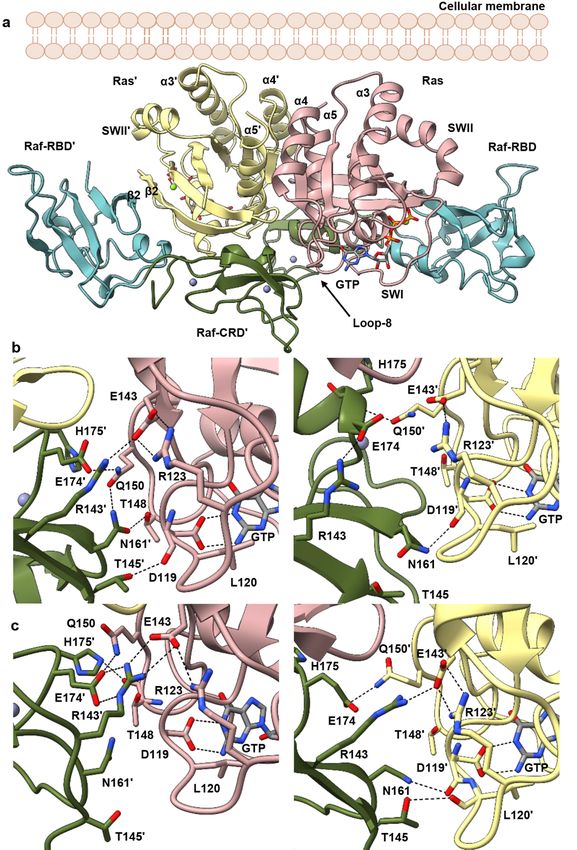
Biomolecules 2021, 11, 996                                                                                            2 of 20

                             interactions with saccharides or peptides/proteins [14]. It is as scaffold proteins inside the
                             cell that Galectin-1 and Galectin-3 have been associated with HRas and KRas activities,
                             respectively [15,16]. The demonstration that this activity is through direct interaction
                             between Galectin-1 dimers and CRaf-RBD was shown for HRas nanoclustering and signal-
                             ing [13]. However, Galectin-3 readily dimerizes in the absence of glycans [17] and one can
                             envision an analogous role in its signaling through KRas.
                                   While the interaction between Ras and Raf-RBD has been well studied, the trans-
                             formation potential of Raf requires interaction with Ras through both the Raf-RBD and
                             Raf-CRD [2,18,19]. The recent crystal structure of KRas–CRaf_RBD_CRD (PDB ID 6XI7) has
                             contributed to our understanding of the weaker micromolar affinity interaction between
                             Ras and the CRD [20]. However, questions remain about a possible role of Ras dimerization
                             and the function of the CRD in this context. The Raf-CRD requires coordination of two
                             zinc ions and has been shown to interact with various phospholipids and supported lipid
                             bilayers in vitro [3,21–23]. This interaction is driven primarily by electrostatic attraction
                             between the positively charged CRD and negative phospholipid headgroups on the mem-
                             brane [22]. The CRD membrane-binding role has further been studied by MD simulations
                             and recently supported by NMR-guided characterization of nanodisc-bound KRas–CRaf-
                             RBD_CRD complexes [24–28]. However, interaction of the CRD with the membrane has
                             only been observed in vitro or in silico in the absence of other signaling components, with
                             no evidence that this interaction is biologically relevant in cells.
                                   In addition to the N-terminal Ras-binding conserved region (CR1) (residues 52–194,
                             CRaf numbering), the three Raf isoforms (ARaf, BRaf, and CRaf) share two regions with
                             high similarity that are connected through unstructured linker regions [5]. The second
                             conserved region (CR2) (residues 254–269) contains a serine/threonine-rich domain with
                             key phosphorylation sites [29–31] and the third conserved region (CR3) comprises the
                             C-terminal kinase domain (residues 349–609) (Figure 1a). Prior to its recruitment to the
                             membrane and interaction with Ras, Raf is held in an autoinhibited state through the aid
                             of 14-3-3 scaffold proteins that bind motifs located in the CR2 and CR3 regions [32–34].
                             Recruitment of Raf to the membrane through Ras results in dimerization of the Raf ki-
                             nase domain, leading to its activation and subsequent phosphorylation of MEK to propa-
                             gate signaling [35,36]. Recent cryo-EM structures of inactive and active BRaf/14-3-3 and
                             BRaf/MEK1/14-3-3 complexes have shed light into this mechanism revealing a critical role
                             for the Raf-CRD in stabilizing the autoinhibited state [32,37,38]. It has been noted that the
                             Raf-RBD is exposed in the complex with Ras, allowing its interaction with Ras to displace
                             the CRD from the kinase/14-3-3 complex [20]. From there, the proposed model for Ras
                             activation of Raf is built on the premise that the CRD must both bind Ras and insert into
Biomolecules 2021, 11, x     the membrane upon release from its autoinhibition role, leading to a signaling state    3 of in
                                                                                                                           20
                             which Ras is monomeric on the membrane [20].

                             Figure 1. Conts.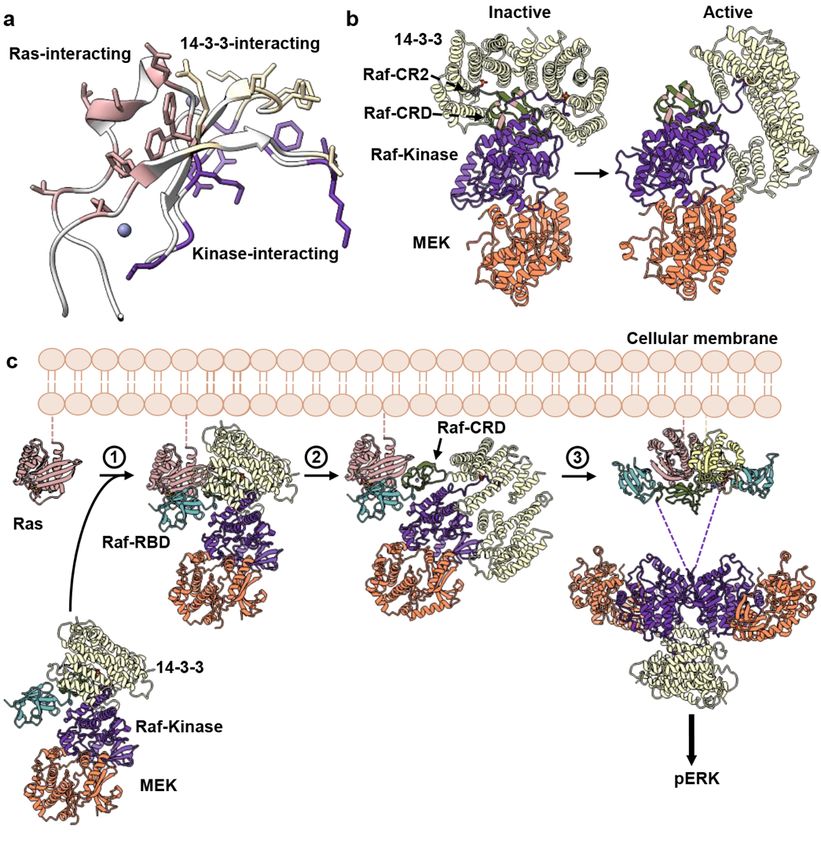
Biomolecules 2021, 11, 996                                                                                                               3 of 20

                                  Figure 1. Structure of the
       Figure 1. Structure of the HRas–CRaf-RBD_CRD            HRas–CRaf-RBD_CRD
                                                           complex.   (a) Schematic diagramscomplex.   (a) Schematic
                                                                                                   of HRas  and CRaf diagrams     of HRas and
                                                                                                                        provide structural
       boundaries for the Ras G domain     and hypervariable
                                  CRaf provide                  region, and
                                                  structural boundaries    for the Ras
                                                                                    threeG conserved
                                                                                            domain and regions found inregion,
                                                                                                         hypervariable    Raf kinases.
                                                                                                                                 and the(b)three
       Cartoon and surface representations
                                  conservedof   the HRas–CRaf-RBD_CRD
                                              regions  found in Raf kinases. complex      reveal
                                                                                (b) Cartoon   anda surface
                                                                                                   continuous  binding surface
                                                                                                           representations       spanning
                                                                                                                            of the HRas–CRaf-
       Ras (pink), residues 23 through  48 forcomplex
                                  RBD_CRD      both thereveal
                                                         Raf-RBD   (teal), andbinding
                                                               a continuous     Raf-CRD    (green).
                                                                                         surface    (c) TheRas
                                                                                                 spanning   Raf-CRD
                                                                                                                (pink),interacts
                                                                                                                        residues with resi- 48
                                                                                                                                 23 through
       dues in Ras α1, α5 and the interswitch region through hydrogen bonds and hydrophobic contacts. The symbol ♦ identi-
                                  for both the Raf-RBD (teal), and Raf-CRD (green). (c) The Raf-CRD interacts with residues in Ras α1,
       fies residues that show chemical shift perturbations in the analysis described by Feng et al. [24].
                                  α5 and the interswitch region through hydrogen bonds and hydrophobic contacts. The symbol
                                  identifies residues that show chemical shift perturbations in the analysis described by Feng et al. [24].
                                          Here, we present the crystal structure of HRas bound to a construct containing both
                                     the CRaf-RBD    and CRD
                                          Here, we present   theat  2.8 Å structure
                                                                  crystal  resolutionof(PDB
                                                                                        HRasID   7JHP)to(Figure
                                                                                               bound              1b,c containing
                                                                                                          a construct   and Supplemen-
                                                                                                                                   both
                                     tary Material  Table S1)  and   build  on  our  recently  proposed   model    for Ras
                                    the CRaf-RBD and CRD at 2.8 Å resolution (PDB ID 7JHP) (Figure 1b,c and Supplementary  activation of
                                     Raf [8]. Our model  is based   on  the discovery   that, on supported   lipid  bilayers,
                                    Material Table S1) and build on our recently proposed model for Ras activation of Raf [8].Ras dimer-
                                     izes model
                                    Our   upon binding  to on
                                                 is based   Raf,the
                                                                  and  allows forthat,
                                                                    discovery      the possibility
                                                                                       on supportedthatlipid
                                                                                                        previously   observed
                                                                                                              bilayers,        insertion
                                                                                                                         Ras dimerizes
                                    upon binding to Raf, and allows for the possibility that previously observed insertion
                                    of the CRD into the membrane is an artifact of experimental conditions. Our crystal
                                    structure of the HRas–CRaf-RBD_CRD is nearly identical to that of the recently published
                                    KRas–CRaf-RBD_CRD structure [20], adding to evidence that the Ras isoforms activate
                                    Raf kinase through a common mechanism [8]. This structure reveals that the Raf-CRD
                                    interacts with Ras residues at a surface contiguous to the Raf-RBD along β2, stretching to
                                    loop 3 at the interswitch region. As the dimer is not present in our crystals (Figure S1a),
                                    we used this structure to build a model of the HRas–CRaf-RBD_CRD dimer based on our
                                    HRas–CRaf-RBD structure, which places the Raf-CRD at the base of the two Ras protomers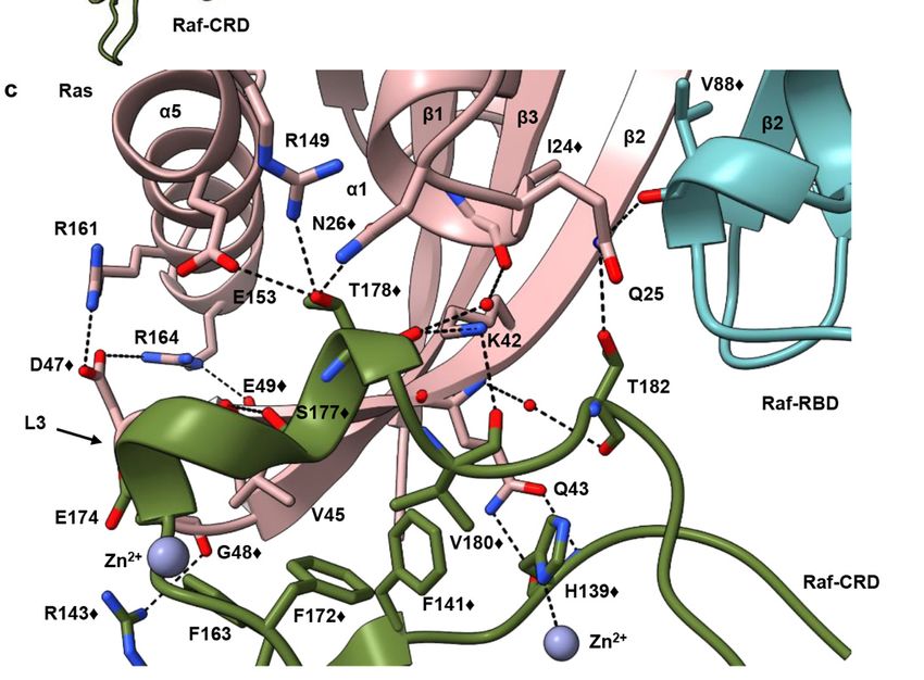
Biomolecules 2021, 11, 996                                                                                         4 of 20

                             opposite the C-terminal end of α5 and the HVR, facing away from the expected location of
                             the membrane. In this model, the CRD is poised to have an impact on both the Ras active
                             site and allosteric connections in the signaling complex. Significantly, although not noted
                             in the publication [20], this dimer is present through a two-fold crystallographic symmetry
                             axis in the higher resolution crystal structure of the KRas–CRaf-RBD_CRD complex (PDB
                             ID 6XI7) (Figure S1b), consistent with our model and the analysis presented here. Based on
                             our crystal structure and recent developments in the literature, we propose an alternate
                             mechanism in which binding of Raf-RBD promotes dimerization of the Ras–Raf-RBD_CRD
                             complex, with the CRD positioned at the base between the two Ras molecules in the dimer,
                             where it has the potential to allosterically affect regulation and signaling output.

                             2. Materials and Methods
                             2.1. Protein Expression/Purification
                                  The HRas–CRaf-RBD_CRD complex was crystallized using the HRasR97C mutant.
                             The HRasR97C construct containing residues 1–166 (henceforth referred to as HRas) was
                             generated by PCR site-directed mutagenesis from the wild-type HRas construct similarly
                             truncated. HRas protein was expressed, purified, and GDP was exchanged for the non-
                             hydrolyzable GTP analog GppNHp, as previously described [39]. The GB1_Raf-RBD-CRD
                             (52–184) construct, as previously published [7,40], was extended utilizing PCR site-directed
                             mutagenesis to generate the construct of CRaf-containing residues 52–187 used for the
                             experiments described in this paper. Cys 184 coordinates a Zn2+ ion and truncation at this
                             position led to a disordered CRD in our previously published structure [7]. CRaf protein
                             was transformed into Escherichia coli BL21(DE3) cells and grown at 37 ◦ C at 220 rpm in the
                             presence of 50 mg/L ampicillin. Expression was induced in the presence of 20 µM ZnCl2 at
                             an OD600 of 0.6–0.8 with 0.5 mM IPTG. The temperature was lowered to 32 ◦ C and cells
                             were harvested after 5 h by centrifugation.
                                  Cells were resuspended in 50 mM Tris-HCl pH 7.4, 500 mM NaCl, 1 mM BME, 5% v/v
                             glycerol, and 20 µM ZnCl2 in the presence of 1 mg/mL leupeptin, 1 mg/mL pepsatin, and
                             12 mg bezamidine. Cells were lysed by sonication and the insoluble fraction was separated
                             by centrifugation at 13,500 rpm, 4 ◦ C for 30 min. The supernatant was syringe filtered
                             through a 0.45 µm membrane before performing Ni NTA affinity chromatography (HisTrap
                             HP, GE Lifesciences). CRaf protein was eluted with 200 mM imidazole. Fractions were
                             pooled in the presence of 1 molar equivalent HRas and concentrated to 2 mL. Protein was
                             dialyzed overnight into cleavage buffer (20 mM Tris-HCl pH 8.1, 100 mM NaCl, 1 mM DTT,
                             25 mM CaCl2 , 5 mM MgCl2 , 20 µM ZnCl2, 5% v/v glycerol) at 4 ◦ C. Ten units of thrombin
                             were added per mg of CRaf_52-187 and incubated for 18 h at room temperature. The
                             cleaved complex was further purified by size exclusion chromatography (16/60 Sephacryl
                             S100, GE Lifesciences).

                             2.2. Protein Crystallization, Data Collection, and Refinement
                                   The HRas–CRaf_52-187 complex containing RBD and CRD was concentrated to
                             7 mg/mL for crystallization trials. Crystals of the complex were obtained by sitting-
                             drop vapor diffusion with a reservoir solution of 0.1 M ammonium acetate, 0.1 M Bis-Tris
                             pH 5.5, and 17% w/v PEG 10K. Drops were prepared with 1 µL protein and 1 µL reservoir
                             solution and crystals were grown at 18 ◦ C over ten weeks. Data collection was performed
                             on a home source MicroMax007HF with a Cu2+ anode, tungsten filament, and R-AxisIV2+
                             detector from Rigaku. Data were indexed, integrated, and scaled with the HKL3000 soft-
                             ware package [40]. Molecular replacement was performed in PHENIX [41] utilizing the
                             HRas–CRaf-RBD crystal structure (PDB ID 4G0N) as the search model. After correct place-
                             ment of Ras and Raf-RBD molecules, an additional search was performed for the Raf-CRD
                             utilizing the Raf-CRD NMR structure (PDB ID 1FAR). Further structure refinement was
                             performed with PHENIX [41] and COOT [42].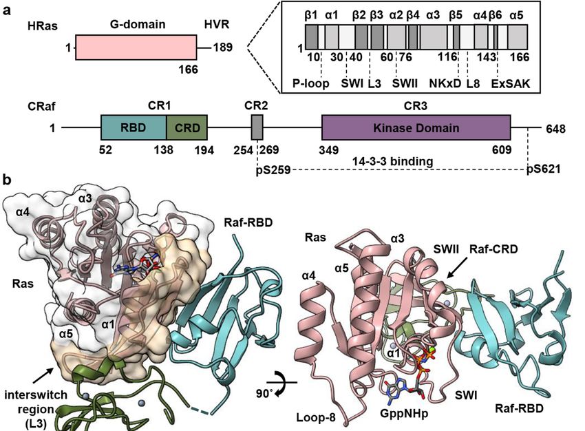
Biomolecules 2021, 11, 996                                                                                                  5 of 20

                             2.3. Model Preparation for Molecular Dynamics Simulations
                                  The monomer of the HRas–CRaf-RBD_CRD complex was prepared by substituting the
                             fully ordered wild-type HRas G domain (PDB ID 3K8Y) for the RasR97C G domain in the
                             HRasR97C/CRaf-RBD_CRD crystal structure. Loop 4 of the Raf-RBD (residues 103–109)
                             was modelled using the coordinates of the Raps/Raf-RBD crystal structure (PDB ID 1C1Y).
                             The Raf-RBD_CRD linker region and unmodelled side chains were built using guidance
                             from the 2Fo -Fc electron density map contoured at low sigma levels.
                                  To obtain the dimer model with the α4–α5 interface frequently observed in crystal
                             structures, the dimer generated by 2-fold crystallographic symmetry from our structure
                             of HRas–CRaf-RBD (PDB ID 4G0N) was used as a template for superposition of two
                             HRas–Raf-RBD_CRD complexes to generate the dimer containing the CRD.
                                  The six cysteines located in the CRaf-CRD for all systems were patched with CYN (de-
                             protonated Cys residues) for stable coordination of the two Raf-CRD zinc ions throughout
                             the simulations. All histidines in both HRas and CRaf molecules were in the uncharged
                             HSE form.

                             2.4. Molecular Dynamics Simulations
                                   Each system was solvated in a TIP3P water box and sodium and chloride ions were
                             added to a final concentration of 150 mM to neutralize the system. Each system was
                             minimized for 5000 steps and gradually heated from 50 to 250 K prior to the production
                             runs. Production runs were performed at constant temperature and pressure of 300 K and
                             1.01325 bar with periodic boundary conditions. The first 30 ns of each simulation had a
                             time step of 1 fs and the remaining simulation time was performed with a time step of
                             2 fs. Simulations were performed with the NAMD package [43] and CHARMM27 force
                             field [44]. The Particle Mesh Ewald method with a grid size of 1 Å was used to calculate
                             long-range electrostatics. Simulations for the HRas–CRaf-RBD_CRD dimer model were
                             performed in three independent runs, each for a duration of 350 ns, for a total of 1.05 µs.

                             2.5. Trajectory Analysis
                                  Dynamical network analysis was performed using the Carma [45] and Catdcd software
                             packages. Each protein residue within the simulation is assigned a spherical node centered
                             on its alpha carbon. Edges are drawn between nodes that stay within 4.5 Å of each other
                             for at least 75% of the trajectory and are weighted utilizing pairwise cross correlation data.
                             Optimal and suboptimal path calculations can also be used to identify residues important
                             in allosteric communication. The optimal path is calculated as the path containing the
                             fewest number of edges connecting user specified “source” and “sink” nodes. Nodes
                             occurring most frequently in the calculated optimal and suboptimal paths are important
                             for allosteric communication between the two specified “source” and “sink” nodes [46].
                                  Root-mean square deviation (RMSD) and root-mean square fluctuation (RMSF) calcu-
                             lations were performed using the Bio3d software package [47]. Distance calculations were
                             performed in VMD [48] by sourcing the distance.tcl script that can be found in the VMD
                             script library.

                             2.6. Protein Interfaces Surfaces and Assemblies (PISA)
                                  PISA is a program for analyses of PDB structures including surfaces associated with
                             crystal contacts to determine the likelihood that an interface is an artifact of crystal packing [49].
                             For this, it uses the interface free-energy ∆i G p-value as a measure of whether the interface
                             hydrophobicity is consistent with the average of biologically relevant interfaces. A p-value
                             < 0.5 means that the interface is more hydrophobic than the average and is likely to be
                             biologically relevant, whereas a p-value > 0.5 indicates that the surface is probably an artifact
                             of crystallization. Further detail can be found at the European Bioinformatics Institute
                             webpage: http://www.ebi.ac.uk/pdbe/prot_int/pistart.html (accessed on 1 June 2021).
Biomolecules 2021, 11, 996 6 of 20
3. Results
3.1. Overall Structure of Ras–Raf-RBD_CRD Complex
The Raf-RBD and CRD interfaces are contiguous surfaces spanning Ras residues 23
through 48, including the C-terminal end of the Ras helix α1, switch I, β2 and loop 3
(Figure 1b). The Raf-RBD interacts with Ras at switch I and the N-terminal portion of
β2 (residues 27–41), as previously described [7], and the short linker connecting the Raf-
RBD and Raf-CRD (residues 133–137) is partially disordered in our structure, with poor
electron density for residues 134–136. The Raf-CRD interaction involves hydrogen bonds
and hydrophobic contacts with C-terminal residues in Ras α1 (residues L23-N26) and β2,
extending to loop 3 in the interswitch region (residues K42-G48), with direct and allosteric
connections to α5 (Figure 1c). The Raf-CRD interface involves residues near the C-terminal
end of the CRD in the stretch from F172 to T182, and nearby N-terminal residues H139, F141,
R143, as well as F163. This interface includes the helix adjacent to the first Zn2+ -binding
site and extends toward the C-terminus, where the second Zn2+ ion is coordinated by
residues C184 as well as by the interface residue H139, linking the N and C-terminal ends
of the Raf-CRD. The position of the CRD observed in our crystal structure is as recently
reported [20] and consistent with published NMR chemical shift perturbation (CSP) data
(Figure 1c and Figure S2a), and with Ras N26G and V45E mutations shown to disrupt the
Ras–Raf-CRD interaction [19,24]. Given its close contact with the interswitch loop 3 on Ras,
the Raf-CRD is allosterically connected to α5 through salt bridges that form between Ras
D47 and E49 on loop 3 and R161 and R164 on α5 (Figure 1c). This binding mode of the
Raf-CRD to Ras is distinct from that described in the NMR data-driven nanodisc-bound
KRas–CRaf-RBD_CRD complexes (Figure S2b), where the Ras–Raf-CRD interaction was
modelled utilizing chemical shift perturbation data that are also consistent with our model
(Figure S2a) and paramagnetic relaxation enhancement (PRE) data with placement of the
probe at a cysteine engineered in place of Q43 [24], a residue with multiple interactions
at the Ras–Raf-CRD interface (Figure 1c). Only the RBD-CRD configuration in the crystal
structure is consistent with the expected location of the Raf-RBD when superimposed on
the cryo-EM structure for the inactive BRaf/MEK1/14-3-3 complex (PDB ID 6NYB), in
which the Raf-CRD is sandwiched between the Raf-kinase domain and 14-3-3 dimer and the
Raf-RBD is solvent exposed, absent from the final cryo-EM reconstruction [32] (Figure S2c).
3.2. Raf-CRD Links to the Active Site through Loop 8 across the Dimer Interface
To study Ras–Raf in the context of the dimer and determine possible roles for the
CRD, we modelled the HRas–CRaf-RBD_CRD dimer utilizing a two-fold symmetry axis
conserved across many Ras crystal structures, including our structure of the HRas–CRaf-
RBD complex (PDB ID 4G0N) containing the α4–α5 dimer interface [7,50,51]. In this
model, the Raf-CRD is positioned for interaction with both Ras protomers in the Ras
dimer, bridging the interswitch region of one to loop 8 of the other at the base of the
Ras dimerization interface (Figure 2a), as is found in the recently published KRas–CRaf-
RBD_CRD crystal structure containing the dimer (PDB ID 6XI7) [20]. We performed three
independent replicates of molecular dynamics (MD) simulations, each for a duration of
350 ns, totaling just over 1 µs. The model equilibrated quickly and was stable throughout
the trajectories, with average RMSD near 2.5 Å from the starting model in each case
(Figure S3a). The simulations show greater fluctuations for Raf-CRD than observed for
the rest of the complex (Figure S3b), with sampling of interactions seen in the KRas–CRaf-
RBD_CRD crystals where the dimer is present.independent replicates of molecular dynamics (MD) simulations, each for a duration o
350 ns, totaling just over 1 μs. The model equilibrated quickly and was stable throughou
the trajectories, with average RMSD near 2.5 Å from the starting model in each case (Fig
Biomolecules 2021, 11, 996 ure S3a). The simulations show greater fluctuations for Raf-CRD than observed
7 of 20 for th
rest of the complex (Figure S3b), with sampling of interactions seen in the KRas–CRa
RBD_CRD crystals where the dimer is present.
Figure 2. HRas–CRaf-RBD_CRDFigure 2.dimer complex. (a) Modelled
HRas–CRaf-RBD_CRD HRas–CRaf-RBD_CRD
dimer complex. dimer complex. Approximately
(a) Modelled HRas–CRaf-RBD_CRD dimer com-
perpendicular orientation of helices
plex. 3, 4 and 5 perpendicular
Approximately with respect to the membrane
orientation does
of helices 3, 4not
andallow
5 withthe Raf-CRD
respect to theto interact with
membrane
the membrane. (b) Frame taken from
does not the replicate
allow 2 HRas–CRaf-RBD_CRD
the Raf-CRD simulation
to interact with the membrane. (b)after
Frame60taken
ns shows
from sample Ras–Raf-
the replicate
CRD’ (left) and Ras’/Raf-CRD (right) interactions. (c)
2 HRas–CRaf-RBD_CRD Frame taken
simulation from
after 60 replicate
ns shows 3 HRas–CRaf-RBD_CRD
sample Ras–Raf-CRD’ (left) andsimulation
Ras’/Raf-after
140 ns shows sample Ras–Raf-CRD’
CRD (right) (left) and Ras’/Raf-CRD
interactions. (right)
(c) Frame taken frominteractions. Right and left panels simulation
replicate 3 HRas–CRaf-RBD_CRD in b and c illustrate
after
the asymmetry introduced140 through the Raf-CRDs’ dynamic interaction with the opposing Ras
ns shows sample Ras–Raf-CRD’ (left) and Ras’/Raf-CRD (right) interactions. Right protomers. One
andprotomer
left
of HRas in the dimer is shown in in
panels pink andc illustrate
b and the other,the
HRas’ in yellow.
asymmetry Both CRDs
introduced in the
through dimer, CRD
the Raf-CRDs’ and CRD’
dynamic are shown
interaction
in dark green. with the opposing Ras protomers. One protomer of HRas in the dimer is shown in pink and the other,
HRas’ in yellow. Both CRDs in the dimer, CRD and CRD’ are shown in dark green.Biomolecules 2021, 11, 996 8 of 20
In the starting model for simulations of the HRas–CRaf-RBD_CRD dimer, Raf-CRD
residues R143, N161, and E174 are within interacting distance of loop 8 residue R123, the
nucleotide-binding residue D119, and Q150 at loop 10 immediately preceding α5 in the
opposing Ras protomer. Ras D119 is part of the NKxD motif (residues 116–119), and di-
rectly binds the guanine base in the Ras active site (Figure 2b,c). This motif is followed
by loop 8, containing residue R123 which makes a salt bridge interaction with E143 of
the ExSAK motif (residues 143–147), linking two highly conserved regions important for
active site stability [52]. The Ras R123–E143 salt bridge remains present throughout the
simulations, even as R143 of the Raf-CRD moves to interact with Ras E143 (Figure 2b,c,
Figure S4a (replicate 1), Figure S5a (replicate 2) and Figure S6a (replicate 3)), contributing
to the interface with the opposing Ras promoter. Raf-CRD residue N161 samples various
states in which it can make contacts with the side chains of Ras T148 and Q150 in loop 10
(Figure 2b,c, Figures S4b, S5b and S6b) or with the backbone of loop 8 residues C118, D119,
and L120 (Figure 2b,c, Figures S4c, S5c and S6c). Ras Q150 also interacts with Raf-CRD E174
and H175 (Figure 2b,c, Figures S4d, S5d and S6d). Raf-CRD T145 is also observed to inter-
act with the loop 8 residues D119 and L120 (Figure 2b,c). Various interactions revealed in
the simulations, although not initially present in the model built from our structure of the
HRas–CRaf-RBD_CRD in which the dimer is not present (PDB ID 7JHP), are observed in the
recently published structure of the KRas–CRaf-RBD_CRD dimer (PDB ID 6XI7). Most no-
tably, the interactions between the Raf-CRD and both the glutamate and lysine of the ExSAK
nucleotide-binding motif are observed both in our simulations and KRas–CRaf-RBD_CRD
dimer structure (PDB ID 6XI7). It is clear that the interactions between the CRD and Ras across
the dimer interface are dynamic, with the potential of stabilizing different sets of interactions
depending on the state of the signaling complex. The dynamic ability of the two Raf-CRDs
to interact with the opposing Ras molecule provides a means to introduce some local asym-
metry into the Ras–Raf dimer complex (Figure 2b,c, Figures S4a–d, S5a–d and S6a–d). There
are several isoform-specific residues in this region of Ras (Figure S7a,c) [52], thus increased
dynamics of the Raf-CRD may be a key feature allowing the three Ras isoforms to have
specific interactions with each of the Raf proteins [53]. Furthermore, the asymmetry and
flexibility that we observe in the dimer may facilitate a given Ras isoform to accommodate
heterodimers of Raf [54], as CRaf residues N161, E174, and H175 are isoform-specific and
correspond to Q257, Q270, and R271 in BRaf (Figure S7b,c). Overall, in the Ras–Raf dimer,
the Raf-CRD has access to the Ras dimerization interface through its interactions with loop 3
and α5, and to the Ras active site through loop 8, poised to affect key allosteric connections
involved in the regulation of Ras [55].
3.3. Allosteric Communication across the Ras–Raf-RBD_CRD Dimer Complex
We recently demonstrated that the Raf-RBD promotes Ras dimerization and used
dynamical network analysis and optimal/suboptimal path calculations based on MD
trajectories to identify allosteric connections linking both Ras dimerization and Raf-RBD
interfaces in the HRas–CRaf-RBD dimer [8]. We use the same type of analysis here to iden-
tify pathways of allosteric connections in the HRas–CRaf-RBD_CRD dimer and compare
them with those we found in the absence of the CRD. Briefly, a network is constructed
of edges drawn between residues that stay within 4.5 Å of each other for at least 75% of
the simulation time and edges are weighted as a function of pairwise correlations [46].
The resulting connectivity network allows for the calculation of allosteric paths through
residues that link distant regions of the protein or protein complex, with the optimal path
representing the shortest sum of edge weights between two residues and suboptimal paths
deviating from the optimal path within a user-specified threshold. In our previous work,
we studied the paths linking the two Raf-RBD residues D113 in the dimer of the HRas–
CRaf-RBD complex due to its positioning, along with Raf-RBD D117, in the Raf-RBD pocket
for the scaffold protein Galectin-1, critical for the activation of the MAPK pathway [13]. We
observed strong allosteric linkages between the D113 residues extending 85 Å across the
Ras–Raf-RBD dimer complex, and identified residues D47, located in loop 3 of one RasBiomolecules 2021, 11, 996 9 of 20
protomer, and E143, in β6 of the other, as a critical point of information transfer across the
Ras dimerization interface [8].
It is intriguing that the Raf-CRD is located at the base of the Ras–Raf dimer to in-
teract with both loop 3 of one Ras molecule and residue E143 of the other, therefore in
contact with residues identified as critical for extending allosteric communication across
the Ras dimerization interface [8]. Dynamical network analysis was performed for each
simulation replicate to identify residues with correlated motions within the dimer of the
HRas–CRaf-RBD_CRD complex and involved in intermolecular information transfer across
the complex [56,57]. The edge weights, determined as a function of pairwise correlations
and used for optimal and suboptimal path calculations, for residues that cross the Ras–Raf-
CRD’, Ras–Raf-RBD_CRD and Ras–Ras’ interfaces in the three MD simulation replicates,
demonstrate the variability across both Ras–Raf-CRD interfaces in the global analysis
(Tables S2–S4), consistent with the high RMSF associated with the Raf-CRD (Figure S3b).
Due to longer simulation run times and the dynamic nature of the Ras–Raf-CRD inter-
actions across the dimer interface (Figures S4a–d, S5a–d and S6a–d), dynamical network
analysis was also performed by dividing each simulation into seven segments of 50 ns
each in order to capture the diversity in communication extending across the complex
(Figure 3a). Analysis of optimal and suboptimal path calculations between the two D113
residues of each Raf-RBD in the networks constructed over 350 ns of simulation time, are
consistent with our HRas–CRaf-RBD dimer simulations [8], demonstrating the conserva-
tion of intermolecular information transfer involving the interswitch region of one Ras
protomer and β6 of the other across the Ras dimerization interface (Figure 3a,b, orange
path). Additionally, by constructing networks for smaller simulation segments, we capture
four distinct modes of intermolecular information transfer that are consistent across the
three replicates (Figure 3a and Figure S8) (Tables S5–S7). The first mode of information
transfer involves the interswitch region and β6 as described above (Figure 3b, orange path).
The second mode involves α5 and β6 as described for KRas–CRaf-RBD dimer simulations
modelled on the membrane [8] (Figure 3b, red path). The remaining two means for in-
formation transfer directly involve the Raf-CRD, with one utilizing the Raf-CRD’s close
association with loop 8, creating paths that wrap around the Ras active site (Figure 3c); and
the other alternatively using the interconnectivity between the Raf-CRD and interswitch
region to link the two Ras protomers again through loop 3 and β6 (Figure 3d).
The allosteric connections linking residues D113 at the Galectin-binding pocket of
each Raf-RBD molecule in the dimer, which also includes residue D117 [13] (Tables S5–S7),
suggest a model where Ras–Raf-RBD_CRD dimers couple with Galectin dimers to form
a higher-order macromolecular platform involved in signal amplification and kinetic
proofreading [8] (Figure 3e), similar to that described for the LAT/Grb2/SOS system [58].
This model is consistent with the functional importance of Galectins 1 and 3 for signaling
through the MAPK pathway [15,16,59,60] and with results from single molecule tracking
experiments correlating immobile species of Ras observed in live cells to active signaling
through the Ras–Raf–MEK–ERK pathway [61–63].
Our molecular dynamics simulation data suggest that the interconnectivity between
the Raf-CRD and the interswitch region of one Ras molecule and loop 8 of the other, in the
context of its dynamic nature, optimally positions the Raf-CRD to diversify the paths of
allosteric communication across the Ras–Raf complex linking the Galectin-binding sites
at the two ends of the dimer, not only by further bridging the two Ras molecules in the
Ras dimer, but also serving as a link between the Raf-RBD and second Ras molecule in the
complex, with a direct connection to the Ras active site. Simulations of the KRas–CRaf-RBD
dimer complex modelled on the membrane demonstrate that this complex is flexible at the
interface [8]. The Raf-CRD may accommodate the complex’s dynamic nature by providing
alternative routes of communication between the two Galectin-binding regions at opposite
ends of the dimer. The prominence of each of the alternate allosteric paths facilitated by the
CRD may be modulated by isoform-specific residues and sensitive to oncogenic mutations
in the active site.Biomolecules 2021, 11, 996 10 of 20
Biomolecules 2021, 11, x 10 of 2
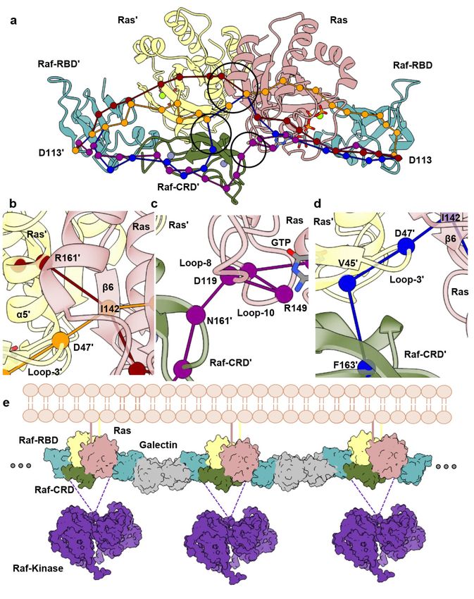
Figure 3. Dynamical network analysis performed for HRas–CRaf-RBD_CRD dimer simulations.
Figure 3. Dynamical network analysis performed for HRas–CRaf-RBD_CRD dimer simulations. (a) Allosteric paths calcu-
(a) Allosteric paths calculated between D113 and D113’ of the Raf-RBD molecules. (b) Close-up view
lated between D113 and D113’ of the Raf-RBD molecules. (b) Close-up view showing path for intermolecular information
showing path for intermolecular information involving Ras loop 3/β6 (orange) and Ras α5/β6 (red)
involving Ras loop 3/β6 (orange) and Ras α5/β6 (red) across the Ras dimerization interface. (c) Close-up view showing
across the Ras dimerization interface. (c) Close-up view showing path for intermolecular information
path for intermolecular information involving Raf-CRD/Ras loop 8 (purple). (d) Close-up view showing path for intermo-
involving Raf-CRD/Ras loop 8 (purple). (d) Close-up view showing path for intermolecular informa-
lecular information involving Raf-CRD/Ras interswitch region (blue). (e) Proposed Ras–Raf–Galectin assemblies adapted
tion involving Raf-CRD/Ras interswitch region (blue). (e) Proposed Ras–Raf–Galectin assemblies
from Packer et al. [8] with inclusion of the Raf-CRD. Galectin (grey) (PDB ID 3W58) and Raf-kinase (purple) (PDB ID 6Q0J)
adapted from Packer et al. [8] with inclusion of the Raf-CRD. Galectin (grey) (PDB ID 3W58) and
were downloaded from the Protein Data Bank. The scaffold protein 14-3-3 stabilizes the Raf kinase dimer (not shown).
Raf-kinase (purple) (PDB ID 6Q0J) were downloaded from the Protein Data Bank. The scaffold
Given that the structure of the CR2 is unknown, it is also possible that kinase domains from distinct Ras–Raf-RBD_CRD
protein 14-3-3 stabilizes the Raf kinase dimer (not shown). Given that the structure of the CR2 is
dimers bridged by Galetin come together to form the activated Raf dimer.
unknown, it is also possible that kinase domains from distinct Ras–Raf-RBD_CRD dimers bridged by
Galetin come together to form the activated Raf dimer.
The allosteric connections linking residues D113 at the Galectin-binding pocket o
each Raf-RBD molecule in the dimer, which also includes residue D117 [13] (Tables S5
S7), suggest a model where Ras–Raf-RBD_CRD dimers couple with Galectin dimers t
form a higher-order macromolecular platform involved in signal amplification and kineti
proofreading [8] (Figure 3e), similar to that described for the LAT/Grb2/SOS system [58
This model is consistent with the functional importance of Galectins 1 and 3 for signalin
through the MAPK pathway [15,16,59,60] and with results from single molecule trackin
experiments correlating immobile species of Ras observed in live cells to active signalin
through the Ras–Raf–MEK–ERK pathway [61–63].Biomolecules 2021, 11, 996 11 of 20
4. Discussion
Ras nanoclustering on the membrane modulates Ras activation of MAPK signaling,
but the significance of Ras dimers as functional signaling units is still not fully resolved
in the literature [64]. Weak binding is predicted for Ras dimers in the absence of other
factors [25,64–67], putting in question its viability as a robust signaling unit. However, we
have recently shown that Raf-RBD promotes robust dimerization of Ras on supported lipid
bilayers even at very low surface densities, in contrast with Ras in the absence of Raf-RBD,
for which no dimers can be detected [8,68]. This is consistent with observation of dimeric
Ras in live cells [12] and with experiments that suggest a requirement for Ras dimerization
in the activation of Raf [9–12].
The recently published crystal structures of the KRas–CRaf-RBD_CRD complex, each
with one complex monomer in the asymmetric unit in two different crystal forms (PDB
IDs 6XI7 and 6XHB), led authors to suggest a model in which Ras acts as a monomer in
the complex with Raf [20], building on an emerging model for Ras activation of Raf [69].
Here, the kinase domains of two nearby complexes dimerize to activate the signal with-
out the need for the Ras molecules to dimerize. This model is consistent with years of
research showing interaction of the CRD with the membrane in vitro and in silico [3,21–28].
Interestingly, one of the published crystal forms (PDB ID 6XI7) has the dimer in the crystal
with a structure that converges on our model of the dimer built from the crystal structure
of the HRas–CRaf-RBD_CRD (PDB ID 7JHP). Here, we offer an alternative model of Raf
activation in which Ras dimerization upon binding Raf-RBD is a key step, with placement
of the CRD at the base of the dimer interface, allosterically connecting elements of α5, the
interswitch region and two highly conserved elements of the nucleotide-binding site: the
NKxD and ExSAK motifs. Within the bigger picture of signaling through Ras–Raf–MEK–
ERK, the primary function of Ras in our model is not in promoting Raf dimerization per
se, but in providing an essential building block for a signaling platform in which Ras–Raf
dimers couple with Galectin dimers, leading to synchronized activation of multiple Raf
kinase dimers, stabilized by 14-3-3, for signal amplification (Figure 3e). Binding of short
oligosaccharides to Galectin-1 has recently been shown to promote allosteric connections
to the dimer interface [70]. Given that the proposed site for Raf-RBD partially overlaps
the glycan ligand-binding site [13], the binding of HRas–CRaf-RBD_CRD to Galectin-1
as we propose could result in allosteric connections across the entire signaling platform.
Galectin-3, known to be monomeric when bound to glycans, readily self-dimerizes in
the apo form [17], such that one could envision an analogous KRas–Raf-RBD–Galectin-3
signaling platform. The Raf-CRD in this context is centrally and flexibly located to affect
regulation both through diversifying the paths of allosteric connections in the signaling
complex and possibly affecting active site function, such as GTP hydrolysis, through its
proximity to the nucleotide-binding pocket.
We recognize that the specific interface through which Ras dimerizes has not yet
been unequivocally determined [64]. However, both the HRas–CRaf-RBD (PDB ID 4G0N)
and the KRas–CRaf-RBD_CRD (PDB ID 6XI7) dimer structures with the α4–α5 Ras dimer
interface, as well as the many studies supporting Ras dimerization through this inter-
face [8,9,50,51,71], point to our proposed model for the dimer. Given this dimer, we can
hypothesize its placement with respect to the membrane in a signaling context. The ev-
idence supporting an approximately perpendicular orientation of α3, α4 and α5 with
respect to the membrane [8,50], and the fact that Ras is tethered to the membrane through
the HVR following the C-terminal end of α5, places the dimer with the Raf-CRD away from
the membrane. This dimer model, which is derived from crystal structures and remains to
be confirmed experimentally on the membrane, would preclude the CRD insertion into the
membrane. We thus question the presumed membrane-binding function of the Raf-CRD
in MAPK signaling, characterized solely in the context of monomeric Ras–Raf-RBD_CRD
complexes [24,26,27]. It has been shown in live cells that the CRD is important for the re-
cruitment of Raf to the membrane in the presence of activated Ras [72]. However, the study
does not observe insertion of the CRD in the membrane, leaving open other possible mech-Biomolecules 2021, 11, 996 12 of 20
anisms through which the CRD can have an effect. We also point out that experimental
results where the Ras farnesyl group interacts in vitro with the CRD in solution [2,73], are
countered by experiments in cells that suggest that the farnesyl group does not contribute
to the Ras–Raf-RBD_CRD interaction [74] and recent SPR experiments showing that the
farnesyl group does not affect the affinity between KRas and CRaf-RBD_CRD [20]. Thus, it
is not clear whether the farnesyl group is involved in the Ras–Raf-RBD_CRD interaction.
The SPR experiments that accompany the KRas–CRaf-RBD_CRD structures validate the
primary interaction between Ras and the CRD as critical for signaling through MAPK [20],
consistent with common interactions in the two alternate models discussed here.
We now know that the surface of the Raf-CRD contains non-overlapping binding sites
for Ras (in the monomer of the complex), the Raf kinase domain and the scaffold protein
14-3-3 (Figure 4a). The CRD segments involved in the latter interactions have hydrophobic
and positively charged residues that are thought to insert into the membrane once the
CRD binds Ras and is released from its autoinhibited state [20,21,24,75,76]. They include
residues 143-RKTFLKLAF-151 and 157-KFLLNGFR-164, with the intermitting residues
152-CDICN-156 along with H173 and C176 coordinating the first Zn2+ -binding site at
the N-terminal end of the helix that forms the primary Ras-binding surface of the CRD
(Figure 1). These segments are found in the interactions across the Ras dimer interface
in our model that provides an alternative role for the CRD in the activated state of Raf.
Although these regions are dynamic in NMR structures of the CRD both in solution [3]
and in nanodiscs [24], in the crystal structure of the KRas–CRaf-RBD_CRD complex (PDB
ID 6XI7), residues 142–146 and 160–164 are part of a β-sheet that presents R143, T145,
L160 and N161 for interaction with loop 8 and the ExSAK nucleotide-binding motif of the
opposing Ras protomer in the dimer (Figure S9a), consistent with our simulations. L160
in particular makes van der Waals contact with the aliphatic portion of Ras K147 of the
ExSAK motif across the dimer interface, which in turn interacts with Ras F28 in switch
I. Both K147 and F28 are in direct contact with the guanine base of the nucleotide [77].
Several of the Raf-CRD residues in the two segments mentioned above form a core layer of
hydrophobic interactions in the CRD that includes packing of L160 at the dimer interface,
F151, F158, L159, F146 and the aliphatic portions K144 and R164 (Figure S9b). K144 and
R164 have the charged portion of their side chains exposed to solvent, capping the end
of this hydrophobic cluster opposite from L160. Thus, with the exception of K144 and
R164, the polar groups in the supposed membrane-binding segments are arranged at the
interface between the CRD and Ras, while the hydrophobic ones form a second layer
stabilizing the core structure that presents the Ras-interacting residues. The question of
whether the ordering of these key CRD segments is an artifact of our MD simulation
model (HRas–CRaf-RBD_CRD) or crystallization (KRas–CRaf-RBD_CRD), or a biologically
relevant consequence of dimerization is at the core of the present discussion. We have
used the Protein Interfaces Surfaces and Assemblies (PISA) [49] website at the European
Bioinformatics Institute to calculate the surface area buried in the KRas–CRaf-RBD_CRD
dimer interface (PDB ID 6XI7) as 1005 Å2 , with a ∆ι G p-value of 0.496, just within the cutoff
indicative of an interaction-specific interface unlikely to be an artifact of crystal packing
(see Methods section).
It has been recently demonstrated that mutations of Raf-CRD residues at the primary
Ras–Raf-CRD interface only marginally affect the affinity of the Ras–Raf_RBD_CRD com-
plex, dominated by the interaction with Raf-RBD, while these same mutations lead to
reduction in the kinase activity in cells [20]. This is consistent with S177A, T182A and
M183A variants at the primary Ras-binding site of the CRD inhibiting kinase activity in
a previous alanine-scanning mutagenesis study aimed at probing binding epitopes on
the surface of the Raf-CRD [74]. Interestingly, point mutations K144A, L160A and R164A
also weakly inhibit Raf activation, with the double mutant K144A/L160A completely
abrogating signal through Raf [74]. Here, we have two possible interpretations for these
results. It can be argued that the inhibitory effects of the K144, L160 and R164 variants are
due to disruption of the CRD interaction with the membrane. However, the data are alsoBiomolecules 2021,
Biomolecules 2021, 11,
11, 996
x 13 of 20 13 of
consistent
interfacewith
(PDBdisruption
ID 6XI7)ofas
key1005
interactions
Å2, withbetween the CRD of
a ΔιG p-value and0.496,
Ras loop
just8within
and ExSAK
the cutoff in
motifs across the dimer interface due to destabilization of the hydrophobic core maintained
dicative of an interaction-specific interface unlikely to be an artifact of crystal packing (se
by key residues in the 143–151 and 157–164 segments of the CRD as described above.
Methods section).
Figure 4. Proposed Mechanism for Ras-mediated activation of Raf. (a) Residues involved in the
Figure 4. Proposed Mechanism for Ras-mediated activation of Raf. (a) Residues involved in the primary Ras–Raf-CRD
primary Ras–Raf-CRD interaction (pink) do not overlap with Raf-CRD residues used to stabilize
interaction (pink) do not overlap with Raf-CRD residues used to stabilize contacts with the kinase domain (purple) and
contacts with the kinase domain (purple) and 14-3-3 proteins (wheat) in the autoinhibited state.
14-3-3 proteins (wheat) in the autoinhibited state. (b) Comparison of Raf-kinase domain (purple) and 14-3-3 (wheat) con-
(b) Comparison of Raf-kinase domain (purple) and 14-3-3 (wheat) conformations in the inactive (PDB
formations in the inactive (PDB ID 6NYB) and active Raf structure (PDB ID 6Q0J). Note the shift in the 14-3-3 dimer in the
ID 6NYB) and active Raf structure (PDB ID 6Q0J). Note the shift in the 14-3-3 dimer in the absence of
absence of the Raf CR2 motif and release of the Raf-CRD. (c) Proposed structural model for the Ras-mediated release of
the Raf CR2 motif and release of the Raf-CRD. (c) Proposed structural model for the Ras-mediated
Raf autoinhibition and activation of MAPK signaling. Raf-RBD (cyan) is exposed in the autoinhibited state, disordered in
release of Raf autoinhibition and activation of MAPK signaling. Raf-RBD (cyan) is exposed in the
the cryo-EM structure, likely due to flexibility in its linker to the CRD (dark green). The high-affinity binding of Raf-RBD
autoinhibited state, disordered in the cryo-EM structure, likely due to flexibility in its linker to the
to Ras places the exposed area of the CRD in its Ras-binding site, while steric hindrance is expected to displace the Raf C-
CRD (dark green). The high-affinity binding of Raf-RBD to Ras places the exposed area of the CRD
terminus (purple) and promote the shift of the 14-3-3 dimer (wheat) towards its active state interaction with a second Raf
in its Ras-binding site, while steric hindrance is expected to displace the Raf C-terminus (purple) and
kinase domain (purple). These two features of the Ras–Raf interaction are shown in steps 1 and 2. Binding of Raf-RBD
promote the shift of the 14-3-3 dimer (wheat) towards its active state interaction with a second Raf
concurrently promotes dimerization of the complex as shown in step 3, which we propose is a key step in binding Galectin
kinase domain (purple). These two features of the Ras–Raf interaction are shown in steps 1 and 2.
dimers to build a platform for signal amplification (see Figure 3e). We propose that steps 1, 2 and 3 occur concertedly to
Binding of Raf-RBD concurrently promotes dimerization of the complex as shown in step 3, which
activate Raf.
we propose is a key step in binding Galectin dimers to build a platform for signal amplification (see
Figure 3e). We propose that steps 1, 2 and 3 occur concertedly to activate Raf.
It has been recently demonstrated that mutations of Raf-CRD residues at the primar
Ras–Raf-CRD interface only marginally affect the affinity of the Ras–Raf_RBD_CRD com
plex, dominated by the interaction with Raf-RBD, while these same mutations lead to r
duction in the kinase activity in cells [20]. This is consistent with S177A, T182A and M183
variants at the primary Ras-binding site of the CRD inhibiting kinase activity in a previou
alanine-scanning mutagenesis study aimed at probing binding epitopes on the surface
the Raf-CRD [74]. Interestingly, point mutations K144A, L160A and R164A also weak
inhibit Raf activation, with the double mutant K144A/L160A completely abrogating signBiomolecules 2021, 11, 996 14 of 20
In addition to the inhibitory mutations, the alanine-scanning mutagenesis study iden-
tified a number of activating mutations [74], all of which are in residues at or near the
binding interface with 14-3-3 and Raf-kinase domain in the autoinhibited state. However,
most of these variants, N140A, R143A, T145A, Q156A, K157A, Q166A, T167A, K171A,
H175A are activating only in the context of RasG12V, suggesting a Ras dependence on the
resulting increased Raf activity. The variants F151A and D153A have significant effects in
the context of WT Ras, with a drastic increase in the basal Raf activity that the other mutants
do not show [74]. These two residues make key interactions in the inactive state of the
kinase [32] and activation is likely due to release of autoinhibition in the absence of Ras. Of
the RasG12V dependent activating variants of Raf-CRD, Q156, K157 and Q166, T167 are
pairs of hydrophilic residues exposed to solvent that follow one of the cysteine residues in
the two loops containing the Zn2+ -binding pockets. Residue N140 is also exposed to solvent
and follows H139 that coordinates one of the Zn2+ ions. The mechanism through which
mutations of these residues lead to RasG12V-dependent activation of the kinase is currently
not known. However, residue K157 is at the beginning of one of the segments thought to
interact with the membrane. If this interaction were important for activity, one would expect
that the K157A variant would inhibit rather than activate signaling through Raf.
The remaining activating residues in the alanine-scanning mutagenesis study, R143,
T145 and H175, make interactions across the dimer interface in the Ras–Raf-RBD_CRD
dimer and K171 is part of the packing that stabilizes T145. We have shown that the
CRD interactions across the dimer interface are dynamic in the Ras–Raf-RBD_CRD dimer.
The activating variants other than F151A and D153A have no effect on WT Ras signaling.
However, specific mutations at the dimer interface may synergize with RasG12V to stabilize
the dynamic CRD for more efficient information transfer through the allosteric pathways
with direct access to the active site and the Ras–Raf-RBD interface. Interestingly, variants
associated with RASopathies are also weakly activating [78,79]. In particular, BRaf T241P
and Q257R are found in individuals with LEOPARD syndrome and cardiofacialcutaneous
(CFC) syndrome, respectively [78]. These residues mediate contacts with the 14-3-3 scaffold
dimer in the autoinhibited state. They correspond to T145 and N161 in CRaf, and in the
context of the activated Ras–Raf dimer are located at the interface between the CRD and
loop 8 across the Ras–Raf-RBD_CRD dimer interface (Figure 2b,c).
The active BRaf/MEK1/14-3-3 structure, with a dephosphorylated 14-3-3-binding
motif on the CR2 [32], displays a shift of the 14-3-3 dimer with respect to the Raf-kinase
domain relative to the inactive state, accompanied by conformational rearrangement of
the Raf-kinase C-terminus, which now redirects away from the Ras-binding surface of
the Raf-CRD (Figure 4b). The absence of the Raf-RBD from the inactive BRaf cryo-EM
reconstruction suggests it does not contribute toward stabilizing the autoinhibited state
and therefore must remain accessible for Ras-binding [32]. This is consistent with a highly
dynamic RBD observed by MD simulations [80]. Indeed, by aligning the CRaf-CRD from
our Ras–Raf-RBD_CRD structure with the BRaf-CRD in the inactive complex, we observe
that the short Raf-RBD_CRD linker is sufficient for the Raf-RBD to be accessible for inter-
action with Ras (Figure 4c and Figure S2c). We propose that Ras-binding to the Raf-RBD
results in the dissociation of the 14-3-3 subunit from the CR2-binding site and redirection
of the Raf C-terminus. This would expose the CR2 site for dephosphorylation, preventing
association of the 14-3-3 dimer to reestablish Raf autoinhibition. This is consistent with an
increase in Raf-kinase activity in cells upon dephosphorylation of the CR2 site [5,29,33].
The displacement of 14-3-3 from the CR2-binding site would promote its interaction with a
second Raf kinase to facilitate kinase domain dimerization and further expose the Raf-CRD
for extraction (Figure 4b,c), resulting in collapse of the autoinhibited conformation and
progression toward Raf activation.Biomolecules 2021, 11, 996 15 of 20
Our proposed model for Ras-mediated activation of the MAPK pathway (Figure 4c) is
similar to the recently published model [20] in the mechanism by which Raf-RBD binds
to Ras. However, the two models deviate in that, in our model, binding of Raf-RBD to
Ras promotes Ras dimerization, permitting allosteric modulation of the Galectin-binding
regions on the Raf-RBD, to form a platform of Ras–Raf and Galectin dimers for signal
amplification (Figure 3e). During this process the CRD is released from its autoinhibitory
role, allowing the shift in 14-3-3 and stabilization of the Raf dimer. Given the high-affinity
dimerization of Ras promoted by Raf-RBD that we previously reported [8], we propose
that these events occur concertedly, resulting in simultaneous release of autoinhibition and
allosteric modulation of the Galectin-binding site on Raf-RBD, with robust activation of the
Ras–Raf–MEK–ERK pathway.
Our model of activation of Raf through formation of the Ras dimer upon binding
Raf-RBD serves as a viable alternative to the recently published model of a monomeric Ras–
Raf-RBD_CRD complex in which the CRD is inserted in the membrane [20]. Both models
remain to be tested and validated, as there is currently not enough experimental evidence to
prove either one. Although not discussed here, a third model of Ras activation of Raf was
published as a preprint in BioRXiv [81] and that model too remains to be tested. We, in the
Ras research community, have made exciting progress toward a better understanding of the
Ras–Raf interaction and of the components important in the release of autoinhibition and
activation of the Raf kinase domain. We must now stay open-minded as we consider the
mechanistic link that lies between the binding of Raf-RBD to Ras and the resulting robust
signaling output in cells. Given the artificial contexts in which the CRD has been observed
to bind membranes, and the fact that there are examples in the literature where artificial
protein–membrane interactions have been observed [82–84], the question of whether the
CRD contributes to membrane insertion or to interaction across the Ras–Raf-RBD_CRD
dimer interface remains open and must be further explored. Our recent demonstration that
Raf-RBD promotes dimerization of Ras on the membrane and that this results in strong
allosteric connections across the dimer formed through α4 and α5, provides an important
set of constraints in going forward with refining the model for Ras activation of Raf. The
placement of the CRD in this dimer at a strategic position to affect allosteric communication
between functionally important regions of Ras, and beyond to the Galectin-binding regions of
Raf-RBD, calls for serious consideration as we move forward.
5. Conclusions
The crystal structure of the HRas–CRaf-RBD_CRD complex presented here with our
analysis of the dimer model based on MD simulations supported by a recently published
structure of the KRas–CRaf-RBD_CRD in which the dimer is present provide a key missing
link in our understanding of Ras-mediated Raf activation. They show a continuous surface
for Ras–Raf interaction that links the Ras active site and dimerization interface through
switch I, loop 3, and α5. In the context of the dimer, the CRD also contacts loop 8 and
residues in two highly conserved nucleotide-binding motifs, centrally positioned to access
regulatory and functional regions of Ras. This is consistent with the observation that the
Raf-CRD is required for Raf activation in vivo. Our analysis of these structures in the
context of other recent structural breakthroughs in the literature provides an emerging
model of Ras activation of Raf, where formation of the Ras–Raf complex is linked to
both dimerization and release of Raf autoinhibition to form a platform for amplified
synchronized signaling. This model builds upon our recent proposal of higher-order Ras–
Raf–Galectin signaling assemblies, paving forward a new research direction paramount
to our understanding of signaling through the Ras–Raf–MEK–ERK pathway [8]. Because
the affinity between Ras and the Raf-CRD alone is relatively weak, the disruption of its
interactions with Ras, either through the interswitch region involving loop 3 or through
loop 8, holds strong potential as a novel approach for targeting Ras- and Raf-related cancers.
The structural analysis presented here, with release of the requirement for insertion of the
CRD in the membrane, provides an alternate framework, consistent with available dataYou can also read