SURVEY AND SUMMARY DNA G-quadruplex structures: more than simple roadblocks to transcription?
←
→
Page content transcription
If your browser does not render page correctly, please read the page content below
Published online 13 July 2021                                                    Nucleic Acids Research, 2021, Vol. 49, No. 15 8419–8431
                                                                                                       https://doi.org/10.1093/nar/gkab609

SURVEY AND SUMMARY
DNA G-quadruplex structures: more than simple
roadblocks to transcription?
Jenna Robinson1,2 , Federica Raguseo1,2 , Sabrina Pia Nuccio1,2 , Denise Liano1,2 and
Marco Di Antonio 1,2,3,*

                                                                                                                                                              Downloaded from https://academic.oup.com/nar/article/49/15/8419/6320416 by guest on 08 November 2021
1
 Imperial College London, Chemistry Department, Molecular Sciences Research Hub, 82 Wood Lane, London W12
0BZ, UK, 2 Institute of Chemical Biology, Molecular Sciences Research Hub, 82 Wood Lane, London W12 0BZ, UK
and 3 The Francis Crick Institute, 1 Midland Road, London NW1 1AT, UK

Received May 26, 2021; Revised July 01, 2021; Editorial Decision July 01, 2021; Accepted July 05, 2021

ABSTRACT                                                                        GRAPHICAL ABSTRACT
It has been >20 years since the formation of G-
quadruplex (G4) secondary structures in gene pro-
moters was first linked to the regulation of gene
expression. Since then, the development of small
molecules to selectively target G4s and their cel-
lular application have contributed to an improved
understanding of how G4s regulate transcription.
One model that arose from this work placed these
non-canonical DNA structures as repressors of tran-
scription by preventing polymerase processivity. Al-
though a considerable number of studies have re-
cently provided sufficient evidence to reconsider this
simplistic model, there is still a misrepresentation
of G4s as transcriptional roadblocks. In this review,
we will challenge this model depicting G4s as sim-                              INTRODUCTION
ple ‘off switches’ for gene expression by articulating
how their formation has the potential to alter gene                             G-quadruplexes (G4s) are DNA structures that can arise
expression at many different levels, acting as a key                            under physiological conditions from guanine-rich DNA se-
regulatory element perturbing the nature of epige-                              quences (Figure 1) (1–4). Although the ability of guanine
                                                                                analogues to assemble into tetrameric structures has been
netic marks and chromatin architecture.
                                                                                known since 1962 (5), the idea that G4 structures could form
                                                                                in the context of genomic DNA was only seriously consid-
                                                                                ered 40 years later when the crystal structure of a G4 formed
                                                                                by the human telomeric DNA sequence was reported for the
                                                                                first time (6). The very same year a different G4-forming se-
                                                                                quence was described in the promoter region of the onco-
                                                                                gene MYC by Hurley et al. (7). Importantly, this study was
                                                                                not limited to the structural characterization of the quadru-
                                                                                plex, but also provided evidence that such a G4 was biolog-
                                                                                ically active by showing MYC-downregulation as a direct
                                                                                consequence of G4 stabilization by the porphyrin-based lig-
                                                                                and TMPyP4 (7). This observation sparked interest in non-

* To   whom correspondence should be addressed. Tel: +44 20 7594 5866; Email: m.di-antonio@imperial.ac.uk


C The Author(s) 2021. Published by Oxford University Press on behalf of Nucleic Acids Research.
This is an Open Access article distributed under the terms of the Creative Commons Attribution License (http://creativecommons.org/licenses/by/4.0/), which
permits unrestricted reuse, distribution, and reproduction in any medium, provided the original work is properly cited.
8420 Nucleic Acids Research, 2021, Vol. 49, No. 15

                                                                                investigation of the association between endogenous G4-
                                                                                formation and gene expression, revealing an almost inverted
                                                                                picture to the earlier investigations. Indeed, G4 ChIP-Seq
                                                                                performed in keratinocytes (15) and human xenografts
                                                                                (16) using the G4-selective antibody BG4, has demon-
                                                                                strated that G4s are prevalently found at gene promoters
                                                                                of transcriptionally active genes, acting as transcriptional
                                                                                enhancers rather than repressors. The link between G4 for-
                                                                                mation and active transcription has been confirmed beyond
                                                                                keratinocytes, in liposarcoma cells still using BG4-based
                                                                                ChIP-Seq (17), as well as in a range of mammalian cell
                                                                                lines using a G4-selective peptide probe (18), and even with
                                                                                single-molecule detection of G4s within live cells under non-

                                                                                                                                                  Downloaded from https://academic.oup.com/nar/article/49/15/8419/6320416 by guest on 08 November 2021
                                                                                perturbative conditions (19). Furthermore, G4 formation
                                                                                seems to contribute to activated gene expression by means
                                                                                of many different mechanisms that span from transcription
Figure 1. Schematic representation of G-quadruplex structures. G4s are          factor binding (20) to guanine oxidation (21). Despite this,
constituted of four guanine bases arranged in a square planar conforma-         there is still a misrepresentation in the scientific community
tion (G-tetrad) held together by Hoogsteen hydrogen bonding and further
stabilized by alkali cation such as K+ . Specific G4 topologies can be formed   of G4s acting as transcriptional roadblocks and repressors,
and include anti-parallel, parallel and hybrid structures, depending on the     which is reminiscent of the initial hypothesis postulated 20
relative orientation of the DNA strand within the structure. Intermolecu-       years ago. In this review, we aim to provide a critical assess-
lar G4 structures can also be formed when more than one DNA strand is           ment of the latest research relating G4s to transcriptional
used to generate the final structure (red and blue strands).
                                                                                regulation, discussing the genome-wide effects of G4 for-
                                                                                mation on chromatin architecture, long-range interactions,
                                                                                phase separation and DNA oxidation.
telomeric G4s and suggested that these structures could act
as transcriptional repressors, a concept supported in several
                                                                                G4 formation in the chromatin context
following studies on G4s formed in other oncogene promot-
ers, such as c-KIT, BCL-2, KRAS and VEGF (8). This gen-                         To fully appreciate and investigate the role G4 formation
eral notion was further consolidated by multiple bioinfor-                      may play in gene regulation, we need to consider the ge-
matic studies revealing that G4s are enriched at gene pro-                      nomic context in which G4s exist. The 3D organization of
moters, particularly in oncogenes (9–11), supporting a role                     genetic material in cells presents a high level of complex-
of these structures in regulating gene-expression and their                     ity, where the canonical double helix of DNA is wrapped
potential to be targeted by small-molecules for therapeutic                     around proteins in a macromolecular structure known as
intervention.                                                                   chromatin. Chromatin is made of nucleosomes: a repeti-
   However, most of the initial studies that portrayed G4s                      tive unit consisting of histone proteins which wrap around
as antagonists to gene expression relied on the use of syn-                     a stretch of DNA (ca. 146 bp) contributing to the folding
thetic molecules as G4-stabilizers or in vitro models of tran-                  of DNA inside the nucleus (Figure 2A) (22,23). However,
scription (12), typically plasmid-based, which do not take                      genome sequencing and microarray hybridization technolo-
into account the complexity of endogenous gene expres-                          gies have revealed that nucleosome occupancy is not ho-
sion within chromatin. Indeed, direct evidence to support                       mogenous across DNA and that there are certain genomic
the idea that endogenous G4 formation at gene-promoters                         regions depleted of nucleosomes (24). Nucleosome depleted
results in transcriptional repression is still lacking. In 2012                 regions (NDRs) are stretches of more accessible DNA
Rodriguez et al. challenged the notion of G4s as direct tran-                   which act as hubs for protein binding, including those that
scriptional repressors, by revealing that treatment with the                    regulate transcription (24). Interestingly, the location of
potent G4 ligand pyridostatin (PDS) caused DNA-damage                           NDRs within the genome is highly dynamic and dependent
at G4 sites in a transcription and replication dependent                        on chemical modifications to histones, such as methylation
fashion (13). In this key study, which generated the first                      or acetylation, which change the interaction of histones
genome-wide G4 map, it was demonstrated that the DNA                            with key proteins involved in regulating gene expression
damage elicited by PDS at G4 sites caused gene down-                            (25). Therefore, nucleosome positioning across the genome
regulation, rather than the formation of the G4 structure it-                   is key to define the epigenetic status of a cell and this orga-
self. The same year, Hurley and co-workers challenged their                     nization appears to also be associated with the formation of
own observations made with TMPyP4 10 years earlier, re-                         certain DNA secondary structures, including G4s.
porting how treatment with G4 ligands could lead to MYC                            The first evidence that G4s may have a role in shaping
downregulation without direct targeting and stabilization                       chromatin was highlighted in a study focused on REV1, a
of the MYC G4, but rather as an indirect consequence of                         protein belonging to the Y family of DNA polymerases,
global G4 stabilization elicited by the ligand (14).                            that has an essential role in ensuring replication proceeds
   More recently, the development of G4-selective antibod-                      when DNA damage occurs (26). Cells containing a mutant
ies has enabled genome-wide mapping of G4s by immuno-                           form of REV1 were characterized by delayed or fully com-
precipitation experiments followed by sequencing (ChIP-                         promised DNA replication, particularly at G4-forming se-
Seq). These studies have allowed for a far more direct                          quences (26). The unresolved G4 structures in REV1 mu-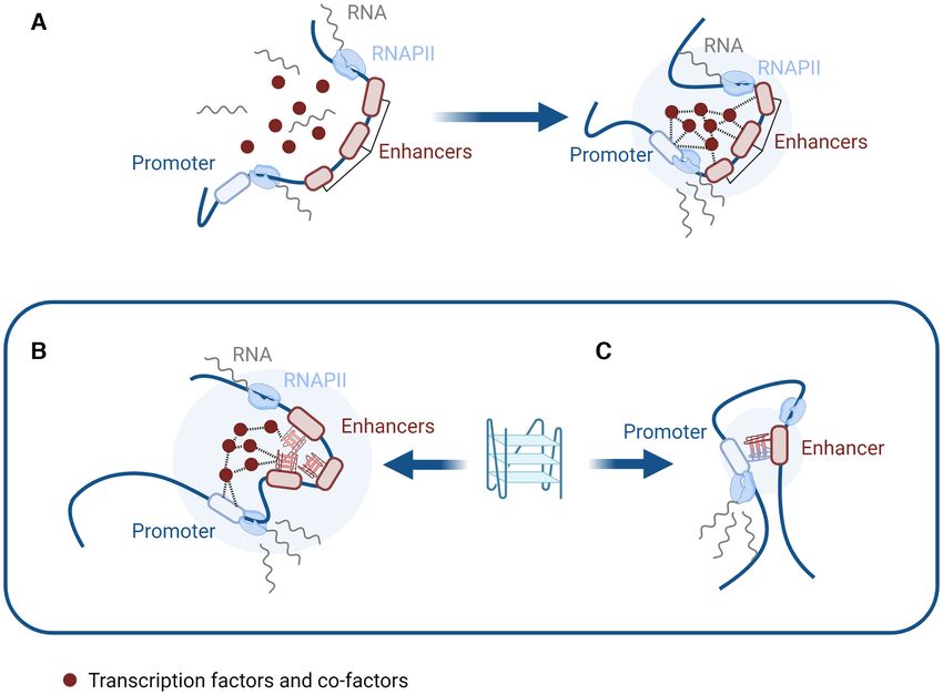
Nucleic Acids Research, 2021, Vol. 49, No. 15 8421

                                                                                                                                                           Downloaded from https://academic.oup.com/nar/article/49/15/8419/6320416 by guest on 08 November 2021
Figure 2. G4s and chromatin structure. (A) Structure of chromatin comprised of DNA wrapped around nucleosome complexes. (B) REV1 mutants unable
to resolve G4s during DNA replication exhibit altered histones modifications in the newly synthesized strand and a consequent loss of epigenetic memory.
(C) G4s can interact with a wide panel of proteins, including chromatin remodelers such as BRD3. (D) G4s are strongly associated with nucleosome
depleted regions (NDRs) as confirmed by BG4-ChIP.

tants led to replication fork stalling, but surprisingly also                 recently confirmed by immunofluorescence studies (28). In
had a significant impact on the epigenetic status of the cell                 this latest work, colocalization between G4 structures and
(Figure 2B). In particular, an increase in the expression of                  histone modifications found in nucleosome-depleted eu-
the p-globin locus was observed. This upregulation was due                    chromatin regions was observed, underlining the potential
to the loss of a histone modification negatively associated                   of G4s in moulding the epigenetic landscape.
with transcription: H3K9 dimethylation, as well as incorpo-                      From these findings, some open questions arise: are G4s
ration of newly acetylated histones around the G4-forming                     a passive element in the deposition of histones marks or do
site, promoting active transcription.                                         they have a mechanistic role in shaping chromatin that is
   However, a later study of a separate locus revealed that                   not limited to replication stalling? To unravel any mechanis-
unresolved G4 structures in REV1 mutants could also lead                      tic link between G4s and chromatin structure, it may be key
to the loss of distinct histone marks that promote transcrip-                 to investigate G4-interacting proteins especially those that
tion, suggesting that the role of G4s in modifying the his-                   are known for having an active role in chromatin remod-
tone code is dependent on their genomic context (27). Al-                     elling. An example is the protein BRD3 that was found to
though, these findings were limited to chicken DT40 cells,                    be one of the top hits in an unbiased microarray screening of
they suggested for the first time a relationship between G4s                  the G4-interactome (29). BRD3 contains a bromo-domain
and the installation of epigenetic marks, which was more                      capable of binding acetylated histones, allowing for the re-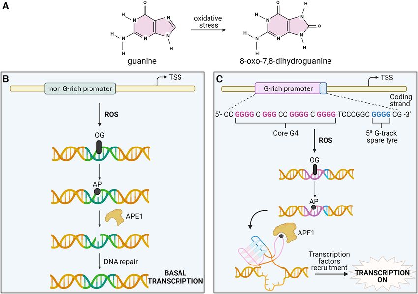
8422 Nucleic Acids Research, 2021, Vol. 49, No. 15

cruitment of RNA polymerase and initiation of transcrip-        C-rich regions on the template strand of DNA (35). Thus,
tion (30). The idea that G4s interact with BRD3 within cells    the creation of R-loops often displaces a single-stranded
has been substantiated by BG4 ChIP-seq and BRD3 ChIP-           G-rich sequence which may be primed for folding into a
seq analysis, which revealed a significant colocalization of    G-quadruplex (36–39). This can occur from the formation
G4 motifs within BRD3 occupancy sites (31). Additionally,       of an intramolecular G4 on the displaced strand (Figure
the interaction between G4s and BRD3 does not prevent its       3A) or a DNA:RNA hybrid intermolecular G4 (Figure 3B)
binding to acetylated histones or other chromatin remod-        (40,41).
elers, suggesting that BRD3-G4 interactions might instead          Such R-loops form significantly in actively transcribed
guide the recruitment of chromatin remodelling complexes        genes and interestingly, genome-wide studies have also
to favour transcription at G4-sites (Figure 2C).                confirmed formation of G4-structures in transcriptionally
   These findings highlighted the relevance that G4s may        active genes (15,17,18). Furthermore, it appears that G-
have in engaging key regulators of chromatin architecture,      quadruplexes may have a functional role in mediating tran-
leading to the hypothesis that global changes in chromatin      scription by actively stabilizing the R-loop (42), allowing

                                                                                                                                   Downloaded from https://academic.oup.com/nar/article/49/15/8419/6320416 by guest on 08 November 2021
structure could be directly caused by G4 formation in the       for enhanced transcription. A recent study demonstrated
genome. This hypothesis is supported by bioinformatic as-       this by showing that the placement of G4s on the non-
sociations made on a genome-wide scale, which demon-            template strand enhances R-loop formation resulting in sig-
strated that G4-forming sequences are highly enriched at        nificantly increased transcript output, RNA polymerase ini-
nucleosome depleted regions (Figure 2D) (10,32). Although       tiation and elongation (43). This study also revealed that
these first studies were based on computational predictions     such an effect was strand dependent, as no such increases
of G4s, such results have recently been validated directly in   in transcription are seen when the G4 motif was found on
the chromatin context by using the G4-selective antibody        the template strand. Despite these interesting results, this
BG4 which confirmed the enrichment of G4s at nucleosome         work was limited to in vitro measures of transcription which
free sites (15). Specifically, 98% of G4s identified by BG4-    may not encapsulate the complex interactions of G4s within
ChIP coincided with NDRs, an observation that has now           cells. Thus, it would be worth further investigating if the
been recapitulated in independent studies (17). This exem-      presence of G4s in R-loops is similarly correlated with in-
plifies the interconnectedness between G4 formation and         creased transcriptional output within a chromatin DNA
chromatin structure and strongly suggests that these struc-     context that is more representative of transcriptional pro-
tures might mark transcriptionally active regions and pos-      cesses in living cells.
sibly influence nucleosome positioning.                            In contrast to this work suggesting G4s have a positive ef-
   Despite the fact that there is now significant evidence      fect on transcription by stabilizing R-loop formation, it has
suggesting that G4s may act as an active factor in shap-        been noted that over-stabilization of G4s can have deleteri-
ing the structure of chromatin, for example by altering hi-     ous effects on transcription. For instance, artificially enrich-
stone modifications and nucleosome placement, it is also        ing G4 content either by exposing cells to G4-ligands (44)
conceivable that G4s form at open chromatin sites as a sim-     or down-regulating G4 helicases (42), increases the number
ple consequence of DNA accessibility and negative super-        of R-loops due to the positive relationship between G4 and
coiling that is also required for active transcription (33).    R-loop formation (Figure 3C). These unscheduled R-loops
Future experiments must focus on assessing a direct cau-        can collide and stimulate DNA-damage in the form of dou-
sation between chromatin remodelling and G4 formation,          ble strand breaks as demonstrated by 53BP1 and ␥ H2AX
underpinning whether these structures can act as recruiters     marks that are enriched after G4 ligand incubation. In turn,
of epigenetic enzymes to actively shape chromatin structure.    such DNA damage and transcription-replication fork colli-
Previous reports on BG4 ChiP-Seq have suggested that ac-        sions can significantly hinder successful transcription, par-
cessible chromatin is necessary but not sufficient for G4s to   ticularly in cells deficient in DNA damage repair proteins
form (15), which indicates that these structures do not form    such as BRCA2 which are also known to be highly sensi-
as a simple consequence of chromatin accessibility. Identify-   tive to G4 ligands (44,45). This is a useful example of how
ing the regulators of their formation within open chromatin     disrupting the natural homeostasis of G4 formation within
sites will be key to underpin the relevance of G4s in mould-    cells can have antagonistic effects on gene expression that
ing the epigenetic landscape.                                   are not otherwise observed, highlighting the need to distin-
                                                                guish between such studies and those considering the epige-
                                                                netic effects of endogenously formed G4s.
G4 formation stabilizes R-loops
In addition to nucleosome free DNA, a further structural
                                                                G4 formation can promote long-range DNA interactions
requirement of transcription is the ability of duplex DNA
to be unwound into single strands, one of which acts as         Although the enrichment of G4s in promoters of highly
the template strand for RNA polymerase, whilst the other        transcribed genes sprouted the notion that G4s may me-
is denoted the non-template strand. As polymerase tran-         diate transcriptional control (potentially by shaping local
scribes the template strand, a three-stranded intermediate is   chromatin structure and R-loop formation), it is possible
formed comprising the two original DNA strands and the          that this role expands beyond the local context and into
newly transcribed mRNA sequence, which make up a so-            additional long-range mechanisms of epigenetics. In fact,
called R-loop (Figure 3A) (34). Interestingly, the require-     considering promoter sites directly proceeding genes con-
ments for R-loop generation are also compatible with G4-        stitute only a fraction of the human genome, research in
formation, as RNA:DNA hybrids form most stably with             this area may represent just the tip of the iceberg when it
Nucleic Acids Research, 2021, Vol. 49, No. 15 8423
Downloaded from https://academic.oup.com/nar/article/49/15/8419/6320416 by guest on 08 November 2021
Figure 3. G4s and R-loops. (A) Intramolecular G4s formed on the non-template strand stabilize R-loops and increase transcriptional output in vitro. (B)
Intermolecular DNA:RNA hybrid G4s may also form stabilizing R-loops on the template strand. (C) The use of G4-stabilizing small-molecules increases
the prevalence of G4s and R-loops within cells. This can lead to the collision of nearby unscheduled R-loops, resulting in DNA-damage which hinders
transcription.
comes to investigating how G4s control gene expression. (53) thus it has long been speculated that distal interactions
For example, a single gene may have its transcription regu- in DNA may be facilitated by the folding of DNA into sec-
lated by multiple regulatory sites littered all throughout the ondary structures such as loops to allow normally separated
genome––sites which are able to exert their regulatory pow- sequences to meet (Figure 4A) (49).
ers over long-distances and are often dysregulated in disease To test this hypothesis, countless chromatin conforma-
states (46–48). In recent years, compelling research has been tion capture (3C/Hi-C) experiments have been conducted,
conducted suggesting that G4s may not only be involved which allow for the cross-linking and downstream sequenc-
in proximal transcriptional control, but part of these long- ing of DNA regions known as ‘topologically associated do-
range mechanisms that define an important component of mains’ (TADs) that are connected not in sequence, but in
the cell’s epigenetic toolkit. space (54). The results of which have shown that DNA
One of the most important long-range interactions uti- looping occurs across the genome and can allow for dis-
lized for epigenetic control is between transcriptional pro- tal interactions between regulatory sites. However, this work
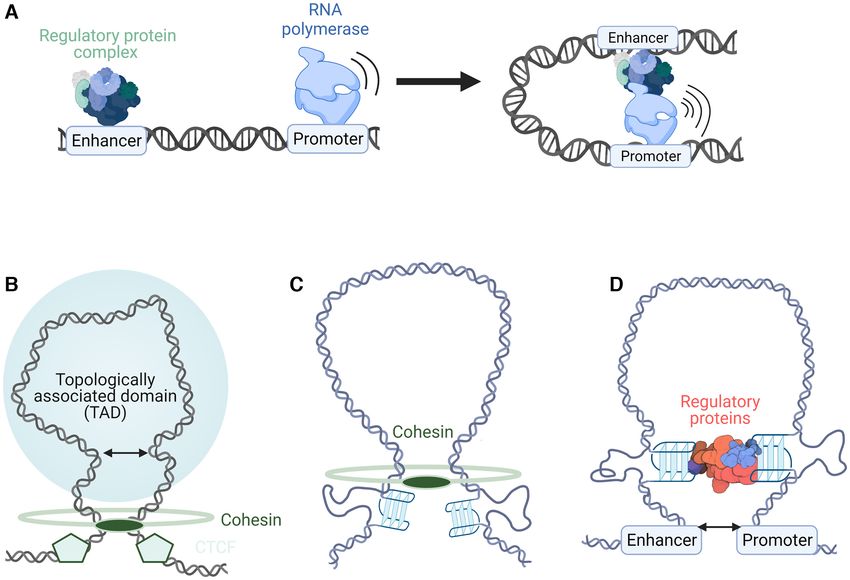
moter and enhancer sites of DNA (Figure 4A) (49). While also revealed that regions where loops form in the genome
promoters mark the beginning points of transcription––the are in fact not random, but instead occur in a sequence-
region at which RNA polymerase binds and begins its jour- specific fashion (55). The mechanism for this specificity is
ney transcribing the code of DNA––enhancers act as aux- widely agreed to be achieved through the cooperation of
iliary regulatory regions, recruiting additional proteins to two proteins: cohesin and CTCF (Figure 4B). Cohesin is
carefully control the extent to which genes are expressed a ring-like protein, initially discovered for its role clasping
(50). This enhancer-mediated control occurs through the together sister chromatids during replication; however, it
binding of large protein complexes containing transcription has a secondary function translocating down stretches of
factors and cofactors, which increase the ability of RNA DNA to form transient loops (56). In order to prevent co-
polymerase II to initiate and sustain transcription (50). So hesin sliding down DNA indefinitely, the cell has utilized
although promoter regions are sufficient to achieve a basal cohesin stop points in the form of binding to the tran-
level of gene expression (51), it is through the utilization of scription factor CTCF which is named after its interac-
enhancers that increases of transcription as high as 100-fold tion with CCCTC sequences in DNA. CTCF binds to and
can be achieved (52). Interestingly, this control of transcrip- blocks the releasing function of cohesin (57), which means
tion can occur over hundreds of thousands of base pairs; its presence defines the start and end points of loops and in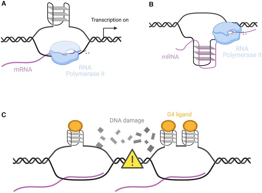
8424 Nucleic Acids Research, 2021, Vol. 49, No. 15

                                                                                                                                                       Downloaded from https://academic.oup.com/nar/article/49/15/8419/6320416 by guest on 08 November 2021
Figure 4. G4s in enhancer-promoter loop formation. (A) Schematic of how enhancers interact with their respective promoters to increase polymerase
activity and transcription. (B) Cohesin and CTCF cooperate to form stable loops (topologically associated domains) in DNA. (C) G4 enrichment at loop
boundaries possibly stalls cohesin. (D) G4s act as recruiters of regulatory proteins such as transcription factors that stabilize loops.

turn where the boundaries lie for topologically associated                  somewhat indirectly as G4s are correlated with more open,
domains.                                                                    accessible chromatin and also reduced DNA methylation
   However, CTCF binding motifs are not the only se-                        both of which promote protein-DNA interactions (59,60).
quences enriched at loop boundaries as G-quadruplex se-                     However, various in vitro studies have demonstrated the
quences are also abundant at the edges of loops (58). More-                 high affinity of multiple regulatory proteins for G4 struc-
over, it was found that the presence of G4s at loop bound-                  tures including, eukaryotic transcription factors SP1 (61),
aries increases the stability of DNA loops and in turn en-                  MAZ (62) and YY1 (63) and viral regulatory proteins such
hances long-distance DNA interactions (58). Although the                    as the transcription factor ICP4 (64). Additionally, proteins
mechanism for this correlation has yet to be fully explored,                that form part of large transcription-enhancing complexes
the authors of this study speculated that G4s may act anal-                 such as the transcriptional co-activator BRD3 (29,31) and
ogously to CTCF, stalling the progression of cohesin and                    the chromatin regulating protein PARP-1 (65) have been
thus defining the boundaries of distal interactions (Figure                 shown to interact strongly with G4s. Within cells, enhancers
4C). This is further supported by the fact that G4 motifs                   and promoters containing G4-forming sequences addition-
at loop boundaries are significantly enriched on the same                   ally have significantly higher levels of transcription factor
strand as the CTCF binding motif, suggesting some co-                       binding, which is accompanied by increased levels of RNA
operation between the action of CTCF and G4 formation                       pol II occupancy and transcriptional activity (17,20,58).
(58). This work demonstrates, perhaps counter-intuitively,                  This enrichment is seen even when controlling for differ-
that the protein-stalling capabilities of G4s that have been                ences in chromatin accessibility (58) and the G-richness (20)
demonstrated in vitro with RNA polymerase, may in vivo be                   of a given DNA region suggesting that it is the formation
used to enhance rather than inhibit transcription and poly-                 of the G-quadruplex structure itself that is responsible for
merase activity by enriching loop formation. Furthermore,                   enhanced transcription, as opposed to a simple correlation
these findings highlight even more how the simple strategy                  with open chromatin or GC content.
of considering individual G4 formation at specific gene pro-                   These findings support a model of G-quadruplex struc-
moters might significantly underestimate the potential roles                tures as scaffolds for protein binding, which may promote
of G4s in the regulation of gene expression.                                both local and remote interactions of gene regulation sites
   Beyond the initial extrusion of enhancer-promoter loops                  (Figure 4D). This hypothesis has been substantiated fur-
in DNA, G4s may further stabilize these loops by promot-                    ther with work studying the interactions of G-quadruplexes
ing the binding and recruitment of key regulatory proteins                  with the transcription factor ying-yang 1 (YY1), a protein
which inhibit loop collapse. It is possible that this is done               well-established for its importance in creating promoter-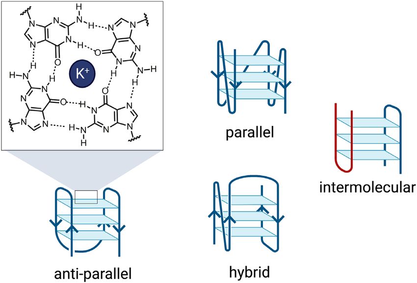
Nucleic Acids Research, 2021, Vol. 49, No. 15 8425
enhancer loops (63). In order to establish more directly the For instance, protein-free aggregation has been observed
role that G4s may have on transcription factor binding and in vitro for RNA repeat expansions, nucleobase homopoly-
DNA looping, this study utilized three distinct methods mers and some mRNAs by means of RNA–RNA interac-
of G4 perturbation and subsequently measured associated tions including base pairing, base stacking and other long-
changes to gene expression within cells. This included: (i) range promiscuous interactions (71). Similarly, G4s have
the over-expression of a G4-helicase which conventionally the ability to form networks by connecting multiple nu-
unwinds G4 secondary structures; (ii) the disruption of G4- cleic acid strands in an intermolecular configuration or by
forming sequences through subtle CRISPR-mediated gene -stacking between G-tetrads of different G4s, suggesting
editing; and iii) the use of G4-binding ligands to displace that they have the potential to cause phase separation ex-
native G4-protein interactions. ploiting a similar mechanism (Figure 5B). This was exempli-
In each case, perturbation of G4 formation reduced YY1 fied in work directly associating G4s with LLPS in live cells
binding and looping interactions at G4-sites by an order of within short root RNA (72) and the C9Orf72 expansion re-
magnitude. This was additionally accompanied by signifi- peat (73). In these studies, it was shown that G4-triggered
Downloaded from https://academic.oup.com/nar/article/49/15/8419/6320416 by guest on 08 November 2021
cantly altered expression of genes not only directly associ- phase separation is dependent on exposure of the system
ated with G4 regions through their promoters, but through to G4-favouring conditions, such as increasing potassium
distal enhancer sites that interact with said genes via loops concentration, local concentration of nucleic acids and rela-
as visualized with Hi-C experiments (63). This work is an tive guanine content. Furthermore, guanine-rich sequences
encouraging example of how it is possible to design exper- have been proposed to stimulate the formation of stress
iments to test, in a cellular context, the link between G4s granules in the cytoplasm that regulate gene translation, of-
and gene expression, the results of which highlight the cur- fering yet another piece of evidence to correlate G4 forma-
rently under-studied proposition that G4s may act as long- tion to LLPS (74).
distance regulators of transcription. The hypothesis that G4s stimulate LLPS by forming in-
termolecular networks has also been supported by work
considering the potential formation of intermolecular G4s
G4s may trigger liquid–liquid phase separation events
within regulatory regions of the genome. For instance, one
In recent years it has been discovered that the expression computational study showed that ‘half-G4’ sequences (con-
of genes can be mediated by phase separation events that taining two instead of four guanine runs) are enriched in
allow for the concentration of transcriptional machinery promoter and enhancer regions of DNA. It is speculated
within membrane-less organelles inside the nucleus (66,67). that when the two guanine runs are brought into close
Liquid-liquid phase separation (LLPS) relies on a combina- proximity, they are able to hydrogen bond together and
tion of weakly interacting forces between nucleic acids and thus assemble into a ‘full-G4’, that may mediate promoter-
the low-complexity domains of DNA/RNA-binding pro- enhancer interactions (75). Similarly, it has been found that
teins (68). This occurs, for instance, in the nucleolus, which regions with a high density of guanine runs are capable
is a dense region within the nucleus that contain clusters of forming quadruplexes over unconventionally long dis-
of ribosomal DNA (69). However, LLPS can also occur tances and are significantly enriched within super-enhancer
outside of the nucleolus in ‘super-enhancer’ regions of the regions (76).
genome characterized by closely localized transcriptional Thus, the formation of intermolecular or long-distance
enhancer sequences that bind transcription factors, chro- G4 structures in such regions may be a mechanism of DNA
matin remodelling proteins and RNA polymerase II and networking that stimulates LLPS and promotes enhancer
thus activate transcription (Figure 5A) (70). activity (Figure 5C). This is particularly relevant in light of
This was first demonstrated with the transcriptional co- our recent findings on the nucleolar protein CSB, which we
activator proteins BRD4 (bromodomain-containing pro- found to exhibit picomolar affinity for intermolecular G4s
tein 4) and MED1 (Mediator 1) which are able to link whilst displaying negligible binding to intramolecular G4s
via intermolecular interactions at their disordered domains (77). The characterization of CSB as the first selective inter-
to create liquid-condensates specifically at super-enhancers molecular G4 interactor combined with the known nucle-
(66). The liquid-like state of the BRD4 and MED1 ag- olar localization of this protein suggests that intermolecu-
gregates presents several parallels to membrane-less or- lar G4s may form within the nucleoli and contribute to its
ganelles such as nucleoli and P-granules that are also phase-separated state, however, this is yet to be explicitly
seen in the nucleus. Additionally, disruption of liquid con- demonstrated.
densates via treatment with hexanediol, significantly re- As such long-range and intermolecular interactions are
duced the recruitment of BRD4 and MED1 to super- favoured in crowded environments (78), it is possible that
enhancers (66). As such co-factors are essential for super- G4s are even more likely to form in the crowded bubbles of
enhancer activity, it is thought that the formation of these liquid condensates (79). Thus, this work brings forward a se-
liquid-condensates may be a unique mechanism to recruit ries of evidence suggesting that G4s might act as nucleation
proteins to enhancer regions and achieve transcriptional points for LLPS and in turn regulatory elements that may
enhancement. influence transcriptional control. As LLPS offers an inter-
Although many of the studies considering LLPS in a bi- esting mechanism by which the cell increases the local con-
ological context have historically focused on the protein centration of regulatory proteins and is subsequently able
component of the aggregate, recent work has revealed that to increase the transcription rate of a given gene, exploring
certain nucleic acid structures can also act as nucleation such mechanisms in relation to G4s may yield insightful re-
sites for phase-separation, even in the absence of proteins. sults. However, investigations into this area are still in their8426 Nucleic Acids Research, 2021, Vol. 49, No. 15
Downloaded from https://academic.oup.com/nar/article/49/15/8419/6320416 by guest on 08 November 2021
Figure 5. G4s and phase separation. (A) Super-enhancers interact with promoter regions via transcription factors, co-factors and chromatin remodelers
triggering LLPS that enhances transcription. (B) G4s at super-enhancer sites may promote aggregation via intermolecular interactions such as -stacking
between quadruplexes. (C) ‘half-G4’ sequences can assemble intermolecularly to mediate enhancer-promoter interactions and phase separation.
infancy and demonstrate the need for additional work on This has been explored in recent studies which demon-
the diverse roles that physical and chemical changes associ- strated that OG formation in gene promoters containing
ated with G4 formation might play in the broader context G4-forming sequences can stimulate DNA repair mecha-
of gene regulation. nisms and promote gene activation (87–89). In one study, a
luciferase reporter assay was employed to investigate the ef-
fects on gene expression when inserting OG bases into the
G4s stimulate expression by promoting DNA oxidation and
VEGF and NTHL1 promoters (87). Surprisingly, a 300%
repair
increase in luciferase expression was measured with the OG
An additional area in which epigenetic control appears at plasmid compared to the plasmid without the oxidized gua-
the G4 level can surprisingly be seen when considering how nine and this activation was related to the formation of G4s
cells respond to environmental stresses. An example of this in these promoters. Under normal circumstances, the pres-
stress is demonstrated by guanine oxidation induced by re- ence of an OG would trigger a DNA damage repair pathway
active oxygen species (ROS) that arise from multiple exoge- known as base excision repair (BER) in which the oxidised
nous and endogenous factors such as exposure to radiation base is removed leaving an apurinic (AP) site in the DNA
and metabolism (80,81). Among the four DNA bases, gua- sequence (90). Subsequently, another BER-related protein
nine has the lowest redox potential, therefore, guanines are known as APE1 would cleave the apurinic region so it can
one of the most frequent sites to be oxidized by ROS gen- be repaired by downstream polymerase action (Figure 6B).
erating 8-oxo-7,8-dihydroguanine (OG) DNA (Figure 6A) However, in this study it was shown that when this oxidation
(82,83). Consequently G4s, being composed of several runs occurs in a G4-forming region that contains a fifth extra run
of guanines, are easy targets for guanine oxidation (84). As (‘spare tyre’) of guanines, an alternative G4 structure can
oxidizing species can potentially affect many cellular pro- form which causes the AP site to be extruded into a loop.
cesses, resulting in ageing or cancer (85,86), exposure to This looped G4 structure is then recognized and bound
ROS is a constant challenge for cells which have developed by APE1, which reduces the cleaving ability of APE1 and
efficient repair mechanisms to cope with such mutagenic instead unmasks a separate function of the protein as a
stress (81). However, the stimulation of these DNA damage recruiter of transcription factors (Figure 6C) (87,91). In-
repair pathways is not always a negative event and may in deed, previous data reported APE1 as a multifunctional en-
fact have important implications for transcriptional control zyme which can form part of a large transcriptional com-
particularly in relation to G4s. plex which includes HIF-1, STAT3 and CBP/p300 leadingNucleic Acids Research, 2021, Vol. 49, No. 15 8427
Downloaded from https://academic.oup.com/nar/article/49/15/8419/6320416 by guest on 08 November 2021
Figure 6. G4s and DNA damage. (A) Guanine is frequently oxidized by oxidative stress generating 8-oxo-7,8-dihydroguanine (OG). (B) OG formation
upon ROS damage in a gene promoter without a G4 causes the formation of an apurinic (AP) site that is recognized and cleaved by APE1. This cleaved
site is subsequently repaired without increasing the gene transcription level. (C) OG formation in a gene promoter containing a core G4 sequence (in pink)
and a fifth G-track (spare tyre in blue) causes the formation of an alternative G4-structure and the extrusion of the AP site into a loop. The structure
is recognized and bound by APE1 which reduces the cleaving ability of APE1 and stimulates the recruitment of transcription factors with consequent
transcriptional activation.
to gene activation (92,93). They hypothesize that the accu- more, a separate analysis considered endogenously formed
mulation of transcription factors through the ref-1 domain cellular G4s using G4 ChIP-Seq and showed a significant
of APE1 explains the transcriptional enhancement that is enrichment of APE1 at G4 sites within human cells (96).
seen only when this alternative G4 forms in vicinity to OG In this study they additionally verified that the binding of
sites and hinders the usual nuclease function of APE1 (87). APE1 to G4s not only stabilizes the G4 structure but in-
In support of this, they did not observe increased luciferase creases the residence time of the protein at sites of DNA
expression when the OG damage was inserted in VEGF pro- damage (96), further consolidating a mechanism linking G4
moter with only four G-tracks, suggesting that the new G4 oxidation with transcriptional enhancement within cells.
conformation cannot be formed once the AP is extruded In addition to this, APE1 is not the only protein that
into a loop. Similar results were also observed in subsequent appears to be recruited to OG DNA by G4s: a recent re-
studies on other gene promoters such as PCNA, as well as in port showed that oxidized quadruplexes can be bound by
studies considering the location and strand dependency of and enhance Poly ADP-ribose polymerase-1 (PARP-1) ac-
this phenomenon, providing additional examples of the role tivity (97). Similarly to APE1, PARP-1 is an important ef-
of G4s as regulators of gene expression upon G oxidation fector of the BER pathway which recognizes various DNA
(88,89,94). lesions (98) and seems to respond to the formation of the
Whilst mechanistically informative, a limitation of these looped G4 structure that arises during DNA damage. Addi-
studies is the reliance on plasmid systems with synthetically tionally, ChIP-qPCR studies within pancreatic cancer cells
inserted G4-rich promoters which may not be representa- suggested that OG formation promotes the recruitment of
tive of the role of G4s in a cellular and chromatin context. transcription factor proteins MAZ and hnRNP A1 to the
Despite this, similar results were observed when considering G4-rich KRAS promoter and may promote the transcrip-
plasmids transfected into human glioblastoma cells where tion process (99).
guanines were oxidized using the cytokine TNFa which Altogether this work demonstrates that an apparently
naturally stimulates oxidative stress within cells, suggest- mutagenic event such as the oxidation of guanine bases by
ing that these findings are consistent even when tested un- ROS, in a G4 context, could actually be considered as a way
der more physiologically relevant conditions (95). Further- to epigenetically control gene expression. Such DNA dam-8428 Nucleic Acids Research, 2021, Vol. 49, No. 15
age coincides with relaxed helical tension and is particularly multi-faceted regulatory elements in a complex cellular en-
likely to occur in guanine-rich regions (87), thus promoting vironment.
the formation of G4s which in turn appear to tune the ac-
tivity and recruitment of transcriptional regulators. In this ACKNOWLEDGEMENTS
way, DNA damage in the form of guanine oxidation at G4-
sites might represent a novel mechanism by which G4s natu- All figures were created on Biorender.com
rally contribute towards transcriptional control within cells. The authors are grateful to Professor Ramon Vilar for help-
ful discussions on the manuscript.
CONCLUSIONS
FUNDING
In this review, we have explored a number of pathways by
which quadruplex DNA may regulate gene transcription M.D.A. is grateful to BBSRC that fully supports his
with an emphasis on how G4 formation may actually as- research group with a BBSRC David Phillips Fellow-
Downloaded from https://academic.oup.com/nar/article/49/15/8419/6320416 by guest on 08 November 2021
sociate with transcriptional activation. This is in direct con- ship [BB/R011605/1]; J.R. is supported by an EPSRC
trast to the initial and perhaps outdated perspective where and NIHR Imperial Biomedical Research Centre schol-
G4s are labelled as direct inhibitors of polymerase activity arship [EP/S023518/1]; F.R. is supported by a Lever-
and transcription. Despite this, the goal of this review is not hulme Trust, Leverhulme Cellular Bionics scholarship
to argue that G4s act solely as transcriptional activators, as [EP/S023518/1]; S.N. is supported by an EPSRC schol-
there may be individual cases where G4s are repressive in arship [EP/S023518/1]; D.L. is supported by an Imperial
nature (100), but to critically highlight the substantial ev- College DTP scholarship funded by the Chemistry Depart-
idence showing that G4s are globally correlated with tran- ment. Funding for open access charge: Imperial College
scriptional enhancement rather than repression. This forces London.
us to assess the way in which we have been investigating G4s Conflict of interest statement. None declared.
in terms of gene regulation.
Early studies considered how individual G4s behave REFERENCES
within plasmid systems or relied on ligands that artificially
stabilize dynamic G4 structures and may additionally dis- 1. Raguseo,F., Chowdhury,S., Minard,A. and Di Antonio,M. (2020)
Chemical-biology approaches to probe DNA and RNA
place natural G4-binding partners. This work contributed G-quadruplex structures in the genome. Chem. Commun., 56,
to a narrative that depicts G4s as transcriptional roadblocks 1317–1324.
and might not reflect at all the endogenous role of these 2. Spiegel,J., Adhikari,S. and Balasubramanian,S. (2020) The structure
structures. Such a narrative is still often re-iterated in the and function of DNA G-Quadruplexes. Trends Chem., 2, 123–136.
3. Kwok,C.K. and Merrick,C.J. (2017) G-Quadruplexes: prediction,
current literature and reflects a simplistic view of G4 for- characterization, and biological application. Trends Biotechnol., 35,
mation and transcriptional regulation that we strongly feel 997–1013.
needs to be updated in light of recent studies. Going for- 4. Mukherjee,A.K., Sharma,S. and Chowdhury,S. (2019) Non-duplex
ward, it is important to consider the G4 not as an isolated G-Quadruplex structures emerge as mediators of epigenetic
entity within a specific genomic location, but as a structure modifications. Trends Genet., 35, 129–144.
5. Gellert,M., Lipsett,M.N. and Davies,D.R. (1962) Helix formation
that exists as part of an interconnected network of other by guanylic acid. Proc. Natl. Acad. Sci. U.S.A., 48, 2013–2018.
biomolecules in living cells. 6. Parkinson,G.N., Lee,M.P.H. and Neidle,S. (2002) Crystal structure
Recent research conducted in this spirit has revealed mul- of parallel quadruplexes from human telomeric DNA. Nature, 417,
tiple exciting mechanisms that may explain how G4s act 876–880.
7. Siddiqui-Jain,A., Grand,C.L., Bearss,D.J. and Hurley,L.H. (2002)
as transcriptional regulators. This includes the role of G4s Direct evidence for a G-quadruplex in a promoter region and its
in mediating the placement of histone marks and in inter- targeting with a small molecule to repress c-MYC transcription.
acting with chromatin remodelling proteins, thus shaping Proc. Natl. Acad. Sci. U.S.A., 99, 11593–11598.
the higher order structure of chromatin. Additionally, the 8. Balasubramanian,S., Hurley,L.H. and Neidle,S. (2011) Targeting
notion that G4s can influence the formation and stability G-quadruplexes in gene promoters: A novel anticancer strategy?
Nat. Rev. Drug Discov., 10, 261–275.
of transcriptional loops including R-loops and long-range 9. Huppert,J.L. and Balasubramanian,S. (2007) G-quadruplexes in
enhancer-promoter loops has been demonstrated. Even rel- promoters throughout the human genome. Nucleic Acids Res., 35,
atively new mechanisms considering intermolecular G4s as 406–413.
triggers of liquid-liquid phase separation in combination 10. Halder,K., Halder,R. and Chowdhury,S. (2009) Genome-wide
analysis predicts DNA structural motifs as nucleosome exclusion
with selective interactors (such as CSB) and DNA-damage signals. Mol. Biosyst., 5, 1703–1712.
induced transcriptional activation are being explored. Alto- 11. Eddy,J. and Maizels,N. (2008) Conserved elements with potential to
gether these novel mechanisms strongly challenge the sim- form polymorphic G-quadruplex structures in the first intron of
ple ‘on/off’ switch role that it is still often associated to G4 human genes. Nucleic Acids Res., 36, 1321–1333.
formation. 12. Kim,N. (2017) The interplay between G-quadruplex and
transcription. Curr. Med. Chem., 26, 2898–2917.
By considering G4s as more than simple knot-like imped- 13. Rodriguez,R., Miller,K.M., Forment,J.V., Bradshaw,C.R.,
iments for RNA polymerase to overcome, it will be possi- Nikan,M., Britton,S., Oelschlaegel,T., Xhemalce,B.,
ble to develop these new avenues of research which may be Balasubramanian,S. and Jackson,S.P. (2012)
particularly important for investigations of transcriptional Small-molecule-induced DNA damage identifies alternative DNA
structures in human genes. Nat. Chem. Biol., 8, 301–310.
dysfunction in pathologies such as cancer and ageing. It fol- 14. Boddupally,P.V.L., Hahn,S., Beman,C., De,B., Brooks,T.A.,
lows that further research in this area may unravel a distinct Gokhale,V. and Hurley,L.H. (2012) Anticancer activity and cellular
layer of epigenetic regulation in which G4s are implicated as repression of c-MYC by the G-quadruplex-stabilizingNucleic Acids Research, 2021, Vol. 49, No. 15 8429
11-piperazinylquindoline is not dependent on direct targeting of the 35. Huppert,J.L. (2008) Thermodynamic prediction of RNA-DNA
G-quadruplex in the c-MYC promoter. J. Med. Chem., 55, duplex-forming regions in the human genome. Mol. Biosyst., 4,
6076–6086. 686–691.
15. Hänsel-Hertsch,R., Beraldi,D., Lensing,S.V., Marsico,G., Zyner,K., 36. Miglietta,G., Russo,M. and Capranico,G. (2020)
Parry,A., Di Antonio,M., Pike,J., Kimura,H., Narita,M. et al. G-quadruplex-R-loop interactions and the mechanism of anticancer
(2016) G-quadruplex structures mark human regulatory chromatin. G-quadruplex binders. Nucleic Acids Res., 48, 11942–11957.
Nat. Genet., 48, 1267–1272. 37. Duquette,M.L., Handa,P., Vincent,J.A., Taylor,A.F. and Maizels,N.
16. Hänsel-Hertsch,R., Simeone,A., Shea,A., Hui,W.W.I., Zyner,K.G., (2004) Intracellular transcription of G-rich DNAs induces
Marsico,G., Rueda,O.M., Bruna,A., Martin,A., Zhang,X. et al. formation of G-loops, novel structures containing G4 DNA. Genes
(2020) Landscape of G-quadruplex DNA structural regions in Dev., 18, 1618–1629.
breast cancer. Nat. Genet., 52, 878–883. 38. Zhao,Y., Zhang,J.Y., Zhang,Z.Y., Tong,T.J., Hao,Y.H. and Tan,Z.
17. Lago,S., Nadai,M., Cernilogar,F.M., Kazerani,M., Domı́niguez (2017) Real-Time detection reveals responsive cotranscriptional
Moreno,H., Schotta,G. and Richter,S.N. (2021) Promoter formation of persistent intramolecular DNA and intermolecular
G-quadruplexes and transcription factors cooperate to shape the DNA:RNA Hybrid G-quadruplexes stabilized by R-Loop. Anal.
cell type-specific transcriptome. Nat. Commun., 12, 3885. Chem., 89, 6036–6042.
Downloaded from https://academic.oup.com/nar/article/49/15/8419/6320416 by guest on 08 November 2021
18. Zheng,K.W., Zhang,J.Y., He,Y.De, Gong,J.Y., Wen,C.J., Chen,J.N., 39. Zheng,K.W., He,Y.De, Liu,H.H., Li,X.M., Hao,Y.H. and Tan,Z.
Hao,Y.H., Zhao,Y. and Tan,Z. (2020) Detection of genomic (2017) Superhelicity constrains a localized and R-loop-dependent
G-quadruplexes in living cells using a small artificial protein. Nucleic formation of G-Quadruplexes at the upstream region of
Acids Res., 48, 11706–11720. transcription. ACS Chem. Biol., 12, 2609–2618.
19. Di Antonio,M., Ponjavic,A., Radzevičius,A., Ranasinghe,R.T., 40. Wanrooij,P.H., Uhler,J.P., Shi,Y., Westerlund,F., Falkenberg,M. and
Catalano,M., Zhang,X., Shen,J., Needham,L.M., Lee,S.F., Gustafsson,C.M. (2012) A hybrid G-quadruplex structure formed
Klenerman,D. et al. (2020) Single-molecule visualization of DNA between RNA and DNA explains the extraordinary stability of the
G-quadruplex formation in live cells. Nat. Chem., 12, 832–837. mitochondrial R-loop. Nucleic Acids Res., 40, 10334–10344.
20. Spiegel,J., Cuesta,S.M., Adhikari,S., Hänsel-Hertsch,R., 41. Zheng,K.W., Xiao,S., Liu,J.Q., Zhang,J.Y., Hao,Y.H. and Tan,Z.
Tannahill,D. and Balasubramanian,S. (2021) G-quadruplexes are (2013) Co-transcriptional formation of DNA: RNA hybrid
transcription factor binding hubs in human chromatin. Genome G-quadruplex and potential function as constitutional cis element
Biol., 22, 117. for transcription control. Nucleic Acids Res., 41, 5533–5541.
21. Fleming,A.M. and Burrows,C.J. (2020) Interplay of guanine 42. Tan,J., Wang,X., Phoon,L., Yang,H. and Lan,L. (2020) Resolution
oxidation and G-Quadruplex folding in gene promoters. J. Am. of ROS-induced G-quadruplexes and R-loops at transcriptionally
Chem. Soc., 142, 1115–1136. active sites is dependent on BLM helicase. FEBS Lett., 594,
22. Kornberg,R.D. (1974) Chromatin structure: a repeating unit of 1359–1367.
histones and DNA. Science, 184, 868–871. 43. Lee,C.Y., McNerney,C., Ma,K., Zhao,W., Wang,A. and Myong,S.
23. Noll,M. (1974) Subunit structure of chromatin. Nature, 251, (2020) R-loop induced G-quadruplex in non-template promotes
249–251. transcription by successive R-loop formation. Nat. Commun., 11,
24. Jiang,C. and Pugh,B.F. (2009) Nucleosome positioning and gene 3392.
regulation: advances through genomics. Nat. Rev. Genet., 10, 44. De Magis,A., Manzo,S.G., Russo,M., Marinello,J., Morigi,R.,
161–172. Sordet,O. and Capranico,G. (2019) DNA damage and genome
25. Bannister,A.J. and Kouzarides,T. (2011) Regulation of chromatin by instability by G-quadruplex ligands are mediated by R loops in
histone modifications. Cell Res., 21, 381–395. human cancer cells. Proc. Natl. Acad. Sci. U.S.A., 116, 816–825.
26. Sarkies,P., Reams,C., Simpson,L.J. and Sale,J.E. (2010) Epigenetic 45. Xu,H., Di Antonio,M., McKinney,S., Mathew,V., Ho,B.,
instability due to defective replication of structured DNA. Mol. Cell, O’Neil,N.J., Santos,N.D., Silvester,J., Wei,V., Garcia,J. et al. (2017)
40, 703–713. CX-5461 is a DNA G-quadruplex stabilizer with selective lethality
27. Schiavone,D., Guilbaud,G., Murat,P., Papadopoulou,C., Sarkies,P., in BRCA1/2 deficient tumours. Nat. Commun., 8, 14432.
Prioleau,M., Balasubramanian,S. and Sale,J.E. (2014) Determinants 46. Field,A. and Adelman,K. (2020) Evaluating enhancer function and
of G quadruplex-induced epigenetic instability in REV1-deficient transcription. Annu. Rev. Biochem., 89, 213–234.
cells. EMBO J., 33, 2507–2520. 47. Herz,H.M. (2016) Enhancer deregulation in cancer and other
28. Komůrková,D., Svobodová Kovařı́ková,A. and Bártová,E. (2021) diseases. Bioessays, 38, 1003–1015.
G-Quadruplex structures colocalize with transcription factories and 48. Bradner,J.E., Hnisz,D. and Young,R.A. (2017) Transcriptional
nuclear speckles surrounded by acetylated and dimethylated addiction in cancer. Cell, 168, 629–643.
histones H3. Int. J. Mol. Sci., 22, 1995. 49. Schoenfelder,S. and Fraser,P. (2019) Long-range
29. Vlasenok,M., Levchenko,O., Basmanov,D., Klinov,D., Varizhuk,A. enhancer–promoter contacts in gene expression control. Nat. Rev.
and Pozmogova,G. (2018) Data set on G4 DNA interactions with Genet., 20, 437–455.
human proteins. Data Br., 18, 348–359. 50. Stadhouders,R., van den Heuvel,A., Kolovos,P., Jorna,R., Leslie,K.,
30. Lamonica,J.M., Deng,W., Kadauke,S., Campbell,A.E., Grosveld,F. and Soler,E. (2012) Transcription regulation by distal
Gamsjaeger,R., Wang,H., Cheng,Y., Billin,A.N., Hardison,R.C., enhancers: Who’s in the loop? Transcription, 3, 181.
Mackay,J.P. et al. (2011) Bromodomain protein Brd3 associates with 51. Haberle,V. and Stark,A. (2018) Eukaryotic core promoters and the
acetylated GATA1 to promote its chromatin occupancy at erythroid functional basis of transcription initiation. Nat. Rev. Mol. Cell Biol.,
target genes. Proc. Natl. Acad. Sci. U.S.A., 108, E159–E168. 19, 621–637.
31. Pavlova,I.I., Tsvetkov,V.B., Isaakova,E.A., Severov,V.V., 52. Beagan,J.A., Pastuzyn,E.D., Fernandez,L.R., Guo,M.H., Feng,K.,
Khomyakova,E.A., Lacis,I.A., Lazarev,V.N., Lagarkova,M.A., Titus,K.R., Chandrashekar,H., Shepherd,J.D. and
Pozmogova,G.E. and Varizhuk,A.M. (2020) Phillips-Cremins,J.E. (2020) Three-dimensional genome
Transcription-facilitating histone chaperons interact with genomic restructuring across timescales of activity-induced neuronal gene
and synthetic G4 structures. Int. J. Biol. Macromol., 160, 1144–1157. expression. Nat. Neurosci., 23, 707–717.
32. Du,Z., Zhao,Y. and Li,N. (2009) Genome-wide colonization of gene 53. Higgs,D.R. (2020) Enhancer–promoter interactions and
regulatory elements by G4 DNA motifs. Nucleic Acids Res., 37, transcription. Nat. Genet., 52, 470–471.
6784–6798. 54. Dixon,J.R., Selvaraj,S., Yue,F., Kim,A., Li,Y., Shen,Y., Hu,M.,
33. Selvam,S., Koirala,D., Yu,Z. and Mao,H. (2014) Quantification of Liu,J.S. and Ren,B. (2012) Topological domains in mammalian
topological coupling between DNA superhelicity and G-quadruplex genomes identified by analysis of chromatin interactions. Nature,
formation. J. Am. Chem. Soc., 136, 13967–13970. 485, 376–380.
34. Belotserkovskii,B.P., Tornaletti,S., D’Souza,A.D. and Hanawalt,P.C. 55. Ren,G., Jin,W., Cui,K., Rodrigez,J., Hu,G., Zhang,Z., Larson,D.R.
(2018) R-loop generation during transcription: formation, and Zhao,K. (2017) CTCF-mediated enhancer-promoter interaction
processing and cellular outcomes. DNA Repair (Amst.)., 71, 69–81. is a critical regulator of cell-to-cell variation of gene expression.
Mol. Cell, 67, 1049–1058.8430 Nucleic Acids Research, 2021, Vol. 49, No. 15
56. Chien,R., Zeng,W., Kawauchi,S., Bender,M.A., Santos,R., 76. Williams,J.D., Houserova,D., Johnson,B.R., Dyniewski,B.,
Gregson,H.C., Schmiesing,J.A., Newkirk,D.A., Kong,X., Ball,A.R. Berroyer,A., French,H., Barchie,A.A., Bilbrey,D.D., Demeis,J.D.,
et al. (2011) Cohesin mediates chromatin interactions that regulate Ghee,K.R. et al. (2020) Characterization of long G4-rich
mammalian -globin expression. J. Biol. Chem., 286, 17870–17878. enhancer-associated genomic regions engaging in a novel loop:loop
57. Li,Y., Haarhuis,J.H.I., Sedeño Cacciatore,Á., Oldenkamp,R., van ‘G4 Kissing’ interaction. Nucleic Acids Res., 48, 5907–5925.
Ruiten,M.S., Willems,L., Teunissen,H., Muir,K.W., de Wit,E., 77. Liano,D. and Di Antonio,M. (2021) Cockayne Syndrome B protein
Rowland,B.D. et al. (2020) The structural basis for selectively interacts and resolves intermolecular 1 DNA
cohesin–CTCF-anchored loops. Nature, 578, 472–476. G-quadruplex structures. bioRxiv doi:
58. Hou,Y., Li,F., Zhang,R., Li,S., Liu,H., Qin,Z.S. and Sun,X. (2019) https://doi.org/10.1101/2021.03.25.436565, 25 March 2021, preprint:
Integrative characterization of G-quadruplexes in the not peer reviewed.
three-dimensional chromatin structure. Epigenetics, 14, 894–911. 78. André,A.A.M. and Spruijt,E. (2020) Liquid–liquid phase separation
59. Bell,A.C. and Felsenfeld,G. (2000) Methylation of a in crowded environments. Int. J. Mol. Sci., 21, 5908.
CTCF-dependent boundary controls imprinted expression of the 79. Zheng,K., Chen,Z., Hao,Y. and Tan,Z. (2009) Molecular crowding
Igf2 gene. Nature, 405, 482–485. creates an essential environment for the formation of stable
60. Medvedeva,Y.A., Khamis,A.M., Kulakovskiy,I.V., Ba-Alawi,W., G-quadruplexes in long double-stranded DNA. Nucleic Acids Res.,
Downloaded from https://academic.oup.com/nar/article/49/15/8419/6320416 by guest on 08 November 2021
Bhuyan,M.S.I., Kawaji,H., Lassmann,T., Harbers,M., 38, 327–338.
Forrest,A.R.R. and Bajic,V.B. (2014) Effects of cytosine methylation 80. Ward,J.F. (2000) Complexity of damage produced by ionizing
on transcription factor binding sites. BMC Genomics, 15, 119. radiation. In: Cold Spring Harbor Symposia on Quantitative Biology.
61. Eun-Ang,R., Ramon,K., Enid,L., Mehran,N. and Cold Spring Harbor Laboratory Press, Vol. 65, pp. 377–382.
Balasubramanian,S. (2012) A non-canonical DNA structure is a 81. Markkanen,E. (2017) Not breathing is not an option: how to deal
binding motif for the transcription factor SP1 in vitro. Nucleic Acids with oxidative DNA damage. DNA Repair (Amst.)., 59, 82–105.
Res., 40, 1499–1508. 82. Steenken,S. and Jovanovic,S.V. (1997) How easily oxidizable is
62. Cogoi,S., Paramasivam,M., Membrino,A., Yokoyama,K.K. and DNA? One-electron reduction potentials of adenosine and
Xodo,L.E. (2010) The KRAS promoter responds to Myc-associated guanosine radicals in aqueous solution. J. Am. Chem. Soc., 119,
zinc finger and poly(ADP-ribose) polymerase 1 proteins, which 617–618.
recognize a critical quadruplex-forming GA-element. J. Biol. Chem., 83. Cadet,J., Wagner,J.R., Shafirovich,V. and Geacintov,N.E. (2014)
285, 22003–22016. One-electron oxidation reactions of purine and pyrimidine bases in
63. Li,L., Williams,P., Ren,W., Wang,M.Y., Gao,Z., Miao,W., cellular DNA. Int. J. Radiat. Biol., 90, 423–432.
Huang,M., Song,J. and Wang,Y. (2020) YY1 interacts with guanine 84. Clark,D.W., Phang,T., Edwards,M.G., Geraci,M.W. and
quadruplexes to regulate DNA looping and gene expression. Nat. Gillespie,M.N. (2012) Promoter G-quadruplex sequences are targets
Chem. Biol., 17, 161–168. for base oxidation and strand cleavage during hypoxia-induced
64. Frasson,I., Soldà,P., Nadai,M., Lago,S. and Richter,S.N. (2021) transcription. FreeRadic. Biol. Med., 53, 51–59.
Parallel G-quadruplexes recruit the HSV-1 transcription factor ICP4 85. Finkel,T., Serrano,M. and Blasco,M.A. (2007) The common biology
to promote viral transcription in herpes virus-infected human cells. of cancer and ageing. Nature, 448, 767–774.
Commun. Biol., 4, 510. 86. Tubbs,A. and Nussenzweig,A. (2017) Endogenous DNA damage as
65. Cogoi,S., Paramasivam,M., Spolaore,B. and Xodo,L.E. (2008) a source of genomic instability in cancer. Cell, 168, 644–656.
Structural polymorphism within a regulatory element of the human 87. Fleming,A.M., Ding,Y. and Burrows,C.J. (2017) Oxidative DNA
KRAS promoter: Formation of G4-DNA recognized by nuclear damage is epigenetic by regulating gene transcription via base
proteins. Nucleic Acids Res., 36, 3765–3780. excision repair. Proc. Natl. Acad. Sci. U.S.A., 114, 2604–2609.
66. Sabari,B.R., Dall’Agnese,A., Boija,A., Klein,I.A., Coffey,E.L., 88. Fleming,A.M., Zhu,J., Ding,Y. and Burrows,C.J. (2019) Location
Shrinivas,K., Abraham,B.J., Hannett,N.M., Zamudio,A.V., dependence of the transcriptional response of a potential
Manteiga,J.C. et al. (2018) Coactivator condensation at G-quadruplex in gene promoters under oxidative stress. Nucleic
super-enhancers links phase separation and gene control. Science, Acids Res., 47, 5049–5060.
361, eaar3958. 89. Fleming,A.M., Zhu,J., Ding,Y. and Burrows,C.J. (2017) 8-Oxo-7,
67. Boija,A., Klein,I.A., Sabari,B.R., Dall’Agnese,A., Coffey,E.L., 8-dihydroguanine in the context of a gene promoter G-quadruplex is
Zamudio,A.V., Li,C.H., Shrinivas,K., Manteiga,J.C., Hannett,N.M. an on-off switch for transcription. ACS Chem. Biol., 12, 2417–2426.
et al. (2018) Transcription factors activate genes through the 90. David,S.S., O’Shea,V.L. and Kundu,S. (2007) Base-excision repair of
phase-separation capacity of their activation domains. Cell, 175, oxidative DNA damage. Nature, 447, 941–950.
1842–1855. 91. Broxson,C., Hayner,J.N., Beckett,J., Bloom,L.B. and Tornaletti,S.
68. Wolozin,B. and Ivanov,P. (2019) Stress granules and (2014) Human AP endonuclease inefficiently removes abasic sites
neurodegeneration. Nat. Rev. Neurosci., 20, 649–666. within G4 structures compared to duplex DNA. Nucleic Acids Res.,
69. McStay,B. (2016) Nucleolar organizer regions: genomic ‘dark 42, 7708–7719.
matter’ requiring illumination. Genes Dev., 30, 1598–1610. 92. Bhakat,K.K., Mantha,A.K. and Mitra,S. (2009) Transcriptional
70. Wang,X., Cairns,M.J. and Yan,J. (2019) Super-enhancers in regulatory functions of mammalian AP-endonuclease
transcriptional regulation and genome organization. Nucleic Acids (APE1/Ref-1), an essential multifunctional protein. Antioxidants
Res., 47, 11481–11496. Redox Signal., 11, 621–637.
71. Van Treeck,B. and Parker,R. (2018) Emerging roles for 93. Gray,M.J., Zhang,J., Ellis,L.M., Semenza,G.L., Evans,D.B.,
intermolecular RNA-RNA interactions in RNP assemblies. Cell, Watowich,S.S. and Gallick,G.E. (2005) HIF-1␣, STAT3, CBP/p300
174, 791–802. and Ref-1/APE are components of a transcriptional complex that
72. Zhang,Y., Yang,M., Duncan,S., Yang,X., Abdelhamid,M.A.S., regulates Src-dependent hypoxia-induced expression of VEGF in
Huang,L., Zhang,H., Benfey,P.N., Waller,Z.A.E. and Ding,Y. pancreatic and prostate carcinomas. Oncogene, 24, 3110–3120.
(2019) G-quadruplex structures trigger RNA phase separation. 94. Redstone,S.C.J., Fleming,A.M. and Burrows,C.J. (2019) Oxidative
Nucleic Acids Res., 47, 11746–11754. modification of the potential G-Quadruplex sequence in the PCNA
73. Fay,M.M., Anderson,P.J. and Ivanov,P. (2017) gene promoter can turn on transcription. Chem. Res. Toxicol., 32,
ALS/FTD-associated C9ORF72 repeat RNA promotes phase 437–446.
transitions in vitro and in cells. Cell Rep., 21, 3573–3584. 95. Fleming,A.M., Zhu,J., Howpay Manage,S.A. and Burrows,C.J.
74. Byrd,A.K., Zybailov,B.L., Maddukuri,L., Gao,J., Marecki,J.C., (2019) Human NEIL3 gene expression regulated by epigenetic-like
Jaiswal,M., Bell,M.R., Griffin,W.C., Reed,M.R., Chib,S. et al. oxidative DNA modification. J. Am. Chem. Soc., 141, 11036–11049.
(2016) Evidence that G-quadruplex DNA accumulates in the 96. Roychoudhury,S., Pramanik,S., Harris,H.L., Tarpley,M., Sarkar,A.,
cytoplasm and participates in stress granule assembly in response to Spagnol,G., Sorgen,P.L., Chowdhury,D., Band,V., Klinkebiel,D.
oxidative stress. J. Biol. Chem., 291, 18041–18057. et al. (2020) Endogenous oxidized DNA bases and APE1 regulate
75. Hegyi,H. (2015) Enhancer-promoter interaction facilitated by the formation of G-quadruplex structures in the genome. Proc. Natl.
transiently forming G-quadruplexes. Sci. Rep., 5, 9165. Acad. Sci. U.S.A., 117, 11409–11420.You can also read