DNA damage shifts circadian clock time via Hausp-dependent Cry1 stabilization
←
→
Page content transcription
If your browser does not render page correctly, please read the page content below
RESEARCH ARTICLE
           elifesciences.org

                                   DNA damage shifts circadian clock time
                                   via Hausp-dependent Cry1 stabilization
                                   Stephanie J Papp†, Anne-Laure Huber†, Sabine D Jordan, Anna Kriebs,
                                   Madelena Nguyen, James J Moresco, John R Yates III, Katja A Lamia*

                                   Department of Chemical Physiology, Scripps Research Institute, La Jolla,
                                   United States

                                   Abstract The circadian transcriptional repressors cryptochrome 1 (Cry1) and 2 (Cry2) evolved
                                   from photolyases, bacterial light-activated DNA repair enzymes. In this study, we report that while
                                   they have lost DNA repair activity, Cry1/2 adapted to protect genomic integrity by responding to
                                   DNA damage through posttranslational modification and coordinating the downstream
                                   transcriptional response. We demonstrate that genotoxic stress stimulates Cry1 phosphorylation
                                   and its deubiquitination by Herpes virus associated ubiquitin-specific protease (Hausp, a.k.a Usp7),
                                   stabilizing Cry1 and shifting circadian clock time. DNA damage also increases Cry2 interaction with
                                   Fbxl3, destabilizing Cry2. Thus, genotoxic stress increases the Cry1/Cry2 ratio, suggesting distinct
                                   functions for Cry1 and Cry2 following DNA damage. Indeed, the transcriptional response to
                                   genotoxic stress is enhanced in Cry1−/− and blunted in Cry2−/− cells. Furthermore, Cry2−/− cells
                                   accumulate damaged DNA. These results suggest that Cry1 and Cry2, which evolved from DNA
                                   repair enzymes, protect genomic integrity via coordinated transcriptional regulation.
                                   DOI: 10.7554/eLife.04883.001

*For correspondence: klamia@
scripps.edu
†
                                   Introduction
 These authors contributed         Mammalian circadian clocks involve transcriptional feedback loops (Green et al., 2008): Brain and
equally to this work
                                   muscle ARNT-like protein 1 (BMAL1) and ‘circadian locomotor output cycles kaput’ (CLOCK) activate
Competing interests: The           expression of many transcripts including the period (Per1, Per2, and Per3) and cryptochrome
authors declare that no            (Cry1 and Cry2) genes, whose protein products (PERs and CRYs) inhibit CLOCK and BMAL1,
competing interests exist.         resulting in rhythmic expression. Posttranslational modifications reset the clock (Green et al., 2008),
Funding: See page 15               including ubiquitination and subsequent degradation of CRYs by Skp-Cullin-Fbox (SCF) E3 ligases in
                                   which substrates are recruited by F-box and leucine-rich repeat proteins 3 (FBXL3) (Busino et al.,
Received: 23 September 2014
                                   2007; Siepka et al., 2007) and 21 (FBXL21) (Dardente et al., 2008; Hirano et al., 2013; Yoo et al.,
Accepted: 10 February 2015
                                   2013). Phosphorylation of CRY1 by AMP-activated protein kinase (AMPK) increases its association
Published: 10 March 2015
                                   with FBXL3 (Lamia et al., 2009) by disrupting interaction with PER (Xing et al., 2013). CRY stability
Reviewing editor: Joseph S         seems to be a key factor in circadian period determination: several mutants identified in forward
Takahashi, Howard Hughes           genetic screens selected by robust changes in circadian period have been alleles of FBXL3 or
Medical Institute, University of   FBXL21 (Godinho et al., 2007; Siepka et al., 2007; Yoo et al., 2013).
Texas Southwestern Medical
                                      In addition to their roles in circadian clock negative feedback, Cry1 and Cry2 are key effectors of
Center, United States
                                   a variety of physiological pathways. In mammals, Cry1 and Cry2 modulate glucose homeostasis by
    Copyright Papp et al. This     repressing the transcriptional activity of the glucocorticoid receptor (Lamia et al., 2011) and the CRE-
article is distributed under the   responsive element binding protein (CREB) (Zhang et al., 2010). Consistent with these results, small
terms of the Creative Commons      molecules that stabilize Cry1/2 depress glucose production in hepatocytes and may be useful in the
Attribution License, which         treatment of hyperglycemia (Hirota et al., 2012). Genetic disruption of both Cry1 and Cry2 also
permits unrestricted use and
                                   alters the expression of proinflammatory cytokines (Narasimamurthy et al., 2012), the severity of
redistribution provided that the
                                   arthritis (Hashiramoto et al., 2010), and salt-induced blood pressure elevation (Doi et al., 2010).
original author and source are
credited.
                                   Genetic inactivation of Cry1 and/or Cry2 has also been reported to alter rates of tumor formation

Papp et al. eLife 2015;4:e04883. DOI: 10.7554/eLife.04883                                                                           1 of 19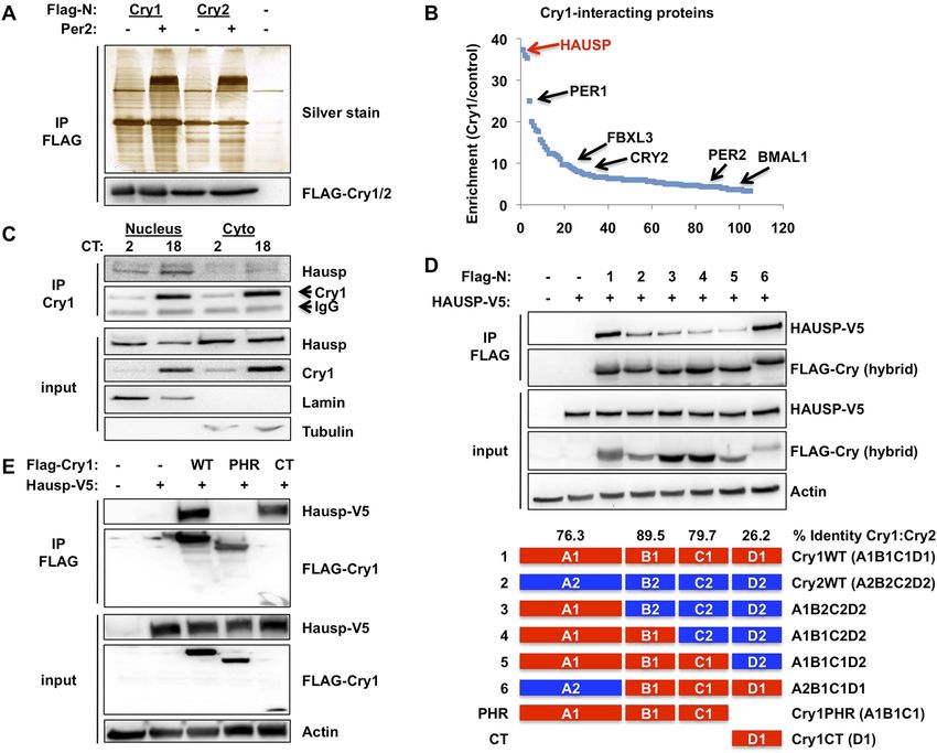
Research article                                                                                     Biochemistry | Neuroscience

                                   eLife digest Many aspects of our physiology and behavior, most notably our patterns of sleep
                                   and wakefulness, are synchronized with the day–night cycle. These circadian rhythms are generated
                                   and maintained by the circadian clock, which consists of positive and negative feedback loops
                                   formed by a large number of genes and proteins. The end result is that the rates at which thousands
                                   of proteins are produced varies rhythmically over the course of the day–night cycle.
                                       It has long been suspected that one of the functions of this circadian clock is to control the timing
                                   of cell division. Moreover, since UV radiation can give rise to genetic mutations when cells divide, it is
                                   thought that the circadian clock limits the amount of DNA damage that occurs during daytime. Papp,
                                   Huber et al. have now confirmed that the circadian clock does indeed participate in the DNA damage
                                   response and have revealed that two proteins known to be involved in the circadian clock-
                                   —Cryptochrome 1 and 2—have a central role in protecting the integrity of the genetic information in
                                   the cell. These proteins evolved from light-activated enzymes that repair DNA in bacteria.
                                       While mammalian cryptochromes have lost their ability to repair DNA, they still prefer to bind to
                                   genetic material that has been damaged by UV radiation. Papp, Huber et al. show that DNA damage
                                   triggers cryptochrome 1 to bind to a protein called Hausp, which stabilizes the cryptochrome and
                                   prevents it from being broken down. By contrast, DNA damage triggers cryptochrome 2 to bind to
                                   a protein called Fbxl3, which has a destabilizing effect on the cryptochrome and promotes its
                                   degradation. Since the cryptochromes regulate the activity of BMAL1 and CLOCK, the proteins
                                   associated with the two master clock genes, these changes can have a significant effect on the
                                   circadian clock of an organism.
                                       Further experiments are needed to work out how these proteins influence the activity of BMAL1
                                   and CLOCK, and to investigate the seemingly conflicting roles of the two cryptochromes and the
                                   interactions between them.
                                   DOI: 10.7554/eLife.04883.002

                                   (Ozturk et al., 2009), though the reported effects have varied (Fu and Kettner, 2013). In addition,
                                   Cry-deficient mice are resistant to genotoxic stress in the context of cyclophosphamide treatment
                                   (Gorbacheva et al., 2005). Consistent with the idea that Cry1/2 may be promiscuous transcriptional
                                   repressors involved in a wide variety of physiological pathways, a recent study found that Cry1 and
                                   Cry2 each bound thousands of chromatin sites independently of other clock transcription factors in
                                   mouse liver (Koike et al., 2012).
                                      Though Cry1 and Cry2 are mostly believed to associate with chromatin via binding a variety of
                                   transcription factors, they can also interact directly with DNA. Cry1 and Cry2 evolved from prokaryotic
                                   light-activated DNA repair enzymes, known as photolyases. While they seem to have lost the [6-4]
                                   photolyase catalytic activity characteristic of their ancestral homologs (Ozturk et al., 2007), they
                                   retain the ability to bind preferentially to UV-damaged DNA containing a [6-4]photoproduct
                                   (Ozgur and Sancar, 2003). The three-dimensional structures of Cry1 and Cry2 resemble those
                                   of photolyases, including the DNA binding surfaces (Maul et al., 2008; Czarna et al., 2013;
                                   Xing et al., 2013). Together, these properties suggest that Cry1 and Cry2 could retain a residual
                                   role in sensing or responding to damaged DNA. Such conservation of function by divergent
                                   molecular mechanisms has been seen previously between cryptochromes derived from different
                                   species (Yuan et al., 2007; Lamia et al., 2009; Kim et al., 2014).
                                      Ubiquitination of substrate proteins by E3 ligases, like SCFFbxl3 and SCFFbxl21, is reversed by
                                   ubiquitin-specific proteases (USPs) (Eletr and Wilkinson, 2014). Herpes virus associated ubiquitin-
                                   specific protease (Hausp; a.k.a. Usp7) was first identified as the cellular partner of the herpes virus
                                   protein Vmw110 (Everett et al., 1997). Hausp modulates proliferation by catalyzing the removal of
                                   polyubiquitin chains from the tumor suppressor p53 and from the p53-destabilizing E3 ligases Mdm2
                                   and MdmX (Li et al., 2002, 2004). The affinity of Hausp for p53 is increased and for Mdm2/MdmX is
                                   decreased in response to DNA damage (Khoronenkova et al., 2012), contributing to stabilization of
                                   p53. Knockout of Hausp in mice is lethal (Kon et al., 2010), probably due to disrupted cell
                                   proliferation. A growing list of Hausp substrates has been identified recently, including several
                                   components of DNA damage response and DNA repair pathways (Nicholson and Suresh Kumar,
                                   2011; Schwertman et al., 2012; Jacq et al., 2013; Eletr and Wilkinson, 2014). In this study, we

Papp et al. eLife 2015;4:e04883. DOI: 10.7554/eLife.04883                                                                             2 of 19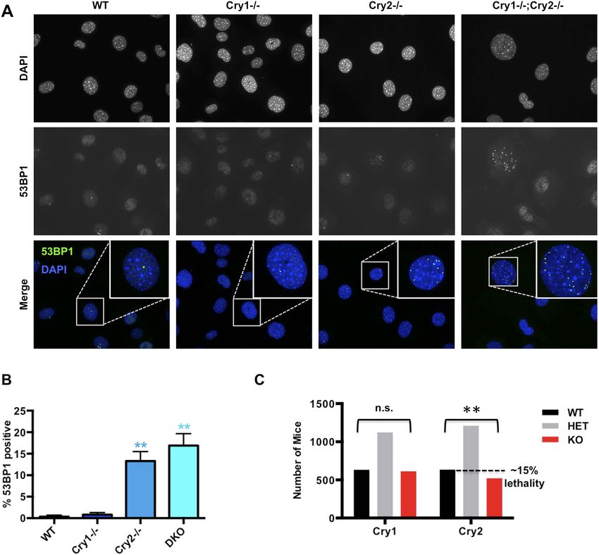
Research article                                                                                   Biochemistry | Neuroscience

                                   demonstrate that Hausp participates in DNA damage-induced resetting of circadian clock time by
                                   stabilizing Cry1.

                                   Results
                                   Identification of Hausp as a novel regulator of Cry1 stability
                                   In an ongoing effort to understand the molecular determinants of cryptochrome stability, we used
                                   mass spectrometry to identify novel protein partners of mammalian CRYs and found Hausp to be
                                   the most highly enriched protein in Cry1-containing complexes (Figure 1A–B, Supplementary file 1).
                                   Co-immunoprecipitation of endogenous (Figure 1C) and overexpressed (Figure 1D–E) Cry1 and
                                   Hausp confirmed the specificity of this interaction. Interestingly, Hausp interacts much more strongly
                                   with Cry1 than with the closely related Cry2 (Figure 1D). Indeed, the divergent Cry1 C-terminus is
                                   necessary and sufficient for strong interaction with Hausp (Figure 1D–E).
                                       The regulation of Cry1/2 protein stability is complex and involves differential expression and
                                   localization of the E3 ligase subunits Fbxl3 and Fbxl21 that compete for Cry binding and have
                                   different rates of ubiquitin conjugation (Hirano et al., 2013; Yoo et al., 2013). Similar to what has
                                   been described for Fbxl3 and Fbxl21, we found no significant tissue specificity or circadian rhythm of
                                   expression or localization for Hausp (Figure 1—figure supplements 1, 2). However, while both Hausp
                                   and Cry1 are more abundant in the cytoplasm, their interaction is stronger in the nucleus, regardless
                                   of circadian phase (Figure 1C, Circadian Time, CT, denotes hours after dexamethasone-induced
                                   synchronization of circadian cycles).
                                       Because Hausp is an ubiquitin-specific protease, its interaction with Cry1 in the nucleus seemed
                                   likely to stabilize nuclear Cry1 by removing polyubiquitin chains. We used small hairpin RNA (shRNA)-
                                   expressing viruses to demonstrate that Hausp depletion led to decreased Cry1 protein primarily in
                                   the nuclear compartment in mouse embryonic fibroblasts (MEFs) independent of circadian phase, as
                                   expected from the ubiquity of Hausp expression (Figure 2A, Figure 2—figure supplement 1).
                                   Treatment of cells with pharmacological inhibitors of Hausp (Nicholson and Suresh Kumar, 2011;
                                   Weinstock et al., 2012) also decreases Cry1 protein, especially in the nucleus (Figure 2B),
                                   consistent with the hypothesis that Hausp stabilizes nuclear Cry1 in vivo. (Note that compound 7
                                   also inhibits Usp47.)
                                       Recombinant Hausp can deubiquitinate Cry1 in vitro (Figure 2—figure supplement 2). To examine
                                   Cry1 ubiquitination in vivo, we measured ubiquitinated Cry1 in MEFs expressing control or
                                   Hausp-targeting shRNA in the presence or absence of the proteasome inhibitor MG132 to stabilize
                                   ubiquitinated proteins. Cry1 from Hausp-depleted cells was much more highly ubiquitinated than
                                   Cry1 from control cells (Figure 2C, Figure 2—figure supplement 3) as expected if Hausp catalyzes the
                                   removal of polyubiquitin chains from Cry1 in vivo. (Note that while Cry1 is decreased in Hausp-depleted
                                   cells, we used a limiting amount of anti-Cry1 antibody for immunoprecipitation to normalize the amount
                                   of Cry1 and enable comparison between samples. Cry−/− cells were used as a control to subtract the
                                   non-specific ubiquitin signal and enable quantitative comparison of control and Hausp-depleted cells.)

                                   Hausp activity alters circadian period length
                                   Cryptochrome stability is a critical determinant of circadian period length, though the direction and
                                   magnitude of the period change associated with altered expression or stability of Cry1 and/or Cry2
                                   seems to depend on the mechanism and context of altered stability (Vitaterna et al., 1999; Hirota
                                   et al., 2012; St John et al., 2014). Nonetheless, if Hausp stabilizes Cry1 by removing ubiquitin chains,
                                   reducing Hausp expression or activity is expected to alter circadian rhythms. In immortalized
                                   fibroblasts expressing a Per2-Luciferase fusion from the endogenous Per2 locus (Per2::Luc [Yoo et al.,
                                   2004]), shRNA-mediated depletion of Hausp increased circadian period (Figure 2D,E). We also
                                   observed period lengthening in immortalized Per2::Luc MEFs when Hausp activity was inhibited
                                   pharmacologically (Figure 2F,G). Because our data suggest that Hausp inhibition and AMPK
                                   activation each destabilizes nuclear Cry1, we examined whether they could synergistically increase
                                   circadian period. Using cells stably expressing luciferase under a circadian promoter (U2OS-B6
                                   [Vollmers et al., 2008]), we observed that activation of AMPK increased the circadian period as
                                   expected (Lamia et al., 2009), inhibition of Hausp also increased period, and combined activation of
                                   AMPK and inhibition of Hausp led to a dramatic increase in period, perhaps reflecting synergistic
                                   destabilization of nuclear Cry1 (Figure 2H,I).

Papp et al. eLife 2015;4:e04883. DOI: 10.7554/eLife.04883                                                                           3 of 19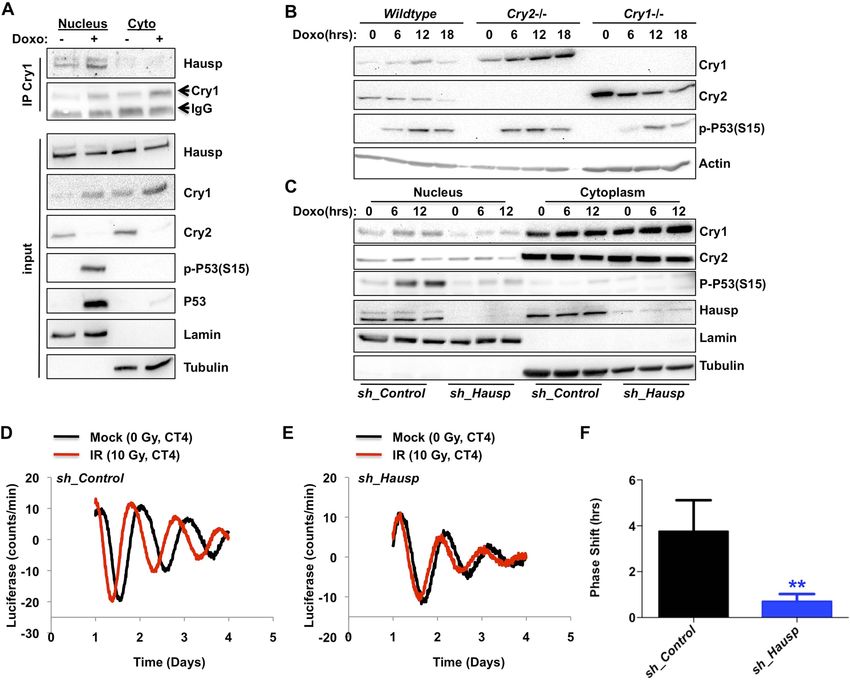
Research article                                                                                                 Biochemistry | Neuroscience

                                   Figure 1. Hausp interacts with Cry1. (A and B) Lysates from 293T cells expressing pcDNA3-2xFLAG with no insert (−),
                                   Cry1, or Cry2 after the FLAG tag with (+) or without (−) co-expression of Per2 were used to purify control, Cry1, or
                                   Cry2-containing complexes by immunoprecipitation (IP) of the FLAG tag. 5% of each purification was analyzed by
                                   SDS-PAGE and silver stain (A) and components of the resulting complexes were identified by mass spectrometry
                                   performed on the remaining 95% of the sample. The experiment was performed in triplicate and Pattern Lab for
                                   Proteomics (Carvalho et al., 2012) was used to identify statistically enriched partners. In (B) Enrichment is the ratio
                                   of spectral counts in Cry1 vs control samples for all statistically enriched partners over three experiments (e.g., lane 1
                                   vs lane 5 from [A]). Arrows depict several established partners for Cry1 as well as the observed 37-fold enrichment for
                                   Hausp in Cry1-containing samples. (C) Endogenous Hausp bound to endogenous Cry1 was detected by
                                   immunoblot (IB) following IP from nuclear and cytoplasmic fractions of mouse embryonic fibroblasts (MEFs)
                                   harvested at the indicated times (CT, hours) following circadian synchronization by dexamethasone. (D) Top: Hausp-
                                   V5 bound to FLAG-Cry1/2 hybrids was detected by IB following IP from 293T cells. Bottom: schematic diagram
                                   showing the composition of the Cry1/2 hybrids and domains used in D and E. (E) Hausp-V5 bound to FLAG-Cry1 full
                                   length or isolated domains was detected by IB following IP.
                                   DOI: 10.7554/eLife.04883.003
                                   The following figure supplements are available for figure 1:
                                   Figure supplement 1. Circadian measurement of Hausp mRNA expression in mouse tissues.
                                   DOI: 10.7554/eLife.04883.004
                                   Figure supplement 2. Circadian measurement of Hausp protein expression in mouse tissues.
                                   DOI: 10.7554/eLife.04883.005

                                   DNA damage increases the Cry1/Cry2 ratio
                                   Given that the Cry1–Hausp interaction occurs primarily in the nucleus and that Hausp interaction with
                                   other partners is regulated by DNA damage, we examined the impact of DNA damage on the
                                   Hausp–Cry1 association and found that it increases the interaction (Figure 3A, Figure 3—figure
                                   supplements 1, 2). Because Hausp catalyzes the removal of polyubiquitin chains from Cry1 thereby
                                   decreasing its proteasomal degradation (Figure 2), increased Cry1–Hausp association in response to
                                   genotoxic stress leads to a prediction that DNA damage should increase Cry1 protein levels.

Papp et al. eLife 2015;4:e04883. DOI: 10.7554/eLife.04883                                                                                            4 of 19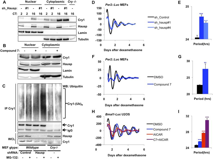
Research article                                                                                            Biochemistry | Neuroscience

                                   Figure 2. Hausp stabilizes Cry1 via deubiquitination and alters circadian rhythms. (A) Wild-type or Cry1−/−;Cry2−/−
                                   (Cry−/−) MEFs stably expressing a control sequence (−) or shRNA targeting Hausp (#1) were subjected to nuclear
                                   and cytoplasmic fractionation. Cry1, Hausp, Lamin, and Tubulin were analyzed by IB from fractions harvested at the
                                   indicated times following circadian synchronization with dexamethasone (CT, hours). (B) Cry1, Hausp, Lamin and
                                   Tubulin were detected by IB in nuclear and cytoplasmic fractions from MEFs treated with vehicle (DMSO, −) or
                                   Compound 7 (+). (C) Wild-type MEFs stably expressing control or Hausp-targeting shRNA or Cry−/− MEFs were
                                   treated with vehicle (DMSO, −) or MG132 (+) for 6 hr, and lysed in RIPA buffer containing iodoacetamide. 6 mg of
                                   RIPA lysates from each condition was subjected to IP with 5 μg of anti-Cry1 antibody. Ubiquitinated Cry1 (Cry1−
                                   (Ub)N), Cry1, and Hausp were detected by IB in IPs and whole cell lysates (WCL). (D, F, H) Typical results of
                                   continuous monitoring of luciferase activity from MEFs expressing Per2-luciferase fusion protein from a knock-in
                                   allele (D and F) or from U2OS cells stably expressing luciferase under the control of the Bmal1 promoter (H) with
                                   stable expression of control or either of two shRNA sequences targeting Hausp (D) or treated with Compound 7
                                   and/or AICAR (F and H). (E, G, I) Quantitation of the circadian period of luciferase activity from experiments
                                   performed as described in (D, F, H). Data represent the mean ± s.d. for 4–8 samples per condition. **p < 0.01, ***p
                                   < 0.001 vs control samples (control shRNA for E or DMSO-treated cells for G and I).
                                   DOI: 10.7554/eLife.04883.006
                                   The following figure supplements are available for figure 2:
                                   Figure supplement 1. Validation of shRNA targeting Hausp.
                                   DOI: 10.7554/eLife.04883.007
                                   Figure supplement 2. In vitro deubiquitination of Cry1 by recombinant Hausp.
                                   DOI: 10.7554/eLife.04883.008
                                   Figure supplement 3. Quantitation of in vivo Cry1 ubiquitination.
                                   DOI: 10.7554/eLife.04883.009

                                   Consistent with this hypothesis, we found that exposure to DNA damage transiently stabilized
                                   endogenous Cry1 in primary MEFs (Figure 3A–C). Intriguingly, Cry2 was destabilized following
                                   exposure to DNA damage, demonstrating that the increase in Cry1 does not merely reflect a change
                                   or synchronization of the circadian rhythm and suggesting differential regulation of these highly
                                   homologous family members, consistent with our observation that Hausp preferentially interacts with
                                   Cry1. Because Cry1 and Cry2 each can repress the other’s expression, Cry2 protein could decrease in
                                   response to damage secondary to stabilization of Cry1. However, Cry2 protein decreases and Cry1

Papp et al. eLife 2015;4:e04883. DOI: 10.7554/eLife.04883                                                                                     5 of 19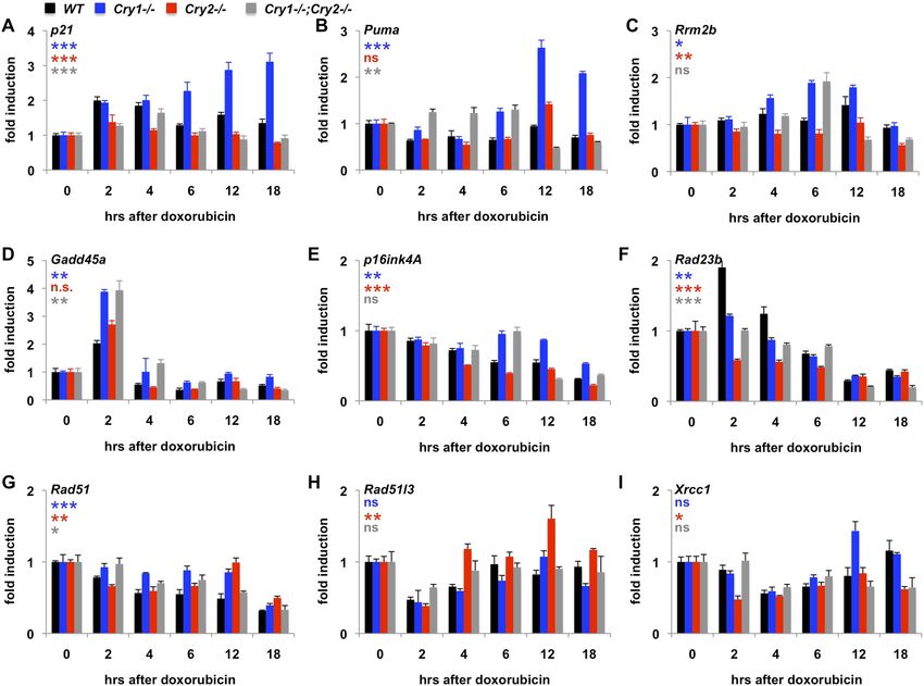
Research article                                                                                                  Biochemistry | Neuroscience

                                   Figure 3. DNA damage resets the clock via Hausp-dependent stabilization of nuclear Cry1. (A) Endogenous Hausp,
                                   Cry1, Cry2, phospho-P53 (Ser15), P53, Lamin, and Tubulin were detected by IB in Cry1 immunoprecipitates or input
                                   samples from nuclear and cytoplasmic fractions of primary MEFs treated with vehicle (−) or doxorubicin (+). (B) Cry1,
                                   Cry2, phospho-P53 (Ser15), and Actin were detected by IB in lysates from wildtype (WT), Cry1−/− or Cry2−/− MEFs
                                   treated with doxorubicin for the indicated times. (C) Cry1, Cry2, phospho-P53 (Ser15), Hausp, Lamin, and Tubulin
                                   were detected by IB in nuclear and cytoplasmic fractions from MEFs expressing control or Hausp-targeting shRNA
                                   and treated with doxorubicin for the indicated times. (D and E) Typical results of continuous monitoring of luciferase
                                   activity from primary adult ear fibroblasts expressing Bmal1-luciferase and control or Hausp-targeting shRNA and
                                   treated with 0 (black) or 10 Gy (red) irradiation 3 hr after circadian synchronization with dexamethasone. Data
                                   represent the mean luciferase counts of eight samples per condition from one of four independent experiments.
                                   (F) Quantitation of the differences in initial circadian phase of luciferase activity caused by irradiation calculated from
                                   experiments performed as described in (D and E). Data in (D–F) represent the mean ± s.d. of phase shifts observed
                                   in four independent experiments. **p < 0.01 vs control samples.
                                   DOI: 10.7554/eLife.04883.010
                                   The following figure supplements are available for figure 3:
                                   Figure supplement 1. Effect of DNA damage on Cry1-Hausp interaction in transfected 293T cells.
                                   DOI: 10.7554/eLife.04883.011
                                   Figure supplement 2. Proteostasis and/or membrane stress increase the Cry1-Hausp interaction.
                                   DOI: 10.7554/eLife.04883.012
                                   Figure supplement 3. Circadian time of exposure determines phase shift in response to DNA damage.
                                   DOI: 10.7554/eLife.04883.013

                                   protein increases in response to DNA damage in MEFs expressing only a single Cry paralog (i.e., Cry2
                                   in Cry1−/− MEFs and vice versa; Figure 3B). Thus, DNA damage acutely regulates Cry1 and Cry2
                                   protein levels independently.
                                      To determine the contribution of Hausp to the damage-induced stabilization of nuclear Cry1, we
                                   examined nuclear Cry1 protein levels following DNA damage in MEFs expressing either control
                                   sequences or Hausp-targeting shRNA. Indeed, depletion of Hausp prevents damage-induced
                                   stabilization of nuclear Cry1, similar to the effect of Hausp depletion on p53 accumulation

Papp et al. eLife 2015;4:e04883. DOI: 10.7554/eLife.04883                                                                                             6 of 19
Research article Biochemistry | Neuroscience
(Figure 3C). Note that the weaker interactions that we observed between Hausp and Cry2 or hybrid
constructs containing the C-terminus of Cry2 compared to those containing the Cry1 C-terminus
(Figure 1D) are likely artefacts of overexpression in 293T cells since we did not observe destabilization
of Cry2 upon Hausp depletion (Figure 3C).
Hausp is required for clock resetting in response to DNA damage
It has been reported that DNA damage causes phase shifts of circadian rhythms (Oklejewicz et al.,
2008; Engelen et al., 2013). Consistently, we observed phase shifts in primary MEFs with a peak shift
following irradiation at CT2-4, (Figure 3—figure supplement 3). The requirement for Hausp in
stabilization of nuclear Cry1 after DNA damage suggested Hausp could contribute to phase shifts in
response to DNA damage. By examining the circadian phase of control and Hausp-depleted
fibroblasts after exposure to irradiation at CT3, we found that although the circadian phase of the
non-irradiated cells is similar (Figure 2D), DNA damage-induced phase shifts were greatly diminished
in Hausp-deficient fibroblasts (Figure 3D–F).
Phosphorylation of both partners influences the association between
Cry1 and Hausp
ATM- and PPM1G-dependent dephosphorylation of serine 18 in the N-terminus of Hausp has been
reported to drive the DNA damage dependent disruption of Hausp interaction with Mdm2 and MdmX
(Khoronenkova et al., 2012). Conversely, S18 de-phosphorylation may increase Hausp–Cry1
association because mutation of S18 to the non-phosphorylatable amino acid alanine (S18A)
increases interaction and mutation to aspartic acid, which is chemically similar to phospho-serine,
decreases the interaction (Figure 4A). However, S18 dephosphorylation cannot fully explain DNA
damage induction of Cry1–Hausp interaction as evidenced by persistent stimulated association
between Cry1 and Hausp S18A after DNA damage. Intriguingly, we (Figure 4—figure supplement
1, Supplementary file 2) and others (Gao et al., 2013) find that Cry1 and Cry2 interact with kinases
that are activated by DNA damage and phosphorylate serine or threonine followed by glutamine,
(S/T)-Q (Kim et al., 1999; O’Neill et al., 2000). Cry1 and Cry2 contain several such sequences
(Figure 4—figure supplement 2), including three serines in the Cry1 C-terminal tail that are not
conserved in Cry2.
Using an antibody that recognizes phospho-(S/T)Q, we determined that Cry1 is rapidly
phosphorylated in response to either chemically or radiation-induced DNA damage, while
damage-induced phosphorylation of Cry2 on (S/T)Q was reduced and delayed compared to that
of Cry1 (Figure 4B and not shown), indicating that the rapid phosphorylation of Cry1 in response
to DNA damage likely occurs on the non-conserved C-terminal tail. Mutating each or all of the
Cry1 C-terminal SQ serines to alanine decreased or abolished, respectively, the phospho-(S/T)Q
signal after DNA damage, indicating that these are the sites in Cry1 that are rapidly phosphorylated
in response to DNA damage (Figure 4C, and not shown). Notably, S588 is the only one of these
sites on which phosphorylation has been directly detected in vivo (Lamia et al., 2009;
Hegemann et al., 2011). We generated an antibody that specifically recognizes Cry1
phosphorylated on S588 and measured a rapid increase in the presence of this phosphorylated
species after exposure to DNA damage (Figure 4D). Consistent with the reported stabilization
of Cry1 by mimicking phosphorylation at S588, mutation of this site to aspartic acid increased its
association with Hausp (Figure 4E, left).
DNA damage stimulates the association of Cry1/2 with Fbxl3
Because the effects of DNA damage on Cry1 and Cry2 stability are not fully explained by
regulated interaction with Hausp, we examined the effect of DNA damage and subsequent
phosphorylation events on the interactions between Cry1/2 and Fbxl3. To our surprise, prolonged
exposure to DNA damage increases the interactions of Cry1 and especially Cry2 with Fbxl3
(Figure 4F), probably contributing to the transient nature of the Cry1 stabilization and to Cry2
destabilization following damage. Notably, mutation of Cry1 S588 to aspartic acid, which
increases Cry1 interaction with Hausp, decreases the association of Cry1 with Fbxl3 (Figure 4E,
right) suggesting that phosphorylation of the unique Cry1 C-terminus may oppose the increase in
Fbxl3 binding to Cry1, possibly explaining the preferential induction of Fbxl3 binding to Cry2
compared to Cry1.
Papp et al. eLife 2015;4:e04883. DOI: 10.7554/eLife.04883 7 of 19Research article Biochemistry | Neuroscience
Figure 4. DNA damage induced signaling modulates interactions of Cry1/2, Hausp, and Fbxl3. Hausp-V5, FLAG-
Cry1/2, phospho-P53 (Ser15), Phospho-SQ/TQ, Phospho-Cry1S588 (P-S588), Fbxl3-V5, and Actin were detected by
IB in IPs and lysates (WCL) from 293T cells transfected with the indicated plasmids and lysed at the indicated times
following treatment with doxorubicin (doxo) or irradiation (IR).
DOI: 10.7554/eLife.04883.014
The following figure supplements are available for figure 4:
Figure supplement 1. Composition of Cry1- and Cry2-associated protein complexes.
DOI: 10.7554/eLife.04883.015
Figure supplement 2. Conserved SQ/TQ motifs present in Cry1 and/or Cry2.
DOI: 10.7554/eLife.04883.016
Genetic disruption of Cry1/2 alters the transcriptional response to DNA
damage
Given that Cry1 and Cry2 are transcriptional repressors and that we found a robust regulation of their
stability by DNA damage, we asked whether the transcriptional response to DNA damage is altered
by genetic disruption of Cry1 or Cry2. By measuring the induction of transcripts activated by DNA
damage in fibroblasts (Kenzelmann Broz et al., 2013), we found that genetic loss of Cry1 or Cry2
enhances or suppresses, respectively, the induction of Cdkn1a (p21) by genotoxic stress and alters the
dynamic response of other established damage responsive transcripts as well (Figure 5A–E and
Figure 5—figure supplement 1). Although the chromatin association of cryptochromes may be
different in different cell types, both Cry1 and Cry2 bind some of these loci in mouse liver (Koike
et al., 2012) (Figure 5—figure supplement 2). In addition, Cry1 and Cry2 bind to chromatin regions
near several genes encoding proteins that participate in DNA repair (Supplementary file 3).
Interestingly, the expression of several of those genes in response to DNA damage is also altered by
genetic loss of Cry1 and/or Cry2 (Figure 5F–I), suggesting that cryptochromes may modulate the
activation of DNA repair in response to damage. The regulation of some transcripts in Cry1−/−;Cry2−/−
cells resembles that in Cry1−/− cells (e.g., Rrm2b, Gadd45a, p16ink4a), suggesting that Cry1 is more
relevant to their regulation than is Cry2. For other transcripts (e.g., p21, Puma, Xrcc1), the response to
DNA damage in Cry1−/−;Cry2−/− cells is closer to the response in Cry2−/− cells suggesting that Cry2 is
Papp et al. eLife 2015;4:e04883. DOI: 10.7554/eLife.04883 8 of 19Research article Biochemistry | Neuroscience
Figure 5. Cry1/2 deficiency alters transcriptional response to DNA damage. Expression of the indicated transcripts
was measured by quantitative PCR (qPCR) in cDNA from wildtype (black), Cry1−/− (blue), Cry2−/− (red), and Cry1−/−;
Cry2−/− (gray) fibroblasts treated with doxorubicin for the indicated times. *p < 0.05, **p < 0.01, ***p < 0.001 for
effect of genotype by repeated measures ANOVA analysis (blue—WT vs Cry1−/−; red—WT vs Cry2−/−; gray—WT vs
Cry1−/−;Cry2−/−).
DOI: 10.7554/eLife.04883.017
The following figure supplements are available for figure 5:
Figure supplement 1. Transcriptional response to irradiation-induced DNA damage.
DOI: 10.7554/eLife.04883.018
Figure supplement 2. Circadian pattern of Cry1 and Cry2 binding to selected chromatin sites.
DOI: 10.7554/eLife.04883.019
more important for regulation of those targets. A full understanding of how Cry1 and Cry2 influence
gene expression following DNA damage will require further study.
Cry2−/− cells accumulate DNA damage
We next asked whether disruption of Cry1/2-dependent transcriptional regulation causes a functional
deficit in the cellular response to DNA damage in cryptochrome-deficient cells. Indeed, Cry2−/− and
Cry1−/−;Cry2−/− fibroblasts accumulate damaged DNA, reflected by an increased percentage of non-
dividing cells containing multiple 53BP1-positive foci (Figure 6A,B). Accumulation of damaged DNA
in cells lacking Cry2 was surprising given that Cry2−/− mice are viable and fertile and that genetic
disruption of Cry1 and Cry2 decreases tumor formation in p53-deficient animals (Ozturk et al., 2009).
To determine whether the increased accumulation of DNA damage that we observed in cells could
possibly be relevant in vivo, we analyzed breeding records of a large number of progeny from Cry1+/−;
Cry2+/− mice over several years: while Cry1 genotypes segregate in the expected Mendelian ratios,
the Cry2−/− genotype is significantly underrepresented (Figure 6C). This is similar to reduced survival
observed in mice with genetic defects in established components of the DNA damage response or
DNA repair pathways (Tsai et al., 2005; Mukherjee et al., 2010; Crossan et al., 2011) and is
consistent with the possibility that animals lacking Cry2 are prone to genetic instability, though we
cannot exclude other possible explanations for the reduced viability of Cry2−/− mice.
Papp et al. eLife 2015;4:e04883. DOI: 10.7554/eLife.04883 9 of 19Research article Biochemistry | Neuroscience
Figure 6. Cry2−/− cells accumulate damaged DNA. (A) Representative early passage (P3–4) primary wildtype (WT),
Cry1−/−, Cry2−/−, and Cry1−/−;Cry2−/− adult ear fibroblasts stained with anti-53BP1 antibody (green) and DAPI (blue).
Insets show enlarged view of indicated cells. (B) Quantitation of 53BP1-positive cells prepared as described in (A).
Nuclei containing more than five 53BP1 puncta and negative for BrdU labeling were considered positive for DNA
damage. Data represent the mean ± s.d. for at least 200 cells per genotype. (C) Chi-squared analysis of the
distributions of Cry1 and Cry2 wildtype (black), heterozygous (gray), and homozygous knockout (red) genotypes
establishes a significantly reduced survival of Cry2−/− mice. **p < 0.01 by chi-squared analysis with 2 degrees of
freedom (χ2 = 10.39).
DOI: 10.7554/eLife.04883.020
Discussion
Diverse organisms use circadian clocks to optimize the timing of physiological processes in relation to
predictable diurnal changes in the external environment (Dodd et al., 2005; Lamia et al., 2008;
Marcheva et al., 2010; Sadacca et al., 2011). It has long been suspected that clocks influence the
timing of cell division to temporally separate DNA replication from predictably recurring exposure to
DNA damage (Sahar and Sassone-Corsi, 2009; Sancar et al., 2010). This hypothesis is supported by
the non-random distribution of cell division within circadian cycles (Nagoshi et al., 2004).
Theoretically, in order for clocks to enable such a separation, their timing must be responsive to
genotoxic stress, analogous to entrainment by metabolic signals, which enables clocks to optimally
coordinate cellular metabolism with externally fluctuating metabolic demands (Ramsey and Bass,
2011; Jordan and Lamia, 2013). Indeed, others have shown that DNA damage shifts circadian clock
time (Oklejewicz et al., 2008). In this study, we confirm that phenomenon and describe a molecular
mechanism by which Hausp-mediated deubiquitination and stabilization of Cry1 contributes to it.
Papp et al. eLife 2015;4:e04883. DOI: 10.7554/eLife.04883 10 of 19Research article Biochemistry | Neuroscience
Furthermore, we provide evidence for specific and divergent roles of the circadian transcriptional
repressors Cry1 and Cry2 in modulating the transcriptional response to DNA damage, thus addressing the
longstanding question of whether circadian rhythmicity per se is sufficient to minimize the occurrence or
accumulation of DNA damage. Indeed, our results suggest that circadian rhythmicity as such is not
protective because Cry2−/− cells maintain robust circadian rhythms (Khan et al., 2012), but they
accumulate DNA damage at rates comparable to arrhythmic Cry1−/−;Cry2−/− cells. Therefore, it appears
that the genome-protective function of normal circadian rhythms is dependent on the expression of
specific clock components, including Cry2. This distinction may underlie some of the controversy over the
importance of circadian clocks for maintaining genomic integrity (Fu and Kettner, 2013).
Increased accumulation of DNA damage in Cry2−/− cells would be expected to lead to an increased
mutation rate; consistent with that hypothesis, we observed sub-Mendelian inheritance of the Cry2−/−
genotype. Though the potential for circadian clocks to influence the cellular response to DNA damage
has been controversial (Gaddameedhi et al., 2012), our results are also consistent with several lines of
evidence supporting a conserved role for clocks in modulating the DNA damage response and/or
DNA repair (Kang et al., 2009, 2010; Cotta-Ramusino et al., 2011; Gaddameedhi et al., 2011).
Interestingly, we identified several proteins that specifically bind damaged DNA or participate in DNA
repair in Cry1- and/or Cry2-associated complexes (Supplementary files 1, 2), suggesting that
cryptochromes may influence DNA repair by non-transcriptional mechanisms as well.
While Cry1 and Cry2 have long been established as repressors of Clock:Bmal1-driven gene
expression, and we observe altered expression of several transcripts in response to DNA damage in
Cry1/2-deficient cells, it remains unclear how Cry1/2 regulate gene expression. Though it is not the
focus of this study, the composition of the Cry1- and Cry2-associated protein complexes suggests
that regulation of mRNA processing may be an important function for Cry1 and Cry2: a large number
of RNA binding and RNA processing factors were found associated with Cry1 and Cry2
(Figure 4—figure supplement 2; Supplementary files 1, 2). This is consistent with other recent
literature describing the association of RNA processing machinery in complex with Per proteins
(Padmanabhan et al., 2012) and the importance of post-transcriptional regulation in circadian clock
function generally (Kojima et al., 2011).
It has long been hypothesized that the C-termini of cryptochromes are important for distinguishing
their species-specific functions (Green, 2004) and for enabling regulated interactions with protein and
possible nucleic acid partners (Czarna et al., 2011; Zoltowski et al., 2011; Engelen et al., 2013).
Here, we identify a Cry1-specific partner (Hausp) that interacts with the C-terminus in isolation.
Furthermore, we describe phosphorylation events in Cry1, Cry2, and Hausp that are influenced by
DNA damage and contribute to their regulated interactions. It appears that DNA damage initiates
a complex cascade of signal transduction that alters circadian clock dynamics in a complicated
manner. The large number of phosphorylation events in Cry1, Cry2, and Hausp induced by genotoxic
stress supports an important role for these proteins in sensing or responding to such stress.
Understanding the specific functions of each of these modifications will require further study.
These results suggest that mammalian cryptochromes, Cry1 and Cry2, represent a node of
interaction between circadian clocks and the cellular response to genotoxic stress. Cry1 and Cry2 play
non-redundant roles in this pathway, highlighting the importance of analyzing their roles
independently rather than relying on the exclusive use of doubly deficient cells and animals to
understand their functions. Further study will determine whether Cry1 and Cry2 modulate the
transcriptional response to DNA damage via sequence-specific DNA binding transcription factors or
through direct binding of damaged DNA as has been observed in vitro (Ozgur and Sancar, 2003).
Finally, accumulation of damaged DNA in Cry2−/− cells suggests that Cry2 is a particularly important
integrator of circadian rhythms and genomic integrity. Taking all of these data together, we conclude
that Cry1 and Cry2 cooperatively regulate the transcriptional response to genotoxic stress and the
inverse relationship of their stability in response to DNA damage contributes to transient activation of
gene networks that protect genome integrity (Figure 7).
Materials and methods
Mass spectrometry
For mass spectrometry analysis, samples were denatured, reduced, and alkylated before an overnight
digestion with trypsin. Peptide mixtures were analyzed by liquid chromatography mass spectrometry
Papp et al. eLife 2015;4:e04883. DOI: 10.7554/eLife.04883 11 of 19Research article Biochemistry | Neuroscience
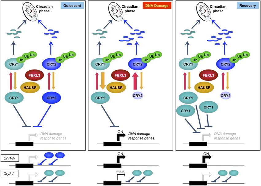
Figure 7. Model depicting a novel mechanism by which the regulation of Cry1 and Cry2 enables coordination of the transcriptional response to genotoxic
stress. In quiescent cells, Cry1 and Cry2 repress transcription of target genes. Upon DNA damage, Cry2 is degraded, relieving repression. As Cry1
accumulates, it replaces Cry2 and returns gene expression to normal levels resulting in transient activation. In Cry1−/− cells, gene expression is enhanced,
while in Cry2−/− cells, damage-induced transcription is suppressed. Note that this model does not explain the dynamics of altered response observed for
all transcripts but may apply to the average change in the transcriptional response to DNA damage in Cry1/2-deficient cells.
DOI: 10.7554/eLife.04883.021
using an Accela pump and LTQ mass spectrometer (Thermo Fisher Scientific, Waltham, MA) using a four-
step multidimensional protein identification technology (MudPIT) separation (MacCoss et al., 2002).
Tandem mass spectrometry spectra were collected in a data-dependent fashion and resulting spectra
were extracted using RawXtract. Protein identification was done with Integrated Proteomics Pipeline (IP2)
by searching against the UniProt Human database and filtering toResearch article Biochemistry | Neuroscience
Ionizing radiation exposure was performed using a 137Cs γ-radiation source at the indicated times
after dexamethasone synchronization. Transfections were carried out using calcium phosphate or
polyethylenimine (cat #23966-2; PEI; Polysciences Inc, Warrington, PA) by standard protocols.
Plasmids and shRNA
pcDNA3-2xFlag-mCRY1, pcDNA3-2xFlag-mCRY2, and pcDNA3-Fbxl3-v5 are as described previously
(Lamia et al., 2009). pCl-neo Flag HAUSP deposited by Dr Bert Vogelstein was purchased
from Addgene (Addgene plasmid 16655) (Cummins and Vogelstein, 2004) and cloned into pcDNA
3.2/V5/GW-CAT purchased from Invitrogen (cat #K244020) using standard protocols. Lentiviruses
expressing Bmal1-luciferase and Per2-luciferase were from Dr Satchidananda Panda. Five shRNAs
against Hausp and one shRNA against Gapdh were purchased from Open Biosystems. pLKO.1
sh_scramble deposited by Dr David Sabatini was purchased from Addgene (Addgene plasmid 1864)
(Sarbassov et al., 2005). Either sh_Scramble or sh_Gapdh was used as controls for sh_Hausp. pLenti-
lox-GFP shRNA p19-2 for immortalizations deposited by Dr Tyler Jacks was purchased from Addgene
(Addgene plasmid 14091) (Sage et al., 2003). psPAX plasmid (Addgene plasmid 12,260) and pMD2.G
plasmid (Addgene plasmid 12259) deposited by Dr Didier Trono used for infection also purchased
from Addgene. Cry hybrid constructs were a gift from Dr Andrew C Liu (Khan et al., 2012); the hybrid
coding sequences were transferred to pcDNA3-2xFlag using standard protocols; several observed
mutations in the hybrid coding sequences were corrected by site-directed mutagenesis. All mutations
were generated using Agilent Site-Directed Mutagenesis kit and protocols (cat #200521).
Cell lines
HEK 293T cells were from the American Type Culture Collection (ATCC, Manassas, VA). U2OS-B6 cells
were a gift from Dr Satchidananda Panda. MEFs were isolated from embryos of the indicated
genotypes at E15.5 and were used as primary (passaged no more than 10 times and grown in 3%
oxygen), immortalized with pLenti-lox-GFP shRNA p19-2, or spontaneously immortalized. Ear
Fibroblasts were isolated from 3-month-old littermates. Ear punches were put in 70% ethanol for 2
min, washed in PBS, cut into small pieces using a scalpel and transferred to a 15-ml tube. 2 ml of
trypsin 0.25% was added and samples were incubated for 1 hr at 37˚C in a water bath, vortexing
briefly every 10 min. The trypsin was inactivated with 8 ml of EF media (DMEM/15%FBS/PS1%). Cells
were spun down 5 min at 1000 rpm, re-suspended in 3 ml of EF media and transferred into a well of
a 6-well plate. The medium was changed the next day. Fibroblasts grew after 3–5 days.
Generation of viruses and stable cell lines
Lentiviral shRNA were transfected into HEK 293T cells using psPAX and pMD2.G packaging
plasmids for virus generation. Viral supernatants were collected 48 hr after transfection, filtered
through a 0.45-μm filter, supplemented with 6 μg/ml polybrene and added to parental cell lines.
After 4 hr, additional media were added to dilute the polybrene toResearch article Biochemistry | Neuroscience
Technology (Danvers, MA). Anti-Cry1-phosphoS588 antibody was affinity purified from rabbit antisera
raised against a phospho-S588 containing peptide.
Immunofluorescence
Cells were grown on glass coverslips and pulse-labeled for 30 min by adding 10 μM of BrdU to the cell
culture medium, washed three times with PBS before fixation with 4% (wt/vol) paraformaldehyde in
PBS for 15 min at room temperature (RT) and permeabilized with 0.5% (vol/vol) Triton X-100 in PBS for
10 min at RT. Coverslips were blocked with 1% BSA in PBS for 30 min at RT. For BrdU co-staining, cells
were subjected to a DNase I treatment (Sigma; 200 U/ml in 30 mM Tris HCl pH 8.1, 0.33 mM MgCl2,
0.5 mM Mercaptoethanol, 1% BSA, and 0.5% Glycerol) for 1 hr at 37˚C in the presence of anti-BrdU 1/
50 (BD Pharmingen). Then, coverslips were washed three times with PBS prior to incubation with
primary antibodies (anti-53BP1; 1/3000) for 2 hr at RT in blocking buffer. Cells were washed with PBS
and incubated with secondary antibodies (Alexa Fluor 488 goat anti-rabbit 1/150 and Alexa Fluor 594
goat anti-mouse 1/150) for 1 hr at RT in blocking buffer. Cells were then washed three times with PBS
and stained 15 min with DAPI (0.4 μg/ml in PBS1X) to visualize DNA. The coverslips were mounted
onto glass slides with Fluoromount G (Electron microscopy Science). For quantification, at least 200
cells were counted following IF analysis. Cells with at least five 53BP1 foci and negative for BrdU
labeling were considered positive for DNA damage. Images were processed using Image J software.
Nuclear and cytoplasmic fractionation of cultured cells
Cells were washed once with ice cold PBS, fresh cold PBS was added and the cells were transferred to
a 5-ml tube and centrifuged 5 min at 2000 rpm. The resulting pellets were washed with cold PBS and
transferred to 1.5-ml eppendorf tubes and centrifuged 5 min at 2000 rpm. The resulting pellets were
resuspended in Solution A (10 mM Hepes pH 8, 1.5 mM MgCl2, 10 mM KCl, plus protease and
phosphatase inhibitors), and incubated for 15 min at 4˚C. An equal volume of Solution B (solution A +
1% NP40) was added and the samples were further incubated for 5 min at 4˚C and centrifuged 5 min
at 3000 rpm. Supernatants from this step represent the cytoplasmic fraction. The remaining nuclear
pellets were then washed twice with cold PBS, lysed in RIPA buffer and either used directly (nuclear
lysates) or diluted sixfold into IP buffer for immunoprecipitation.
Lumicycle analysis of circadian period and phase shifts
U2OSB6 cells, MEFs, or adult ear fibroblasts were plated at 100% confluency in 35-mm dishes (cat #82050-
538; VWR, Radnor, PA). The next day, cells were treated for 1–2 hr in normal growth medium containing 1
mM dexamethasone and 100 μM D-luciferin. Media were removed and replaced with media containing
DMEM, 5% FBS, 1% penicillin-streptomycin, 15 mM Hepes, pH 7.6, and 100 μM D-luciferin. Plates were
sealed with vacuum grease (Dow Corning high vacuum grease; cat #59344-055; VWR) and glass cover slips
(cat #22038999; 40CIR-1, Fisher Scientific) and placed into the Lumicycle 32 from Actimetrics, Inc.
(Wilmette, IL). Data were recorded using Actimetrics Lumicycle Data Collection software and analyzed
using Actimetrics Lumicycle Analysis program. Background subtraction of the recorded data was
performed with Running Average setting, and fit by least mean squares calculation to a damped sine wave
to calculate the period, amplitude, and phase of the curves. Only data with a goodness of fit percentage of
80 or above was included in the analysis.
Quantitative RT-PCR
RNA was extracted from mouse tissues or cells with Qiazol reagent using standard protocols
(cat #799306; Qiagen, Germany). cDNA was prepared using QScript cDNA Supermix (cat #101414-
106; VWR) and analyzed for gene expression using quantitative real-time PCR with iQ SYBR Green
Supermix (cat #1708885; Biorad, Hercules, CA). For analysis of transcriptional response to DNA
damage (doxorubicin or irradiation), cells were used at approximately 70% confluency (Table 1).
In vitro deubiquitination assay
293T cells transiently expressing Flag-tagged Cry1 were treated with 10 μM MG132 for 18 hr and lysed in
RIPA buffer containing Roche complete protease inhibitors, 1 mg/ml iodoacetamide, and 50 μM PMSF.
Flag-Cry1 was immunoprecipitated for 2 hr with M2-agarose (Sigma A2220), washed five times in RIPA
buffer and three times in reaction buffer, and eluted for 1 hr in reaction buffer (60 mM Hepes pH 7.4,
5 mM MgCl2, 4% glycerol, 2 μg/ml aprotinin, 50 μM PMSF, 2 mg/ml BSA) containing 3XFLAG peptide.
Papp et al. eLife 2015;4:e04883. DOI: 10.7554/eLife.04883 14 of 19Research article Biochemistry | Neuroscience
Table 1. Primers used for qPCR
Cdkn2a (p21): Fwd: CCAGGCCAAGATGGTGTCTT Rev: TGAGAAAGGATCAGCCATTGC
Mdm2: Fwd: CTGTGTCTACCGAGGGTGCT Rev: CGCTCCAACGGACTTTAACA
Rrm2b: Fwd: GACAGCAGAGGAGGTTGACTTG Rev: AAAACGCTCCACCAAGTTTTCA
Puma: Fwd: GTACGGGCGGCGGAGACGAG Rev: GCACCTAGTTGGGCTCCATTTCTG
Gadd45a: Fwd: AAGACCGAAAGGATGGACACG Rev: CAGGCACAGTACCACGTTATC
Rad23b: Fwd: ACCTTCAAGATCGACATCGACC Rev: ACTTCTGACCTGCTACCGGAA
Rad51l3: Fwd: GGAGCTTTGTGCCCAGTACC Rev: TCCCCAATGTCCCAATGTCTAT
Xrcc1: Fwd: AGCCAGGACTCGACCCATT Rev: CCTTCTCCAACTGTAGGACCA
p16ink4a: Fwd: GTGTGCATGACGTGCGGG Rev: GCAGTTCGAATCTGCACCGTAG
Rad51: Fwd: TCACCAGCGCCGGTCAGAGA Rev: CCGGCCTAAAGGTGCCCTCG
DOI: 10.7554/eLife.04883.022
Equal volumes of eluted Flag-Cry1 were combined with the indicated amounts of recombinant Hausp
(cat #E-519; USP7, Boston Biochem) or USP8 (cat #E-520; Boston Biochem, Cambridge, MA) and the
reactions were incubated for 30 min at 30˚C before adding SDS sample buffer and boiling for 5 min. The
resulting samples were separated by 8% SDS-PAGE and Cry1 was detected by immunoblot.
Mice
Cry1−/−;Cry2−/− mice were from Dr Aziz Sancar (Thresher et al., 1998); Per2::Luciferase mice (Yoo
et al., 2004) were purchased from Jackson laboratories (Bar Harbor, ME). All animal care and
treatments were in accordance with The Scripps Research Institute guidelines for the care and use of
animals under protocol #10-0019.
Acknowledgements
We thank Drs Andrew Liu (University of Memphis), Satchidananda Panda (The Salk Institute),
Eros Lazzerini Denchi (The Scripps Research Institute), and Ben Nicholson (Progenra, Inc.) for
providing materials and reagents and Drs Eros Lazzerini Denchi, Supriya Srinivasan, Reuben
Shaw, and Joseph Bass for helpful discussions and critical reading of the manuscript. KAL, SJP,
ALH, AK, SDJ, and MN were supported by the Searle Scholars Fund, the Sidney Kimmel
Foundation for Cancer Research, the Lung Cancer Research Foundation, the National Institute of
Diabetes, Digestive and Kidney Diseases (K01DK090188-03 and R01DK097164-01), and
a research fellowship from the Deutsche Forschungsgemeinschaft (DFG, to SDJ). JJM and JRY
were supported by the National Institute of General Medical Sciences (8 P41 GM103533) and the
National Institute on Aging (R01AG027463).
Additional information
Funding
Funder Grant reference number Author
National Institutes of Health (NIH) K01, DK090188 Katja A Lamia
Kinship Foundation Searle Scholars Award Katja A Lamia
Sidney Kimmel Foundation for Cancer Scholar Award Katja A Lamia
Cancer Research
Lung Cancer Research research project grant Katja A Lamia
Foundation (LCRF)
National Institutes of Health (NIH) R01, DK097164 Katja A Lamia
National Institutes of Health (NIH) R01, AG027463 YatesJohn R III
Papp et al. eLife 2015;4:e04883. DOI: 10.7554/eLife.04883 15 of 19Research article Biochemistry | Neuroscience
Funder Grant reference number Author
National Institutes of Health (NIH) P41, GM103533 YatesJohn R III
Deutsche research fellowship Sabine D Jordan
Forschungsgemeinschaft
The funders had no role in study design, data collection and interpretation, or the
decision to submit the work for publication.
Author contributions
SJP, A-LH, SDJ, KAL, Conception and design, Acquisition of data, Analysis and interpretation of
data, Drafting or revising the article; AK, Acquisition of data, Analysis and interpretation of data,
Contributed unpublished essential data or reagents; MN, JJM, JRY, Acquisition of data, Analysis and
interpretation of data
Author ORCIDs
Sabine D Jordan, http://orcid.org/0000-0001-8974-8522
Ethics
Animal experimentation: This study was performed in strict accordance with the recommendations in
the Guide for the Care and Use of Laboratory Animals of the National Institutes of Health. All of the
animals were handled according to approved institutional animal care and use committee (IACUC)
protocols (#10-0019) of The Scripps Research Institute.
Additional files
Supplementary files
· Supplementary file 1. Cry1-associated proteins. Lysates from 293T cells expressing pcDNA3-
2xFLAG with no insert (control) or Cry1 after the FLAG tag were used to purify control or Cry1-
containing complexes by immunoprecipitation (IP) of the FLAG tag. Components of the resulting
complexes were identified by mass spectrometry. The experiment was performed in triplicate and
PatternLab for Proteomics (Carvalho et al.) was used to identify statistically enriched partners in Cry1-
associated complexes compared to the control. Enrichment (Cry1/control) is the ratio of spectral
counts in Cry1 vs control samples for all statistically enriched partners over three experiments.
DOI: 10.7554/eLife.04883.023
· Supplementary file 2. Cry2-associated proteins. Lysates from 293T cells expressing pcDNA3-
2xFLAG with no insert (control) or Cry2 after the FLAG tag were used to purify control or Cry1-
containing complexes by immunoprecipitation (IP) of the FLAG tag. Components of the resulting
complexes were identified by mass spectrometry. The experiment was performed in triplicate and
PatternLab for Proteomics (Carvalho et al.) was used to identify statistically enriched partners in Cry2-
associated complexes compared to the control. Enrichment (Cry2/control) is the ratio of spectral
counts in Cry2 vs control samples for all statistically enriched partners over three experiments.
DOI: 10.7554/eLife.04883.024
· Supplementary file 3. Chromatin binding of circadian transcription factors to loci encoding DNA
repair proteins. Published data (Koike et al., 2012, Table S2) was searched for the text string ‘repair’
to make a preliminary identification of chromatin regions near genes involved in DNA repair that were
found to be associated with each of the seven circadian transcription factors Cry1, Cry2, Per1, Per2,
Clock, Npas2, and Bmal1.
DOI: 10.7554/eLife.04883.025
References
Busino L, Bassermann F, Maiolica A, Lee C, Nolan PM, Godinho SI, Draetta GF, Pagano M. 2007. SCFFbxl3 controls
the oscillation of the circadian clock by directing the degradation of cryptochrome proteins. Science 316:
900–904. doi: 10.1126/science.1141194.
Carvalho PC, Yates JR III, Barbosa VC. 2012. Improving the TFold test for differential shotgun proteomics.
Bioinformatics 28:1652–1654. doi: 10.1093/bioinformatics/bts247.
Papp et al. eLife 2015;4:e04883. DOI: 10.7554/eLife.04883 16 of 19Research article Biochemistry | Neuroscience
Cotta-Ramusino C, McDonald ER III, Hurov K, Sowa ME, Harper JW, Elledge SJ. 2011. A DNA damage response
screen identifies RHINO, a 9-1-1 and TopBP1 interacting protein required for ATR signaling. Science 332:
1313–1317. doi: 10.1126/science.1203430.
Crossan GP, van der Weyden L, Rosado IV, Langevin F, Gaillard PH, McIntyre RE, Sanger Mouse Genetics Project,
Gallagher F, Kettunen MI, Lewis DY, Brindle K, Arends MJ, Adams DJ, Patel KJ. 2011. Disruption of mouse Slx4,
a regulator of structure-specific nucleases, phenocopies Fanconi anemia. Nature Genetics 43:147–152. doi: 10.
1038/ng.752.
Cummins JM, Vogelstein B. 2004. HAUSP is required for p53 destabilization. Cell Cycle 3:689–692. doi: 10.4161/
cc.3.6.924.
Czarna A, Berndt A, Singh HR, Grudziecki A, Ladurner AG, Timinszky G, Kramer A, Wolf E. 2013. Structures of
Drosophila cryptochrome and mouse cryptochrome1 provide insight into circadian function. Cell 153:
1394–1405. doi: 10.1016/j.cell.2013.05.011.
Czarna A, Breitkreuz H, Mahrenholz CC, Arens J, Strauss HM, Wolf E. 2011. Quantitative analyses of
cryptochrome-mBMAL1 interactions: mechanistic insights into the transcriptional regulation of the mammalian
circadian clock. The Journal of Biological Chemistry 286:22414–22425. doi: 10.1074/jbc.M111.244749.
Dardente H, Mendoza J, Fustin JM, Challet E, Hazlerigg DG. 2008. Implication of the F-Box Protein FBXL21 in
circadian pacemaker function in mammals. PLOS ONE 3:e3530. doi: 10.1371/journal.pone.0003530.
Dodd AN, Salathia N, Hall A, Kevei E, Tóth R, Nagy F, Hibberd JM, Millar AJ, Webb AA. 2005. Plant circadian
clocks increase photosynthesis, growth, survival, and competitive advantage. Science 309:630–633. doi: 10.
1126/science.1115581.
Doi M, Takahashi Y, Komatsu R, Yamazaki F, Yamada H, Haraguchi S, Emoto N, Okuno Y, Tsujimoto G, Kanematsu
A, Ogawa O, Todo T, Tsutsui K, van der Horst GT, Okamura H. 2010. Salt-sensitive hypertension in circadian
clock-deficient Cry-null mice involves dysregulated adrenal Hsd3b6. Nature Medicine 16:67–74. doi: 10.1038/nm.
2061.
Eletr ZM, Wilkinson KD. 2014. Regulation of proteolysis by human deubiquitinating enzymes. Biochimica Et
Biophysica Acta 1843:114–128. doi: 10.1016/j.bbamcr.2013.06.027.
Engelen E, Janssens RC, Yagita K, Smits VA, van der Horst GT, Tamanini F. 2013. Mammalian TIMELESS is involved
in period determination and DNA damage-dependent phase advancing of the circadian clock. PLOS ONE 8:
e56623. doi: 10.1371/journal.pone.0056623.
Everett RD, Meredith M, Orr A, Cross A, Kathoria M, Parkinson J. 1997. A novel ubiquitin-specific protease is
dynamically associated with the PML nuclear domain and binds to a herpesvirus regulatory protein. The EMBO
Journal 16:1519–1530. doi: 10.1093/emboj/16.7.1519.
Fu L, Kettner NM. 2013. The circadian clock in cancer development and therapy. Progress in Molecular Biology and
Translational Science 119:221–282. doi: 10.1016/B978-0-12-396971-2.00009-9.
Gaddameedhi S, Reardon JT, Ye R, Ozturk N, Sancar A. 2012. Effect of circadian clock mutations on DNA damage
response in mammalian cells. Cell cycle 11:3481–3491. doi: 10.4161/cc.21771.
Gaddameedhi S, Selby CP, Kaufmann WK, Smart RC, Sancar A. 2011. Control of skin cancer by the circadian
rhythm. Proceedings of the National Academy of Sciences of USA 108:18790–18795. doi: 10.1073/pnas.
1115249108.
Gao P, Yoo SH, Lee KJ, Rosensweig C, Takahashi JS, Chen BP, Green CB. 2013. Phosphorylation of the
cryptochrome 1 C-terminal tail regulates circadian period length. The Journal of Biological Chemistry 288:
35277–35286. doi: 10.1074/jbc.M113.509604.
Godinho SI, Maywood ES, Shaw L, Tucci V, Barnard AR, Busino L, Pagano M, Kendall R, Quwailid MM, Romero MR,
O’neill J, Chesham JE, Brooker D, Lalanne Z, Hastings MH, Nolan PM. 2007. The after-hours mutant reveals a role
for Fbxl3 in determining mammalian circadian period. Science 316:897–900. doi: 10.1126/science.1141138.
Gorbacheva VY, Kondratov RV, Zhang R, Cherukuri S, Gudkov AV, Takahashi JS, Antoch MP. 2005. Circadian
sensitivity to the chemotherapeutic agent cyclophosphamide depends on the functional status of the CLOCK/
BMAL1 transactivation complex. Proceedings of the National Academy of Sciences of USA 102:3407–3412.
doi: 10.1073/pnas.0409897102.
Green CB. 2004. Cryptochromes: tail-ored for distinct functions. Current Biology 14:R847–R849. doi: 10.1016/j.
cub.2004.09.040.
Green CB, Takahashi JS, Bass J. 2008. The meter of metabolism. Cell 134:728–742. doi: 10.1016/j.cell.2008.08.
022.
Hashiramoto A, Yamane T, Tsumiyama K, Yoshida K, Komai K, Yamada H, Yamazaki F, Doi M, Okamura H,
Shiozawa S. 2010. Mammalian clock gene Cryptochrome regulates arthritis via proinflammatory cytokine TNF-
alpha. Journal of immunology 184:1560–1565. doi: 10.4049/jimmunol.0903284.
Hegemann B, Hutchins JR, Hudecz O, Novatchkova M, Rameseder J, Sykora MM, Liu S, Mazanek M, Lénárt P,
Hériché JK, Poser I, Kraut N, Hyman AA, Yaffe MB, Mechtler K, Peters JM. 2011. Systematic phosphorylation
analysis of human mitotic protein complexes. Science Signaling 4:rs12. doi: 10.1126/scisignal.2001993.
Hirano A, Yumimoto K, Tsunematsu R, Matsumoto M, Oyama M, Kozuka-Hata H, Nakagawa T, Lanjakornsiripan D,
Nakayama KI, Fukada Y. 2013. FBXL21 regulates oscillation of the circadian clock through ubiquitination and
stabilization of cryptochromes. Cell 152:1106–1118. doi: 10.1016/j.cell.2013.01.054.
Hirota T, Lee JW, St John PC, Sawa M, Iwaisako K, Noguchi T, Pongsawakul PY, Sonntag T, Welsh DK, Brenner DA,
Doyle FJ III, Schultz PG, Kay SA. 2012. Identification of small molecule activators of cryptochrome. Science 337:
1094–1097. doi: 10.1126/science.1223710.
Jacq X, Kemp M, Martin NM, Jackson SP. 2013. Deubiquitylating enzymes and DNA damage response pathways.
Cell Biochemistry and Biophysics 67:25–43. doi: 10.1007/s12013-013-9635-3.
Papp et al. eLife 2015;4:e04883. DOI: 10.7554/eLife.04883 17 of 19You can also read