The function of twister ribozyme variants in non-LTR retrotransposition in Schistosomamansoni
←
→
Page content transcription
If your browser does not render page correctly, please read the page content below
Published online 22 September 2021                                              Nucleic Acids Research, 2021, Vol. 49, No. 18 10573–10588
                                                                                                         https://doi.org/10.1093/nar/gkab818

The function of twister ribozyme variants in non-LTR
retrotransposition in Schistosoma mansoni
Getong Liu1,2 , Hengyi Jiang1,2 , Wenxia Sun1,2 , Jun Zhang1,2 , Dongrong Chen1,2,* and
Alastair I. H. Murchie 1,2,*
1
 Fudan University Pudong Medical Center, and Institutes of Biomedical Sciences, Shanghai Medical College, Key
Laboratory of Medical Epigenetics and Metabolism, Fudan University, Shanghai 200032, China and 2 Key Laboratory
of Metabolism and Molecular Medicine, Ministry of Education, School of Basic Medical Sciences, Fudan University,
Shanghai 200032, China

                                                                                                                                                                  Downloaded from https://academic.oup.com/nar/article/49/18/10573/6374148 by guest on 18 October 2021
Received March 25, 2021; Revised August 23, 2021; Editorial Decision September 04, 2021; Accepted September 08, 2021

ABSTRACT                                                                          in bacteria and diverse eukaryotic genomes; including
                                                                                  yeasts, plants, insects and worms. The twister RNA is com-
The twister ribozyme is widely distributed over                                   posed of highly conserved structural domains that have self-
numerous organisms and is especially abundant                                     cleavage ribozyme activity in vitro (1). Crystal structures of
in Schistosoma mansoni, but has no confirmed bio-                                 the RNA, supported by biochemical data, confirm that the
logical function. Of the 17 non-LTR retrotransposons                              four helical stems (P1 to P4), two internal loops (L1 and
known in S. mansoni, none have thus far been as-                                  L2) and hairpin loop (L4) adopt a compact fold stabilized
sociated with ribozymes. Here we report the identifi-                             by two pseudoknots (T1 and T2) with the U–A cleavage
cation of novel twister variant (T-variant) ribozymes                             site buried in the center (2–6). Within the RNA sequence,
and their function in S. mansoni non-LTR retrotrans-                              ten nucleotides are >97% conserved. A highly conserved
position. We show that T-variant ribozymes are lo-                                Guanosine plays a key catalytic role in cleavage of the scis-
cated at the 5 end of Perere-3 non-LTR retrotrans-                               sile U–A bond. A function for the twister ribozyme has yet
                                                                                  to be shown.
posons in the S. mansoni genome. T-variant ri-
                                                                                     Retrotransposons are transposable genetic elements that
bozymes were demonstrated to be catalytically ac-                                 require an RNA intermediate for transposition (7,8). They
tive in vitro. In reporter constructs, T-variants were                            are abundant in the genomes of organisms across all king-
shown to cleave in vivo, and cleavage of T-variants                               doms of life, for example, 45% of the human genome and at
was sufficient for the translation of downstream re-                              least 50% of the maize genome are made up of retrotrans-
porter genes. Our analysis shows that the T-variants                              poson sequences (9,10). Retrotransposon insertion con-
and Perere-3 are transcribed together. Target site du-                            tributes to genomic diversity and complexity (11,12). In
plications (TSDs); markers of target-primed reverse                               contrast to LTR retrotransposons (13) non-LTR retro-
transcription (TPRT) and footmarks of retrotranspo-                               transposons, long interspersed nuclear elements (LINEs)
sition, are located adjacent to the T-variant cleavage                            and non-autonomous short interspersed nuclear elements
site and suggest that T-variant cleavage has taken                                (SINEs) and SVA (SINE/VNTR/Alu) elements lack long
                                                                                  terminal repeats at each end (14–18). In general, the non-
place in S. mansoni. Sequence heterogeneity in the
                                                                                  LTR retrotransposons may contain an internal promoter
TSDs indicates that Perere-3 retrotransposition is not                            and open reading frames (ORFs) that encode reverse tran-
site-specific. The TSD sequences contribute to the 5                             scriptase (RT) and/or endonuclease domains and short
end of the terminal ribozyme helix (P1 stem). Based                               sequence repeats at their 3 boundary (19,20). The pro-
on these results we conclude that T-variants have a                               moter sequences of functional non-LTR retrotransposons
functional role in Perere-3 retrotransposition.                                   are not conserved across species (21,22) and some ele-
                                                                                  ments lack internal promoters and are transcribed as in-
INTRODUCTION                                                                      trons of larger host transcripts (23). Some elements may be
                                                                                  transcribed by a nearby upstream cellular promoter, while
The twister ribozyme was originally identified by bioinfor-                       some elements specifically insert into genes and may be ex-
matics. Twister RNA sequences are remarkably widespread,                          pressed as precise cotranscripts (24). The features and regu-
with close to 2700 twister ribozyme RNA sequences present                         lation of the transcription of non-LTR retrotransposons are

* To
   whom correspondence should be addressed. Tel: +86 215 423 7517; Email: AIHM@fudan.edu.cn
Correspondence may also be addressed to Dongrong Chen. Email: drchen@fudan.edu.cn

C The Author(s) 2021. Published by Oxford University Press on behalf of Nucleic Acids Research.
This is an Open Access article distributed under the terms of the Creative Commons Attribution-NonCommercial License
(http://creativecommons.org/licenses/by-nc/4.0/), which permits non-commercial re-use, distribution, and reproduction in any medium, provided the original work
is properly cited. For commercial re-use, please contact journals.permissions@oup.com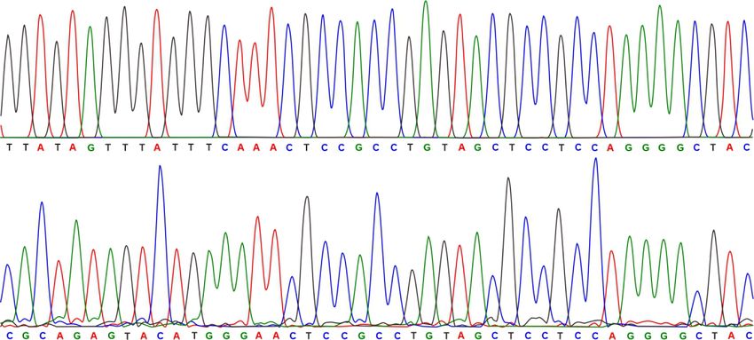
10574 Nucleic Acids Research, 2021, Vol. 49, No. 18

likely to vary from species to species and within particular         The human parasite, Schistosoma mansoni, causes Schis-
retrotransposon clades (25). The main feature of the non-         tosomiasis, a disease that affects ∼250M people worldwide
LTR retrotransposons is the presence of a reverse tran-           in more than 70 countries (49). The parasite has a com-
scriptase (RT)/endonuclease domain (8,26,27), which gen-          plex life cycle with snail and human hosts mediating the
erates DNA copies from the retrotransposon RNA tran-              six stages of its life-cycle: egg, miracidia, sporocysts, cer-
scripts for insertion of a transposon DNA copy into the           caria, schistosomula and adult. The S. mansoni genome se-
new genomic target (25,28,29). For transposition, the non-        quence is available (50,51) and transcriptome profiles and
LTR retrotransposons undergo a replicative cycle, the broad       EST of S. mansoni have been reported (52,53). More than
features of which are outlined in Supplementary Figure S1         20% of S. mansoni genome is considered to be composed
(25,30,31). The mRNA is exported from the nucleus and             of retrotransposons and reverse transcriptase activity has
the RT/endonuclease domains translated in the cytoplasm,          been detected in S. mansoni extracts (54,55). Studies have
mRNA and proteins are subsequently assembled into ri-             identified 28 different S. mansoni retrotransposon elements
bonucleoprotein particles (RNP) (32). Translation of the          including members of LTR and non-LTR retrotransposon.

                                                                                                                                   Downloaded from https://academic.oup.com/nar/article/49/18/10573/6374148 by guest on 18 October 2021
ORFs may be cap dependent (33) or through internal ri-            The members of the S. mansoni non-LTR retrotransposon
bosomal entry (23). Ribonucleoprotein particles are then          elements belong to the RTE (Perere-3), the CR1 (Perere,
transported into the nucleus, for retrotransposon insertion       Perere-2, Perere-4, Perere-5, Perere-6 and Perere-7) clade,
at a new site in the host genome (34,35). Non-LTR retro-          the R2 (Perere-9) and the Jockey clade (56,57). Perere-3 is
transposon integration into the host genome is thought to         a member of the RTE family of non-LTR retrotransposons
take place by a multi-step process termed target-primed re-       elements and has a single ORF coding for a protein with en-
verse transcription (TPRT) (15,36).                               donuclease and reverse transcriptase domains (58). Perere-3
   A simplified TPRT model has the following steps:               has an estimated genomic copy number of 2400–24 000 and
   Firstly, a free 3 hydroxyl group is generated by an ini-      is transcriptionally active (56). All the S. mansoni non-LTR
tial endonucleolytic cleavage at the target site on the bot-      retrotransposon elements are archived in the Repbase (59).
tom strand, by a retrotransposon encoded endonuclease             Although the twister ribozyme is abundantly present in S.
(7,15). Non-LTR retrotransposons can be grouped into 2            mansoni (1), no association of non-LTR retrotransposon el-
functional classes; either encoding restriction enzyme-like       ements and the twister ribozyme has been reported so far.
endonucleases (RLE), or apurinic/apyrimidic endonucle-               Historically, self-cleaving ribozymes were identified
ases (APE). Non-LTR retrotransposition can be either site-        through their association with biological functions (60).
specific or non-specific (26–28). The 3 -hydroxyl (3 -OH)       Analysis of the well characterized R2 LINE retrotrans-
product of the endonucleolytic cleavage serves as a priming       poson that inserts into the 28S rRNA of Drosophila
site for the reverse transcriptase at the target site (7,8,30).   melanogaster showed that the 5 junction of the retrotrans-
RT initiates reverse transcription using the exposed 3 end       poson contained an embedded self-cleaving ribozyme that
as a primer and the mRNA of the non-LTR retrotranspo-             was similar to the previously characterized hepatitis delta
son as a template (25,28–30,37–40).                               virus (HDV) ribozyme and was proposed to have a role in
   The subsequent integration of the freshly synthesised          5 processing of the R2 RNA for insertion (61–65).
LINE DNA is not fully understood (41,42). A second cleav-            Here, we have investigated the function of novel twister
age on the top strand is then introduced for the synthesis of     ribozyme variants in non-LTR retrotransposon RNA pro-
the second cDNA (30). This cleavage may generate blunt,           cessing. We show biochemically that the twister ribozyme
5 or 3 overhangs, and insertion at 3 overhangs leads to        variants are active in vitro and in reporter constructs and
target site duplication (TSD), and at 5 overhangs to tar-        present evidence that twister ribozyme variants process the
get site truncation (TST) (25,43,44). For either TSDs or          RNA of non-LTR retrotransposons in schistosoma mansoni
TSTs endogenous repair enzymes are believed to contribute         by specific ribozyme cleavage.
to the final transposon integration (45). The presence of a
TSD in an integrated transposon is therefore a consequence
                                                                  MATERIALS AND METHODS
of target-primed reverse transcription and also a footprint
characteristic of TPRT (46). The asymmetry and sequence           The materials used in this study were obtained from the
differences between the initial target endonucleolytic cleav-     following sources. 5 6-FAM labeled RNA were synthe-
age site and the second cleavage site, support a role for ad-     sized by Takara. DNA primers for T-variant in vitro tran-
ditional factors or changes to the DNA tertiary structure in      scription template amplification is purchased from Sangon
the selection and cleavage of the second site (as discussed       Biotech (Shanghai, China). Phanta max DNA polymerase
in (42)). Synthesis of the second strand, has not yet been        Mix was purchased from Vazyme (Nanjing, China). dNTP
efficiently verified in vitro (38,42). Second strand synthesis    and NTP were purchased from Sangon Biotech. T7 RNA
by the LINE reverse transcriptase ‘template jumping’ has          polymerase was produced in our lab. Plasmid insertion frag-
been proposed to take place through priming at the 3 -OH         ments for reporter assay and real-time PCR were synthe-
of the second endonuclease cleavage site, the biochemical         sized by GenScript (Nanjing, China). Yeast extract, glucose,
complexities and specificities of this reaction have been dis-    leucine, tryptone, agar and thiamine for strain culture were
cussed (38,42). In some cases host polymerase activities may      purchased from Sigma. Phenol (pH 4.3 ± 0.2) and EDTA
account for second strand synthesis as with the analogous         for RNA extraction were purchased from Sigma. Acetic
group II intron retrohoming reverse splicing reaction, or by      acid for RNA extraction were purchased from Sinopharm
strand invasion through the host repair/recombination ma-         (China). DNase I for genome DNA digestion was pur-
chinery (47,48).                                                  chased from Thermo Fisher.
Nucleic Acids Research, 2021, Vol. 49, No. 18 10575
T-variant search and sequence function prediction T-variant in vitro cleavage in presence of divalent metal ions
The RNABOB program (66) was used to search genome se- 10 M ribozyme and 200nM 6-FAM-labeled substrate
quence data from the NCBI Refseq database (release 90, strands were annealed separately with 30 mM HEPES,
https://ftp.ncbi.nlm.nih.gov/genomes/refseq) using the de- pH7.5, 100 mM KCl, the mixture was heated at 95◦ C for 1.5
scriptor detailed in Figure 1C for the T-variant search- min, and cooled to room temperature for over 2 h. MgCl2
ing; the sequences are listed Supplementary Document or other metal ions were then added to a final concentra-
1. The secondary structure was built using information tion of 10 mM. After incubation at 25◦ C for 15 min the
from the twister ribozyme covariance model (1). Down- cleavage reaction was initiated by mixing the two solutions.
stream and upstream 10kb sequences were extracted from After incubation at 37◦ C for 15 min or 2 h as indicated,
Refseq database and coding sequences were identified by the cleavage reactions were stopped by adding 1 volume of
GENSCAN (67) and ExPASy translate tool (68). Predicted stop buffer (80% v/v deionized formamide, 50 mM EDTA
amino acids sequence identities were further compared at pH 8.0, 0.025% w/v bromophenol blue, 0.025% w/v xy-
with known functional proteins by BLAST searching the lene cyanol). Substrate and cleavage products were sepa-
Downloaded from https://academic.oup.com/nar/article/49/18/10573/6374148 by guest on 18 October 2021
UniProt protein database (69). Conserved protein domains rated on 20% polyacrylamide/8 M urea gels, and the frac-
were identified by SMART (Simple Modular Architecture tion of substrate cleaved was quantitated by using ImageJ
Research Tool) (70). 1.51j8. The observed rate constant for the cleavage reaction
To obtain the 10 kb sequences downstream of the T- was obtained using GraphPad Prism 6.01.
variants, sequences were extracted from the NCBI nu-
cleotide database using an in-house script. The extracted T-variant single-turnover kinetics
T-variant (10 kb downstream) sequences, accession num-
bers and locations were assembled into a FASTA for- For twister ribozyme and T-variant kinetics under single-
mat database. The in-house script is available at https:// turnover conditions, 10 M ribozyme and 200 nM 6-FAM-
github.com/threadtag/SPSA/tree/main/snippet. A common labeled substrate strands were annealed separately as pre-
endonuclease-reverse transcriptase nucleic acid sequence viously described (72). The cleavage reaction was initi-
was obtained by alignment of six endonuclease-reverse ated by mixing the two solutions. At each time point, the
transcriptase DNA sequences downstream of T-variants 3– cleavage reactions were stopped by adding 1 volume of
8. The Alignment of T-variant and amino acid sequences stop buffer (80% deionized formamide, 50 mM EDTA at
were performed by UniProt Align (https://www.uniprot. pH 8.0, 0.025% bromophenol blue, 0.025% xylene cyanol).
org/align). The Alignment parameters are as follows: Se- Substrate and cleavage products were separated on 20%
quence Type (DNA), Dealign Input Sequences (no), Out- polyacrylamide/8 M urea gels, and the fraction of substrate
put Alignment Format (clustal num), mBed-like Cluster- cleaved was quantitated by using ImageJ 1.51j8 software.
ing Guide-tree (true), mBed-like Clustering Iteration (true), The first order rate constants (kobs ) with and without an-
Number of Combined Iterations (Values 0), Max Guide tibiotic were calculated by plotting the fraction of substrate
Tree Iterations (Values -1), Max HMM Iterations (Val- cleaved (ft ) versus time (t) and fitting to the equation ft = 1
ues –1), Order (Aligned). The common endonuclease- – exp(kobs t) with GraphPad Prism 6.01.
reverse transcriptase nucleic acid sequence was then used to
BLAST against the 10Kb downstream sequence database T-variant in vitro cleavage site mapping
to predict bulk sequence function. Promoter prediction of
upstream sequences was implemented on the neural net- For each T-variant transcription product, 500 ng was an-
work promoter prediction server (71): (https://www.fruitfly. nealed with 1 M T-variant-RT-primer, and reverse tran-
org/seq tools/promoter.html). scribed using the SuperScript III Reverse Transcriptase
Kit (Invitrogen). Sequence markers were generated by re-
verse transcription of the RNA in the presence of ddNTPs.
Determination of TSD cDNA sequences were analyzed by capillary electrophoresis
(TsingKe, Beijing).
TSDs were determined individually by searching for identi-
cal nucleotide sequences at the 5 and 3 end of the sequences
that were located 5 to the cleavage site. Reporter plasmid constructs
For the wild type T-variant 3A reporter plasmid, T-variant
3A, T-variant 3A with the original 5 -UTR of the pre-
Synthesis and purification of oligoribonucleotides
dicted endonuclease-reverse transcriptase and T-variant se-
RNA was prepared by in vitro transcription using T7 RNA quences 1-N were synthesized with Xho I complemen-
Polymerase. The reaction contained 0.4 M dsDNA tem- tary ends and cloned into Xho I-digested REP41X-lacZ
plate, 40 mM Tris–HCl, 40 mM KCl, 10 mM MgCl2 , 2.5 (73,74). For plasmids without the T-variant 3A, the 5 -UTR
mM DTT, 1 mM rNTP, and 3000 U/ml T7 RNA poly- of the predicted endonuclease-reverse transcriptase lacking
merase at pH 8. After incubating the mixture at 42◦ C for the T-variant 3A was cloned into Xho I-digested REP41X-
3 h, the DNA template was digested by DNase I at 37◦ C lacZ. An HCV-IRES was cloned into the Xho I-digested
for 1 h. RNA transcripts were purified on 8%, 8M urea de- REP41X-lacZ as an additional control and a further five
naturing polyacrylamide gel and eluted with 0.3 M sodium genomic T-variant sequences were also cloned as T-variant
acetate at pH 5.2 with 1 mM EDTA. It was precipitated with controls (all sequences are given in Supplementary Table
ethanol and dissolved finally in sterile water. S4).10576 Nucleic Acids Research, 2021, Vol. 49, No. 18
A B C D
E F
Downloaded from https://academic.oup.com/nar/article/49/18/10573/6374148 by guest on 18 October 2021
G
Figure 1. Identification of T-variants in Schistosoma mansoni. (A) Covariance model of twister ribozyme. (B) Twister ribozyme lacking the P1 helix. (C)
RNABOB descriptor of twister with seven ‘A’s neighbouring the cleavage site. (D) Covariance model of twister ribozyme variants with altered helix P1. (E)
Distribution of T-variants by organism. (F) Overlap between published twister sequences (1) and T-variants in S. mansoni. (G) Primary sequence alignment
of typical T-variant-0A∼7A in S.mansoni, compared to published N. vitripennis twister ribozyme.
5 RACE detection of in vivo cleavage site ing PrimeScript RT Regent Kit (Takara, RR037A) accord-
ing to the manufacturer’s instructions. Messenger RNA
The wild type T-variant 3A-REP41X-lacZ plasmid was
abundance of lacZ (-galactosidase reporter) from the re-
transformed into fission yeast hleu1-32 competent cells by
porter plasmid was detected by real-time PCR (oligonu-
electroporation, and cultured on an EMM plate at 30◦ C
cleotide PCR primer sequences are detailed in Supporting
for 3–5 days. Positive clones were transferred into fresh
data using SYBR Premix Ex Mix II (Takara, RR820A) with
EMM, and cells were grown to OD600 = 0.5, 10ml of cul-
Amp as an internal reference. Error bars are the mean ± SD
ture was used for total RNA extraction. DNA was removed
of three biological replicates.
by DNase I (Thermo Fisher Scientific) from the RNA sam-
ple. Reverse transcription and PCR was carried out using
SMARTer RACE 5 /3 kit (Clotech). Genespecific primer Reporter assays
P1 and P2 were respectively used for T-variant-3A cleav-
age site and transcription start site identification. The T- Fission yeast hleu1-32 competent cells transformed with
variant 3A cleavage site and transcription start sites were the wild type T-variant 3A REP41X-lacZ plasmid and two
determined from the DNA sequence. control plasmids containing no T-variant and HCV-IRES
were initially grown on EMM plates for 3∼5 days, fol-
lowed by transfer to EMM liquid medium. Cells were di-
Real-time PCR analysis
luted to OD600 = 0.1 in 3 × 10 ml of EMM. Cells were
The wild type T-variant 3A-REP41X-lacZ plasmid and harvested and resuspended in 1 ml of Z buffer (60 mM
three control plasmids were transformed into fission yeast Na2 HPO4 , 40 mM NaH2 PO4 , 10 mM KCl, 1 mM MgSO4 ,
hleu1-32 competent cells by electroporation and cultured on 50 mM 2-mercaptoethanol, pH 7.0). Cells were diluted
EMM plates. Total RNA was extracted by the hot phenol thrice with Z buffer, and 600 l of cell suspension was
protocol and DNase I digested. cDNA was synthesized us- mixed with 70 l of chloroform and 60 l of 0.1% SDS,Nucleic Acids Research, 2021, Vol. 49, No. 18 10577
followed by mixing for 10 s and incubated at 30◦ C for 15
UUGG
UUGG
UUGG
UUGG
UUGG
UUGA
UUAU
min, after adding 120 l of 4 mg/ml o-nitrophenyl -D-
P1
galactopyranoside (ONPG), and further incubated for 15–
20 min (30◦ C). The reaction was quenched by the addition
GAGGG
GAGGG
GAGGG
GAGGG
GAGGG
GAGGG
GAGGG
of 400 l of 1 M sodium carbonate. The OD420 and OD600
L1
were measured, and Miller units were calculated from the
formula: U = 1000 × OD420 /(time) × (volume) × OD600
(75). Error bars are the mean ± SD from three individual
GGAG
GGAG
GGAG
GGAG
GGAG
GGAG
GGAG
replicates.
P2
S. mansoni transcriptome data analysis
GUGAA
GUAAA
GUAAA
GUAAA
GUAAA
GUAAA
AUAAA
L2
The RNA-seq data of the six developmental stages of
Downloaded from https://academic.oup.com/nar/article/49/18/10573/6374148 by guest on 18 October 2021
S. mansoni was obtained from the NCBI SRA database,
with the following accession numbers; Egg (SRR2245469),
Miracidia (SRR922067), Sporocyst (SRR922068), Cercaria
CCGG
CCGG
CCGG
CCGG
CCGG
CCGG
CCGG
P4
(SRR5860351), Schistosomula (SRR5054493) and Adult
(SRR2245496) (50–52). The RNABOB descriptor was built
to search T-variants with different numbers of ‘A’s around
GUUCCAAGC
the cleavage site. The T-variant candidate sequences were
GUCCCAAGC
GUCCCAAGC
GUCCCAAGC
GUCCCAAGC
GUCCCAAGC
GUCCCAAGC
mapped to the S.mansoni genome (NCBI Genome Ac-
L4
cession number: Assembly ASM23792v2) by GMAP (76),
then base quality control implemented using Trimmomatic
(Parameter:LEADING: 3TRAILING:3 SLIDINGWIN-
DOW:4:15 AVGQUAL:20) (77), the positions of T-variant
CCG
CCG
CCG
CCG
CCG
CCG
CCG
sample sequences were mapped onto the genome using
P4
hisat2 (78). Counts were based on htseq-count, and calcu-
lated as FPKM (fragments per kb per million reads) by the
following formula:
GGGGUUACUG
GGGGUUACUG
GGGGCUACUG
GGGGCUACUC
GAGUUACUG
AGUUACUUG
GGGCCACUG
Fragments Count of Mapped Gene A
Table 1. Published twister ribozyme candidate sequences with different As neighbouring the cleavage site
FPKM(A) =
P3
Fragments Count of All Mapped Gene × the Length of Gene A
×109
The distance between the 345 T-variants and the AUG of
the downstream RT domains were each analysed manually.
CUUCUAA
CUCCUA
UUCUA
UCUG
UCUG
UCCG
UCCG
The AUGs of the downstream RT domains are divided in
L3
three main groups: reported AUGAGGCCGAUGCACC
UUCUU (56), predicted AUGACGUCUCAUGAUGAA
and predicted II AUGCACCUUCU by ExPASy translate
tool (68).
GUCUGUAGCUCC
CUGUAGCUCC
CUGUAGCUCC
CUGUAGCUC
CUGUAGCUC
CUGUAGCU
GUAGCUCC
RESULTS
P3
Identification of twister ribozyme variant sequences
Twister ribozymes self-cleave at the U–A position within the
(UAA) L1 loop of the ribozyme; one nucleotide 3 to the P1
GC
GC
GC
GC
GC
GC
GC
helical stem (Figure 1A) (1,4). The P1 stem typically con-
L2
tains at least two base pairs, although mutational analysis
of the ribozyme has shown that inefficient ribozyme cleav-
CUCC
CUCC
CUCC
CUCC
CUCC
CUCC
CUCC
age can take place in the absence of the P1 stem (Figure 1B)
P2
(79). The P1 stem is immediately adjacent to the cleavage
site in the (UAA) L1 loop. The majority of the sequences
contain 2A’s in L1 at the cleavage site and a P1 stem (Figure
UCA
AAA
AAA
AAA
AAA
AAA
UAA
L1
1A), on closer examination of the published natural twister
ribozyme sequences (1), a small number of the sequences
contain fewer or more than 2 adenines (0,1, 3–7A) in the
UGCU
UGAA
UUUA
AAAA
UAAA
AGAU
CCUA
P1
L1 loop that overlap the position of the cleavage site and
impinge upon the stem P1 (Table 1).
The variation in the number of A’s in L1, neighbour-
Sma-1–680
Sma-1–119
Sma-1–146
Sma-1–102
Twister ID
ing the cleavage site was intriguing to us and, based on
Sma-1–15
Sma-1–94
Sma-1–73
the known twister ribozyme sequence domains, a further
search was initiated using RNABOB (http://eddylab.org/10578 Nucleic Acids Research, 2021, Vol. 49, No. 18
software.html) (80) (the exemplar syntax for the T-variant the genome, are found downstream of T-variant sequences
containing seven As at the cleavage site is shown in Fig- (Figure 2A, B and Supplementary Figure S3). Target Site
ure 1C) to search for sequences that retained the conserved Duplications (TSDs) are the end product of non-LTR retro-
twister ribozyme sequence domains, but had 0–7A’s in the transposon replication in the genome and are evidence that
L1 loop next to the cleavage site with an allowance of up to retrotransposition has taken place. TSDs are found flank-
four mismatches in the stem P1 (Figure 1D). A total of 2060 ing the Perere-3 and the T-variant sequences, confirming
twister-like variant sequences were identified in vertebrate, that these sequences are the product of retrotransposition.
invertebrate, plant and bacterial genomes and their distri- Note that TSD is immediately adjacent to the AA at cleav-
bution is displayed (Supplementary Figure S2 and Sup- age site of the T-variant (Figure 2A, B and Supplementary
plementary Document 1). To distinguish these sequences Figure S3). In the case of the T-variants 0A and 1A where
from the characterized twister ribozyme and to avoid confu- there is no evidence of TSD, it may be that for these se-
sion, these twister-like variant sequences were designated as quences, retrotransposition has taken place with deletion
twister-variant (T-variant (n)A where n = 0–7), in this study. of the target site (25). In addition, short repeats of the se-
Downloaded from https://academic.oup.com/nar/article/49/18/10573/6374148 by guest on 18 October 2021
Examples of T-variant 0–7A sequences are listed in Supple- quence GTAA are found at the 3 boundary of non-LTR
mentary Table S1. The distribution of T-variants by organ- retrotransposons (Figure 2B), which may be an additional
ism is shown in Figure 1E. In invertebrates, the majority of feature of Perere-3 retrotransposons, and may be analogous
T-variant sequences are found to be in Schistosoma man- to the tandem UAA repeats at the 3 -end of the transcripts
soni. There are 813 S. mansoni T-variant sequences, most of non-LTR retrotransposons in Drosophila melanogaster
of which contain 2As at the cleavage site and the distribu- (82).
tion of numbers of A are shown in Supplementary Doc- The analysis of the downstream sequences of the eight ex-
ument 2. Out of the 813 S. mansoni T-variant sequences, emplar T-variant sequences revealed characteristic Perere-3
422 sequences had been previously identified in the pub- APE/RT domains. The numbers of the RT domains down-
lished twister ribozyme sequences (1), a further 391 novel stream of the total 813 S. mansoni T-variant sequences
T-variant sequences were identified in this study (Figure were next investigated. All of the 813 T-variant downstream
1F, Supplementary Document 3). Examples of published S. 10 kb sequences were collated using an in-house script (Ma-
mansoni twister sequences that have T-variant sequences (0– terials and Methods). Although protein domain prediction
7A) at the cleavage site are displayed as Figure 1G. is feasible on a gene-by-gene basis, it is challenging to pre-
dict protein domains on bulk sequences due to the ab-
T-variant ribozymes are associated with Perere-3 non-LTR sence of prediction tools that can directly annotate func-
retrotransposon elements in the S. mansoni genome tional protein domains from a large number of DNA se-
quences. However, we found that at the DNA level the re-
Among the 813 T-variants, there are T-variant sequences verse transcriptase domain sequences downstream of the
that lack an A at the cleavage site. The T-variant 0A consists T-variants share high sequence identities. The downstream
of only the highly conserved region lacks both a P1 stem DNA sequences of the 813 T-variants were then searched
and an A adjacent to the scissile bond (Figure 1G), which for the presence of RT domains by 90% similarity. In Schis-
had not been previously reported. These observations led tosoma mansoni, of the 813 T-variants 42% (345) contain
us to investigate the origin of such sequences. We randomly RT domains downstream (Figure 2D). In contrast, no RT
selected fifty T-variant sequences (0–7A). Ten kilobase of domains were identified in the sequences up to 10 kb up-
the downstream sequences of these T-variants (0–7A) were stream of the T-variants (Supplementary Figure S4). In ad-
searched for proximal protein coding sequences. Because dition, only 18% of published Twister sequences (1) con-
the majority of the sequences had not been annotated, two tain RT domains downstream (Figure 2D and Supplemen-
independent peptide prediction programs (GENSCAN (67) tary Document 4). The downstream T-variant sequences
and ExPASy translate tool (68) were used to predict pep- that have RT domains include the majority of the known
tide sequences. The potential protein coding sequences were Twister sequences with RT-domains (180 of 190) (Supple-
further blasted against the UniProt protein database (69). mentary Figure S5). Twister was initially identified by a
For one subset of the T-variant downstream sequences, we bioinformatics pipeline based on sequence homology and
identified potential protein domains that shared high iden- the T-variants were found by adding additional searching
tities with known apurinic/apyrimidinic endonucleases and criteria based on the conserved structural components of
reverse transcriptases (APE and RT Domain) (UniProtKB Twister (both search strategies for Twister/T-variant and
Code: Q4QQE8), that are key components of S. mansoni downstream protein domain in this study are displayed in
Perere-3 non-LTR retrotransposon elements (56,81). Since Figure 2D). By using search criteria that focus on allow-
this subset of the T-variant downstream sequences are en- ing up to four mismatches in the P1 stem, the downstream
riched with the RT domain of Perere-3, we subsequently sequences of the T-variants were found to be enriched in
choose 8 examples sequences to analyse the association be- RT domains, suggesting an association between T-variant
tween T-variants (0–7) A and the RT domain of Perere-3. ribozyme and the RT domains, a key component of Perere-
A schematic representation of genomic organization of T- 3 non-LTR retrotransposons. Although 42% (>90% iden-
variants and Perere-3 is shown in Figure 2A (Supplemen- tity) of T-variants contain downstream RT domains this
tary Figure S3) and the sequence alignments in Figure 2B. was probably an underestimate of the true RT content. Fur-
The high similarity of protein domains downstream of T- ther sequence analysis (83,84) of the downstream sequences
variants (0–7A) to known APE and RT domains are listed of the remaining 58% of T-variants revealed a further 13.2%
in Figure 2C. The Perere-3 APE and RT domains, which Perere-3 (90%-60% identity) encoded RT domains, 11.4%
play central roles in TPRT during retrotransposition into other, 1.6% LTR RT domains, 12.8% known protein do-Nucleic Acids Research, 2021, Vol. 49, No. 18 10579
A C
D
B
E
F
Downloaded from https://academic.oup.com/nar/article/49/18/10573/6374148 by guest on 18 October 2021
G
H
Figure 2. Genomic location of T-variant and Perere-3. (A) Schematic representation of the Perere-3 non-LTR retrotransposable element (UniProtKB
Code: Q4QQE8) containing T-variant. T-variant sequences at the retrotransposon 5 ends are marked as light grey boxes, with the different numbers of
As at the cleavage site highlighted in the red box. The single open reading frame (ORF) of perere-3 is indicated as a turquoise box, with the embedded
gradient boxes denoting the APE (pea green) and RT domains (sky blue). The light green arrow after the ORF represents the short repeats at the 3 end.
The TSDs flanking the whole retrotransposon element are marked as navy-blue boxes. (B) Alignment of representative (0–7A) T-variant sequences with
accession numbers and genomic locations. The site of ribozyme self-cleavage is marked with the red arrow. TSDs are shown in shaded boxes at the 5 and
3 ends and the short 3 sequence repeats indicated. The predicted amino acid sequences of the APE and RT domains downstream of the T-variants in
Perere-3 retrotransposable elements are aligned. Similarities between the two domains are indicated as a grey shadow below the sequences. (C) Identity of T-
variant downstream endonuclease-reverse transcriptases compared to the reported perere-3 non-LTR retrotransposon (56). (D) Pipeline for identification
of twister (upper path) and T-variant sequences (lower branch-point). T-variants were identified by retaining the conserved structural components of
Twister and relaxing the constraints on the P1 stem as an additional search criterion. The pie charts indicate the percentage of the published twister (top)
and the enrichment of T-variant (bottom) sequences in S. mansoni that possess RT domains within 10kb downstream of the ribozyme sequence (marked as
charcoal grey). (E) Analysis of the 813 T-variant downstream sequences in S. mansoni by domain identity: Perere-3 RT domains ≥ 90% (Blue segments),
Perere-3 RT domains 60–90% (Red segments), LTR RT domains (green), other RT domains (light blue), other protein domains (light green) and no
conserved domain (sand). Chromosomal locations and accession numbers are listed in Supplementary Table S2. (F) Pipeline for the reciprocal searching
of all 17 non-LTR retrotransposons classes in S.mansoni. The full-length published non-LTR retrotransposon sequences were obtained from Repbase
(https://www.girinst.org/repbase) and searched against the S.mansoni genome. The upstream sequences (1 kb) of these non-LTR retrotransposons were
searched for T-variants with RNABOB. The pie charts indicate the percentage of the full-length Perere-3 (100%) and other non-LTR retrotransposons in S.
mansoni that possess T-variants up to 1kb upstream (marked as charcoal gray). (G) Counts of each full-length non-LTR retrotransposons with respective
identities and their upstream T-variants. (H) The Possible function of T-variants in the Perere-3 non-LTR retrotransposon replication cycle.10580 Nucleic Acids Research, 2021, Vol. 49, No. 18
mains and 18.6% contained no conserved domain (Figure position their ribozyme activity must be established. The
2E). RT domain, known protein domains, chromosomal lo- potential ribozyme activity of the representative T-variants
cations and accession numbers are listed in Supplementary (0–7A) (Figure 3A) was investigated and compared to pre-
Table S2. viously characterised twister ribozymes in vitro in Figure 3.
Here we have identified RT domains that belong to T-variants (0–7A) were separated into substrate and enzyme
Perere-3 non-LTR retrotransposons by searching down- strands based on twister. The FAM labeled substrate strand
stream sequences of T-variants. Alternatively, a recipro- was mixed with the enzyme strand and the ribozyme cleav-
cal approach is to search the upstream sequences of all S. age was measured by gel electrophoresis. No cleavage was
mansoni non-LTR retrotransposon elements for T-variant detected for either T-variant 0A or T-variant 1A under stan-
sequences. In Repbase, there are 17 S. mansoni non-LTR dard twister ribozyme cleavage conditions compared to the
retrotransposon elements based on RT domain similar- control (Figure 3B). However, for the T-variants (2–7A), en-
ity (59) (Figure 2F, G). The numbers of the full-length S. zyme strand dependent cleavage of the substrate RNA was
mansoni non-LTR retrotransposons and their relative RT observed under the same conditions, confirming ribozyme
Downloaded from https://academic.oup.com/nar/article/49/18/10573/6374148 by guest on 18 October 2021
sequence identities are listed in Figure 2F, G. There are activity (Figure 3C).
113 full-length Perere-3, all of which contain T-variant se- For the T-variants (2A–7A), when compared to the
quences upstream (Supplementary Document 5). Complete twister control, broadly similar divalent cation dependent
conservation of T-variant sequences upstream of Perere-3 ribozyme activity was observed for Mg2+ , Mn2+ , Ca2+ and
implies a functional role for T-variants in Perere-3 retro- Sr2+ , but different specificities were observed for Co2+ ,
transposition. However, no T-variant sequence was found Zn2+ , Ni2+ and Cd2+ (1) (Figure 3D and Supplementary
upstream of the other 16 non-LTR retrotransposons ele- Figure S6). For the T-variants (2A–7A) time courses were
ments, for example 301 full-length SR2B non-LTR retro- used to measure ribozyme kinetics, in comparison with a
transposons were found but no T-variant sequences can be twister ribozyme control (Figure 3F and Supplementary
detected upstream (Figure 2G). The analysis in Figure 2F Figure S7). Plots of cleavage versus time yield ribozyme
was performed on full-length non-LTR retrotransposon el- cleavage rates, showing that all of the T-variants catalyze
ements that contain the whole protein including RT and En- RNA self-cleavage on a similar time-frame to known ri-
donuclease domains. This excludes the possibility that ele- bozymes (Figure 3F, G, H, I and Supplementary Figure S7).
ments containing only the RT domains can associate with T-variants 2A and 5A, have similar activities to twister. Al-
T-variants. When the sequences of all of the other 16 non- though the T-variants 3A, 4A and 7A have lower efficien-
LTR retrotransposons that contain only RT domains were cies, they show typical ribozyme activity (Figure 3H and I).
collected and used to search for T-variants, no T-variant se- To investigate and map the potential cleavage sites of the
quences were found upstream of the RT domains (Supple- T-variants, 6-FAM labeled substrate strands were also ana-
mentary Table S3). Therefore, there appears to be a specific lyzed by capillary gel electrophoresis. The positions of cleav-
association between the T-variants and Perere-3 that is un- age (red arrows) were resolved by capillary electrophore-
likely to have occurred at random in the genome. sis (russet trace) and mapped relative to sequence markers
Taken together, the bidirectional searching results con- (Figure 3J). The cleavage positions of the T-variants (2A–
firms the genomic association of the T-variant and Perere- 7A) are the same as for the established twister ribozyme,
3 non-LTR retrotransposons element. T-variants are po- such that cleavage of the RNA generates a 5 -AA end. Struc-
tential self-cleaving ribozymes. The presence of TSDs are tural, modelling and mechanistic studies have shown that
footprints and evidence of Perere-3 non-LTR retrotranspo- the product of phosphodiester bond scission; the free 5
sition. In our analysis, the TSDs are positioned right next to HO-AA, is generated through acid-base catalysis utilising
the potential T-variant cleavage sites (Figure 2B). We spec- the N3 of the terminal A as a proton donor, and the con-
ulate that T-variants may function during the life cycle of served catalytic G of loop 1 as a general base (2–6,85). Anal-
Perere-3 non-LTR retrotransposon elements (Figure 2H). ysis of the T-variant (2A) sequences identified T-variant
T-variant ribozyme cleavage of RNA transcript would gen- substrate sequences composed of C*AA, G*AA and A*AA
erate a 5 AA at the cleavage site for TPRT genome insertion (as T-variant (3A)) (where * indicates the position of the
with TSD. The location of TSD in the genomic sequence di- scissile bond) as potential T-variant ribozymes, in addition
rectly correlates to the nucleotides of the T-variant P1 stem to the well characterised (U*AA). For these RNAs, enzyme
in the RNA, which is ultimately related to the self-cleavage strand dependent cleavage of the substrate RNA also took
activity of the T-variant. The efficiency of Perere-3 non- place under ribozyme cleavage conditions, confirming ri-
LTR retrotransposition may be affected by the sequence at bozyme activity (Figure 3E) and suggesting that ribozyme
the genomic insertion site (TSD) which forms P1 of the T- activity is not contingent on the identity of the nucleobase
variant. There may be a close relationship between the ac- 5 to the scissile bond. Thus, the T-variant sequences are
tivity of T-variant and efficiency of the Perere-3 non-LTR catalytically active ribozymes.
retrotransposition.
T-variant ribozyme activity and function in reporter con-
T-variant ribozyme activity in vitro structs
The T-variants identified here have not previously been To investigate T-variant ribozyme function and its effect on
shown to have ribozyme activity and differ, compared to downstream gene translation, a reporter plasmid was con-
previously characterized twister ribozymes, in the sequences structed using the plasmid REP41X-LacZ in fission yeast.
neighbouring the scissile position in the P1 stem. For the T- The plasmid REP41X-LacZ contains the thiamine repres-
variants to have a function in Perere-3 non-LTR retrotrans- sive NMT41 promoter, the polylinker sites for insertionNucleic Acids Research, 2021, Vol. 49, No. 18 10581
A
B G J
Downloaded from https://academic.oup.com/nar/article/49/18/10573/6374148 by guest on 18 October 2021
C
D H
E
I
F
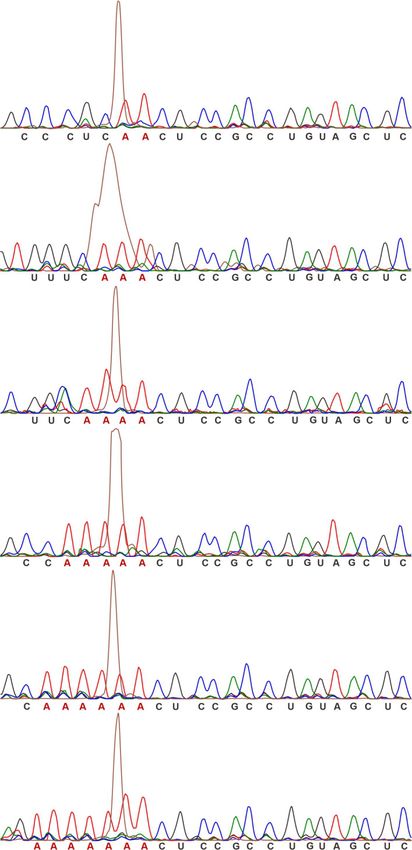
Figure 3. T-variant catalytic activity in vitro. (A) Sequences of typical T-variants in S.mansoni for in vitro cleavage activity investigation, compared to
published N. vitripennis twister ribozyme. Sequence accession numbers and locations are shown. The 5 end and 3 ends of the P1 stem are marked as red
shadow. (B) Test of in vitro cleavage activity of T-variants 1A and 0A, compared to the N. vitripennis twister ribozyme, based on the structure of the N.
vitripennis ribozyme, T-variants 1A and 0A RNAs were divided into substrate (S) and enzyme (E) strands. Purified strands were mixed in the combinations
shown in the figure and incubated at 37◦ C for 2 h in 30 mM HEPES, pH 7.5, 100 mM KCl and 10 mM Mg2+ , the cleavage products (5 Clv) of 5 6-FAM-
labeled substrate RNA samples were resolved on 8% denaturing polyacrylamide gels. (C) T-variant-2A-7A and N. vitripennis ribozyme cleavage activities
in vitro, T-variants 2A-7A RNAs were divided into substrate (S) and enzyme (E) strands and cleavage products identified as before. (D) T-variant activity
in the presence of divalent metal ions. The 5 6-FAM-labeled substrate was incubated with excess enzyme RNA for 15 min in the absence (–) or presence
of 10 mM divalent metal ion as indicated. (E) Comparison of in vitro cleavage activity of T-variant sequences 5 to the cleavage site for the cleavage triplet
NAA where N = A, G, C or U. (F) T-variant-2A and 5A and N. vitripennis ribozyme cleavage kinetics in vitro. Time courses were performed; E + S
strands were mixed and incubated at 37◦ C in 30 mM HEPES, pH 7.5, 100 mM KCl and 10 mM Mg2+ , and samples removed after incubation at the
given times (t). (G) Plots of ribozyme cleavage for the N. vitripennis twister, T-variant-2A and 5A ribozymes taken from Supplementary Figure S7, the
first order rate constants (kobs ) of T-variant were calculated by plotting the fraction of substrate cleaved (ft) versus time (t) and fitting to the equation
ft = 1 – exp(kobs t) with GraphPad Prism 6.01. Error bars are the standard deviation of three independent experiments. (H) Comparison of kobs for each
T-variant (2A–7A) and N. vitripennis twister ribozyme as measured in (E), relative kobs , krel = kT-variant/k N. vitripennis twister. (I) Maximal cleavage
(cleavage plateau) for the N. vitripennis and T-variant-2A to 7A and ribozymes. (J) Cleavage site mapping of T-variants 2A–7A by capillary electrophoresis.
The purified transcription products of self-cleaved T-variant 2A–7A RNAs were reverse transcribed and subjected to capillary electrophoresis, relative to
dideoxy markers. In each panel the peak corresponding to self-cleavage is shown in russet and the location of the cleavage site marked with a red arrow
above the marker peaks.10582 Nucleic Acids Research, 2021, Vol. 49, No. 18
and the LacZ protein coding sequence. The transcription 3A mRNA, due to T-variant 3A mRNA in vivo cleav-
of downstream sequences is dependent on the NMT41 pro- age activity that is not present in the T-variant 3A (Figure
moter in the absence of thiamine. In the presence of thi- 4E). In contrast, similar amounts of LacZ mRNA were de-
amine, transcription from the NMT41 promoter should tected in cells with T-variant 3A mRNA or T-variant 3A
be significantly repressed although incomplete repression mRNA using (F2 + R2) primer pairs (Figure 4E). Although
with reduced levels of transcription has been reported (86– similar amounts of LacZ mRNA were observed, the LacZ
88). Thiamine repression can be used as a control for re- protein expression was still much higher in the T-variant
porter assays. The DNA fragment corresponding to the ac- 3A constructs in the reporter assay (Figure 4C). These re-
tive T-variant 3A sequence and control sequences was in- sults suggest that the expression of LacZ is dependent on
serted downstream of the NMT41 promoter and upstream whether the T-variant is cleaved. 5 RACE was further used
of LacZ (Figure 4A and B). The effect of T-variant on to map the T-variant 3A cleavage site in vivo. The reverse
downstream gene expression can be measured through ex- primer P2 in the 5 RACE generated a fragment of 310 nt
pression of the LacZ reporter. Control constructs, consist- up to the transcription start site and the reverse primer P1
Downloaded from https://academic.oup.com/nar/article/49/18/10573/6374148 by guest on 18 October 2021
ing of T-variant 3A that lacks a cleavage site (T-variant in the 5 RACE generates a fragment of 625 nt from the
3A), a deletion of the T-variant 3A which has only the up- cleavage site for T-variant 3A in vivo (Figure 4B, F). The
stream and downstream sequences (No T-variant 3A) were 5 RACE sequence (Figure 4G) revealed the cleavage site
constructed in parallel (Supplementary Table S4). The HCV of the T-variant 3A in vivo to be the same as observed in
internal ribosomal entry site (HCV-IRES) RNA is a highly vitro (Figure 3J). T-variant cleavage removes the 5 end of
structured RNA (65) that is unrelated to the T-variant se- the RNA including the 5 -cap but leaves the residual struc-
quences in S. mansoni was also used as a control (Supple- tured ribozyme. These results suggest that T-variant cleaves
mentary Table S4). in vivo and that the cleaved T-variant is sufficient for the
The plasmids were transformed into the host strain hleu1- translation of its downstream genes, implying that cleav-
32 (a gift from Jurg Bahler) and grown in the presence or ab- age of T-variants in S. mansoni is required for translation
sence of thiamine. In the absence of thiamine (NMT41 pro- of APE/RT the key protein for Perere-3 retrotranspostion.
moter is active), the expression of Lac Z was detected in the
construct containing active T-variant 3A, while only very
T-variant ribozyme activity and Perere-3 non-LTR retro-
low levels of Lac Z expression were detected in the construct
transposon element replication in S. mansoni
with the inactive T-variant 3A that lacks cleavage site or
in the construct with only the upstream and downstream se- If T-variants function as ribozymes to process RNA in
quences (No T-variant 3A) in which the T-variant 3A has Perere-3 non-LTR retrotransposon elements in S. man-
been deleted (Figure 4C), suggesting that LacZ expression soni, as proposed in Figure 2H, two primary require-
is associated with the activity of the T-variant 3A. However, ments have to be met. The T-variants and Perere-3 el-
in the presence of thiamine when the NMT41 promoter is ements must be transcribed together into RNA, and
repressed, reduced levels of Lac Z expression were observed the T-variants must subsequently cleave the transcribed
in the constructs containing active T-variant 3A, inactive T- RNA. To identify RNA transcripts for T-variants and
variant3A or No T-variant 3A, compared to the samples Perere-3, we carried out a search in the Ensembl EST
in the absence of thiamine (Figure 4C). These control re- database that contains the assembled RNA transcripts
sults suggest that the expression of Lac Z is dependent on in S. mansoni (ftp://ftp.ensemblgenomes.org/pub/metazoa/
both the transcription and the activity of T-variant 3A. No current/fasta/schistosoma mansoni/cdna). The S. mansoni
Lac Z expression was observed in the control construct con- genome is 65% AT rich (50), promoter prediction identi-
taining the unrelated HCV-IRES sequence in the presence fied a number of possible promotor sequences upstream of
or absence of thiamine (Figure 4C). To further investigate the T-variant sequences (71) (Supplementary table S5). The
if T-variants have effects on downstream gene expression in cDNA data in the Ensembl EST database confirms the tran-
general, constructs containing 5 genomic Perere-3 T-variant scription of active promoters in vivo. The results for rep-
sequences were made and the LacZ expression measured. In resentative transcript RNA sequences are shown in Figure
each case LacZ expression of the additional sequences was 5A. The genomic locations of these RNA transcripts and
observed. The LacZ expression in the constructs containing the transcript IDs are shown in Figure 5B and C. These
T-variants 1–5 and is comparable with that of T-variant 3A results reveal that there are indeed RNA transcripts for T-
(Supplementary Table S4, Figure 4D). variants with Perere-3, suggesting that there is an active pro-
These results indicate a relationship between the activ- moter upstream of the T-variants. In Perere-3 the APE/RT
ity of T-variant 3A RNA and the LacZ expression in vivo. domain is a single ORF in S. mansoni (58).
To further investigate the in vivo cleavage activity of the T- For each sequence, TSDs are observed flanking the
variant 3A, RNA was extracted from the strains express- APE/RT/Perere-3 element, and the TSDs are directly adja-
ing the active T-variant 3A and cleavage site deletion T- cent to the T-variant cleavage site AA (Figure 5A, D). TSDs
variant 3A RNAs. Real-time PCR experiment was per- are a footprint for TPRT and evidence that retrotranspo-
formed to compare the amount of T-variant 3A mRNA and sition of the Perere-3 element has taken place. Each TSD
T-variant 3A mRNA by using cross cleavage site primer is generated by the insertion of double-stranded DNA that
pairs (F1 + R1) (Figure 4B). The amount of LacZ mRNA was synthesized from the cleaved T-variant RNA template.
was measured in each of the two constructs by primer pairs The observation that the TSDs are immediately next to
(F2 + R2) (Figure 4B). The results show that the amount of the T-variant cleavage sites provides evidence that T-variant
T-variant 3A mRNA is less than half (49%) of T-variant cleavage has taken place in vivo.Nucleic Acids Research, 2021, Vol. 49, No. 18 10583
A C D
Downloaded from https://academic.oup.com/nar/article/49/18/10573/6374148 by guest on 18 October 2021
B E F
G
Figure 4. Reporter constructs and T-variant in vivo catalytic activity. (A) Sequence and secondary structure of T-variant 3A. (B) The reporter plasmid
constructs. The wild type T-variant 3A for 5 RACE is located behind an NMT41 thiamine repressible promoter and in front of a lacZ reporter gene. The
gene specific primers P1 and P2 were used to map the T-variant 3A in vivo cleavage site and transcription start sites respectively. The T-variant 3A cleavage
site as determined in vitro is marked by the red arrow and the predicted P1 and P2 primer fragment sizes of cleaved and whole transcript shown. The control
plasmid constructs for real-time PCR and Miller assay are shown in the grey box: In parallel with T-variant 3A (red box), a sequence containing only the
peripheral sequence of the Perere-3 5 -UTR without the ribozyme (No T-variant 3A) or with an HCV-IRES were substituted into the position of the red box
in the lacZ reporter. (C) Miller assay of lacZ reporter activity ± thiamine for T-variant 3A, no T-variant and HCV IRES plasmid constructs. Note the high
levels of reporter gene expression for T-variant 3A relative to the controls when the ribozyme is active. (D) Miller assay of lacZ reporter activity ± thiamine
for T-variant sequences 1 to 5, relative to the control constructs T-variants 3A and 3A. (E) Real-time PCR analysis of mRNA abundance of the lacZ RNA
(F2 and R2 primers) relative to the Ampicillin internal reference, showing the level of mRNA abundance of lacZ in the wild type T-variant 3A remains
stable after T-variant cleavage, compared to the controls. Error bars represent the standard deviation of three independent experiments. (F) Agarose gel of
PCR product of 5 RACE; the P1 primer generates a 625 nt T-variant cleavage product. (G) Capillary electrophoresis sequencing of original plasmid DNA
compared to the 5 RACE of cleaved T-variant 3A in vivo, the T-variant 3A cleavage site is marked by the red arrow.
The TSD for each of the sequences is different, suggest- insertion is closely linked to the activity of the T-variant
ing that insertion of the cleaved T-variant associated with through the P1 stem (Figure 5D). TSD generation in the
Perere-3 is not site-specific (Figure 5A, D). Since the S. man- replication cycle is located in the position that corresponds
soni genome is AT rich, the insertion would be predicted to to the 5 end of P1 stem of T-variant. The P1 stem is formed
be more likely next to sequences with an A or T (50). Be- by base paring of the 5 and 3 ends of the T-variant. Since
cause the TSDs are located 5 to the genomic position cor- the 3 end of the P1 stem is imbedded within Perere-3, it
responding to the T-variant cleavage site in the RNA, they remains unchanged during retrotransposition. However, 5
also form the P1 stem of the T-variant and the sequence TSD can generate variable sequences in the 5 strand of the
composition of the P1 stem also affects the activity of the T- P1 stem. Changes in base-pairing between the variable 5
variants (Figure 3F, G, H, I and Supplementary Figure S7). strand and the unchanged 3 strand of P1 may stabilise or
Thus, the TSD sequence generated by T-variant/Perere-3 destabilise P1, potentially enhancing or inhibiting T-variant10584 Nucleic Acids Research, 2021, Vol. 49, No. 18
A D
Downloaded from https://academic.oup.com/nar/article/49/18/10573/6374148 by guest on 18 October 2021
B
C
Figure 5. T-variant and Perere-3 retrotransposition. (A) cDNA sequences of four T-variants and downstream ORFs containing APE domain and RT
domain from Ensembl cDNA database. The T-variant sequences are highlighted in pink, with secondary structural stems and loops marked. The arrow
marks the T-variant cleavage site. The 5 TSD next to the cleavage site and the 3 TSDs next to the short repeats are marked with red boxes. The APE
and RT domains are highlighted in turquoise and blue. (B) The location of the sequences (from A) on S. mansoni chromosomes. (C) Genomic accession
number, location number and transcript location number of the sequences in (A). (D) Schematic of the possible process by which T-variant cleavage
and retrotransposon insertion in the Perere-3 replication cycle leads to TSD and the formation of a new ribozyme P1 stem. The formation of an active
ribozyme generates a viable retrotransposon that is available for further retrotransposition, conversely the formation of a lower efficiency ribozyme would
be predicted to lead to a reduction in retrotransposon activity.
cleavage (Figure 5D), which is consistent the T-variant ac- DISCUSSION
tivities observed in vitro (Figure 3F, G, H, I and Supple-
Here, we have identified over 800 T-variant sequences (Fig-
mentary Figure S7). A T-variant/Perere-3 retrotransposi-
ure 1) and investigated their potential function in S. Man-
tion event that makes a TSD sequence and P1 stem that
soni. Several lines of evidence point to an important func-
generates an active T-variant would enable Perere-3 to re-
tional role for T-variant ribozymes in the non-LTR retro-
main active during the replication cycle (Figure 5D). In con-
transposon replication cycle: (1) The genomic location of
trast, a T-variant/Perere-3 retrotransposition that generates
T-variant ribozyme is upstream of the Perere-3 retrotrans-
a TSD that leads to a loss of T-variant activity would poten-
poson element containing APE/RT domains (Figures 2A-
tially impact Perere-3 replication through effects on down-
G and 5). (2) T-variant ribozymes were shown to be ac-
stream gene expression as seen in the reporter assays (Figure
tive in vitro and in vivo (Figures 3 and 4). (3) Reporter as-
4). Therefore, the dependence of T-variant activity on the
says show that T-variant cleavage is required for transla-
insertion site (by TSD) through retrotransposition is linked
tion of the downstream gene (Figure 4). (4) T-variants and
to the activity of Perere-3 during its replication cycle.
Perere-3 are cotranscribed in S. Mansoni (Figure 5A). TSDs
The proposed model in Figure 5D explains the function
are generated by the repair of the intermediates of retro-
of the T-variant in the Perere-3 replication based on these
transposon DNA insertion in the final integration step and
results. If the proposed model is reasonable, the distances
are therefore evidence of retrotransposon insertion (53,89).
between the T-variant and the AUG of the Perere-3 ele-
TSDs flank the inserted retrotransposons of T-variant se-
ments are expected to be similar. The distances between
quences (Figures 5A, 2A, B) and are positioned right next
the T-variant and the AUG of all 345 Perere-3 elements
to the T-variant cleavage sites, suggesting T-variant cleav-
was analysed manually. For the majority of the Perere-3 el-
age in vivo and active ribozyme sequences were involved in
ements (∼306 out of 345 with RT domain), the distance be-
successful Perere-3 retrotransposition. Differences in TSDs
tween the T-variant and the reported AUG is ∼147 nt (211
suggest that Perere-3 transposition is not site-specific. The
sequences) and there are 95 sequences with distances ∼112
TSDs contribute to the 5 P1 stem of the T-variants and may
nt (T-variant to predicted AUG I). There are small num-
effect T-variant structure and function which can in turn
bers of other distances (Supplementary Table S6). The av-
impact Perere-3 transposition. Together this evidence sug-
erage and mean distances between T-variant and the AUG
gests a function for the T-variants in Perere-3 retrotranspo-
are shown in (Supplementary Table S6). The distances be-
son replication (Figure 5D).
tween the T-variants and the AUG naturally falls into two
An understanding of the RNA template that is involved
main groups (Supplementary Table S6), supporting the no-
in reverse transcription is required for dissection of retro-
tion that within each group the proposed model explains the
transposon integration reaction. There are similarities and
function of the T-variant in Perere-3 replication.You can also read