Functional detection of botulinum neurotoxin serotypes A to F by monoclonal neoepitope-specific antibodies and suspension array technology
←
→
Page content transcription
If your browser does not render page correctly, please read the page content below
www.nature.com/scientificreports

                        OPEN       Functional detection of botulinum
                                   neurotoxin serotypes A to F by
                                   monoclonal neoepitope-specific
Received: 20 December 2018
Accepted: 15 March 2019            antibodies and suspension array
                                   technology
Published: xx xx xxxx

                                   Laura von Berg1, Daniel Stern 1, Diana Pauly1,4, Stefan Mahrhold2, Jasmin Weisemann2,
                                   Lisa Jentsch1, Eva-Maria Hansbauer1, Christian Müller3, Marc A. Avondet3, Andreas Rummel                        2
                                                                                                                                                    ,
                                   Martin B. Dorner1 & Brigitte G. Dorner1
                                   Botulinum neurotoxins (BoNTs) are the most potent toxins known and cause the life threatening disease
                                   botulism. Sensitive and broad detection is extremely challenging due to the toxins’ high potency and
                                   molecular heterogeneity with several serotypes and more than 40 subtypes. The toxicity of BoNT is
                                   mediated by enzymatic cleavage of different synaptic proteins involved in neurotransmitter release
                                   at serotype-specific cleavage sites. Hence, active BoNTs can be monitored and distinguished in vitro
                                   by detecting their substrate cleavage products. In this work, we developed a comprehensive panel
                                   of monoclonal neoepitope antibodies (Neo-mAbs) highly specific for the newly generated N- and/or
                                   C-termini of the substrate cleavage products of BoNT serotypes A to F. The Neo-mAbs were implemented
                                   in a set of three enzymatic assays for the simultaneous detection of two BoNT serotypes each by
                                   monitoring substrate cleavage on colour-coded magnetic Luminex-beads. For the first time, all relevant
                                   serotypes could be detected in parallel by a routine in vitro activity assay in spiked serum and food
                                   samples yielding excellent detection limits in the range of the mouse bioassay or better (0.3–80 pg/mL).
                                   Therefore, this work represents a major step towards the replacement of the mouse bioassay for botulism
                                   diagnostics.

                                   Botulinum neurotoxins (BoNTs) represent the most poisonous biological substances known today and are the causative
                                   agents of the rare but severe neurological disease botulism in humans and animals1,2. In humans, botulism
                                   can be caused by ingestion of food contaminated with the toxins (food-borne botulism). Uptake and outgrowth of
                                   bacterial spores may lead to infant or wound botulism3. Botulism is characterised by descending flaccid paralysis
                                   due to a blockage of neurotransmitter release at neuromuscular junctions, which potentially leads to death due to
                                   respiratory failure. The exclusive neurospecificity and paralysing effect of BoNT makes it an effective therapeutic
                                   against a broad range of neurological and non-neurological diseases and in aesthetic medicine4,5.
                                       On the molecular level, BoNTs are a highly variable group of toxins produced by ubiquitously occurring
                                   Gram-positive, spore forming, anaerobe Clostridium spp. (C. botulinum groups I–IV, C. baratii, and C. butyri-
                                   cum). Until now, seven widely accepted “classical” BoNT serotypes designated BoNT/A–G are known of which
                                   six naturally induce botulism in humans (BoNT/A, B, E, F) or animals (predominantly BoNT/C and D and
                                   their mosaic variants CD and DC)6–9. BoNT/G has not been clearly assigned to a natural outbreak in humans or
                                   animals9. Additionally, several novel BoNT molecules have been pronounced: BoNT/HA (also called BoNT/FA
                                   or BoNT/H) which was involved in an infant botulism case and has been characterised as a novel mosaic type
                                   toxin10–12 and BoNT/X,13 both produced or encoded by C. botulinum, as well as eBoNT/J (aka BoNT/En) encoded

                                   1
                                     Biological Toxins (ZBS 3), Centre for Biological Threats and Special Pathogens, Robert Koch Institute, Berlin,
                                   13353, Germany. 2Institut für Toxikologie, Medizinische Hochschule Hannover, 30625, Hannover, Germany.
                                   3
                                     Spiez Laboratory, Federal Office for Civil Protection, Spiez, 3700, Switzerland. 4Department of Ophthalmology,
                                   University Hospital Regensburg, Regensburg, 93053, Germany. Correspondence and requests for materials should
                                   be addressed to B.G.D. (email: DornerB@rki.de)

        Scientific Reports |   (2019) 9:5531 | https://doi.org/10.1038/s41598-019-41722-z                                                              1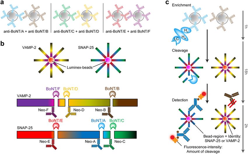
www.nature.com/scientificreports/                                                                  www.nature.com/scientificreports

                           Figure 1. Cleavage sites of BoNT serotypes pathogenic to humans or animals in SNAP-25 or VAMP-2.
                           Serotypes can be distinguished by their substrate specificity and specific cleavage position. After cleavage, neo-
                           epitopes arise on both the C- and N-terminal ends of the cleavage sites that can be detected by highly specific
                           neo-epitope antibodies.

                           by Enterococcus faecium14–16. Advanced whole genome sequencing technologies revealed differences within a
                           given serotype for BoNT/A, B, E and F leading to the introduction of subtypes and adding further variability
                           to the BoNT family6. The subtypes can differ up to 36% on the amino acid level within a given serotype. So far,
                           more than 40 subtypes have been described in the literature9. Subtypes of a given serotype have been shown to
                           differ in their biological activity, e.g. their kinetics of uptake and substrate cleavage, affinity to receptors or overall
                           activity17–21.
                               The mechanism of action is mediated by different domains of the toxin: BoNT is produced as a 150 kDa holo-
                           toxin which, upon proteolytic activation, is cleaved into a 100 kDa heavy chain (HC) and a 50 kDa enzymatically
                           active light chain (LC) which remains connected to HC by a single disulfide bond22. The HC consists of a 50 kDa
                           C-terminal receptor binding domain (HC) and a 50 kDa translocation domain (HN). BoNT exerts its extreme
                           potency by binding to specific neuronal receptors mediated via its HC domain, subsequent uptake into recycling
                           endosomes followed by HN-mediated translocation of the LC into the cytosol, and finally LC-mediated cleavage
                           of soluble N-ethylmaleimide-sensitive-factor attachment receptor (SNARE)-complex proteins thereby blocking
                           neurotransmitter release23–27. Here, different BoNT serotypes cleave different SNARE proteins at distinct sites:
                           synaptosomal-associated protein of 25 kDa (SNAP-25) is cleaved by BoNT/A, C, and E while vesicle-associated
                           membrane protein 2 (VAMP/synaptobrevin-2) is cleaved by BoNT/B, D, F, and G (Fig. 1). BoNT/C additionally
                           cleaves syntaxin and is therefore the only classical serotype that hydrolyses two substrates28. Each serotype has
                           a unique cleavage site on the respective substrate molecule, indicating that the presence of different BoNT sero-
                           types can be detected by analysis of the cleavage products generated26,29. Notably, different subtypes of a given
                           serotype target the very same cleavage site on the respective substrate with one known exception: BoNT/F5 uses
                           a different cleavage site in VAMP-2 than all other BoNT/F subtypes; the same cleavage site is also targeted by the
                           recently identified BoNT/HA11,30.
                               Because of the large variability of BoNTs on the molecular level, its detection is highly challenging. Due to
                           its high potency minute amounts in the low pg per mL range of all different BoNT serotypes must be detected
                           reliably in complex clinical, food or environmental samples31,32. Based on these facts the mouse bioassay (MBA)
                           is still considered the gold standard for diagnostics of botulism. Here, particle-free sample material (e.g. serum,
                           bacterial cultures, homogenised food matrices) diluted in buffer is injected intraperitoneally into mice which are
                           subsequently monitored for up to four days for clinical signs of botulism such as laboured breathing, a wasp-like
                           narrowed waist due to increased respiratory efforts, weakness of limbs that progress to total paralysis and finally
                           death by respiratory failure31,33. The disease causing serotype is determined by toxin-neutralisation employing
                           serotype-specific antibodies which increases the number of animals needed. The MBA is highly sensitive with a
                           50% lethal dose (LD50) in mice corresponding to 5 to 50 pg per mouse for BoNT/A, B, E and F, respectively34,35,
                           reflecting the pharmacokinetics and pharmacodynamics, the latter comprising receptor binding, uptake, trans-
                           location and enzymatic activity of all serotypes. Additionally, the MBA can detect all ‘mouse-toxic’ sero- and
                           subtypes (including any yet unknown subtypes) out of complex matrices, which is crucial for reliable diagnostics.
                           However, drawbacks are long assay duration of up to four days, considerable costs associated with animal hus-
                           bandry, several technical limitations such as species differences between mice and humans36–38 and, importantly,
                           serious ethical concerns due the suffering inflicted on the animals33.
                               Therefore, alternative assays to replace the MBA have long been pursued31–33,39,40. Of those, endopeptidase
                           assays monitor the toxin’s catalytic activity, which displays the major determinant of BoNT toxicity. A highly
                           sophisticated approach successfully employing this principle has been established for BoNT/A to G by moni-
                           toring cleavage products of short serotype-specific peptide substrates by mass spectrometry (MS; Endopep-MS
                           assay)41,42. While the Endopep-MS assay allows for the sensitive detection of all known serotypes with high con-
                           fidence, the applicability of this approach is limited to expert laboratories due to the high level of expertise and
                           expensive technical instrumentation needed. For a broader application, substrate peptides labelled with flanking
                           fluorescence donor and quencher molecules have been designed to measure the catalytic activity of BoNT by
                           Förster resonance energy transfer (FRET)43–49. Advantages of FRET-based approaches are their high sensitivity
                           combined with a simple and fast assay protocol. Furthermore, commonly available laboratory equipment is suf-
                           ficient to perform the assays ensuring a broad applicability in routine laboratories. A major drawback, however,
                           is that serotypes targeting the same substrate cannot be distinguished by this method, since cleavage at every site

Scientific Reports |   (2019) 9:5531 | https://doi.org/10.1038/s41598-019-41722-z                                                                   2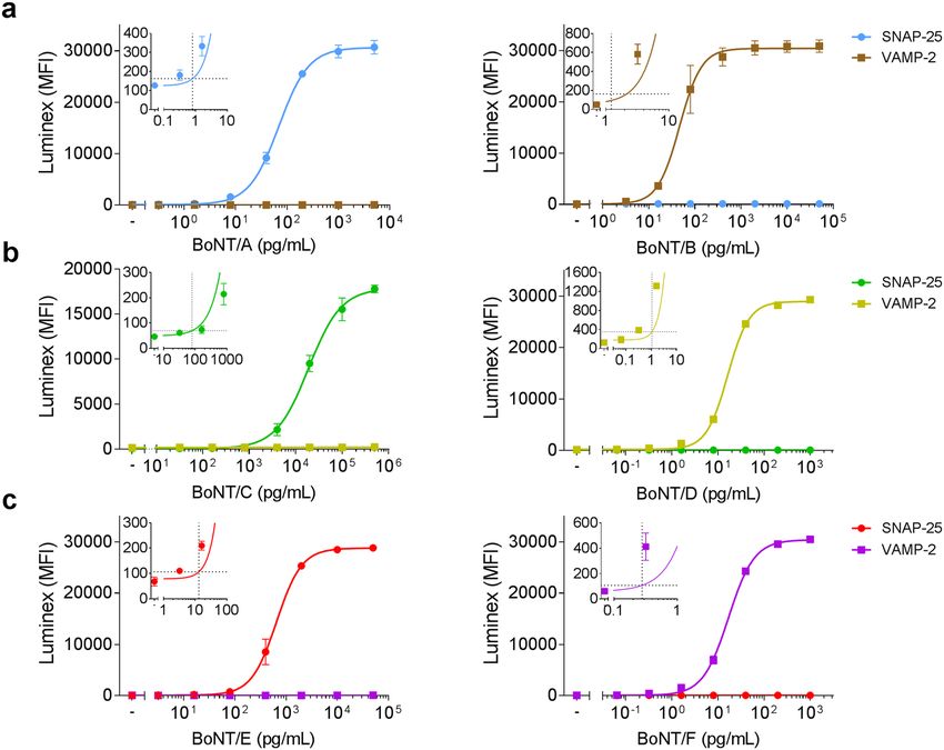
www.nature.com/scientificreports/                                                              www.nature.com/scientificreports

                           between donor and acceptor fluorophore generates a signal. Shorter peptide substrates enabling a discrimination
                           of serotypes E or A/C and B or F/D may be applied, but presumably lead to a loss in sensitivity due to the lack
                           of important exosites49. Still, the discrimination of cleavage by BoNT/A and C or BoNT/F and D is not possible
                           without further substrate modifications, since their respective cleavage sites on SNAP-25 or VAMP-2 are only
                           one amino acid apart (Fig. 1). Noteworthy, cleavage of the substrate peptide used in FRET endopeptidase assays
                           by other proteases present in the sample leads to false positive results. To develop straightforward assays capable
                           of discriminating between all BoNT serotypes, neoepitope specific antibodies (Neo-Abs) have been introduced.
                           Per definition, Neo-Abs exclusively recognise the newly exposed epitopes in the substrate molecules SNAP-25
                           or VAMP-2 after cleavage by a given BoNT, but do not recognise the intact, uncleaved substrate or substrate
                           cleaved at a distinct position50. Different assay platforms employing polyclonal Neo-Abs have been proposed for
                           the detection of BoNT/A, B, C, D, E, and F51–57. However, polyclonal Neo-Abs have major drawbacks for routine
                           diagnostics: they imply the risk of cross-reactivity against uncleaved substrate as well as neighbouring cleavage
                           sites, especially in case of BoNT/A and C or BoNT/D and F, both separated by one amino acid. In addition,
                           detection of BoNT in complex matrices may be hampered by interferences of the polyclonal Neo-Abs with matrix
                           components58,59. Due to their higher specificity monoclonal antibodies can overcome these limitations but their
                           generation is challenging, especially for reagents targeting directly adjacent cleavage sites. So far, assays using
                           monoclonal neoepitope specific antibodies (Neo-mAbs) have been described for BoNT/A and E only60–65.
                               In this work, we generated a unique and comprehensive panel of monoclonal antibodies directed against
                           neoepitopes of VAMP-2 or SNAP-25 after cleavage by BoNT/A, B, C, D, E and F. Out of 20 Neo-mAbs gener-
                           ated, six outperforming antibodies (one for each serotype BoNT/A to F) were selected and implemented in a
                           novel functional suspension array based on the Luminex platform66: The Neo-mAbs were implemented in a set
                           of three enzymatic assays for the simultaneous detection of two BoNT serotypes each by monitoring substrate
                           cleavage on colour-coded magnetic Luminex-beads. After toxin enrichment using monoclonal antibodies cou-
                           pled to paramagnetic beads the assays were capable of detecting enzymatically active toxin with detection limits
                           of 0.3–13 pg/mL for BoNT/A, B, E and F and 79.1 pg/mL and 1.1 pg/mL for BoNT/C and D, respectively, when
                           testing toxin-spiked buffer, serum or food. Based on its sensitivity and specificity, the novel functional suspension
                           array represents a major advancement in BoNT diagnostics with the potential to replace the MBA in routine
                           applications.

                           Results
                           Generation of highly specific monoclonal neoepitope specific antibodies.                        A major aim of this
                           study was to develop a comprehensive panel of Neo-mAbs targeting the newly generated N- or C-termini in the
                           cleavage products of SNAP-25 or VAMP-2 after proteolysis by BoNT/A to F. This task was especially challenging
                           for the serotypes BoNT/A and C as well as BoNT/D and F which target adjacent peptide bonds in SNAP-25 or
                           VAMP-2, resulting in cleavage sites separated by only one amino acid (Fig. 1). Mice were immunised with BSA-
                           coupled 8-mer peptides corresponding to the new N- or C-terminal sequence of the respective cleavage products
                           to induce antibodies restricted to the eight terminal amino acids adjacent to the cleavage site (Fig. 1). Hybridoma
                           clones were subjected to a stringent screening procedure to ensure that only the cleavage products but neither
                           the uncleaved substrate nor the cleavage products of a different serotype were detected.To this aim we tested
                           binding towards the specific peptides as well as peptides covering the other cleavage sites at the same substrate by
                           indirect ELISA. Those peptides were coupled to KLH to exclude antibodies recognising the carrier protein BSA.
                           Furthermore, binding to intact SNAP-25 or VAMP-2 was tested to exclude cross-reactive antibodies from further
                           characterisation. Overall, more than 20,000 hybridoma clones generated in 10 fusions were tested for production
                           of Neo-mAbs. Eventually, 20 promising Neo-mAbs were identified based on their high reactivity against specific
                           peptides targeting the different cleavage products of SNAP-25 and VAMP-2 after proteolysis by BoNT/A to F,
                           respectively, and the lack of cross-reactivity against intact substrate (Table 1 and Fig. S1).
                               To select antibodies most suitable for the implementation in a functional suspension array we compared the
                           performance of all 20 mAbs in an endopeptidase cleavage ELISA (Endopep-ELISA) as described previously by
                           Jones et al.55. VAMP-2 or SNAP-25 was coated on microtitre plates, hydrolysed by serial dilutions of BoNT/A to F
                           and cleavage products were detected by Neo-mAbs. As shown in Fig. S2, remarkable differences in performance
                           were observed for the 20 Neo-mAbs in Endopep-ELISA, although some mAbs were seemingly equivalent in
                           their recognition of 8-mer peptides representing individual neo-epitopes (Fig. S1, e.g. mAb VAMP/D/27 and
                           VAMP/D/29). Based on the Endopep-ELISA results six Neo-mAbs showing superior performance – one for each
                           serotype – were selected for a more detailed characterisation (indicated in bold in Table 1).
                               We first tested specificity towards cleaved but not uncleaved substrate by Western blotting. To this aim,
                           BoNT-cleaved and intact VAMP-2 or SNAP-25 were analysed by SDS-PAGE, Coomassie staining and Western
                           blotting. Here, all six antibodies exclusively recognised the cleaved, but not the intact substrate (Fig. 2a).
                           A crucial requirement of the selected Neo-mAbs was their specific recognition of only one cleavage site,
                           while cross-reactivity towards adjacent cleavage sites had to be excluded. We therefore tested each antibody
                           against substrates cleaved with high concentrations (10 ng/mL) of the next-neighbour BoNT molecules by
                           Endopeptidase-ELISA. Importantly, the six antibodies were exclusively specific for a single neoepitope in cleav-
                           age products of SNAP-25 or VAMP-2, allowing the clear discrimination of catalytic activity of BoNT/A to F. This
                           was particularly notable for BoNT/A and C as well as for BoNT/D and F, since the cleavage sites of these serotypes
                           differ by only one amino acid position (Fig. 2b). Hereby, much of the complexity associated with the comprehen-
                           sive detection of all known and yet unknown subtypes within each serotype is reduced to detection of only six
                           enzymatically active “cleavotypes”9,32.
                               The six selected Neo-mAbs were further used for setting up a novel Luminex-based suspension array for the
                           detection of enzymatically active BoNT/A to F.

Scientific Reports |   (2019) 9:5531 | https://doi.org/10.1038/s41598-019-41722-z                                                             3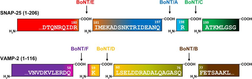
www.nature.com/scientificreports/                                                              www.nature.com/scientificreports

                                                                               Endopep-
                            Serotype   Antibodya      Isotype   Specificityb   ELISAc
                                       SNAP/A/291     IgG1      N              +++
                            BoNT/A
                                       SNAP/A/305     IgG1      N              +++
                                       VAMP/B/1148    IgG1      N              +++
                                       VAMP/B/226     IgG1      N              +++
                            BoNT/B     VAMP/B/151     IgG2b     C              +
                                       VAMP/B/392     IgG1      C              −
                                       VAMP/B/726     IgG3      C              −
                                       SNAP/C/2207    IgG1      C              −
                                       SNAP/C/5593    IgG2a     N              +++
                            BoNT/C
                                       SNAP/C/1844    IgG2a     N              −
                                       SNAP/C/3280    IgG2b     N              −
                                       VAMP/D/27      IgG2b     C              +++
                            BoNT/D
                                       VAMP/D/29      IgG2b     C              ++
                                       SNAP/E/1466    IgG1      C              −
                            BoNT/E
                                       SNAP/E/217     IgG1      N              +
                                       VAMP/F/440     IgG2a     C              +++
                                       VAMP/F/153     IgG1      N              −
                            BoNT/F     VAMP/F/521     IgG1      N              +++
                                       VAMP/F/425     IgG1      N              +++
                                       VAMP/F/1333    IgG1      N              +++

                           Table 1. Overview of all Neo-mAbs generated in this work for the detection of catalytically active BoNT.
                           a
                            Antibodies depicted bold were selected for implementation into the functional Luminex suspension array.
                           b
                             Specificity of Neo-mAbs towards N- or C-terminal fragment generated after cleavage by indicated BoNT. cEC50
                           in Endopep-ELISA: + + + = < 0.1 ng/ml; + + = < 1 ng/ml; + = < 10 ng/ml; −  = signal too weak for detection.

                           Establishment of three Luminex duplex-assays for the simultaneous detection of SNAP-25- and
                           VAMP-2-targeting serotypes. Assay principle and setup. The primary goal of this work was to establish
                           an assay that enables the detection of BoNTs from complex matrices in a routine diagnostic setting as an alter-
                           native to the mouse bioassay. Previous works had encountered matrix interferences when using cleavage based
                           assays for the analysis of complex clinical or food samples (e.g. due to unspecific proteases or incompatible buffer
                           requirements31,61,67). Therefore, a toxin enrichment step was included prior to the Luminex assay to enrich the
                           toxins and to provide optimum buffer conditions for subsequent substrate cleavage. To this end, paramagnetic
                           beads were coupled with monoclonal anti-BoNT antibodies generated in our laboratory targeting the HC or
                           HN domains of BoNT/A to F (Supporting methods, Table S1, Fig. S3). By using the toxins’ receptor-binding or
                           translocation domains for enrichment a potentially negative influence on the enzymatic activity by antibodies
                           targeting the LC was excluded68.
                               To enable the detection of all clinically relevant BoNT serotypes with minimal sample consumption we imple-
                           mented the six candidate Neo-mAbs in a novel enzymatic multiplex suspension assay using the Luminex plat-
                           form. This system uses colour-coded paramagnetic Luminex beads, thereby enabling the detection of multiple
                           analytes in one sample. Initially, we were aiming at coupling the Neo-mAbs to the Luminex beads to capture only
                           cleaved biotinylated SNAP-25 and VAMP-2 substrates from solution. This setup would have enabled a simul-
                           taneous detection of all six serotypes from one sample. However, this hexaplex approach failed, mainly due to
                           unexpectedly strong unspecific binding of VAMP-2 substrate to the Luminex beads (Fig. S4a). Although this
                           unspecific binding could be blocked efficiently by the addition of carboxymethyl-dextran, the overall sensitivity
                           of this assay set-up was unsatisfactory (Fig. S4b).
                               As an alternative, we swapped orientation of the assay set-up and immobilised SNAP-25 and VAMP-2 on
                           two different Luminex bead regions. By adding the immuno-enriched toxins directly to the Luminex beads
                           with immobilised substrates, an on-bead cleavage allows for the discrimination between one SNAP-25- and one
                           VAMP-2-specific BoNT serotype at a time (Fig. 3). Here, the experimental conditions were optimised to ensure
                           maximum sensitivities by identifying a cleavage buffer that allowed for efficient cleavage of all serotypes. In addi-
                           tion, we evaluated different cleavage durations (30 min to 18 h) achieving maximum sensitivity after overnight
                           incubation (Fig. S5). After cleavage, the newly formed neoepitopes were detected by Neo-mAbs. By combining
                           Neo-mAbs against one SNAP-25- and one VAMP-2-cleaving serotype, three duplex-assays for the simultaneous
                           detection of either serotypes BoNT/A and B or BoNT/E and F (all pathogenic to humans) as well as BoNT/C and
                           D as solely veterinarian relevant serotypes were established (Fig. 3).

                           Sensitivity for detection of purified BoNT after enrichment from buffer. To evaluate the assay sensitivity and to
                           exclude cross-reactivity between the SNAP-25 and VAMP-2 coated Luminex beads, the different BoNT sero-
                           types were titrated and detected with the respective duplex-assays (Fig. 4 and Table 2). The three duplex-assays
                           were highly specific for the respective BoNT added since no cross-reactivity between the SNAP-25- and the
                           VAMP-2-cleaving serotype tested simultaneously in one assay was observed. Sensitivity of the two duplex-assays

Scientific Reports |   (2019) 9:5531 | https://doi.org/10.1038/s41598-019-41722-z                                                             4
www.nature.com/scientificreports/ www.nature.com/scientificreports
Figure 2. Selected neoepitope specific monoclonal antibodies exclusively recognise the specific cleavage site
of the corresponding BoNT. (a) Western blot results to compare detection of cleaved vs. uncleaved SNAP-25 or
VAMP-2 by the Neo-mAbs. SNAP-25 or VAMP-2 was incubated with (+) or without (−) BoNT overnight at
37 °C in cleavage buffer, samples were loaded on two identical polyacrylamide gels of which one gel was used
for Coomassie staining, and the second gel was applied for Western blotting using Neo-mAbs. (b) Endopep-
ELISA demonstrating specific recognition of cleavage sites. VAMP-2 or SNAP-25 were coated on microtitre
plates, cleaved with 10 ng/mL BoNT as indicated and cleavage products were detected with Neo-mAbs indicated
above each panel. Results from two independent experiments, each performed in technical duplicates are shown
(n = 4; mean ± SD).
for BoNTs pathogenic to humans (A + B, E + F) was excellent and ranged between 0.3–13 pg/mL whereas the
veterinary relevant serotypes BoNT/C and D were detected with detection limits of 79.1 pg/mL and 1.1 pg/mL,
respectively, which is well in the range of the MBA (Table 2).
To elucidate the robustness of the duplex-assays we performed an initial interlaboratory comparison study
exemplarily for the BoNT/A + B duplex-assay. Here, excellent agreement of results between the two laboratories
was found, showing that the duplex-assay is robust and yields reproducible results independent of the laboratory
or experimenter, a finding that will have to be corroborated in the future more extensively in larger interlabora-
tory exercises (Fig. S6).
Scientific Reports | (2019) 9:5531 | https://doi.org/10.1038/s41598-019-41722-z 5www.nature.com/scientificreports/ www.nature.com/scientificreports
Figure 3. Schematic illustration of the Luminex duplex-assay working principle. (a) Paramagnetic beads
coupled to monoclonal anti-BoNT antibodies are added to a toxin containing sample for toxin enrichment.
For the three duplex-assays paramagnetic beads containing either anti-A and anti-B, or anti-C and anti-D, or
anti-E and anti-F are mixed to enable simultaneous enrichment of two different serotypes in one sample. (b)
SNAP-25 and VAMP-2 are coupled to two different regions of Luminex microspheres. Luminex microspheres
are paramagnetic beads carrying carboxyl groups to enable coupling of molecules via primary amine groups.
Each Luminex bead region carries a distinct ratio of two different fluorescent dyes. Hereby up to 100 colour-
coded bead regions can be discerned via their fluorescence emission pattern after excitation. Different BoNT
serotypes cleave SNAP-25 or VAMP-2 at distinct, individual sites. The resulting serotype-specific neoepitopes
can be detected by Neo-mAbs (Neo-A to Neo-F). The differently coded microspheres for SNAP-25 and VAMP-2
allow for the simultaneous detection of one SNAP- and one VAMP-specific BoNT. (c) Workflow of the duplex-
assay exemplarily shown for the BoNT/A + BoNT/B duplex assay with BoNT/A contained in the sample: After
enrichment captured toxin is added to VAMP-2 and SNAP-25 coupled Luminex beads where in this case only
SNAP-25 is cleaved at the BoNT/A-specific cleavage site. After adding a mixture of Neo-mAbs specific for
BoNT/A and BoNT/B, only BoNT/A-specific Neo-mAbs bind to cleaved SNAP-25 immobilised to the colour-
coded Luminex-beads enabling specific discrimination between one SNAP-25 and one VAMP-2 cleaving
serotype in one assay. The fluorescence-intensity correlates with the amount of cleaved substrate and therefore
indicates the concentration of BoNT in the sample.
Detection from complex matrices. Finally, to evaluate whether the three duplex-assays are applicable for the
functional detection of BoNTs in complex matrices, we tested detection in spiked serum samples as the most
common clinical sample matrix. Furthermore, detection of the most prevalent serotypes BoNT/A and B from dif-
ferent food matrices was evaluated. To cover a broad range of different toxin concentrations, we spiked toxin con-
centrations near the EC50 value (medium toxin concentration), in the lower (low toxin concentration, EC50/10)
and upper (high toxin concentration, EC50 × 10) region of the titration curve for each serotype and determined
recovery rates from spiked serum or food samples compared to spiked BSA/PBS (Table 3 and Figs S7 and S8).
For detection from serum, all toxin concentrations tested were recovered successfully. As expected, excel-
lent recovery rates of nearly 100% were achieved when high toxin concentration were spiked. At medium toxin
concentration almost complete recovery could be achieved for BoNT/A, C, D, and F (81–100%) while slightly
reduced recovery rates were observed for BoNT/B and E correlating with the lower affinities of the monoclonal
antibodies used for enrichment (Table S1). Importantly, even at the lowest concentrations measured (7 pg/mL for
BoNT/A, 5 pg/mL for BoNT/B, 2 ng/mL for BoNT/C, 2 pg/mL for BoNT/D, 70 pg/mL for BoNT/E and 2 pg/mL
for BoNT/F) recovery rates of at least 40% indicated that the duplex assay could be employed successfully for the
detection of all six BoNTs from serum in a diagnostic setting (Table 3 and Fig. S7). This is an important finding,
since serum is the clinical matrix most frequently tested by MBA in reference laboratories in Europe and beyond
during the course of an outbreak investigation.
In the case of three food matrices tested so far, only a slight or no impact on detection of BoNT/A and B was
observed (Table 3 and Fig. S8). Here, BoNT/A was detected from all three matrices at recovery rates of at least
50%. Similarly, BoNT/B was recovered from all food matrices at recovery rates >43% except for fish, which nega-
tively influenced detection. Here, the lowest spiked toxin concentration (5 pg/mL BoNT/B) could not be detected
(Fig. S8). However, medium and high concentrations could still be detected reliably also for BoNT/B showing
Scientific Reports | (2019) 9:5531 | https://doi.org/10.1038/s41598-019-41722-z 6www.nature.com/scientificreports/ www.nature.com/scientificreports
Figure 4. Sensitivity and specificity of BoNT detection by three Luminex duplex-assays. As indicated in
Fig. 3, the different serotypes BoNT/A to F were diluted in BSA/PBS and enriched with monoclonal anti-
BoNT antibodies coupled to paramagnetic beads (duplex enrichment: A + B, C + D, E + F). Captured toxin
was mixed with VAMP-2 and SNAP-25 coupled to Luminex microspheres for substrate cleavage and cleavage
products were subsequently detected by a mixture of Neo-mAbs targeting the cleavage sites of BoNT/A and
B (a), BoNT/C and D (b), or BoNT/E and F (c). Results from two independent experiments with each repeat
performed in technical duplicates are shown. (n = 4; mean ± SD; MFI = Median fluorescent intensity). Inserts
show cut-off values (horizontal dashed lines) used to calculate the limits of detection (vertical dashed lines)
zoomed in on the relevant areas of the graphs.
BoNT/A BoNT/B BoNT/C BoNT/D BoNT/E BoNT/F
EC50 (pg/mL) 70 47 18901 16 654c 17
LOD (pg/mL) 0.8 1.2 79.1 1.1 13 0.3
(95% CI)a (0.6–1.3) (0.7–5.4) (45–227) (0.9–1.3) (9.2–23.7) (0.2–0.4)
LOD (LD50/mL)b 0.211 0.147 2.056 0.18d 0.84 0.005
Table 2. Assay performance of the duplex-assay for detection of BoNT/A-F substrate cleavage. aCutoff:
mean + 3.29 × SD of blank values (determined with 36 blank values for each serotype) including lower and
upper limit of 95% confidence interval (CI; shown in parentheses). bLimit of detection (LOD) in mouse
minimal lethal doses (LD50/mL) according to toxin activity as specified by the manufacturer (see Methods).
c
trypsinated BoNT/E. dCalculated according to Sugiyama35.
that detection from food matrices linked to food botulism was possible for both BoNT/A and BoNT/B using the
duplex assay. Future work will have to extend the assay validation with a broader set of different food matrices.
Discussion
In this work, we established a novel functional suspension array for the detection of all “classical” clinically rel-
evant and catalytically active BoNT serotypes A through F based on the Luminex platform. The assay is highly
sensitive – in the range of the MBA or significantly better – and applicable to clinical and food matrices. As an
asset of the novel approach, a comprehensive panel of Neo-mAbs specifically recognising the individual cleavage
products of the synaptic substrates SNAP-25 or VAMP-2 after proteolysis by BoNT/A to BoNT/F was generated
and characterised.
Scientific Reports | (2019) 9:5531 | https://doi.org/10.1038/s41598-019-41722-z 7www.nature.com/scientificreports/ www.nature.com/scientificreports
Matrix Toxin Lowa,b Mediumb Highb
BoNT/A 58 ± 25.8 85 ± 13.4 99 ± 1.8
BoNT/B 45 ± 3.1 58 ± 10.7 92 ± 1.7
BoNT/C 156 ± 30 100 ± 7.9 98 ± 3.7
Serum
BoNT/D 89 ± 11.9 89 ± 7.1 98 ± 1.9
BoNT/E 40 ± 5.0 34 ± 7.2 96 ± 1.5
BoNT/F 83 ± 4.2 81 ± 7.0 99 ± 2.7
BoNT/A 76 ± 13.5 93 ± 29 100 ± 1.8
Fish
BoNT/B n.d.c 11 ± 8.8 11 ± 1.7
BoNT/A 62 ± 21.4 51 ± 6.9 96 ± 5
Sausage
BoNT/B 78 ± 25.6 48 ± 21.9 74 ± 22.9
BoNT/A 113 ± 32.2 95 ± 18.8 97 ± 3.4
Beans
BoNT/B 72 ± 31 43 ± 14.8 85 ± 12.5
Table 3. Recovery (%) of BoNT from complex matrices at low, medium or high concentration range in the
Luminex duplex-assays. aSpiked low toxin concentrations in serum for BoNT/A to F: 7 (A), 5 (B), 2000 (C),
2 (D), 70 (E), 2 (F) pg/mL. Medium toxin concentrations were near the EC50 value, low toxin concentration:
EC50/10 and high toxin concentration: EC50 × 10. For detection from food matrices, the low concentration
was adjusted to 15 pg/mL for BoNT/A and 5 pg/mL for BoNT/B. bToxin recovery from spiked serum samples
compared to spiked BSA/PBS as reference (100%) was calculated. Results from two independent experiments,
each performed in technical duplicates (n = 4; mean ± SD). cn.d. = not detectable.
The development of replacement methods for the MBA for botulism diagnostics is highly desirable due to
the distress inflicted on the experimental animals. In light of the current legislation in the European Union,
the effective Directive 2010/63/EU of the European Parliament on the protection of animals used for scientific
purposes stipulates in Article 4 the strict adherence to the Russel’s and Burch’s 3 R principles, reduction, refine-
ment and replacement of animals, wherever possible69,70. However, implementing the 3 R principles for botulism
diagnostics is highly challenging due to the molecular variability of pathogenic BoNT serotypes covering several
immunologically distinct serotypes with more than 40 subtypes and mosaics31,32. The analytical challenge is to
comprehensively detect all relevant sero- and subtypes even from complex matrices such as serum or food with-
out risking false negatives. Due to the coverage of the complete mode of action of all mouse-pathogenic sero- and
subtypes, the MBA is still seen as the gold standard assay for botulism diagnostics, despite of its technical and
ethical limitations31,33.
The disadvantages associated with the MBA have driven the development of in vitro methods display-
ing either the abundance of the protein (e.g. ELISA- or MS-based methods), the presence of the toxin genes
(PCR-based assays) or assays depicting partial or full BoNT activity (e.g. Mouse Phrenic Nerve Hemidiaphragma
Assay, Endopep-MS or cell-based assays31,32,71). Still, there is currently no widely accepted, straightforward ani-
mal replacement method available to routine laboratories for botulism diagnostics. A recent first international
proficiency test to evaluate existing BoNT detection methods by comparing in vitro methods with the MBA
showed that among several methods run in parallel on the same sample set, Endopep-MS and Endopep-ELISA
approaches, which both do not require animals or animal tissues, delivered qualitative and/or quantitative results
similar to or better than the MBA59,72. Among those, the applicability of the Endopep-MS assay is somewhat
limited due to the high level of expertise and expensive technical instrumentation needed73. On the other hand,
assays with a broader applicability based on fluorescence or luminescence read-outs after cleavage of modified
peptides usually lack serotype-specificity and/or sensitivity, although an extremely sensitive assay for BoNT/A
has been reported45. Addressing the current limitations, the starting point for this work was the idea that an
Endopep-ELISA based assay using Neo-mAbs would combine the advantage of being applicable in routine
labs using commonly available instrumentation with high sensitivity and specificity, provided that appropriate
Neo-mAbs of high specificity would become available.
Along this line, a major achievement of this work was to generate and characterise a comprehensive panel of
20 Neo-mAbs specifically detecting the cleavage products of the substrates of BoNT/A, B, C, D, E and F, with six
outperforming Neo-mAbs selected for further assay development. BoNT/G was omitted at this stage as no natural
cases of botulism caused by this serotype have been reported so far. One notable characteristic of the Neo-mAbs
is the exquisite specificity for their respective cleavage product with no cross-reactivity to the cleavage products of
other serotypes. This high specificity was especially remarkable for the Neo-mAbs targeting the SNAP-25 cleavage
products of BoNT/A and C or VAMP-2 cleavage products of BoNT/D and F, where the individual cleavage sites
are separated by only one terminal amino acid position. Until then, a similar specificity has only been observed
for polyclonal antibodies targeting BoNT/A or C cleaved SNAP-2554,55. The observed high specificity, together
with the lack of cross-reactivity against intact substrates, indicates a common mode of binding of the Neo-mAbs:
the binding seems to be based on burying the newly exposed termini in deep binding pockets with critical contri-
bution of the terminal amino acids, a feature that has recently been demonstrated for Bapineuzumab, a Neo-mAb
targeting the N-terminal epitope of amyloid-beta peptide in Alzheimer disease74.
In order to detect the catalytic activity of different BoNT serotypes, the precise identification of each cleavage
product by Neo-mAbs has major advantages compared to other detection methods solely analysing substrate
cleavage. FRET based assays, for example, do not allow distinguishing between serotypes targeting the same
Scientific Reports | (2019) 9:5531 | https://doi.org/10.1038/s41598-019-41722-z 8www.nature.com/scientificreports/ www.nature.com/scientificreports
substrate. In addition, those assays bear a higher risk of giving false positive results, as unspecific substrate cleav-
age induced by matrix proteases would result in false positive signals. Assays employing Neo-mAbs, however,
only give positive signals, if the respective BoNT substrate is cleaved at the exact serotype specific position. Here,
false positive results could also occur due to the ability of trypsin to cleave at the cleavage sites for BoNT serotypes
C, D, and E, yet at a lower frequency compared to FRET substrates. Additionally, false positive results would be
identified in the multiplexed approach as simultaneous positive signals for the abovementioned serotypes are
indicative of a contamination with trypsin.
The six selected Neo-mAbs targeting the substrate cleavage site of BoNT/A to F were implemented in a set of
three duplex-assays based on the Luminex platform simultaneously detecting the catalytic activity of two different
serotypes (BoNT/A + B, C + D, or E + F). In fact, our suspension array represents the first implementation of an
endopeptidase assay on the Luminex platform. To date, only few functional Luminex assays have been published
displaying e.g. tyrosine kinase activation by using phospho-tyrosine-specific antibodies75,76. Hence, the Luminex
platform turns out to be instrumental to display abundance, expression pattern, and activation status of target
molecules.
The novel functional suspension array represents a major advancement in BoNT diagnostics since all sero-
types pathogenic to humans can be tested in two parallel tests combining the A + B and the E + F duplex assays
using minimal sample volumes (50–200 µL). In case of veterinary botulism, the duplex assay detecting the enzy-
matic activity of BoNT/C + D complements the set of ELISA and MS-based detection methods previously estab-
lished in our laboratory77. The overall sensitivity of the three duplex-assays was in the range or even higher than
the MBA, is comparable to Endopep-MS based cleavage assays41, and approximately 10-times more sensitive
compared to FRET-or luminescence based assays47,48,78. Furthermore, the functional suspension array reaches this
sensitivity in an assay time of less than 24 h, as compared to the MBA which can take up to 4 days. More sensitive
assays have also been described based on either surface plasmon resonance measurements or Endopep-ELISA for
BoNT/A, BoNT/B and BoNT/E with limits of detections as low as 0.01 LD5055,61,62,79,80. Those higher sensitivities
could in part be explained by omitting enrichment steps necessary for broad detection from complex matrices.
Another factor could be the possibility to optimise the assay conditions regarding cleavage buffer for selected,
individual serotypes whereas our assay was optimised for detection of all six serotypes. However, the excellent
sensitivities obtained in the SPR based assays also indicate that assay specific advantages in conjunction with
excellent cleavage specific mAbs might contribute to the superior sensitivity observed in those assays.
The Neo-mAbs generated in this work might also be useful tools to extend alternative detection platforms, e.g.
surface plasmon resonance sensors based on poly- or monoclonal Neo-Abs binding cleaved SNAP-25 or VAMP-2
for detection of BoNT/A, B, or E62,63,79,81,82. In fact, although in this work we tested detection of cleaved VAMP-2
only, our antibodies should also bind to cleaved VAMP-1 and -3 as the recognition site is conserved across the
three isoforms hereby further broadening the applicability our mAbs. Although the focus of this work was clearly
on the detection of BoNTs in a diagnostic setting, the Neo-mAbs could also be useful for the development of
assays replacing the MBA in potency testing of highly pure pharmaceutical BoNT preparations. Here, an FDA
approved assay for the determination of the specific activity of BoNT/A relies on combining a cell based assay with
a cleavage-based readout by a Neo-mAb specific for the cleavage site for BoNT/A60. In the same context, a recent
bifunctional assay detecting both receptor binding and substrate cleavage based on a polyclonal Neo-Ab has
been proposed for potency testing of BoNT/A and BoNT/B57,83. Here, possible lot to lot variation by polyclonal
Neo-Abs could be avoided by inclusion of Neo-mAbs. Additionally, as both tetanus toxin (TeNT) and BoNT/B
share the same cleavage site on VAMP, our neoepitope antibodies detecting BoNT/B cleavage should be applica-
ble for assays detecting enzymatically active TeNT84.
Similarly, in a diagnostic setting endogenous receptor binding could be an alternative for antibody extraction
of toxin from complex matrices. This principle has been explored for selected serotypes by usage of recombinant
receptor molecules, receptor fragments57,83,85,86, or synaptosomes for BoNT/A, B, or F87–90. However, the imple-
mentation of the BoNT receptor interaction of all serotypes in vitro is highly challenging due to the molecular
structure of the BoNT receptors. Recent evidence extended the long-standing two-receptor binding paradigm,
the binding of BoNTs to a specific transmembrane protein receptor and a ganglioside 25. Additionally to the
known receptors, BoNT/A does need a highly specific post-translational modification, a glycosylation, on the
protein receptor to confer high-affinity binding85,91. Likewise, BoNT/B, DC, and G were shown to employ ternary
interactions by binding to a protein, ganglioside, and membrane lipids92,93. In the future, innovative approaches
are necessary to display all components of the endogenous receptors including any post-translational modifica-
tion in vitro. In a diagnostic setting, only the successful implementation of high-affinity binding would result in
detection limits necessary to replace the MBA.
Very recently, several novel BoNT- and BoNT-like molecules have been identified which all target novel cleav-
age sites in different synaptic substrates10,11,13–15. The relevance of those novel BoNT-like molecules for human
disease is currently under investigation. BoNT/HA which targets the same cleavage site in VAMP-2 as BoNT/F5
has been identified from a bivalent strain co-expressing BoNT/B in a single case of infant botulism10,11. BoNT/X
targets a novel site in VAMP-1,2,3 and several non-canonical substrates and is also present in a bivalent BoNT/B
expressing strain isolated from a single infant botulism case. The LHN of BoNT/X cleaves VAMP-2 and VAMP-4
in cultured neurons and a sortase ligated BoNT/X induces flaccid paralysis in mice at µg dosage, so it could nat-
urally be associated with neurological diseases, although this has not been demonstrated so far13. LC of eBoNT/J
has been shown to cleave both SNAP-25 and VAMP-2 in vitro at sites distinct from known BoNT cleavage sites,
but the host species targeted and any potential association with disease remains to be determined14. As soon as
a link to botulism – or any other human condition – is established, the development of Neo-mAbs targeting the
specific cleavage site(s) could be addressed transferring the current multiplex endopeptidase approach to the
novel BoNT- and BoNT-like molecules. Along this line, work on Neo-mAbs specific for the BoNT/HA / BoNT/
F5 cleavage site is ongoing in our laboratory. In a broader context, our work exemplifies how specific Neo-mAbs
Scientific Reports | (2019) 9:5531 | https://doi.org/10.1038/s41598-019-41722-z 9www.nature.com/scientificreports/ www.nature.com/scientificreports
against enzymatic cleavage sites can be used in multiplexed assays to diagnose human disease – an approach that
can be transferred to other human conditions involving small epitope changes in biomolecules.
In conclusion, the functional suspension array for the detection of all clinically relevant BoNT serotypes A
through F based on the Luminex platform in conjunction with the unique panel of Neo-mAbs represent a signif-
icant advancement towards the replacement of the mouse bioassay for botulism diagnostics. Both the assay and
the Neo-mAbs are now available to routine laboratories upon request strengthening their diagnostic capabilities
and significantly reducing the number of animals needed for botulism diagnostics.
Methods
Chemicals and toxins. The following chemicals were used in the experiments: Coomassie brilliant blue
G250 (Bio-Rad, Munich, Germany), Trimethylamine N-oxide dihydrate (TMAO) (Sigma-Aldrich, Munich,
Germany). All other chemicals and reagents were obtained from Carl Roth, Sigma-Aldrich or Merck. Botulinum
neurotoxin serotypes A1 (2.6 × 10⁸ mouse LD50/mg), B1 (1.2 × 10⁸ mouse LD50/mg), C (Endopep-ELISA: 3 × 10⁷
mouse LD50/mg; Western Blots and duplex-assay: 2.6 × 10⁷ LD50/mg), D (9.6 × 10⁷ mouse LD50/mg; identified
as BoNT/DC94, with BoNT/D and DC using the same cleavage site in VAMP-2), E3 (non-trypsinised: 3 × 10⁵
mouse LD50/mg; trypsinised: 6 × 10⁷ mouse LD50/mg), and F1 (1.8 × 10⁷ mouse LD50/mg) were obtained from
Metabiologics (Wisconsin, USA) as proteins purified from C. botulinum supernatants. Recombinant BoNT/D
for duplex-assay experiments was obtained from Toxogen (Hannover, Germany). For duplex-assay experiments
trypsin activated BoNT/E was used. Trypsin activation was performed prior to the experiments using Mag-
Trypsin (TPCK-trypsin immobilised on magnetic beads; Takara Bio, Heidelberg, Germany) according to the
manufacturer’s instructions. Toxins were always handled in a biosafety cabinet. For inactivation, liquid waste
was decontaminated by incubation with 5% NaOH for at least 24 h. Consumables that were in contact with toxin
containing solutions were flushed with 5% NaOH and autoclaved (60 min at 134 °C).
Expression and purification of full-length rSNAP-25H6 and H6trVAMP-2. Recombinant rat
SNAP-25 amino acid 1–206 fused to a C-terminal His6tag (rSNAP-25H6) was expressed in E. coli M15 strain
as described previously95. The plasmid pET15b-VAMP-2 encoding rat VAMP-2 amino acid 1–97 fused to an
N-terminal thrombin cleavable His6tag (H6trVAMP-2 1–97) was expressed in E. coli BL21DE3 strain upon
induction by IPTG 55. E. coli cells were harvested, resuspended in 50 mM Tris-HCl, pH 8.0, 150 mM NaCl,
5 mM imidazole and protease inhibitor EDTA-free Complete (Roche, Penzberg, Germany) and lysed by ultra-
sound. rSNAP-25H6 and H6trVAMP-2 1–97 were isolated by IMAC using Talon matrix (Takara Bio, Heidelberg,
Germany), washed in resuspension buffer supplemented with 1 M NaCl and eluted in resuspension buffer sup-
plemented with 250 mM imidazole. The proteins were polished by gel filtration (Superdex-75, GE Healthcare,
Freiburg, Germany), rSNAP-25H6 in 20 mM HEPES-KOH, pH 7.4, 150 mM KCl and H6trVAMP-2 1–97 in PBS,
pH 7.4. Desired fractions containing the recombinant proteins were pooled, frozen in liquid nitrogen and kept
at −70 °C. Protein concentrations were determined subsequent to 15% SDS-PAGE and Coomassie blue staining
by using a LAS-3000 imaging system (FUJIFILM Europe GmbH, Düsseldorf, Germany), the AIDA 3.51 software
(Raytest, Berlin, Germany) and BSA (100–1600 ng) as reference protein.
Generation of monoclonal antibodies. Immunisation and euthanisation of mice for the production of
monoclonal antibodies was performed in agreement with the European legislation for the protection of ani-
mals used for scientific purposes (Directive 2010/63/EU). Approval of experiments was given by the State Office
for Health and Social Affairs in Berlin (LaGeSo Berlin, Germany) under the registration number H0109/03.
Monoclonal mouse antibodies used for toxin enrichment were if not published earlier77 raised against recom-
binant HC fragments (Toxogen, Hannover, Germany) of BoNT/A, BoNT/B, or BoNT/F and generated as
described previously58,96. For generating neoepitope specific antibodies, mice (BALB/c or NMRI) bred under
pathogen-free conditions at Charles River (Sulzfeld, Germany) were used for immunisations starting at the age
of eight weeks. For immunisation, BSA-coupled peptides corresponding to the respective cleavage site of each
BoNT molecule were used (BoNT/A: BSA-CTRIDEANQ; BoNT/B: BSA-CALQAGASQ; FETSAAKLC-BSA;
BoNT/C: BSA-CRIDEANQR, ATKMLGSGC-BSA; BoNT/D: LSELDDRAC-BSA; BoNT/E: BSA-CTQNRQIDR,
IMEKADSN-BSA; BoNT/F: BSA-CVDKLERDQ, KLSELDDRC-BSA; Petra Henklein, Institute for Biochemistry,
Charité Berlin). Per immunisation 100 µg BSA-coupled peptides (one single peptide or a mix of up to six dif-
ferent peptides) in complete (first immunisation) or incomplete (booster immunisations) Freund’s adjuvant
(Sigma-Aldrich, München, Germany) were injected intraperitoneally into mice in four-week intervals until a
sufficiently high antibody titre was obtained. Final boosts were performed on days –3, –2, and –1 before fusion
with the same dose of antigens diluted in PBS. On days 10 to 19 post-fusion, specificity of hybridoma clones was
tested by indirect ELISA using the corresponding KLH-coupled peptide (see below). Clones were subcloned
at least twice. IgG-antibodies were purified by affinity chromatography using a HiTrap MabSelect SuRe col-
umn on an Äkta Protein Purification System (GE Healthcare Bio-Sciences AB, Uppsala, Sweden) and subse-
quently dialysed against PBS for storage at 4 °C (short term storage) or –80 °C (long term storage), respectively.
For implementation in the Luminex duplex assay, Neo-mAbs were biotinylated and subsequently desalted using
Biotinamidohexanoyl-6-aminohexanoic acid N-hydroxysuccinimide ester (Sigma Aldrich, Munich, Germany) at
a molar ratio of 20 according to manufacturer’s recommendations. Protein concentrations were determined spec-
trometrically at an Implen Nanophotometer (Munich, Germany) using IgG extinction coefficients and biotiny-
lated mAbs were stored in PBS containing 0.05% (w/v) sodium azide and a final concentration of 0.2% (w/v) BSA.
Indirect enzyme-linked immunosorbent assay (ELISA). To determine specificity towards the
respective BoNT cleavage site and to exclude cross-reactivity towards the full-length substrates, hybridoma
clones and purified Neo-mAbs were analysed by indirect ELISA using the following antigens: KLH-coupled
Scientific Reports | (2019) 9:5531 | https://doi.org/10.1038/s41598-019-41722-z 10www.nature.com/scientificreports/ www.nature.com/scientificreports
peptides corresponding to the respective cleavage site of each BoNT molecule were used (sequences analogue
to the BSA-coupled peptides; obtained from Petra Henklein, Institute for Biochemistry, Charité, Berlin), KLH
(Sigma-Aldrich, Munich, Germany), BSA, H6trVAMP-2, and rSNAP-25H6. For the indirect ELISA 50 µL anti-
gen diluted to 0.5 µg/mL in 1 µg/mL BSA/PBS were coated on MaxiSorp microtitre plates (F96; Nunc, Thermo
Fisher Scientific, Langenselbold, Germany) overnight at 4 °C. Plates were washed (4×) with 300 µL PBS-T (PBS
supplemented with 0.1% Tween 20) and blocked by adding 200 µL per well 2% skimmed milk powder in PBS-T
for 1 h at room temperature. After washing, 50 µL hybridoma supernatants or purified Neo-mAbs (10 µg/mL)
were added and incubated for 1 h before plates were washed again with and incubated with 50 µL per well horse-
radish peroxidase (HRP) goat-anti-mouse IgG (Fcγ) specific antibody (1:2500; Dianova, Hamburg, Germany) for
30 min. Finally, plates were washed 8× and developed using 100 µL per well 3,3′,5,5′-tetramethylbenzidine (TMB,
SeramunBlau slow; Seramun, Heidesee, Germany) for 15 minutes and stopped by adding 100 µL per well 0.25 M
H2SO4. Absorption was read at 420 nm referenced to 620 nm by an ELISA reader (Tecan; Crailsheim, Germany).
SDS-PAGE and Western Blots. Antibodies were tested for their exclusive specificity towards the cleaved
but not the intact substrate using Western blotting. To this end, 6 µM H6trVAMP-2 or rSNAP-25H6 were incu-
bated with 200 nM BoNT/A, B, E or F, 40 nM BoNT/D, or 500 nM BoNT/C in cleavage buffer (50 mM HEPES,
250 µM ZnCl2, 1% Tween 20, 0.75 M TMAO, 25 mM DTT, pH 7) for 18 h at 37 °C under constant agitation in a
thermomixer at 600 rpm (Eppendorf, Hamburg, Germany). Samples were inactivated by heating to 95 °C for
10 minutes. Subsequently, samples were supplemented with 3 × Laemmli loading buffer (150 mM Tris/HCl pH
6.8, 6% SDS, 30% glycerol, 7.5% β-mercaptoethanol, 0.25% bromophenol blue) at a 1:3 ratio, heated for 10 min-
utes at 70 °C and cooled down to 4 °C. For electrophoretic protein separation, samples were loaded on 4–20%
® ™
mini-PROTEAN TGX precast protein gels (Bio-Rad, Munich, Germany; for mAb VAMP/D/27 only) or on
16% polyacrylamide gels97. Gels were then stained with colloidal Coomassie blue98 or applied for Western blot-
ting. For the latter, gels were blotted onto an Immuno-Blot 0.2 µm (VAMP/D/27 only) or 0.45 μm PVDF mem-
brane (Thermo Fisher Scientific, Langenselbold, Germany), blocked in blocking buffer (2% skimmed milk in
PBS-T) and incubated overnight at 4 °C (or 1 h at RT for mAb VAMP/D/27) with 10 µg/mL neoepitope specific
antibody diluted in blocking buffer. After three washing steps with blocking buffer, membranes were incubated
with a horseradish peroxidase (HRP)-coupled goat anti-mouse IgG Fc antibody (Dianova, Hamburg, Germany)
diluted in blocking buffer (1:2500) for 30 minutes (or 1 h for mAb VAMP/D/27) at room temperature, washed
three times with PBS-T, and developed using SuperSignalWest Dura Extended Duration Substrate (Thermo
Fisher Scientific, Langenselbold, Germany). All images were documented at a ChemiDoc workstation (Bio-Rad,
Munich, Germany).
Endopeptidase-ELISA. The endopeptidase-ELISA was performed according to Jones et al.55 using a slightly
modified protocol. 3 µg/mL of rSNAP-25H6 or H6trVAMP-2 diluted in 50 mM carbonate-buffer (pH 9.6) were
coated on MaxiSorp microtitre plates overnight at 4 °C. On the next day, coating solution was decanted and plates
were blocked with 5% skimmed milk powder PBS-T for 90 minutes at room temperature. Subsequently, plates
were washed with ddH2O (4 × ), dried, and toxin diluted in cleavage buffer (see above) was added. Plates were
incubated for 18 h at 37 °C with constant back and forth movement in a hybridisation oven (Biometra GmbH,
Göttingen, Germany) for substrate cleavage. Plates were then washed with PBS-T (4×) and incubated with 100 µL
neoepitope-specific antibodies diluted to 1 µg/mL in 2% skimmed milk powder PBS-T for 90 minutes at room
temperature on a multiwell plate shaker (IKA, Staufen, Germany) at 300 rpm for the detection of cleavage prod-
ucts. After incubation with Neo-mAbs, plates were washed again with PBS-T (4×) and incubated with 100 µL
HRP-coupled goat anti-mouse IgG H + L (1:2500 dilution, Dianova, Hamburg, Germany) secondary antibody
diluted in 2% skimmed milk powder PBS-T for 30 minutes at room temperature on a multiwell plate shaker at
300 rpm. Finally, plates were washed with PBS-T (8×) and developed as described above.
Luminex duplex-assays. For toxin enrichment, 150 µg antibodies specific for BoNT/A to F (Supporting
Table 1) were covalently coupled to 250 µL Dynabeads M-270 carboxylic acid (Thermo Fisher Scientific,
Langenselbold, Germany) according to the manufacturer’s instruction. 200 µL toxin solution diluted either in
0.1%BSA/PBS or serum (pooled from 8 human individuals, not inactivated) or homogenised foods diluted 1:10
with 0.1%BSA/PBS were incubated with 10 µL beads for 1 h at room temperature on a multiwell plate shaker at
600 rpm. Beads were then washed with PBS-T (2×) and ddH2O (2×). After washing, beads were resuspended
in 100 µL cleavage buffer (see above) and applied in the duplex-assay. To detect two distinct BoNT serotypes in
®
one sample, the Bio-Plex Multiplex Immunoassay System (Bio-Rad, Munich, Germany) was used. BoNT sub-
strates SNAP-25 or VAMP-2 were coupled to 1.5 × 10⁶ beads (10 µg substrate, region 008 or 048, respectively) of
®
MagPlex Microspheres (Luminex Corporation, Austin, TX, USA) according to the manufacturer’s instructions.
For BoNT/A + B and BoNT/E + F duplex-assays, the substrates SNAP-25 (ATGen Co. Ltd., Seongnam-si, Korea)
and VAMP-2 (ProSpec-Tany TechnoGene Ltd., Ness Ziona, Israel) were used. For BoNT/C + D duplex-assay
rSNAP-25H6 and H6trVAMP-2 (see above) were used. For substrate cleavage (on beads) toxins bound to enrich-
ment beads resuspended in cleavage buffer were mixed with SNAP-25 and VAMP-2 microspheres (2500 micro-
spheres/100 µL sample) in microtitre plates (96 well Microplate, Greiner Bio One, Frickenhausen, Germany)
and incubated for 18 h at 37 °C under constant back and forth movement in a hybridisation oven. Microspheres
were then washed with PBS-T (2×) and incubated with biotinylated Neo-mAbs SNAP/A/291 + VAMP/B/1148
(1:100 and 1:75), SNAP/C/5593 + VAMP/D/27 (1:7500 and 1:2500), or SNAP/E/217 + VAMP/F/425 (1:1000 and
1:500) diluted in assay buffer (1% BSA/PBS) for 90 minutes on a multiwell plate shaker at 600 rpm. Microspheres
were washed with PBS-T (2×) and incubated with 2 µg/mL streptavidin-R-phycoerythrin (PhycoLink
Streptavidin-R-Phycoerythrin PJRS34; ProZyme, Hayward, USA) diluted in assay buffer for 30 minutes on a
Scientific Reports | (2019) 9:5531 | https://doi.org/10.1038/s41598-019-41722-z 11You can also read