Detection of Helminth Ova in Wastewater Using Recombinase Polymerase Amplification Coupled to Lateral Flow Strips - MDPI
←
→
Page content transcription
If your browser does not render page correctly, please read the page content below
Article
Detection of Helminth Ova in Wastewater Using
Recombinase Polymerase Amplification Coupled to
Lateral Flow Strips
Vivek B Ravindran 1,*, Basma Khallaf 1, Aravind Surapaneni 2, Nicholas D Crosbie 3,
Sarvesh K Soni 1 and Andrew S Ball 1
 1 Centre for Environmental Sustainability and Remediation, School of Science, RMIT University,
   Bundoora West, VIC 3083, Australia; s3178407@hotmail.com (B.K.); sarvesh.soni@rmit.edu.au (S.K.S.);
   andy.ball@rmit.edu.au (A.S.B.)
 2 South East Water, Frankston, VIC 3199, Australia; aravind.surapaneni@sew.com.au

 3 Melbourne Water, Docklands, VIC 3008, Australia; nick.crosbie@melbournewater.com.au

 * Correspondence: vivek.balakrishnanravindran@rmit.edu.au

 Received: 3 February 2020; Accepted: 2 March 2020; Published: 3 March 2020

 Abstract: Ascaris lumbricoides is a major soil-transmitted helminth that is highly infective to humans.
 The ova of A. lumbricoides are able to survive wastewater treatment, thus making it an indicator
 organism for effective water treatment and sanitation. Hence, Ascaris ova must be removed from
 wastewater matrices for the safe use of recycled water. Current microscopic techniques for
 identification and enumeration of Ascaris ova are laborious and cumbersome. Polymerase chain
 reaction (PCR)-based techniques are sensitive and specific, however, major constraints lie in having
 to transport samples to a centralised laboratory, the requirement for sophisticated instrumentation
 and skilled personnel. To address this issue, a rapid, highly specific, sensitive, and affordable
 method for the detection of helminth ova was developed utilising recombinase polymerase
 amplification (RPA) coupled with lateral flow (LF) strips. In this study, Ascaris suum ova were used
 to demonstrate the potential use of the RPA-LF assay. The method was faster (< 30 min) with optimal
 temperature at 37 °C and greater sensitivity than PCR-based approaches with detection as low as 2
 femtograms of DNA. Furthermore, ova from two different helminth genera were able to be detected
 as a multiplex assay using a single lateral flow strip, which could significantly reduce the time and
 the cost of helminth identification. The RPA-LF system represents an accurate, rapid, and cost-
 effective technology that could replace the existing detection methods, which are technically
 challenged and not ideal for on-site detection in wastewater treatment plants.

 Keywords: Ascaris; lateral flow; point-of-care; recombinase polymerase amplification; soil-
 transmitted helminths; wastewater

1. Introduction
      The reuse of wastewater is widespread across the globe, especially in regions with water scarcity
[1,2]. Recycled water can be utilised in irrigation but—if not treated effectively—can pose risk for
public health owing to the pathogens present in the recycled water used for irrigation [3–5]. In
particular, soil-transmitted helminth ova can survive for several months or years in the environment
and are a concern where wastewater and sludge reuse are prominent [6,7].
      In 1989, the World Health Organization (WHO) focused on the helminth-associated infections
that occur due to poor sanitation, poor hygiene, and inadequate water quality [8–10]. Soil-transmitted
helminth (STH) infections are of severe concern, affecting nearly one-third of the world’s population
[9–11]. Ascaris lumbricoides is the major STH, afflicting more than one billion people worldwide [12–
14] and leading to malnutrition in children, cognitive impairment, and gastrointestinal complications

Water 2020, 12, 691; doi:10.3390/w12030691                                      www.mdpi.com/journal/water
Water 2020, 12, 691                                                                                 2 of 12

[15–18]. As a result of their environmental hardiness, the WHO recommends parasitic helminth ova
as an indicator of sanitary risk and water quality parameters [19,20]. WHO recommends an upper
limit of one helminth ova per litre for recycled water to be judged suitable for irrigation and public
use [2,21,22]. Based on such recommendations, the modified Bailenger method was suggested as a
universal method for the detection of one helminth ova per 10 litres of recycled water for urban,
agricultural, industrial, or environmental use [23,24]. However, the method has drawbacks, such as
being time consuming (approximately three days), insensitive, and involves ova recovery,
morphological identification, and enumeration of helminth ova [25–27].
      Diagnostic tests that are sensitive and specific are critical for monitoring Ascaris species in
wastewater and sludge [28–30]. Although polymerase chain reaction (PCR)-based methods provide
sufficient sensitivity, specificity, and throughput [1,31,32], their use is limited due to the requirement
of a sophisticated device, trained personnel, and storage devices, which are not feasible in most of
the endemic countries where poverty is prevalent and resources are limited [33, 34]. Consequently,
inaccurate diagnosis leads to underestimation of the infection intensity. As such, the elimination
goals can only be achieved if more sensitive, rapid, easy to use, and low-cost detection methods are
developed.
      Recombinase polymerase amplification (RPA) is an isothermal amplification method that has
been employed for the detection of various pathogens [35,36]. This approach overcomes the
limitations of existing molecular detection methods [37] offering an affordable, sensitive, specific,
user-friendly, rapid, robust, readily portable equipment and easily deliverable technique, making
this potential technique suitable for onsite diagnosis [38,39].
      The RPA reaction is initiated when recombinase binds to the primers. The recombinase–primer
complex displaces the target’s antisense strand followed by a strand crossover reaction [40]. A single-
stranded DNA-binding protein (SSB) attaches to the parental strand, thereby preventing it from
interacting with the template strand that has been displaced [35]. DNA polymerase initiates the
synthesis of new DNA strands. Since the optimal temperature of the strand synthesis and the
amplification process occurs at a constant temperature (37–42 °C), it is considered as isothermal
amplification. Recently, RPA was utilised to detect bacteria (Neisseria gonorrhoeae, Salmonella enterica),
viruses (Dengue and Yellow Fever virus), and other microbial agents that represent significant public
health risks [41–44].
      The end-point analysis of RPA can be performed using agarose gel electrophoresis (AGE) [36],
real-time quantitative fluorescence [45], and lateral flow strips [46]. AGE takes approximately one
hour and exhibits low sensitivity. In contrast, real time quantitative fluorescence is sensitive and
rapid but is a tedious process requiring well-equipped facilities and trained personnel. Lateral flow
(LF) strips are based on paper oligochromatography and require only 5 to 10 min to obtain the result
[47]. LF strips are cost-effective, yet, with greater accuracy and sensitivity, they offer potential as an
important tool for trace target detection [48]. For the visualisation of bands, LF strips are labelled with
gold nanoparticles, which are amenable to detection by various methods, provide stability, and are
commercially available at a low cost [49]. LF coupled with colloidal gold labelling has been
extensively used for qualitative and semi-quantitative detection of target pathogens by visualisation
or a simple strip reader [50].
      Herein, for the first time, a highly sensitive and rapid recombinase polymerase assay coupled
with lateral flow technique is described for the detection of Ascaris ova in wastewater without the
requirement of complex equipment, thus representing a potential point-of-care detection assay. The
approach focused on the internal transcribed spacer-1 (ITS-1 located between 18 S and 5.8 S rRNA
genes) region to facilitate detection based on high specificity. Additionally, specificity and sensitivity
of this RPA-LF assay were evaluated.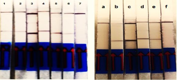
Water 2020, 12, 691                                                                             3 of 12

2. Materials and Methods

2.1. Source of Ascaris Ova
     Ascaris suum (pig roundworm) ova were utilized as surrogate for the human parasite Ascaris
lumbricoides owing to 98.1% genomic similarities and them being morphologically indistinguishable.
Additionally, A. lumbricoides causes enormous risk to public health, while A. suum ova can be easily
obtained and are much safer, as they rarely infect humans [51]. The A. suum infected pig faecal
samples were obtained from an abattoir in Laverton, Australia. The ova of A. suum were recovered
using a modified version of the Tulane method [21,52]. Aliquots of pig faecal samples with
approximately 5 g DS (dissolved solid) were rinsed with milliQ water, homogenised for 1 min with
a blender, and allowed to settle in 1% 7X detergent for 30 min (MP Bio, Australia). The supernatant
was aspirated, and the sediment was washed again with 1% 7X to solubilize the organic matter and
aid the release of ova that remained adhered to larger particles. After settling, the sediment was
poured onto stainless steel sieves of 850 µM and 300 µM pore sizes (Prospector Earth Sciences,
Australia) to remove the larger particles. The filtrate with A. suum ova settled further with the
addition of 7X® for 30 min, and the sediment was mixed and aliquoted in 50 mL falcon tubes, which
were centrifuged at 800 g, and the supernatant removed.
     Ova separation was achieved using flotation with magnesium sulfate (1.20 specific gravity) in
each tube. After centrifugation at 800 g for 3 min, the supernatant was poured on to a 38 µM sieve
and rinsed with water into a 100 mL beaker and later transferred into 15 mL tubes and centrifuged
as mentioned above. The ova were enumerated using an optical microscope (x200 magnification)
with Universal 4 chamber worm egg counting slides (J. A. Whitlock & Co, Melbourne, Australia). It
was then aliquoted (1000 ± 20 ova) into 1 mL volume (1% phosphate buffered saline) in 2 mL
Eppendorf tubes and stored at 4 °C for molecular analysis.

2.2. DNA Extraction
     The recovered Ascaris ova (1000 ± 20) were concentrated in 1% phosphate buffered saline (PBS)
to a final volume of 200 µL. Quick-DNA Fecal/Soil Microbe Miniprep Kit (Zymo Research,
Chatswood, Australia) and FastPrep-24 classic instrument (MP Biomedicals, Irvine, USA) were used
for DNA isolation from ova. The extracted DNA concentration was measured with NanoDrop
spectrophotometer.

2.3. Design and Screening of RPA Primers
     For the singleplex RPA assay, the ITS ribosomal DNA regions of Ascaris suum (Accession number
AB571302) was targeted for amplification. For the multiplex RPA assay, the ITS regions of A. suum
(Accession number AB571302) and T. suis (Accession number AM993008) were the desired targets.
The nucleotide sequences were obtained from Genbank (NCBI). The forward and the reverse RPA
primers for each helminth ova were designed using Primer3 plus (Table 1). RPA primers were
designed as per the guidelines of TwistDxTM guidelines (TwistDx, Cambridge, UK). The primers were
synthesised to produce smaller amplicon size (< 300 bp) to increase the rate of amplification. Between
the primers, a gap of at least 52 bp was maintained for probe design for use in lateral flow detection.
     The screening of primers was performed with a TwistAmpTM Basic kit to select the best primer
pairs that yielded smaller amplicons. The RPA assay was conducted in a pre-PCR chamber to
minimise contamination. For primer screening, 2 ng of A. suum genomic DNA was used as template.
Water 2020, 12, 691                                                                                  4 of 12

                              Table 1. Oligonucleotide primers used for initial screening.

           Assay                     Primer      Helminth                       Sequence (5’ – 3’)
  Recombinase polymerase
                                                 A. suum/A.     CTAATCTATGATTCAATATCTCGTTGTAATTT
    amplification (RPA)              Asc718F
                                                lumbricoides
     primer screening
                                                                 AAATTTTTCATATACATCATTATTGTCACG
                                     Asc881R

                                                                CTTATTTAGCTAATCTATGATTCAATATCTCG
                                     Asc709F

                                                                    GTTATTAACGACCAATGCAGATAAGC
                                     TS596F       T. suis

                                                                   GTTCAAAGTATTCAAGTTCAGTGTGTC
                                     TS764R

                                                                   CATGCTATGTCGGTGAGGTTTAAAGAA
                                     TS510F

     Reactions were performed according to the TwistDxTM Basic RPA protocol, where each reaction
contained 29.5 µL rehydration buffer, 2.4 µL of both forward and reverse primer (10 pmol), 12.2 µL
dH2O, and 1 µL genomic DNA. The RPA pellets in the kit were transferred into 0.2 mL PCR tubes for
easy handling. The reaction mix and 2.5 µL magnesium acetate were added to the lid of each reaction
tube, making a final reaction volume of 50 µL. The tubes were placed into the thermocycler, and the
temperature was set to 37 °C for 20 min. Since this assay can be a potential POC detection assay, the
reaction tubes were also placed in the hands (body temperature) instead of the thermocycler.
Purification of the amplicons was performed using QIAquick PCR purification kit (Qiagen, Germany)
and ran on 2% agarose gel. Negative controls (no template) were kept for every reaction set. Primer
pairs with positive amplification and smaller size amplicons were selected to facilitate rapid
amplification

2.4. Optimisation of Lateral Flow RPA Probe
     The probes targeting the ITS-DNA regions were designed for RPA-LF using the guidelines of
TwistDxTM, with size ranging from 46 to 52 nucleotides, incorporating a 6-carboxy-fluorescein (FAM)
label at the 5’ end. A tetrahydrofuran (THF) residue was added to the probe sequence, and a C3
spacer was incorporated at the 3’ end to prevent extension of any probe that remained unhybridised.
One of the primers, in this case, the reverse primer, was added with biotin to the 5’ end (Table 2).

                      Table 2. Oligonucleotide primers and probe specific for A. suum and T. suis.

     Assay            Primer/Probe                          Sequence (5’ – 3’)
  RPA-lateral                                     CTAATCTATGATTCAATATCTCGTTGTAATTT
                        Asc718F
   flow (LF)
                                                Biotin-AAATTTTTCATATACATCATTATTGTCACG
                       Asc881RB

                                       FAM-ATGAGCGAGAGAGAATATATACATCAAAACG-Tetrahydrofuran-
                        Asc767P                     TCTTAAAAGACGATT-C3 spacer

                                                      GTTATTAACGACCAATGCAGATAAGC
                        TS596F

                                               Digoxigenine-GTTCAAAGTATTCAAGTTCAGTGTGTC
                       TS764RD

                                        FAM-GTGCAGGAACTCTTGAAACATGATGACATT-tetrahydrofuran-
                        TS661P
                                                   CGAACGGCGGATCACTT-C3 spacer

    ITS RPA-LF assays were conducted using TwistDxTM Nfo kit. Each reaction consisted of 2 ng A.
suum genomic DNA, 29.5 µL rehydration buffer, 2.1 µL forward primer, 2.1 µL biotin labelled reverse
primer, 0.6 µL lateral flow probe (10 pmol), and 12.2 µL dH2O. The reaction mix was added to the
Water 2020, 12, 691 5 of 12
RPA pellets in the kit, and 2.5 µL magnesium acetate was transferred to the lids prior to the reaction
tube being placed in the thermocycler at 37 °C for 20 min. Nuclease free water was used as the
negative control for the assay.
2.5. ITS RPA-LF Amplicon Detection
The end-point analysis of the amplicons was performed using the Milenia HybriDetect 2T lateral
flow strips (Milenia Biotec GmbH, Germany). The dipstick is designed for qualitative or quantitative
rapid test systems to detect two different analytes. The assay is based on a “sandwich” format with
the desired target, where the probe conjugates with anti-FAM antibodies present on the sample pad
and captured at the test line by anti-biotin/anti-digoxigenine antibodies, thereby forming a complex
with colloidal gold to produce a coloured signal. A coloured band on the control line prevents false
negatives.
To avoid contamination, post-amplification processing for RPA-LF was carried out in different
rooms of the laboratory. The amplification product (2 µL) was added to 98 µL HybriDetect buffer.
The LF strip was vertically placed into the tube, and the results were read within 3 min.
2.6. Reaction Time and Amplification Temperature
The optimal amplification time for A. suum primers was selected by RPA amplification at 37 °C
for 5, 10, 20, 25, 30, and 40 min using 2 ng A. suum DNA, and for the end-point analysis, the lateral
flow dipsticks were placed vertically on the tubes for less than 3 min at room temperature.
Similarly, the evaluation of the optimal temperature for amplification was performed at different
reaction temperatures of 20, 25, 37, 40, and 45 °C for 20 min. A thermocycler set at the desired
temperature was used for this assay. The tubes were manually mixed in the initial 5 min to avoid
localised depletion of reagents.
2.7. Detection Limit and Specificity
For the lower limit of detection (LLOD), serial dilutions of A. suum were prepared using 20 ng/µL
DNA with reverse osmosis water to give concentrations of 2 ng/µL, 200 pg/µL, 20 pg/µL, 2 pg/µL,
200 fg/µL, 20 fg/µL and 2 fg/µL of DNA. All reactions were run in the thermocycler at 37 °C for 20
min.
For determining the specificity of the RPA primers designed for A. suum, organisms such as
Trichuris suis (whipworm), Haemonchus contortus (sheep hookworms), and A. lumbricoides were
utilised. Faecal samples were collected from pigs that were infected with T. suis. The ova were
recovered using Tulane method with minor modifications (21, 52) and were also used for further
experiments. Genomic DNA of A. lumbricoides and H contortus were obtained from the Faculty of
Veterinary and Agricultural Sciences, The University of Melbourne. Since DNA of human
hookworms Ancylostoma duodenale and Necator americanus were not readily available, NCBI BLAST
was performed to evaluate whether the primers could result in cross reactivity to these organisms.
2.8. Multiplex RPA-LF to Detect Two Different Helminth Ova Genera in Wastewater
A multiplex RPA-LF was performed to detect two different helminth genera, A. suum and T.
suis, in a single lateral flow strip using wastewater from Lang Lang wastewater treatment plant of
South East Water, Victoria. Wastewater (200 µL) was seeded with 200 (±10) A. suum and T. suis ova
each, and DNA was extracted using Isolate II Genomic Spin Column kits (Bioline, USA). The primers
and the probe for T. suis were designed as per the manufacturer’s protocol (TwistDxTM, Cambridge,
UK) and utilised for this assay. The 3’ end of Asc881RB was labelled with biotin, while the 3’ end of
TS764RD was labelled with digoxigenine. To detect the specificity of the primers, DNA of both A.
suum and T. suis (2 ng) were amplified using RPA at 37 °C for 20 min and observed for coloured test
bands using lateral flow dipsticks.Water 2020, 12, 691 6 of 12
3. Results
3.1. Optimisation of A. Suum Primers (Asc718F/Asc881R)
The ITS primer combinations successfully amplified 2 ng of A. suum DNA. The primers also
amplified A. lumbricoides DNA. This could have been due to STH’s morphological and genetic
similarities with A. suum. Agarose gel electrophoresis indicated that the target DNA amplicon lengths
were as expected for each set of primers (Figure 1). The primers (Asc718F/Asc881R) with the best
band and a shorter amplicon (164 bp) were chosen for further assays, as shorter amplicons generate
fewer primer artefacts and thereby provide high sensitivity.
Figure 1. Validation of Asc718F/Asc881R primer pair for target Ascaris DNA: 1) 100 bp Ladder, 2)
Ascaris suum, 3) A. lumbricoides, 4) A. suum (1:10 genomic DNA dilution), 5) A.lumbricoides (1:10
genomic DNA dilution), 6) Trichuris suis, 7) no template (negative control).
3.2.Evaluation of Reaction Time and Amplification Temperature
The RPA assay was performed at 37 °C for 5–40 min, and the LF assay was performed at
approximately 25 °C (room temperature); the results were analysed within 3 min. The colour
observed on the test line was faint at the amplification time of 5 min; however, the colour intensity at
the test line increased at 10 min amplification time with a strong positive signal (Figure 2A). For rapid
and greater sensitivity, a 20 min RPA assay is recommended, though an amplification of 5 min was
sufficient to detect the pathogen. The time taken for RPA-LF was 30 min or less.
The results for the optimal amplification temperature depicted the performance of the assay at
temperatures ranging from 25–45 °C (Figure 2B). The colour intensity in the test line changed with
temperature, suggesting the importance of temperature for efficient amplification. A clear band with
high visibility appeared at 37 °C, and it was chosen at the optimal temperature for amplification of
A. suum DNA.
A BWater 2020, 12, 691 7 of 12
Figure 2. (A) Determination of reaction time for RPA-LF assay using primers for A. suum: 1) 5 min, 2)
10 min, 3) 20 min, 4) 25 min, 5) 30 min, 6) 40 min, and 7) control (no template). (B) Determination of
amplification temperature for RPA-LF assay: a) 20 ⁰C, b) 25 ⁰C, c) 37 ⁰C, d) 40 °C, e) 45 °C, f) no
template control.
3.3. Evaluation of Detection Limit and Specificity
The LLOD of the RPA-LF assay was analysed using 2 ng of A. lumbricoides DNA and dilutions
of A suum DNA as low as 2 fg. The negative control consisted of nuclease free water. The presence of
a band in both the control and the test bands indicated a positive result, while a coloured band at the
control line alone indicated a negative result. As shown, two purple bands on the lateral flow strips
were observed post-amplification from 2 ng to 2 fg but not the negative control (Figure 3). The results
showed that the RPA-LF assay detected as low as 2 fg genomic DNA and has the potential to detect
even one helminth ovum.
a b c d e f g
Figure 3. Sensitivity of RPA-LF primers for Ascaris: a) A. lumbricoides 2 nanogram (ng) DNA, (b-g) A.
suum, b) 2 ng, c) 200 picogram (pg), d) 2 picogram, e) 20 femtogram (fg), f) 2 fg, g) control.
Similarly, the analytical specificity of the assay was tested using 2 ng genomic DNA from A.
suum, A. lumbricoides, T. suis, and H. contortus. The negative control consisted of nuclease free water.
A coloured test band was visualised on the LF strips of A. suum and A. lumbricoides DNA, while other
helminth ova genera were found to be negative (Figure 4), thus revealing that RPA primers showed
high specificity for Ascaris ova.
a b c d e
Figure 4. Evaluating the specificity of Ascaris primers using RPA-LF assay: a) Ascaris lumricoides, b)
Ascaris suum, c) Trichuris suis, d) Haemonchus contortus, e) control.Water 2020, 12, 691 8 of 12
3.4. Multiplex RPA-LF Assay
The results for multiplex RPA-LF to detect A. suum and T. suis in a single strip showed the
capability of the primers specific for the target DNA with no cross reactivity. A clear visible test band
was observed at the bottom, indicating the detection of A. suum, and the test band below the control
band indicates the presence of T. suis. However, the reaction tube containing DNA and primers of
both A. suum and T. suis showed three bands, indicating the control band and both the test bands
were observed and therefore demonstrating the successful detection of both helminth ova genera
(Figure 5).
Figure 5. Multiplex RPA-LF: C) control (no template), A) A. suum and T. suis DNA with T) T. suis and
A. suum DNA with A+T) both A. suum and T. suis DNA and primers.
4. Discussion
Diagnostic tests with greater specificity and sensitivity are crucial to successfully detect the
presence of Ascaris ova in recycled water prior to release for public use [53]. Such a test will also help
to monitor the transmission of ascariasis in endemic and low resource setting areas. Conventional
microscopy and culture methods are laborious and cumbersome [6,54]. Despite the fact that PCR
based methods are specific and sensitive, they possess limitations such as high cost, slow turnaround
times, and difficulty of application in low resource settings [55]. In this study, utilising the TwistDx
platform, RPA methods were coupled with a lateral flow (RPA-LF) assay to detect Ascaris species
(singleplex RPA) in seeded wastewater. Furthermore, a novel duplex RPA-LF assay was performed
to detect ova from two different helminth genera (A. suum and T. suis) in a single LF strip.
Ribosomal ITS DNA of A. suum was utilised in the design of primers and probe, and the best
primer pair that produced shorter amplicons was chosen. The assay was repeated at least twice with
the primer pairs to avoid false positive/negative results, which can arise owing to the formation of
hairpin secondary structure of primer/probe leading to non-specific amplification. The addition of
dimethyl sulfoxide (DMSO) prevented false positives. DMSO has the ability to avoid secondary
structures, especially in GC rich sequences, and is often used in nucleic acid amplification [56].
The RPA-LF assay detected 2 fg genomic DNA of A. suum, thus exhibiting the assay’s potential
in detecting even one ovum in large volumes of wastewater samples. Post-amplification
contamination with RPA-LF assay can be minimised by carefully opening and closing the reaction
tubes, frequently changing the gloves, conducting pre- and post-RPA assays in different chambers or
locations, and reducing the reaction time if required.
Furthermore, this RPA-LF assay showed no cross-reaction with other organisms and exhibited
high specificity for Ascaris ova. Other advantages were the constant (isothermal) amplificationWater 2020, 12, 691 9 of 12
temperature and the shorter reaction time for the assay. Our study indicated that the assay could be
successfully conducted at temperatures ranging from 25 to 45 °C, thus it is effective at various
temperature ranges without the requirement of any heating device; simply placing the reaction tubes
in the hand is sufficient for amplification. Besides, the overall processing time of the RPA-LF assay
from amplification to detection by visualisation of purple coloured bands was 30 min or less,
significantly faster than the currently available isothermal amplification methods, such as loop
mediated isothermal amplification (LAMP), which requires 64 °C for 90 min [29]. Subsequently, the
results for the validation of RPA-LF to detect Ascaris ova in seeded wastewater showed that turbidity
did not have an impact on the sensitivity of this assay, which could be ideal for the onsite detection
of helminth ova in wastewater treatment plants.
Finally, the multiplex RPA-LF assay showed that the assay was effective in detecting the ova of
A. suum and T. suis in a single LF strip. Multiplex RPA-LF based detection of two different pathogens
in a single strip could significantly reduce time, cost, and labour [48,57]. Further optimisation and
validation may prove RPA-LF to be a feasible detection test for Ascaris species in wastewater.
Moreover, the results denoted that the technique is user-friendly and could be performed by
untrained personnel in wastewater treatment plants. No sophisticated devices or training are
required, making this approach ideal for endemic and resource limited areas.
5. Conclusions
The RPA-LF reported in this study was rapid and highly sensitive in the detection of A. suum
ova. Additionally, for the first time, the ova of two different helminth genera were able to be detected
in a single strip using a multiplex approach. Nevertheless, the effectiveness of RPA-LF for the
detection of helminths in wastewater with varying turbidity and from endemic regions must be
evaluated prior to being utilised as a routine point-of-care detection test for STHs in wastewater
treatment plants. Overall, RPA is an efficient isothermal amplification method that can overcome the
limitations encountered by existing molecular diagnostics methods. RPA reactions operate at varying
temperature ranges (25–45 °C), confirming the ability of this assay to be efficient over a range of
temperatures and settings and thereby enhancing the potential of RPA-LF feasible in resource-limited
areas where power supply is a constraint. RPA coupled with LF detection method is very simple and
requires no skilled personnel to interpret the results. Furthermore, development of a device-free,
simple, and rapid DNA extraction method can be combined with RPA-LF for efficient onsite
diagnostic purposes.
Author Contributions: Conceptualization, V.B.R., S.K.S., A.S.B; resources, S.K.S., A.S.B; writing-original draft
preparation, V.B.R., B.K; writing-review and editing, V.B.R., S.K.S., A.S.B., A.S., N.D.C; visualization, V.B.R;
supervision, S.K.S., A.S.B; project administration, A.S.B. All authors have read and agreed to the published
version of the manuscript.
Funding: The research did not receive any specific grant from funding agencies in the public, commercial, or
not-for-profit sectors.
Acknowledgments: The authors acknowledge support from the RMIT Postgraduate International Scholarship,
RMIT-ECP Opportunity Grant and RMIT Research Translation Seed Funding for the development of this
research. We also acknowledge South East Water, Melbourne Water and Rebecca Traub, Faculty of Veterinary
and Agricultural Sciences, The University of Melbourne, Australia, for providing samples and necessary inputs
for this research. Furthermore, we thank the members of Ball’s group, School of Science, RMIT University,
Melbourne, Australia, for providing valuable insights.
Conflicts of Interest: The authors declare no conflict of interest.
References
1. Rocha, M.C.V.D.; Barés, M.E.; Braga, M.C.B. Quantification of viable helminth eggs in samples of sewage
sludge. Water Res. 2016, 103, 245–255.Water 2020, 12, 691 10 of 12
2. Acosta Soto, L.; Santísima-Trinidad, A.B.; Bornay-Llinares, F.J.; Martín González, M.; Pascual Valero, J.A.;
Ros Muñoz, M. Quantitative PCR and Digital PCR for Detection of Ascaris lumbricoides Eggs in Reclaimed
Water. Biomed. Res. Int. 2017, 2017, 9.
3. Gyawali, P.; Sidhu, J.P.; Ahmed, W.; Jagals, P.; Toze, S. Rapid concentration and sensitive detection of
hookworm ova from wastewater matrices using a real-time PCR method. Exp. Parasitol. 2015, 159, 5–12.
4. Gyawali, P. Infectious helminth ova in wastewater and sludge: A review on public health issues and current
quantification practices. Water Sci. Technol. A J. Int. Assoc. Water Pollut. Res. 2018, 77, 1048–1061.
5. Li, D.; Tong, T.; Zeng, S.; Lin, Y.; Wu, S.; He, M. Quantification of viable bacteria in wastewater treatment
plants by using propidium monoazide combined with quantitative PCR (PMA-qPCR). J. Environ. Sci. 2014,
26, 299–306.
6. Collender, P.A.; Kirby, A.E.; Addiss, D.G.; Freeman, M.C.; Remais, J.V. Methods for Quantification of Soil-
Transmitted Helminths in Environmental Media: Current Techniques and Recent Advances. Trends
Parasitol. 2015, 31, 625–639.
7. Amoah, I.D.; Singh, G.; Stenström, T.A.; Reddy, P. Detection and quantification of soil-transmitted
helminths in environmental samples: A review of current state-of-the-art and future perspectives. Acta
Trop. 2017, 169, 187–201.
8. Bethony, J.; Brooker, S.; Albonico, M.; Geiger, S.M.; Loukas, A.; Diemert, D.; Hotez, P.J. Soil-transmitted
helminth infections: Ascariasis, trichuriasis, and hookworm. Lancet 2006, 367, 1521–1532.
9. Hotez, P.J.; Brooker, S.; Bethony, J.M.; Bottazzi, M.E.; Loukas, A.; Xiao, S. Hookworm Infection. N. Engl. J.
Med. 2004, 351, 799–807.
10. Hotez, P.J.; Damania, A.; Barua, A.; Stanaway, J. The first “London Declaration”: The Commonwealth and
its neglected tropical diseases. PLoS Negl. Trop. Dis. 2017, 11, e0005321.
11. Du, R.Y.; Stanaway, J.D.; Hotez, P.J. Could violent conflict derail the London Declaration on NTDs? PLoS
Negl. Trop. Dis. 2018, 12, e0006136.
12. Karkashan, A.; Khallaf, B.; Morris, J.; Thurbon, N.; Rouch, D.; Smith, S.R.; Deighton, M. Comparison of
methodologies for enumerating and detecting the viability of Ascaris eggs in sewage sludge by standard
incubation-microscopy, the BacLight Live/Dead viability assay and other vital dyes. Water Res. 2015, 68,
533–544.
13. Shahsavari, E.; Schmidt, J.; Aburto-Medina, A.; Khallaf, B.; Balakrishnan, V.; Crosbie, N.D.; Surapaneni, A.;
Ball, A.S. A modified assay for the enumeration of ascaris eggs in fresh raw sewage. MethodsX 2017, 4, 186–
190.
14. Mascarini-Serra, L. Prevention of Soil-transmitted Helminth Infection. J. Glob. Infect. Dis. 2011, 3, 175–182.
15. Salam, N.; Azam, S. Prevalence and distribution of soil-transmitted helminth infections in India. BMC
Public Health 2017, 17, 201.
16. Mara, D.; Sleigh, A. Estimation of Ascaris infection risks in children under 15 from the consumption of
wastewater-irrigated carrots. J. Water Health 2010, 8, 35–38.
17. Schmidlin, T.; Hürlimann, E.; Silué, K.D.; Yapi, R.B.; Houngbedji, C.; Kouadio, B.A.; Acka-Douabélé, C.A.;
Kouassi, D.; Ouattara, M.; Zouzou, F.; et al. Effects of hygiene and defecation behavior on helminths and
intestinal protozoa infections in Taabo, Côte d'Ivoire. PLoS ONE 2013, 8, e65722.
18. Strunz, E.C.; Addiss, D.G.; Stocks, M.E.; Ogden, S.; Utzinger, J.; Freeman, M.C. Water, Sanitation, Hygiene,
and Soil-Transmitted Helminth Infection: A Systematic Review and Meta-Analysis. PLoS Med. 2014, 11,
e1001620.
19. Manser, N.D.; Wald, I.; Ergas, S.J.; Izurieta, R.; Mihelcic, J.R. Assessing the fate of Ascaris suum ova during
mesophilic anaerobic digestion. Environ. Sci. Technol. 2015, 49, 3128–3135.
20. Cruz, L.M.; Allanson, M.; Kwa, B.; Azizan, A.; Izurieta, R. Morphological Changes of Ascaris spp. Eggs
During Their Development Outside the Host. J. Parasitol. 2011, 98, 63–68.
21. Bowman, D.D.; Little, M.D.; Reimers, R.S. Precision and accuracy of an assay for detecting Ascaris eggs in
various biosolid matrices. Water Res. 2003, 37, 2063–2072.
22. Maya, C.; Ortiz, M.; Jimenez, B. Viability of Ascaris and other helminth genera non larval eggs in different
conditions of temperature, lime (pH) and humidity. Water Sci. Technol. A J. Int. Assoc. Water Pollut. Res. 2010,
62, 2616–2624.
23. Horiuchi, S.; Uga, S. Modified flotation method, an effective technique for recovering helminth eggs in soil.
Parasitol. Int. 2016, 65, 576–579.Water 2020, 12, 691 11 of 12
24. Sá, M.F.D.; Gonçalves, R.A.; Marder, C.; Baldissera, M.D.; Oliveira, C.B.D.; Noll, J.C.G.; Silva, F.; Monteiro,
S.G. Adapted Bailenger method improves the rate of Ascaris suum eggs recovery from liquid pig manure
compost. Ciência Rural 2017, 47 (4), 1-6.
25. O’Connor, N.A.; Surapaneni, A.; Smith, D.; Stevens, D. Occurrence and fate of Ascaris lumbricoides ova in
biosolids in Victoria, Australia: A human health risk assessment of biosolids storage periods. Water Sci.
Technol. A J. Int. Assoc. Water Pollut. Res. 2017, 76, 1332–1346.
26. Stevens, D.P.; Surapaneni, A.; Thodupunuri, R.; O’Connor, N.A.; Smith, D. Helminth log reduction values
for recycling water from sewage for the protection of human and stock health. Water Res. 2017, 125, 501–
511.
27. Llewellyn, S.; Inpankaew, T.; Nery, S.V.; Gray, D.J.; Verweij, J.J.; Clements, A.C.; Gomes, S.J.; Traub, R.;
McCarthy, J.S. Application of a Multiplex Quantitative PCR to Assess Prevalence and Intensity Of Intestinal
Parasite Infections in a Controlled Clinical Trial. PLoS Negl. Trop. Dis. 2016, 10, e0004380.
28. Wardell, R.; Clements, A.C.; Lal, A.; Summers, D.; Llewellyn, S.; Campbell, S.J.; McCarthy, J.; Gray, D.J.;
Nery, S.V. An environmental assessment and risk map of Ascaris lumbricoides and Necator americanus
distributions in Manufahi District, Timor-Leste. PLoS Negl. Trop. Dis. 2017, 11, e0005565.
29. Shiraho, E.A.; Eric, A.L.; Mwangi, I.N.; Maina, G.M.; Kinuthia, J.M.; Mutuku, M.W.; Mugambi, R.M.;
Mwandi, J.M.; Mkoji, G.M. Development of a Loop Mediated Isothermal Amplification for Diagnosis of
Ascaris lumbricoides in Fecal Samples. J. Parasitol. Res. 2016, 2016, 7376207.
30. Bastos, V.K.; Cutolo, S.A.; Doria, M.d.C.O.; Razzolini, M.T.P. Detection and quantification of viable Ascaris
sp. and other helminth eggs in sewage sludge. Int. J. Environ. Health Res. 2013, 23, 352–362.
31. Pecson, B.M.; Barrios, J.A.; Johnson, D.R.; Nelson, K.L. A Real-Time PCR Method for Quantifying Viable
Ascaris Eggs Using the First Internally Transcribed Spacer Region of Ribosomal DNA. Appl. Environ.
Microbiol. 2006, 72, 7864–7872.
32. Gordon, C.A.; McManus, D.P.; Acosta, L.P.; Olveda, R.M.; Williams, G.M.; Ross, A.G.; Gray, D.J.; Gobert,
G.N. Multiplex real-time PCR monitoring of intestinal helminths in humans reveals widespread
polyparasitism in Northern Samar, the Philippines. Int. J. Parasitol. 2015, 45, 477–483.
33. Drain, P.K., Hyle, E.P., Noubary, F., Freedberg, K.A., Wilson, D., Bishai, W.R., Rodriguez, W. and Bassett,
I.V. Diagnostic point-of-care tests in resource-limited settings. Lancet Infect. Dis. 2014, 14, 239–249.
34. Mugambi, R.M.; Agola, E.L.; Mwangi, I.N.; Kinyua, J.; Shiraho, E.A.; Mkoji, G.M. Development and
evaluation of a Loop Mediated Isothermal Amplification (LAMP) technique for the detection of hookworm
(Necator americanus) infection in fecal samples. Parasit Vectors 2015, 8, 574.
35. Daher, R.K.; Stewart, G.; Boissinot, M.; Bergeron, M.G. Recombinase Polymerase Amplification for
Diagnostic Applications. Clin. Chem. 2016, 62, 947–958.
36. Lau, H.Y.; Botella, J.R. Advanced DNA-Based Point-of-Care Diagnostic Methods for Plant Diseases
Detection. Front. Plant Sci. 2017, 8, 2016.
37. Li, J.; Macdonald, J.; von Stetten, F. Review: A comprehensive summary of a decade development of the
recombinase polymerase amplification. Analyst 2019, 144, 31–67.
38. Yang, Y.; Qin, X.; Song, Y.; Zhang, W.; Hu, G.; Dou, Y.; Li, Y.; Zhang, Z. Development of real-time and
lateral flow strip reverse transcription recombinase polymerase Amplification assays for rapid detection of
peste des petits ruminants virus. Virol. J. 2017, 14, 24.
39. Wang, J.; Liu, L.; Wang, J.; Sun, X.; Yuan, W. Recombinase Polymerase Amplification Assay-A Simple, Fast
and Cost-Effective Alternative to Real Time PCR for Specific Detection of Feline Herpesvirus-1. PLoS ONE
2017, 12, e0166903.
40. James, A.; Macdonald, J. Recombinase polymerase amplification: Emergence as a critical molecular
technology for rapid, low-resource diagnostics. Expert Rev. Mol. Diagn. 2015, 15, 1475–1489.
41. Euler, M.; Wang, Y.; Heidenreich, D.; Patel, P.; Strohmeier, O.; Hakenberg, S.; Niedrig, M.; Hufert, F.T.;
Weidmann, M. Development of a Panel of Recombinase Polymerase Amplification Assays for Detection of
Biothreat Agents. J. Clin. Microbiol. 2013, 51, 1110.
42. Escadafal, C.; Faye, O.; Sall, A.A.; Faye, O.; Weidmann, M.; Strohmeier, O.; von Stetten, F.; Drexler, J.;
Eberhard, M.; Niedrig, M.; et al. Rapid Molecular Assays for the Detection of Yellow Fever Virus in Low-
Resource Settings. PLoS Negl. Trop. Dis. 2014, 8, e2730.
43. Kersting, S.; Rausch, V.; Bier, F.F.; von Nickisch-Rosenegk, M. Multiplex isothermal solid-phase
recombinase polymerase amplification for the specific and fast DNA-based detection of three bacterial
pathogens. Mikrochim. Acta 2014, 181, 1715–1723.Water 2020, 12, 691 12 of 12
44. Teoh, B.T.; Sam, S.S.; Tan, K.K.; Danlami, M.B.; Shu, M.H.; Johari, J.; Hooi, P.S.; Brooks, D.; Piepenburg, O.;
Nentwich, O.; et al. Early Detection of Dengue Virus by Use of Reverse Transcription-Recombinase
Polymerase Amplification. J. Clin. Microbiol. 2015, 53, 830.
45. Zhang, S.; Ravelonandro, M.; Russell, P.; McOwen, N.; Briard, P.; Bohannon, S.; Vrient, A. Rapid diagnostic
detection of plum pox virus in Prunus plants by isothermal AmplifyRP((R)) using reverse transcription-
recombinase polymerase amplification. J. Virol. Methods 2014, 207, 114–120.
46. Tu, P.A.; Shiu, J.S.; Lee, S.H.; Pang, V.F.; Wang, D.C.; Wang, P.H. Development of a recombinase
polymerase amplification lateral flow dipstick (RPA-LFD) for the field diagnosis of caprine arthritis-
encephalitis virus (CAEV) infection. J. Virol. Methods 2017, 243, 98–104.
47. Soliman, H.; Kumar, G.; El-Matbouli, M. Recombinase polymerase amplification assay combined with a
lateral flow dipstick for rapid detection of Tetracapsuloides bryosalmonae, the causative agent of
proliferative kidney disease in salmonids. Parasites Vectors 2018, 11, 234.
48. Tian, A.L.; Elsheikha, H.M.; Zhou, D.H.; Wu, Y.D.; Chen, M.X.; Wang, M.; Chen, D.; Zhang, X.C.; Zhu, X.Q.
A novel recombinase polymerase amplification (RPA) assay for the rapid isothermal detection of Neospora
caninum in aborted bovine fetuses. Vet. Parasitol. 2018, 258, 24–29.
49. Sun, K.; Xing, W.; Yu, X.; Fu, W.; Wang, Y.; Zou, M.; Luo, Z.; Xu, D. Recombinase polymerase amplification
combined with a lateral flow dipstick for rapid and visual detection of Schistosoma japonicum. Parasit
Vectors 2016, 9, 476.
50. Rohrman, B.; Richards-Kortum, R. Inhibition of recombinase polymerase amplification by background
DNA: A lateral flow-based method for enriching target DNA. Anal. Chem. 2015, 87, 1963–1967.
51. Nejsum, P.; Thamsborg, S.M.; Petersen, H.H.; Kringel, H.; Fredholm, M.; Roepstorff, A. Population
dynamics of Ascaris suum in trickle-infected pigs. J. Parasitol. 2009, 95, 1048.
52. Ravindran, V.B.; Surapaneni, A.; Crosbie, N.D.; Schmidt, J.; Shahsavari, E.; Haleyur, N.; Soni, S.K.; Ball,
A.S. A modified approach to recover and enumerate Ascaris ova in wastewater and sludge. PLoS Negl. Trop.
Dis. 2018, 13, 1-6.
53. Steinbaum, L.; Kwong, L.H.; Ercumen, A.; Negash, M.S.; Lovely, A.J.; Njenga, S.M.; Boehm, A.B.; Pickering,
A.J.; Nelson, K.L. Detecting and enumerating soil-transmitted helminth eggs in soil: New method
development and results from field testing in Kenya and Bangladesh. PLoS Negl. Trop. Dis. 2017, 11,
e0005522.
54. Pilotte, N.; Papaiakovou, M.; Grant, J.R.; Bierwert, L.A.; Llewellyn, S.; McCarthy, J.S.; Williams, S.A.
Improved PCR-Based Detection of Soil Transmitted Helminth Infections Using a Next-Generation
Sequencing Approach to Assay Design. PLoS Negl. Trop. Dis. 2016, 10, e0004578.
55. Verweij, J.J.; Stensvold, C.R. Molecular testing for clinical diagnosis and epidemiological investigations of
intestinal parasitic infections. Clin. Microbiol. Rev. 2014, 27, 371–418.
56. Jensen, M.A.; Fukushima, M.; Davis, R.W. DMSO and betaine greatly improve amplification of GC-rich
constructs in de novo synthesis. PLoS ONE 2010, 5, e11024.
57. Pai, N.P.; Vadnais, C.; Denkinger, C.; Engel, N.; Pai, M. Point-of-care testing for infectious diseases:
Diversity, complexity, and barriers in low- and middle-income countries. PLoS Med. 2012, 9, e1001306.
© 2020 by the authors. Licensee MDPI, Basel, Switzerland. This article is an open access
article distributed under the terms and conditions of the Creative Commons Attribution
(CC BY) license (http://creativecommons.org/licenses/by/4.0/).You can also read