Ex Vivo Determination of Broadband Absorption and Effective Scattering Coefficients of Porcine Tissue
←
→
Page content transcription
If your browser does not render page correctly, please read the page content below
hv
               photonics

Article
Ex Vivo Determination of Broadband Absorption and Effective
Scattering Coefficients of Porcine Tissue
Florian Bergmann * , Florian Foschum, Leonie Marzel and Alwin Kienle

                                          Quantitative Imaging and Sensors, Institut für Lasertechnologien in der Medizin und Meßtechnik an der
                                          Universität Ulm, Helmholtzstr. 12, D-89081 Ulm, Germany; florian.foschum@ilm-ulm.de (F.F.);
                                          leonie.marzel@stud.uni-heidelberg.de (L.M.); alwin.kienle@ilm-ulm.de (A.K.)
                                          * Correspondence: florian.bergmann@ilm-ulm.de

                                          Abstract: A novel approach for precise determination of the optical scattering and absorption
                                          properties of porcine tissue using an optimized integrating sphere setup was applied. Measurements
                                          on several sample types (skin, muscle, adipose tissue, bone, cartilage, brain) in the spectral range
                                          between 400 nm and 1400 nm were performed. Due to the heterogeneity of biological samples,
                                          measurements on different individual animals as well as on different sections for each sample type
                                          were carried out. For all samples, we used an index matching method to reduce surface roughness
                                          effects and to prevent dehydration. The derived absorption spectra were used to estimate the
                                          concentration of important tissue chromophores such as water, oxy- and deoxyhemoglobin, collagen
                                          and fat.

                                          Keywords: optical properties; biological tissue; integrating sphere


                                          1. Introduction
Citation: Bergmann, F.; Foschum, F.;            In medical [1–4] and computational [5,6] applications, the optical properties of bi-
Marzel, L.; Kienle, A. Ex Vivo            ological tissue, which include the absorption coefficient µa and the effective scattering
Determination of Broadband
                                          coefficient µs0 , are of high interest. We note that the effective scattering coefficient is often
Absorption and Effective Scattering
                                          more significant for the description of the light propagation in biological media compared
Coefficients of Porcine Tissue.
                                          to the other two optical scattering quantities involved in radiative transport theory, the
Photonics 2021, 8, 365. https://
                                          scattering phase function p(θ ) and the single scattering coefficient µs .
doi.org/10.3390/photonics8090365
                                                The overall amount of studies for determining the optical properties of biological tissue
                                          is relatively large, but the demand in accurate determined properties from the visual (VIS)
Received: 5 August 2021
Accepted: 26 August 2021
                                          to the near infrared (NIR) spectral range is still not met due to sparse, inconsistent, sporadic
Published: 31 August 2021
                                          values [7] and not well validated measurement methods. Reviews of publications dealing
                                          with the determination of the optical properties of tissue were, e.g., given by Cheong
Publisher’s Note: MDPI stays neutral
                                          1990 [8], Kim and Wilson 2011 [9], Sandell and Zhu 2011 [10], Bashkatov et al. 2011 [11]
with regard to jurisdictional claims in
                                          and Jacques 2013 [12]. In the present paper, we mainly refer to recent investigations from
published maps and institutional affil-   Mosca et al. [7] (time and spectral domain) and Foschum [13] (spatial and spectral domain)
iations.                                  to compare our results due to their broadband investigations of similar tissue types as
                                          were investigated in the present study. Our study is based on an optimized integrating
                                          sphere setup having a spectral range from 400 nm to 1400 nm and an innovative two
                                          stage evaluation process [14]. The two stage evaluation uses Monte Carlo simulations
Copyright: © 2021 by the authors.
                                          to describe the exact sample geometry combined with an analytical model that takes the
Licensee MDPI, Basel, Switzerland.
                                          exact sphere geometry into account to correct for the effective detector signal. Thus, the
This article is an open access article
                                          applied theory is based on radiative transfer theory and not on approximations such as
distributed under the terms and           Kubelka–Munk theory or diffusion theory. The used optimized integrating sphere was
conditions of the Creative Commons        thoroughly verified using polystyrene spheres, Al2 O3 and TiO2 nanoparticles in water and
Attribution (CC BY) license (https://     epoxy-based phantoms [15].
creativecommons.org/licenses/by/                In the present paper, we investigated different sample types of the domesticated pig
4.0/).                                    (sus scrofa domesticus), which have common genetic elements to primates, are easily acces-

Photonics 2021, 8, 365. https://doi.org/10.3390/photonics8090365                                          https://www.mdpi.com/journal/photonics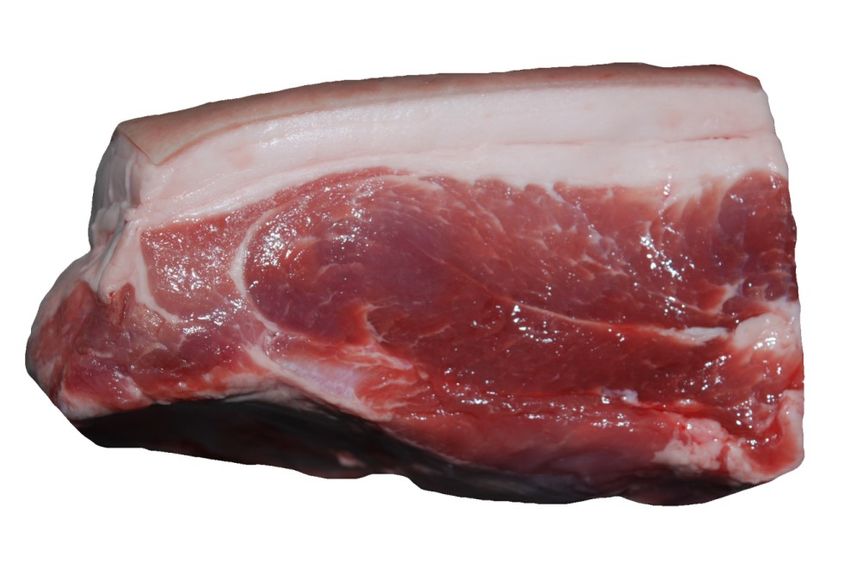
Photonics 2021, 8, 365                                                                                             2 of 19

                         sible and have less pathogenic hazard compared to tissue from primates. In the first part of
                         this paper, we summarize the special features of our novel integrating sphere approach and
                         introduce our sample preparation procedure. The analysis of the influence of an incorrectly
                         assumed refractive index as well as asymmetry factor used for the determination of the
                         optical properties applying an integrating sphere is outlined for adipose tissue in the
                         second part. In the following part of this paper, we present the results of six different tissue
                         types. The results are compared to selected recent literature values, and within this study
                         to different body regions of the same tissue types. Finally, we conclude our results, which
                         may serve as comparison to other setups or investigations.

                         2. Materials and Methods
                         2.1. Experimental Setup and Evaluation Process
                               An integrating sphere setup combined with advanced Monte Carlo simulations was
                         applied for the accurate separation of µs0 and µa . The setup and the evaluation procedure
                         were described in detail by Foschum et al. [14] and Bergmann et al. [15] in previous
                         publications. Briefly, we use a halogen lamp as a “white light source”. The light was
                         separated to a reflection, transmission and normalization beam, see Figure 1. The latter
                         was used to correct the measurement for changes in the throughput of the sphere while
                         applying different samples as well as the calibration standard at the sample port. For
                         detection, two commercial available spectrometers with a nominal spectral range from
                         180 nm to 1100 nm (“Maya2000Pro”, Ocean Insight, Orlando, FL, USA) and from 900 nm
                         to 1700 nm (“NIRQuest512-1.7”, Ocean Insight, Orlando, FL, USA) are connected to two
                         optical fibers. The fiber ends on the north pole of the sphere are imaged onto the sphere
                         wall near the south pole of the sphere. Both spectrometers were corrected for their internal
                         stray light. As a first approximation, we assume that the internal stray light is constant
                         over the entire pixel array [15]. In the VIS, the halogen light source restricted the spectral
                         range to a minimal wavelength of 400 nm, and therefore, we subtracted the internal stray
                         light intensity between 220 nm and 300 nm from each spectrum. For the NIR spectrometer,
                         the internal stray light was considered and corrected by using a short-pass filter in the
                         NIR from 1500 nm to 1585 nm. Due to the large absorption of pure water between 1400 nm
                         to 1500 nm, the evaluated wavelength range was reduced to 400 nm until 1400 nm. The
                         setup is controlled using a self-written C++ software. As mentioned before, we use a
                         two stage evaluation process [14] to solve the inverse problem. This process is based on
                         Monte Carlo simulations to calculate the reflected light from the sample to all parts (port
                         openings and detector field of view) of the integrating sphere. Second, an analytical model
                         of the sphere throughput is used to calculate the effective detector signal after diffusion in
                         the sphere. To enable a fast evaluation of the measurements, look-up tables (LUT) were
                         compiled by performing Monte Carlo simulations of the reflection and transmission from
                         slabs having different effective scattering and absorption coefficients, different thicknesses,
                         different refractive indices, different asymmetry factors and furthermore, considers the
                         refractive index dispersion and the thickness of the cuvette glass [14]. As was stated by
                         Bergmann et al. [15], we constructed and 3D-printed an optimized sphere having an inner
                         diameter of 150 mm. The inner sphere wall was then coated with BaSO4 by Gigahertz-
                         Optik, Germany. For the evaluation of the biological samples, we used the scattering phase
                         function postulated by Henyey and Greenstein [16] with an asymmetry factor of g = 0.9
                         and a refractive index of 1.4 surrounded by water, given by Kedenburg et al. [17], and glass
                         (five layers), except for muscle, brain and adipose tissue, for which 1.4 [18] and 1.46 [19]
                         surrounded by glass (three layers, i.e., it is assumed that there are no water layers) were
                         applied, respectively, see Table 1 and Section 2.2.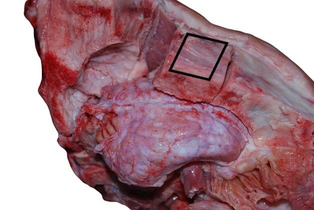
Photonics 2021, 8, 365                                                                                                              3 of 19

      Table 1. Summary of sample types, sample thickness, used refractive index, number of samples (# Samples, 4 repetitions for
      each sample) and the results of the power law fit, Equation (2) as well as Equation (3) in case of small cylindrical scatterers
      (in the latter case marked with an *).

                            Thickness Range                                                 Power Law Fit, Equations (2) and (3),
  Sample Name                                       Refractive Index        # Samples
                            in mm                                                           a as well as a∗ in mm−1
  non-scalded skin          (1.3–1.9)               5 layers, 1.4           3               a = 2.1808, b = 0.4912, c = 0.8663
                                                                                            a∗ = 2.2788, b∗ = 0.8317, c∗ = 0
  adipose tissue            (2.3–4.1)               3 layers, 1.46 [19]     13              a = 1.8468, b = 0.0139, c = 0.5953
  muscle                    (1.0–3.6)               3 layers, 1.4           11              a = 0.5798, b = 0.1647, c = 1.1793
  bone cranium              (0.9–1.9)               5 layers, 1.4           4               a = 1.5963, b = 0.1261, c = 0.5821
  bone mandibula            (0.9–1.4)               5 layers, 1.4           4               a = 1.9909, b = 0.0417, c = 0.5853
  bone scapula              (1.0–1.4)               5 layers, 1.4           4               a = 2.0862, b = 0.1008, c = 0.5473
  bone tibia                (0.9–1.0)               5 layers, 1.4           2               a = 1.9652, b = 0, c = 0.7921
  bone marrow               (2.5–2.6)               5 layers, 1.4           2               a = 1.2177, b = 0.0475, c = 0.3490
  cartilage                 (1.1–1.4)               5 layers, 1.4           3               a = 2.9583, b = 0.1984, c = 0.9418
                                                                                            a∗ = 2.9618, b∗ = 0.4259, c∗ = 0.5777
  brain, white matter       (0.4–2.3)               3 layers, 1.4           6               a = 8.2925, b = 0, c = 0.8290
  brain, gray matter        (1.0–4.0)               3 layers, 1.4           6               a = 1.2345, b = 0.0275, c = 1.3291
  dura mater                (1.3–3.5)               5 layers, 1.4           3               a = 3.8147, b = 0.6265, c = 1.3037
                                                                                            a∗ = 3.9579, b∗ = 0.9605, c∗ = 0
  medulla oblongata         (1.4–1.6)               5 layers, 1.4           2               a = 6.4811, b = 0, c = 0.7731

                                   2.2. Sample Preparation
                                        Besides applying an appropriate experimental setup, also the sample preparation
                                  and the measurement procedure are important for a precise determination of the optical
                                  properties of tissue. Due to the given measurement apparatus, we are limited to studies
                                  that are affected by alterations of the investigated tissue. The tissue preparation may
                                  change, e.g., the water content, the oxygenation state and the amount of blood within
                                  the tissue [20,21], and therefore the optical properties. Besides this, also freezing-thawing
                                  cycles of biological tissues have an influence on the determined optical properties due to
                                  cell membrane disruption and liquid loss [7]. Our investigations on gray brain matter of
                                  a pig, not shown here, pointed out a relative decrease of around 25% in µs0 and a relative
                                  decrease of 5% in µa at around 550 nm (blood loss) after storing the prepared sample
                                  at—14 °C for 15 days.
                                        In following, we therefore used only fresh samples obtained from the slaughterhouse
                                  (Ulmer Fleisch GmbH, Ulm, Germany), prepared and measured within eight hours post-
                                  mortem. For organizational reasons of the time consuming preparation process of our
                                  samples, a small portion of samples were also stored in the fridge, at 4 °C, for a maximum
                                  of two days before measurement. The measurements themselves were performed within
                                  two minutes at room temperature, after sample preparation and temperature adjustment.
                                  We decided to use the cuvettes mentioned by Bergmann et al. [15] using known N-BK7
                                  glass slides (“34–427”, Edmund Optics, Barrington, NJ, USA) for the measurements, to
                                  avoid dehydration, blood loss, surface roughness effects as well as to decrease the influence
                                  of the lacking knowledge of the correct refractive indices of the studied tissues. Therefore,
                                  we filled liquid with a known refractive index in the space between glass slide and sample
                                  surface. This results in a five layered construct surrounded by air, see Figure 1.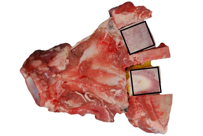
Photonics 2021, 8, 365                                                                                               4 of 19

                                                          y
                                                                         normalisation beam    spacer

                                                              45°
                                           detector FOV                                         glas sample liquid
                                  x
                                                                    5°                  5°
                                                                                               transmission beam
                         reflection beam
                                                                               five layer
                                                                               sample holder

                         Figure 1. Schematic of the used integrating sphere setup in top view including the five layered
                         sample holder. The detector FOV is located perpendicular to the horizontal/equatorial plane near
                         the south pole of the integrating sphere.

                               The refractive index interfaces are then given by air, glass, liquid, sample, liquid, glass,
                         and air. We used a sodium chloride solution (NaCl 0.9%) as a liquid layer, which has almost
                         the refractive index of water and furthermore enables osmotic balance to prevent further
                         blood loss as well as changes in the tissue structure. The thickness of the liquid layer was
                         assumed to be 10−3 mm. For the refractive index of the tissue layer we used n = 1.4. Varying
                         sample refractive indices in the range of 1.3 to 1.5, which seems to be realistic in biological
                         media [18], results in typical relative changes of the determined effective scattering and
                         absorption coefficients of ∆µs0 < 5% and ∆µa < 13%, respectively, according to Monte Carlo
                         simulations. In the case of adipose tissue, muscle and brain matter, the sodium chloride
                         solution was repressed by the soft tissue. Therefore, in the simulation we used a three
                         layered construct with the refractive index value of 1.4 for muscle and brain matter, as well
                         as 1.46 [19] for adipose tissue instead of the five layered construct. Cylindrical spacers were
                         used to enable tissue samples having a defined thickness. For this, we used 3D-printed
                         polylactic acid (PLA) hollow cylinders as well as punched out silicone mats with a sample
                         chamber having a lateral diameter of 30 mm and different axial dimensions. The used
                         silicone mats were applied in case of elastic samples to better control the mechanical stress
                         originating from the glass slides, as, otherwise, there would be a likely change in structural
                         and optical properties. Due to the morphology and constitution of the investigated samples,
                         different preparation methods have been applied. For all the soft tissues like brain matter,
                         dura mater, medulla oblongata and skin, we exclusively used scalpels and tweezers to
                         separate the sample types and in case of dura mater to layer the tissues. For the elastic
                         samples like adipose tissue and muscle, we used a custom made slicer (“universal cutter”,
                         Ritterwerk GmbH, Gröbenzell, Germany), to cut the tissues as plano-parallel as possible.
                         The lateral size was processed with a puncher to produce cylindrical samples with 30 mm
                         in diameter. The harder sample sections of bones were sawed by a saw microtome (“Leica
                         SP1600”, Leica Mikrosysteme Vertrieb GmbH, Wetzlar, Germany). A diamond coated
                         inner-hole saw blade of 280 µm thickness was rotated with 600 rpm and moved with low
                         speed, to obtain plano-parallel samples. Simultaneously, a built-in water cooling device
                         prevented overheating of the saw blade as well as the evaporation of water within the
                         bone tissue. For improved statistics, due to the inhomogeneity of several sample types
                         and to account for the inter-animal variability, repetitions at different lateral positions and
                         different sections as well as different animals were investigated. The maximum thicknesses
                         d are limited by the transmission signal. Therefore, large absorption coefficients of blood
                         and water in the VIS and NIR spectral range, respectively, obliged us to fabricate smaller
                         sample thicknesses. Sometimes, the sample thicknesses are smaller than needed to achieve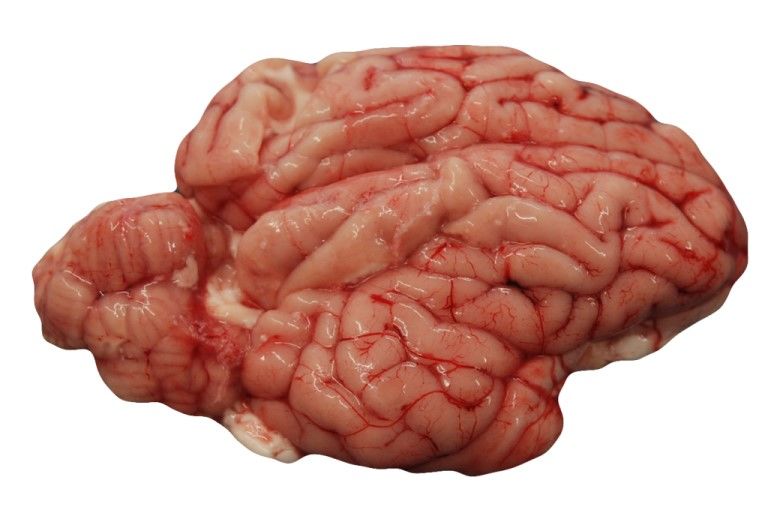
Photonics 2021, 8, 365                                                                                              5 of 19

                         an required optical thickness of τ = µs0 d > 2 [14,15], which is necessary to decrease the
                         influence of the estimated asymmetry factor g and refractive index n.

                         2.3. Main Tissue Constituents and Analysis
                               We compared the obtained absorption spectra with the absorption spectra of the
                         main ingredients, namely de-oxygenated (Hb) and oxygenated (HbO2 ) blood [22], water
                         (VIS [23], NIR [24]), lipid [25] and collagen/elastin [26], see Figure 2. The µa of collagen,
                         elastin and lipid was obtained from the named literature using the free “WebPlotDigitizer”
                         from Rohatgi [27]. Furthermore, we measured collagen (“hydrolysed bovine collagen”, U
                         Nutrition LTD, Urmston, Greater Manchester, UK), and pork lard (“Schweineschmalz”,
                         LARU, Bottrop, Germany), with the integrating sphere setup to double check the absorption
                         spectra of these tissue types. We found a twofold to threefold larger µa of collagen compared
                         to the results from Sekar et al. [26] between 1000 nm and 1400 nm, who measured collagen
                         samples in the time domain. We note that in the spectral range of 1150 nm to 1300 nm
                         the absorption peak of collagen as well as elastin have a smaller spectral width compared
                         to that of water. Due to the similarity of the absorption spectra of elastin and collagen
                         in the spectral range from 400 nm to 1400 nm, it is difficult to differentiate between both
                         absorbers. Therefore, in this study, the estimated collagen content is overestimated and
                         could also be caused by elastin. For lipid, we found a good agreement in µa within the
                         spectral range of 850 nm to 1050 nm, but we obtained a difference by a factor of four at
                         around 600 nm compared to van Veen et al. [25]. The discrepancy in the VIS range could
                         be caused by the difference of the investigated samples. Whereas van Veen et al. used
                         melted fat for the measurements, we used scattering lard at room temperature containing
                         TiO2 particles. The TiO2 particles were added to the liquid phase of lard in a previous step
                         by using a hotplate. Furthermore, we measured the melted lard without TiO2 particles
                         in two separate cuvettes with different thicknesses to validate the measured absorption
                         spectrum. We found a good agreement within our measurements in the range of 420 nm
                         to 1400 nm, whereas the absorption of TiO2 particles lead to a larger µa at around 400 nm
                         (data not shown). The refractive index of the liquid phase was measured at 589 nm with
                         an Abbe refractometer (“120709”, Carl Zeiss AG, Oberkochen, Germany) with n = 1.464
                         and this value as well as the dispersion were measured furthermore with a setup based on
                         the determination of the specular reflection, which was also used in [15] to determine the
                         hemispherical reflectance coefficient of the calibration standard of the integrating sphere.
                         For this measurement, a sample cuvette containing one N-BK7 glass slide, the medium
                         itself and an absorbing backside (black rubber) was used. This was necessary to avoid
                         reflections from the backside. We found a good agreement in the refractive index at 589 nm.
                         The measured dispersion of melted lard can be described by Sellmeier’s equation
                                                              q
                                                                        B1 λ2        B2 λ2        B3 λ2
                                                     n(λ) =       1+            +            +                         (1)
                                                                       λ2 −C1       λ2 −C2       λ2 −C3

                         with the coefficients B1 = 1.9496, C1 = 0.0111 nm2 , B2 = −0.8303, C2 = 0.0139 nm2 , B3 = −1.6134,
                         C3 = −156.5348 nm2 . For turbid lard at room temperature, we used the same procedure apply-
                         ing the specular reflection measurement and got a dispersion described by Equation (1) with
                         B1 = 1.9613, C1 = 0.0109 nm2 , B2 = 0.8168, C2 = 0.0138 nm2 , B3 = −1.6358, C3 = −156.9641 nm2 .
                         At λ = 589 nm we obtain n = 1.473.
                              The absorption coefficients of pure water and blood from pig were also measured
                         in this study (not shown), resulting in very similar results compared to [22–24]. We note
                         that we did not differ the spectra of oxygenated and de-oxygenated blood from that of
                         oxygenated and de-oxygenated myoglobin.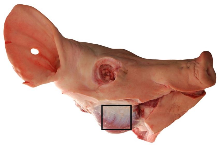
Photonics 2021, 8, 365                                                                                                 6 of 19

                                                               water, Pope/Kou 1997/1993
                                                               HbO 2 0.2%, Bosschaart 2014
                                       100                     Hb 0.2%, Bosschaart 2014
                                                               melted lard, van Veen 2004
                                                               lard
                                                               collagen, Sekar 2017
                                                               elastin, Sekar 2017
                                      10                       collagen, bovine
                         µ a / mm-1

                                      10

                                      10

                                      10
                                           400       600            800       1000                1200         1400
                                                                    wavelength / nm

                         Figure 2. Main absorbers in biological tissue adopted from the literature [22–26] or measured within
                         this study.

                              Besides the absorption coefficient, the effective scattering coefficient is also conclusive
                         for characterizing the investigated tissue. The effective scattering spectra were fitted with
                         two power laws describing two ranges of scattering particle sizes of spherical shape [12,28]

                                                   µs0 (λ) = a · (b · (λ/λ0 )−4 + (1 − b) · (λ/λ0 )−c )                   (2)

                         with the wavelength λ, the scatter amplitude a, the fraction of the Rayleigh scatterers b and
                         the scatter power of the, compared to the wavelength, large scatterers c. In this study, we
                         used λ0 = 630 nm. The three parameters a, b and c are related to the density, size and shape
                         of the scatterers in the turbid media. If no Rayleigh scatterers are present, Equation (2)
                         simplifies to the power law containing only the two parameters a and c for one range
                         of scattering particle size. If Rayleigh scatterers are present, they contribute more to the
                         scattering in the lower VIS range (λ−4 decrease) compared to the longer wavelengths. In
                         most investigated biological tissues, we expect a variety of rather spherical-formed small
                         scatterers and for this it is reasonable to use a generally applicable curve fitting with a λ−4
                         decrease combined with a decrease for larger scatterers using Equation (2). We note that
                         for small cylindrical scatterers, the decrease in µs0 is proportional to λ−3 [29]. Therefore, we
                         additionally fitted with
                                                                                                          ∗
                                                 µs0 (λ) = a∗ · (b∗ · (λ/λ0 )−3 + (1 − b∗ ) · (λ/λ0 )−c )                 (3)

                         in case of skin, cartilage and dura mater due to their high content of cylindrially shaped
                         collagen fibrils, see Table 1 as well as Figures 4, 8 and 9 in the following section.
                              In Table 1, all investigated sample types, sample thicknesses, used refractive indices,
                         number of repetitive measurements (we used samples from a minimum of two animals) as
                         well as the results of the power law fits of the mean µs0 are summarized.
                              To demonstrate the influence of the applied asymmetry factor and refractive index, we
                         used one sample of adipose tissue (three layered construct) from the back of the pig, having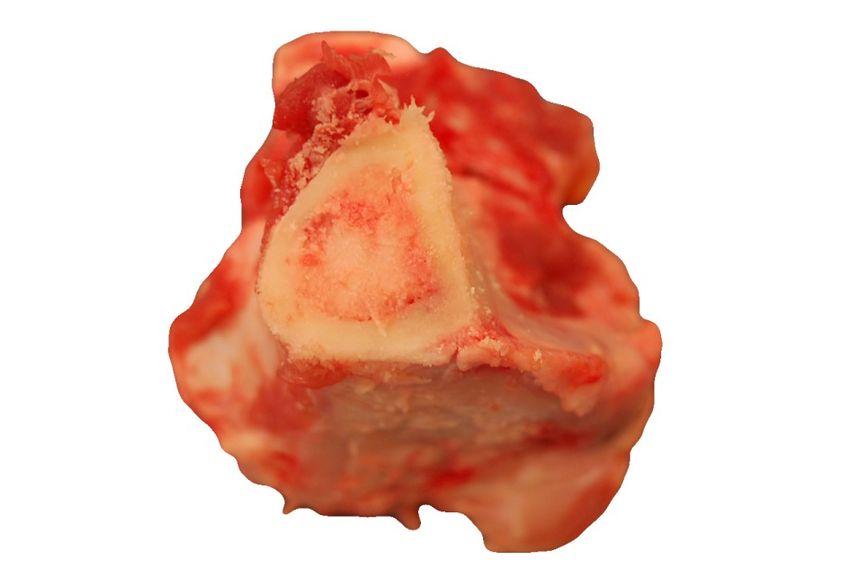
Photonics 2021, 8, 365 7 of 19
a sample thickness of 3.5 mm and varied g as well as n in the Monte Carlo simulations
for fitting the experimental data, see Figure 3. As it was stated by Foschum et al. [14], the
impact on the determined optical properties of n is larger compared to the asymmetry
value g. For example, at 544 nm we obtained a µs0 = 1.429 mm−1 (τ (λ = 544 nm) ≈ 5) and
µa = 0.032 mm−1 for the presumed correct optical properties of g = 0.9 and n = 1.46. A
change in g to 0.75 causes a deviation of 0.24% in µs0 and 0.54% in µa , whereas a change in
n to 1.4 results in a deviation of 4.76% in µs0 and 14.82% in µa .
1.8
n = 1.46, g = 0.9
n = 1.46, g = 0.75
1.7
n = 1.4, g = 0.9
1.6
1.5
µ s / mm-1
1.4
1.3
1.2
1.1
1
0.9
600 800 1000 1200 1400
wavelength / nm
(a)
10
µ a / mm-1
10
n = 1.46, g = 0.9
n = 1.46, g = 0.75
n = 1.4, g = 0.9
600 800 1000 1200 1400
wavelength / nm
(b)
Figure 3. Investigation of the impact of g and n on adipose tissue (three layered construct) with a
sample thickness of 3.5 mm. Figure (a) shows µs0 versus wavelength and (b) depicts µa (λ).
3. Results
3.1. Skin
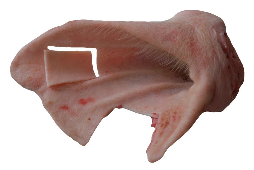
We investigated fresh, non-scalded skin from the ear of four different animals, prepar-
ing in total six samples. First, the hairs were removed with a razor. In the next step, we
used a scalpel and a tweezer to separate the skin from the adjacent subcutis, which is
mainly composed of connective and adipose tissue. The lateral dimension of the sample
was processed by the use of the mentioned puncher with a diameter of 30 mm. Each sample
was measured four times by changing the lateral position as well as flipping the sample
sides. The samples themselves consist of epidermis and dermis, but were evaluated using
a LUT for homogeneous tissue. In Figure 4, the optical properties are given and exemplary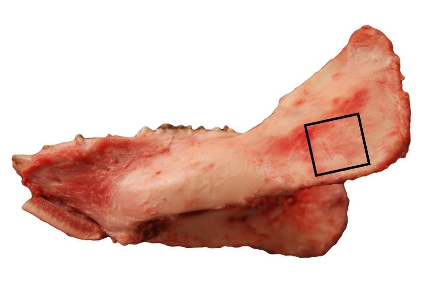
Photonics 2021, 8, 365                                                                                            8 of 19

                         samples, which were prepared for the measurement, are shown under diffuse illumination
                         in reflection and transmission. The skin samples are fairly lateral homogeneous, but a lay-
                         ered structure exists. Regarding the optical properties shown in Figure 4, a strong decrease
                         of µs0 versus wavelength can be seen. We found a good agreement in µs0 between our investi-
                         gations and the values presented by Mosca et al. [7], who used a stack of five skin specimen
                         for their investigation. Larger standard deviations between 400 nm and 600 nm occur due
                         to the difference in reflection of both sides of the layered specimen (epidermis, dermis as
                         well as a possible small amount of subcutis), especially at larger µa , where the reflection
                         values are considerably larger than the transmission values. In the skin dermis collagen
                         fibers with diameters of 0.5–8 µm [30] (measured on human neonatal-skin samples), which
                         are dominated by a general orientation parallel to the skin surface, are located. The collagen
                         fibrils of diameters of around 40 nm to 80 nm [31] (measured on bovine and ovine leather),
                         around 100 nm [30] (measured on neonatal-skin samples), or rather in a range of 25 nm
                         to 250 nm [32] (measured on a rat cornea) are positioned in the collagen fibres. Besides
                         the small cylindrical scatterers, a variety of smaller particles and structures exists in the
                         skin dermis which contribute to the Rayleigh scattering of light [30]. Thus, we used both
                         Equations (2) and (3) to fit the measured effective scattering spectra. We found a much
                         larger fraction of Rayleigh scatterers of around 83% assuming small cylindrical scatters
                         compared to assuming small spherical scatterers, which gives a fraction of around 49%.
                         The subcutis on the other hand consists of connective tissue, adipose tissue, blood vessels
                         and nerves. Therefore, the optical properties of both sample sides are different. As stated
                         before, the smaller particles (collagen fibrils) and structures in the dermis influence more
                         the scattering in the lower VIS (sharp decrease), while larger scatterers (collagen fibres) are
                         dominant in the NIR range. The absorption of skin is mainly influenced by blood (peak
                         around 554 nm), water (peak at about 973 nm) and elastin/collagen (smaller peak-width
                         compared to water at around 1175 nm). The influence of adipose tissue from the subcutis is
                         remarkable small. Unfortunately, it is not easy to separate elastin from collagen absorption
                         spectra, which would be useful as an indicator for dermal skin ageing [33]. Compared to
                         the data presented by Mosca et al. [7], we obtained around two to three times larger µa in
                         the range of 650 nm to 1100 nm. For this reason, we performed also further investigations
                         on scalded skin samples and found a smaller absorption compared to the non-scalded skin
                         in this range. Furthermore, we found a smaller decrease in µs0 versus wavelength for the
                         scalded skin samples, which may be caused by the treatment at the butchery. The animals
                         were treated with approximately 60 °C water or water vapor (steam scalding) for a few
                         minutes to remove the bristles out of the hair follicles. It can be expected that this treat-
                         ment will change the collagen structure, the water content, the surface roughness and the
                         elasticity of the sample. In the range of 450 nm to 500 nm, we found an apparent increase
                         of the absorption of the scalded skin, which may be caused by the change of the collagen
                         structure, but could also be induced by the oxygenation of Fe2+ to Fe3+ in haemoglobin
                         (methaemoglobin), which increases the absorption in the VIS range between 400 nm and
                         670 nm, especially at the Soret band at around 405 nm [34]. We notice a double peak for the
                         non-scalded skin around 550 nm, which indicates the oxygenation of hemoglobin. If higher
                         temperatures above 60 °C are applied to skin samples, the destruction of the epidermis [35]
                         and the coagulation of the proteins are expected to increase.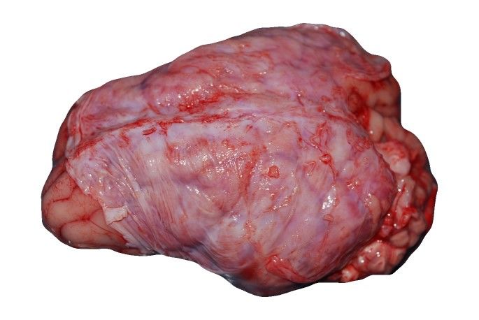
Photonics 2021, 8, 365                                                                                                                                             9 of 19

                               non-scalded skin (ear)                                                   100
                 10            µ s = 2.1808 (0.4912 ( / 0)   + 0.5088 ( / 0)       )
                               µ s = 2.2788 (0.8317 ( / 0)   + 0.1683)
                               scalded skin (belly)
                 8             µ s = 2.3292 (0.2587 ( / 0)   + 0.7413 ( / 0)       )

                               µ s = 2.3377 (0.5665 ( / 0)   + 0.4335 ( / 0)   )
                               Mosca 2020                                                              10
    µ s / mm-1

                                                                                          µ a / mm-1
                 6

                 4

                                                                                                       10
                 2
                                                                                                                                          non-scalded skin (ear)
                                                                                                                                          scalded skin (belly)
                                                                                                                                          Mosca 2020
                 0
                 400     600           800       1000               1200           1400                     400   600   800       1000         1200            1400
                                       wavelength / nm                                                                  wavelength / nm
                                            (a)                                                                           (b)

                                                              (c)                      (d)                        (e)

       Figure 4. Effective scattering coefficient (a) and absorption coefficient (b) versus wavelength for skin. Exemplary tissue
       sample (c), a 1.46 mm thick sample in transmission (d) and in reflection (e) under diffuse illumination.

                                            3.2. Adipose Tissue
                                                  The investigated adipose tissues were obtained near the shoulder joint and from the
                                            back regions of five pigs. As can be seen in Figure 5c,f, there is a visible intersection within
                                            the adipose tissue, and therefore we differentiated between the layer faced to the skin
                                            and the layer faced to the muscle. Apart from this, the adipose samples seemed to be
                                            fairly homogeneous. We found out that fatty tissue near the skin boundary has a smaller
                                            effective scattering coefficient compared to tissue near the muscle boundary (data not
                                            shown). Furthermore, adipose tissue near the shoulder joint had a significantly larger µs0
                                            than that from the back (data not shown). The standard deviation of µs0 for a single adipose
                                            tissue sample is comparable to the standard deviation from non-scalded skin, compare
                                            Section 3.1. In contrast, comparing all measurements of the adipose tissue near the skin
                                            and muscle boundary as well as from the shoulder joint and from the back, we obtain a
                                            larger standard deviation Figure 5a. Compared to the data presented by Mosca [7] and
                                            Foschum [13], we got similar results for µs0 and µa . In the NIR absorption range the main
                                            absorber is fat (in the figure labeled as lard).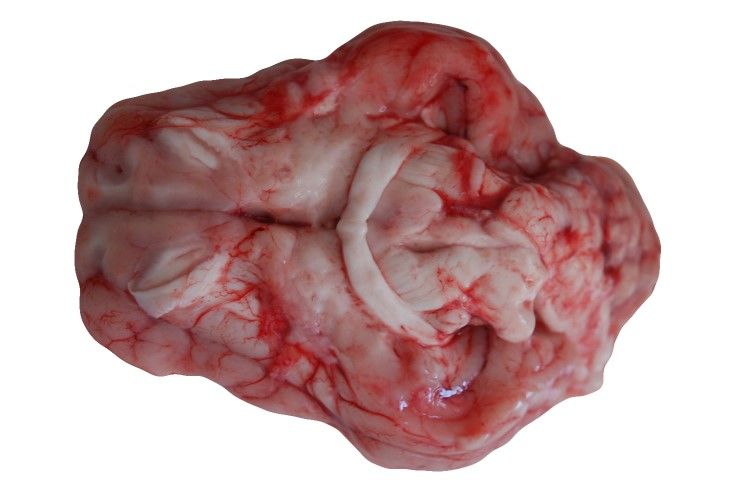
Photonics 2021, 8, 365                                                                                                                                10 of 19

                3.5
                                 adipose tissue
                                 µ s = 1.8468 (0.0139 ( / 0)   + 0.9861 ( / 0)   )

                 3               Foschum 2012
                                 Mosca 2020                                                          10

                2.5
   µ s / mm-1

                                                                                        µ a / mm-1
                 2
                                                                                                     10

                1.5

                                                                                                                                         adipose tissue
                 1
                                                                                                                                         lard
                                                                                                     10
                                                                                                                                         Foschum 2012
                                                                                                                                         Mosca 2020
                0.5
                  400      600          800       1000               1200        1400                     400   600   800       1000    1200          1400
                                        wavelength / nm                                                               wavelength / nm
                                              (a)                                                                       (b)

                (c) back                (d)                       (e)                     (f) near shoulder joint             (g)              (h)

          Figure 5. Effective scattering coefficient (a) and absorption coefficient (b) versus wavelength for adipose tissue near the
          shoulder joint and from the back, facing to muscle tissue as well as to skin tissue. Exemplary tissue samples (c,f), a
          3.42 mm (back) and a 3.81 mm (near shoulder joint) thick sample in transmission (d,g) and in reflection (e,h) under
          diffuse illumination.

                                                  3.3. Muscle
                                                        We performed measurements on masticatory muscle and muscle from the shoulder,
                                                  where it was relatively easy to differentiate the muscle tissue from adjacent tissue. The sam-
                                                  ples were sliced parallel to the skin layer and therefore parallel to the muscles’ longitudinal
                                                  direction. In Figure 6, the heterogeneous structure caused by the combination of muscle
                                                  fibers and connective tissue (endomysium/perimysium) is apparent. However, overall the
                                                  masticatory muscle and the muscle from the shoulder look similar. This was confirmed by
                                                  the resulting optical properties. The effective scattering is comparable with the mentioned
                                                  literature values. Further, we found absorption characteristics caused by de-oxygenated
                                                  blood or rather de-oxygenated myoglobin (around 554 nm [36]), water (around 973 nm) and
                                                  elastin/collagen (around 1175 nm). As was stated before, we did not differ de-oxygenated
                                                  blood from de-oxygenated myoglobin. We found a significant difference in the absorption
                                                  coefficient between 400 nm and 700 nm compared to Foschum [13] and Mosca [7]. Contrary
                                                  to Foschum [13], we found a larger amount of blood. We note that the light propagation
                                                  exhibit an anisotropic light propagation caused by the aligned muscle fibers, which was
                                                  not taken into account in the evaluation process. Thus, the obtained optical properties
                                                  might strongly depend on the alignment of the muscle fibers relative to the slab surface.
Photonics 2021, 8, 365 11 of 19
1.5
muscle
muscle
µ s = 0.5798 (0.1647 ( / 0) + 0.8353 ( / 0) ) Foschum 2012
Foschum 2012 0 Mosca 2020
10
Mosca 2020
1
µ s / mm-1
µ a / mm-1
10
0.5
10
0
400 600 800 1000 1200 1400 400 600 800 1000 1200 1400
wavelength / nm wavelength / nm
(a) (b)
(c) masticatory (d) (e) (f) from shoulder (g) (h)
Figure 6. Effective scattering coefficient (a) and absorption coefficient (b) versus wavelength for muscle. Exemplary
tissue samples (c,f) including region of interest, a 3.53 mm (masticatory muscle) and a 1.86 mm (shoulder) thick sample in
transmission (d,g) and in reflection (e,h) under diffuse illumination.
3.4. Bone
Several bone samples from the cranium, mandibula, scapula and tibia were inves-
tigated. Exemplary sample pictures and determined optical properties are collected in
Figure 7. The samples were separated roughly with hammer and chisel or bone saw.
We fixed the samples with a two-component resin based on methyl methacrylate plastic
(“Technovit 3040”, Kulzer GmbH, Hanau, Germany) to saw small parallel slices having
thicknesses between 0.9 mm to 1.9 mm using a saw microtome. We mainly investigated the
compacta of the bones, which has been cut out parallel to the bone surface as has been illus-
trated in Figure 7. The saw microtome was cooled with water and after the separation we
placed the sample in a closable plastic container filled with sodium chloride solution. This
prevents dehydration, which was the main problem of the bone investigation. Dehydrated
bone tissue leads to a about five-fold larger µs ’, when dried for 48 h (data not shown).
The bone tissues were investigated using the glass plates covering the bone, which was
soaked with the sodium chloride solution. The spectral behavior of the effective scattering
coefficient was similar for the different bone types, except of that of tibia, which revealed no
Rayleigh scattering particles. However, significant differences were found in the absolute
values. A good agreement was found with data presented by Mosca et al. [7] and Fos-
chum [13]. Concerning µa we found absorption characteristics caused by oxygenated blood
(double peak at about 540 nm and 577 nm), collagen (shoulder at around 1175 nm in case of
scapula and mandibula and a slightly reduced peak-width compared to the case when only
a water peak is present), water (around 973 nm) and fat (in case of scapula and mandibula
at about 1209 nm). The tibia bone tissue had less blood content. As expected, we found
the largest amount of blood in the flat bones, where the bone marrow is producing red
corpuscles (haematopoiesis). The absorption coefficient determined by Mosca et al. [7] and
Foschum [13] (bone from cow) is within the range of our results. Remarkably, we foundPhotonics 2021, 8, 365 12 of 19
larger differences for the scapula bone samples resulting in a larger standard deviation. For
the scapula bone, we found regions with a larger amount of oxygenated blood and less fat
content as well as regions with a larger amount of de-oxygenated blood in combination
with a larger fat content (data not shown).
bone, cranium 100
µ s = 1.5963 (0.1261 ( / 0) + 0.87391 ( / 0) )
5
bone, mandibula
4.5 µ s = 1.9909 (0.0417 ( / 0) + 0.95834 ( / 0) )
bone, scapula
4 µ s = 2.0862 (0.1008 ( / 0) + 0.89924 ( / 0) )
bone, tibia
10
µ s / mm-1
3.5 µ s = 1.9652 ( / 0)
µ a / mm-1
Foschum 2012
3 Mosca 2020
2.5
10
2 bone, cranium
bone, mandibula
bone, scapula
1.5 bone, tibia
Foschum 2012
1 Mosca 2020
10
400 600 800 1000 1200 1400 400 600 800 1000 1200
wavelength / nm wavelength / nm
(a) (b)
(c) cranium (d) (e) (f) mandibula (g) (h)
(i) scapula (j) (k) (l) tibia (m) (n)
Figure 7. Effective scattering coefficient (a) and absorption coefficient (b) versus wavelength for bone. Exemplary tissue
samples (c,f,i,l) including region of interest, a 1.05 mm (cranium), a 0.93 mm (mandibula), a 1.33 mm (scapula) and a 0.91 mm
(tibia) thick sample in transmission (d,g,j,m) and in reflection (e,h,k,n) under diffuse illumination.
3.5. Bone Marrow and Cartilage
Bone marrow from the tibia and elastic cartilage from the ear were investigated by
using cuvettes with elastic spacers as described earlier, see Figure 8. Contrary to the
cartilage samples, the bone marrow samples look inhomogeneous due to the blood vessels.
This results in a larger standard deviation of µa , especially between 450 nm and 600 nm.
The elastic cartilage was separated from the non-scalded skin of the ears. For the elastic
cartilage, the sharp decrease in µs0 versus wavelength is caused by the collagen fibrils due
to their small diameter compared to the wavelength of the applied light. Again, applying
Equation (3) results in a larger fraction of Rayleigh scatterers of around 43% compared
to the use of Equation (2), resulting in 20%. Comparing the absorbers from Figure 2, we
found absorption characteristics based on oxygenated blood (double peak at about 540 nm
and 577 nm), fat (about 1209 nm) and elastin/collagen (shoulder at about 1175 nm) for bonePhotonics 2021, 8, 365 13 of 19
marrow and absorption based on water (around 973 nm) and collagen (slightly reduced
peak-width compared to water around 1175 nm) for the cartilage sample.
9 100
bone marrow
8 µ s = 1.2177 (0.0475 ( / 0) + 0.9525 ( / 0) )
cartilage
7 µ s = 2.9583 (0.1984 ( / 0) + 0.8016 ( / 0) )
µ s = 2.9618 (0.4259 ( / 0) + 0.5741 ( / 0) )
6
10
µ s / mm-1
µ a / mm-1
5
4
3
10
2
1 bone marrow
cartilage
0
400 600 800 1000 1200 1400 400 600 800 1000 1200
wavelength / nm wavelength / nm
(a) (b)
(c) bone marrow (d) (e) (f) cartilage (g) (h)
Figure 8. Effective scattering coefficient (a) and absorption coefficient (b) versus wavelength for bone marrow and cartilage.
Exemplary tissue samples (c,f), a 2.54 mm (bone marrow) and a 1.12 mm (elastic cartilage) thick sample in transmission
(d,g) and in reflection (e,h) under diffuse illumination.
3.6. Brain
We obtained three different fresh brains and prepared six samples of gray (substantia
grisea) and white (substantia alba) brain matter out of it. The separation of white and
gray matter was done with scalpel and tweezer. Larger regions of white brain matter were
found at the bottom of the brain, and adjacent on the inner side of the cortex. The gray
brain matter was separated from the inner white matter. Especially for the gray brain
matter, see Figure 9, several pieces were butt-jointed. The resulted samples looked more or
less inhomogeneous due to impurities from adjacent tissue. In addition, we investigated
the dura mater as a stack of thin layers and the medulla oblongata, which we gathered
from two fresh pig heads by separating the skull from the inside organ using hammer and
chisel. Both, the dura mater and the medulla oblongata samples looked homogeneous. The
obtained effective scattering of white matter was much larger compared to the effective
scattering of gray brain. For example, at 633 nm, we found a difference by a factor of
about seven. The white brain matter had less blood and a larger amount of water and fat
compared to the gray brain matter. The larger µs0 of the white brain matter compared to
gray brain matter is the main reason for its whitish appearance. Comparing the results of
the brain tissues with the data presented by Mosca et al. [7], we found a better agreement
for µa than for µs0 . The difference of the µs0 values presented by Mosca et al. compared
to our results depends again on the difference in the investigated sample. Whereas our
samples were separated into the mentioned constituents, Mosca et al. measured the optical
properties from the whole brain including gray and white brain matter. The pronounced
decrease of µs0 for the dura mater in the lower VIS spectral range is dominated by thePhotonics 2021, 8, 365 14 of 19
small substituents like collagen fibrils. Therefore we recommend to use Equation (3) for
the description of the sharp decrease in µs0 , which gives an amount of around 96% of
Rayleigh scatterers compared to the assumption of spherical scatterers, which results in
approximately 63% of Rayleigh scatterers. We found absorption characteristics based on
de-oxygenated blood (around 554 nm), water (around 973 nm), collagen (indicated by a
slightly reduced peak width compared to the case when only a water peak is present) and
fat (peak at about 1209 nm for medulla oblongata and white brain matter).
brain, white matter
µ s = 8.2925 ( / 0) brain, white matter
20 brain, gray matter
100 brain, gray matter
dura mater
µ s = 1.2345 (0.0275 ( / 0) + 0.9725 ( / 0) )
medulla oblongata
dura mater Mosca 2020
µ s = 3.8147 (0.6265 ( / 0) + 0.3735 ( / 0) )
15
µ s = 3.9579 (0.9605 ( / 0) + 0.0395)
µ s / mm-1
µ a / mm-1
medulla oblongata
µ s = 6.4811 ( / 0) 10
Mosca 2020
10
5
10
0
400 600 800 1000 1200 1400 400 600 800 1000 1200 1400
wavelength / nm wavelength / nm
(a) (b)
(c) brain (d) (e) (f) brain (g) (h)
white matter gray matter
(i) dura mater (j) (k) (l) medulla (m) (n)
oblongata
Figure 9. Effective scattering coefficient (a) and absorption coefficient (b) versus wavelength for brain tissue. Exemplary
tissue samples (c,f,i,l), a 1.03 mm (white brain matter), a 1.06 mm (gray brain matter), a 1.37 mm (dura mater) and a 1.54 mm
(medulla oblongata) thick sample in transmission (d,g,j,m) and in reflection (e,h,k,n) under diffuse illumination.
3.7. Quantitative Study
We quantitatively analyzed the concentration of the mentioned main absorbers water,
fat, collagen, de-oxygenated blood and oxygenated blood using the obtained mean absorp-
tion spectra of each tissue type. To this aim, nonlinear regressions (“lsqcurvefit” using
MATLAB R2020a, MathWorks, Natick, MA, USA) to the logarithmized data of the obtained
absorption spectra for each tissue type were performed for wavelengths from 400 nm to
1400 nm using equal weights. We used the mean absorption spectra of water, oxygenated
blood and de-oxygenated blood from the mentioned literature [22–24] and fat as wellPhotonics 2021, 8, 365 15 of 19
as collagen from our own measurements. Fitting parameters were the concentrations of
the main absorbers and a spectral constant offset. The latter was introduced due to the
presence of further absorbing substituent besides the main absorbers. For the average
sum of the main absorber’s concentrations we obtain 85.35%, reaching from 62.45% for
muscle to 105.22% for bone marrow. These values may be influenced by systematic errors
like possibly incorrect absorption spectra of the main absorbers or of the measured mean
absorption spectra of the tissue types, for example due to inhomogeneous samples, the
similarity in the absorption spectra, for example between water and collagen in the NIR
spectral range, or by another, not regarded main absorber, which may be present in the
tissue. The obtained results are given in Figure 10 and listed in Table 2. In general, we
found a good agreement between the derived absorption spectra and the fitted spectra
using the nonlinear regression for non-scalded skin, adipose tissue, bone cranium, bone
mandibula, bone scapula, bone tibia, bone marrow, elastic cartilage, brain white matter
and dura mater. To quantify the remaining deviation between the measured absorption
spectra µa and the absorption spectra determined by the fit algorithm µa,fit , we used
N
µa,fit,i −µa,i
2
1
E= N−P ∑ ∆µa,i , (4)
i =1
with the total number N of 354 spectral values i, P = 6, the number of parameters of the fit
model and the standard deviation of the measured absorption spectra ∆µa [37]. For most
of the investigated tissues we received E < 1 due to the large standard deviation of the
measured absorption spectra. Only for muscle (E = 1.087), brain gray matter (E = 1.403) and
medulla oblongata (E = 1.831) we obtained a larger discrepancy of the measured spectral
absorption using the fit model. For non-scalded skin, we obtained concentrations of 66.31%
water, 3.11% fat, 24.36% collagen, 0.32% de-oxygenated blood and 0.05% oxygenated blood.
The fat content may be influenced from the subcutis, which was not easy to separate from
the skin. Due to further not considered absorbers, we obtained a constant offset factor of
0.012 mm−1 . In adipose tissue we found a pure fat content of 82.21%, which seems to be
realistic. For the fit of the spectral absorption of the adipose tissue, no additional offset
factor was necessary. In muscle, we found a concentration of 59.25% water, 0% fat, 1.55%
collagen and 1.65% de-oxygenated blood. In muscle tissue collagen can be found in the
connective tissue and in tendons. Unfortunately, the similarity in µa between water and
collagen in the NIR spectral range can affect the obtained absolute values of the water
and collagen concentration, especially for larger E. For bone and related tissue we found
a large variety of the concentrations of the main absorbers. We found a water content
between 38.73% and 61.58%, a fat content between 1.5% and 31.68% and a collagen content
between 0% and 17.92%. We mention that the results of Taroni et al. [38] are comparable to
our results for the bone tissue composition. Taroni et al. estimated a content of 41.2% for
water, 20.3% for fat, 18% for collagen, 2% for de-oxygenated blood and 2.3% for oxygenated
blood. In our measurements of the scapula bone tissue we found 2.98% of de-oxygenated
and 0.53% of oxygenated blood. As stated before, due to haematopoiesis we expected a
higher amount of blood in the flat bones. Bone marrow showed a large fat content and
a large content of oxygenated blood, the latter is necessary for the supply of the brain.
Cartilage is mainly composed of water and collagen, whereas the collagen content obtained
in this study is smaller than expected. For brain and related tissue we found again a large
variety, depending on the function of the tissue. We found an approximately twofold larger
de-oxygenated blood content of the gray brain matter compared to the white brain matter.
Rejmstad et al. [39] estimated an oxygen saturation in the white (24%) and gray (52%)
human brain tissue. However, in our study, the de-oxygenated blood content of the white
brain matter was comparable to the medulla oblongata, which was extracted beneath the
brain cortex. We note that we used cuvettes for the sample measurement with sodium
chloride solution between tissue and glass slide. Thus, this additional layer, which was
regarded in the evaluation with refractive index and assumed thickness, may lead to a
larger water content than is expected in physiological conditions. Overall, we estimate aPhotonics 2021, 8, 365 16 of 19
relative error of around 10% for the determined concentrations of absorbers having a large
volume fraction. This error is, e.g., due to the assumption of a homogeneous model, which
neglects the inhomogeneity of the scattering and absorbing structures of the considered
samples, the simplified assumptions concerning the refractive index, and the uncertainty
in the absorption spectra of the main absorbers. Finally, we note that our results were
obtained from fresh samples. Compared to measurements, the results of our measurements
differ, especially the water, oxygenated and de-oxygenated blood content will be affected.
100
skin
fit
bone marrow
fit
µ a / mm-1
10
10
400 600 800 1000 1200 1400
wavelength / nm
Figure 10. Comparison of the absorption spectra from 400 nm to 1400 nm of skin and bone marrow
with the fitted concentrations of water, fat, collagen, de-oxygenated blood and oxygenated blood and
a constant offset factor.
Table 2. Summary of the analyzed concentrations of the mentioned main absorbers water, fat, collagen, de-oxygenated
blood and oxygenated blood, the spectral constant offset and E, Equation (4).
Water Fat Collagen Hb Blood HbO2 Blood Constant µa
Sample Name E
in % in % in % in % in % in mm−1
non-scalded skin 66.31 3.11 24.36 0.32 0.05 0.012 0.29
adipose tissue 10.29 82.21 1.6 0.09 0.05 0 0.266
muscle 59.25 0 1.55 1.65 0 0 1.087
bone cranium 61.58 1.5 17.92 0.09 0.1 0.002 0.346
bone mandibula 43.01 20.24 13.95 0.07 0 0 0.108
bone scapula 48.63 31.68 0 2.98 0.53 0.002 0.382
bone tibia 38.73 11.82 12.43 0.02 0 0.001 0.642
bone marrow 9.96 88.97 5.31 0 0.97 0.006 0.145
cartilage 79.61 3.47 10.72 0 0 0.003 0.155
brain, white matter 67.24 17.18 14.3 0.56 0 0.003 0.222
brain, gray matter 63.47 0 11.82 1.22 0 0 1.403
dura mater 70.27 5.4 6.07 0.33 0.07 0.004 0.284
medulla oblongata 69.55 8.69 17.62 0.54 0 0.006 1.831
4. Conclusions
In this study, an integrating sphere setup was used in the wavelength range between
400 nm to 1400 nm, in combination with an innovative two stage evaluation process, toPhotonics 2021, 8, 365 17 of 19
investigate the intrinsic optical properties µs0 and µa of porcine samples. In more detail, we
investigated various tissue types (skin, muscle, adipose tissue, bone, cartilage, brain) from
different individual animals as well as from different sections. However, this study has
some limitations as we could not perform measurements due to the used setup. Therefore,
e.g., the oxygenation of blood, the amount of blood and water within the tissue and the
tissue microstructure could have been changed and, thus, altered the optical properties. In
order to reduce this, we exclusively used cuvettes filled with sodium chloride solution and
measured the samples postmortem within eight hours. The decrease in µs0 was described by
two different laws consisting of two combined power laws applicable for rather spherical
or cylindrical scatterers. For biological tissue containing small cylindrical scatterers like
collagen fibrils in skin, cartilage and dura mater we found a nearly twofold larger amount
of Rayleigh scatterers when small cylindrical scatterers compared to small spherical scatter-
ers are assumed. All tissue types show characteristic peaks in µa , corresponding to blood,
water, lipid, collagen and elastin. Due to the large standard deviation of the measured
absorption spectra, we quantitatively estimated the constituents mostly with E < 1 between
the measured and the fitted absorption spectra. However, we note that in this study it
was assumed that the considered tissue samples are homogeneous, which is not always a
good approximation, especially for muscle, bone marrow and brain tissue. We compared
the resulted tissue optical properties with two selected publications that measured similar
porcine tissue types in a relatively large spectral range and found, in general, a good agree-
ment. This is remarkable, since we compared measurements using different measurement
principles. Our results can be used, e.g., in the medical field to predict the light distribution
in tissue and therewith to calculate the penetration depths, fluence rate distributions or
to interpret measurement signals, like the reflection and transmission from investigated
biological tissue. Furthermore, the results can help to improve digital visualization of
biological tissue using physics based rendering and to construct realistic tissue phantoms.
Author Contributions: Conceptualization, F.B., F.F. and A.K.; methodology, F.B.; software, F.B. and
F.F.; validation, F.B. and F.F.; formal analysis, F.B.; investigation, F.B. and L.M.; resources, A.K.; data
curation, F.B.; writing—original draft preparation, F.B.; writing—review and editing, F.B., F.F. and
A.K.; visualization, F.B.; supervision, A.K.; project administration, F.B. All authors have read and
agreed to the published version of the manuscript.
Funding: This research received no external funding.
Data Availability Statement: The data presented in this study are openly available and can be found
here: https://www.ilm-ulm.de/publikationen-1-1.html.
Acknowledgments: We acknowledge the Ulmer Fleisch GmbH for their cooperativeness.
Conflicts of Interest: The authors declare that there are no conflict of interest related to this article.
References
1. Fantini, S.; Walker, S.A.; Franceschini, M.A.; Kaschke, M.; Schlag, P.M.; Moesta, K.T. Assessment of the size, position, and optical
properties of breast tumors by noninvasive optical methods. Appl. Opt. 1998, 37, 1982–1989. [CrossRef] [PubMed]
2. Tromberg, B.J.; Shah, N.; Lanning, R.; Cerussi, A.; Espinoza, J.; Pham, T.; Svaasand, L.; Butler, J. Non-invasive characterization of
breast tumors using photon migration spectroscopy. Neoplasia 2000, 2, 26–40. [CrossRef]
3. Ritz, J.P.; Roggan, A.; Isbert, C.; Müller, G.; Buhr, H.J.; Germer, C.T. Optical properties of native and coagulated porcine liver
tissue between 400 and 2400 nm. Lasers Surg. Med. Off. J. Am. Soc. Laser Med. Surg. 2001, 29, 205–212. [CrossRef] [PubMed]
4. Vahrmeijer, A.L.; Frangioni, J.V. Seeing the invisible during surgery. Br. J. Surg. 2011, 98, 749. [CrossRef] [PubMed]
5. Jensen, H.W.; Marschner, S.R.; Levoy, M.; Hanrahan, P. A practical model for subsurface light transport. In Proceedings of the
ACM SIGGRAPH, New York, NY, USA, 1 August 2001; pp. 511–518
6. Zoller, C.J.; Hohmann, A.; Forschum, F.; Geiger, S.; Geiger, M.; Ertl, T.P.; Kienle, A. Parallelized Monte Carlo software to efficiently
simulate the light propagation in arbitrarily shaped objects and aligned scattering media. J. Biomed. Opt. 2018, 23, 065004.
[CrossRef] [PubMed]
7. Mosca, S.; Lanka, P.; Stone, N.; Sekar, S.K.V.; Matousek, P.; Valentini, G.; Pifferi, A. Optical characterization of porcine tissues
from various organs in the 650–1100 nm range using time-domain diffuse spectroscopy. Biomed. Opt. Express 2020, 11, 1697–1706.
[CrossRef] [PubMed]Photonics 2021, 8, 365 18 of 19
8. Cheong, W.F.; Prahl, S.A.; Welch, A.J. A review of the optical properties of biological tissues. IEEE J. Quantum Electron.
1990, 26, 2166–2185. [CrossRef]
9. Kim, A.; Wilson, B.C. Measurement of and tissue optical properties: Methods and theories. In Optical-Thermal Response of
Laser-Irradiated Tissue; Springer: Berlin/Heidelberg, Germany, 2010; pp. 267–319.
10. Sandell, J.L.; Zhu, T.C. A review of optical properties of human tissues and its impact on PDT. J. Biophotonics 2011, 4, 773–787.
[CrossRef]
11. Bashkatov, A.N.; Genina, E.A.; Tuchin, V.V. Optical properties of skin, subcutaneous, and muscle tissues: A review. J. Innov. Opt.
Health Sci. 2011, 4, 9–38. [CrossRef]
12. Jacques, S.L. Optical properties of biological tissues: A review. Phys. Med. Biol. 2013, 58, R37. [CrossRef]
13. Foschum, F. Bestimmung der Optischen Eigenschaften Trüber Medien Mittels Nichtinvasiver Remissionsmessungen.
Ph.D. Thesis, Fakultät für Naturwissenschaften, Universität Ulm, Ulm, German , 2012.
14. Foschum, F.; Bergmann, F.; Kienle, A. Precise determination of the optical properties of turbid media using an optimized
integrating sphere and advanced Monte Carlo simulations. Part 1: Theory. Appl. Opt. 2020, 59, 3203–3215. [CrossRef] [PubMed]
15. Bergmann, F.; Foschum, F.; Zuber, R.; Kienle, A. Precise determination of the optical properties of turbid media using an
optimized integrating sphere and advanced Monte Carlo simulations. Part 2: Experiments. Appl. Opt. 2020, 59, 3216–3226.
[CrossRef] [PubMed]
16. Henyey, L.G.; Greenstein, J.L. Diffuse radiation in the galaxy. Astrophys. J. 1941, 93, 70–83. [CrossRef]
17. Kedenburg, S.; Vieweg, M.; Gissibl, T.; Giessen, H. Linear refractive index and absorption measurements of nonlinear optical
liquids in the visible and near-infrared spectral region. Opt. Mater. Express 2012, 2, 1588–1611. [CrossRef]
18. Khan, R.; Gul, B.; Khan, S.; Nisar, H.; Ahmad, I. Refractive index of biological tissues: Review, measurement techniques, and
applications. Photodiagnosis Photodyn. Ther. 2021, 33, 102192.
19. Shahidi, F. Bailey’s Industrial Oil and Fat Products, Edible Oil and Fat Products: Processing Technologies; John Wiley & Sons:
Hoboken, NJ, USA, 2005; Volume 5.
20. Doiron, D.R.; Svaasand, L.O.; Profio, A.E. Light dosimetry in tissue: Application to photoradiation therapy. Porphyr. Photosensit.
1983, 160, 63–76.
21. Wilson, B.C.; Jeeves, W.P.; Lowe, D.M. In vivo and post mortem measurements of the attenuation spectra of light in mammalian
tissues. Photochem. Photobiol. 1985, 42, 153–162. [CrossRef] [PubMed]
22. Bosschaart, N.; Edelman, G.J.; Aalders, M.C.; van Leeuwen, T.G.; Faber, D.J. A literature review and novel theoretical approach
on the optical properties of whole blood. Lasers Med. Sci. 2014, 29, 453–479.
23. Pope, R.M.; Fry, E.S. Absorption spectrum (380–700 nm) of pure water. II. Integrating cavity measurements. Appl. Opt.
1997, 36, 8710–8723. [CrossRef]
24. Kou, L.; Labrie, D.; Chylek, P. Refractive indices of water and ice in the 0.65-to 2.5-µm spectral range. Appl. Opt.
1993, 32, 3531–3540. [CrossRef]
25. Van Veen, R.; Sterenborg, H.J.; Pifferi, A.; Torricelli, A.; Chikoidze, E.; Cubeddu, R. Determination of visible near-IR absorption
coefficients of mammalian fat using time-and spatially resolved diffuse reflectance and transmission spectroscopy. J. Biomed. Opt.
2005, 10, 054004. [CrossRef]
26. Sekar, S.K.V.; Beh, J.S.; Farina, A.; Dalla Mora, A.; Pifferi, A.; Taroni, P. Broadband diffuse optical characterization of elastin for
biomedical applications. Biophys. Chem. 2017, 229, 130–134. [CrossRef]
27. Rohatgi, A. WebPlotDigitizer. 2020. Available online: https://automeris.io/WebPlotDigitizer/ (accessed on 28 January 2021)
28. Graaff, R.; Aarnoudse, J.; Zijp, J.R.; Sloot, P.; De Mul, F.; Greve, J.; Koelink, M. Reduced light-scattering properties for mixtures of
spherical particles: A simple approximation derived from Mie calculations. Appl. Opt. 1992, 31, 1370–1376. [CrossRef]
29. Bohren, C.F.; Huffman, D.R. Absorption and Scattering of Light by Small Particles; John Wiley & Sons: Hoboken, NJ, USA, 2008.
30. Saidi, I.S.; Jacques, S.L.; Tittel, F.K. Mie and Rayleigh modeling of visible-light scattering in neonatal skin. Appl. Opt.
1995, 34, 7410–7418. [CrossRef]
31. Wells, H.C.; Edmonds, R.L.; Kirby, N.; Hawley, A.; Mudie, S.T.; Haverkamp, R.G. Collagen fibril diameter and leather strength.
J. Agric. Food Chem. 2013, 61, 11524–11531. [CrossRef]
32. Bancelin, S.; Aimé, C.; Gusachenko, I.; Kowalczuk, L.; Latour, G.; Coradin, T.; Schanne-Klein, M.C. Determination of collagen
fibril size via absolute measurements of second-harmonic generation signals. Nat. Commun. 2014, 5, 4920. [CrossRef]
33. Puschmann, S.; Rahn, C.D.; Wenck, H.; Gallinat, S.; Fischer, F.F. Approach to quantify human dermal skin aging using multiphoton
laser scanning microscopy. J. Biomed. Opt. 2012, 17, 036005. [CrossRef] [PubMed]
34. Martínez-Mancera, F.D.; Hernández-López, J.L. In vitro observation of direct electron transfer of human haemoglobin molecules
on glass/tin-doped indium oxide electrodes. J. Mex. Chem. Soc. 2015, 59, 302–307.
35. Worlicek, C. Fluoreszenzinduktion und Relatives Penetrationsverhalten Verschiedener 5-Aminolävulinsäure/-Methylester Formulierun-
gen am Ex-Vivo Schweinehautmodell. Ph.D. Thesis, Medizinische Fakultät, Universität Regensburg, Regensburg, Germany, 2009.
36. Arakaki, L.S.; Burns, D.H.; Kushmerick, M.J. Accurate myoglobin oxygen saturation by optical spectroscopy measured in
blood-perfused rat muscle. Appl. Spectrosc. 2007, 61, 978–985. [CrossRef]
37. Taylor, J.R. An Introduction to Error Analysis: The Study of Uncertainties in Physical Measurements. University Science Books;
Oxford University Press: Mill Valley, CA, USA, 1982You can also read